

Obstetrics_by_Ten_Teachers_19E_-_Kenny_Louise
.pdf56 Antenatal care
and screening tests for these antibodies are performed for a second time in all women at 28 weeks gestation.
Urinalysis
A midstream urine sample should be sent early in pregnancy to detect asymptomatic bacteriuria, which is common in pregnancy. Treating otherwise unidentified urinary infections reduces the chances of developing pyelonephritis.
rate from approximately 30 per cent to less than 1 per cent. Knowledge of HIV infection is therefore vital if the offspring are to be protected. Screening only those women at high risk of HIV infection (e.g. intravenous drug abusers, recent immigrants from central Africa) misses a significant number of cases. The Department of Health guidelines now recommend that all pregnant women should be offered an HIV test at booking.
Rubella
Vertical transmission of rubella from a mother to her fetus carries a high risk of causing serious congenital abnormalities, especially in the first trimester. Women who are found to be rubella non-immune should be strongly advised to avoid infectious contacts and should undergo rubella immunization after the current pregnancy to protect their future pregnancies. The theoretical risk of viral reactivation from the vaccine means it should not be given during pregnancy, and pregnancy should be avoided for the three months following immunization. A history of previous immunization is not a guarantee of permanent immunity.
Hepatitis B
The presence of antibodies to the hepatitis B surface antigen represents immunity resulting either from previous infection or from immunization and should not cause concern. The presence of the surface antigen itself, or the ‘e’ antigen, represents either a recent infection or carrier status. Vertical transmission to the fetus may occur, mostly during labour, and horizontal transmission to maternity staff or the newborn infant can follow contact with bodily fluids. Immunization of the baby after birth minimizes the transmission risk. All babies born to hepatitis B carriers should be actively immunized (vaccination) and those born to women who are highly infective should also receive passive immunization with hepatitis B immunoglobulin (HBIG).
Human immunodeficiency virus
Without screening, less than half of all pregnant women infected with human immunodeficiency virus (HIV) are aware of their status. In known HIV-positive mothers, the use of antiretroviral agents, elective Caesarean section and avoidance of breastfeeding may reduce the vertical transmission
Syphilis
The incidence of syphilis has been rising over the last ten years. It can be vertically transmitted to the fetus, with serious consequences. This transmission can be prevented simply by treatment of the mother with antibiotics, and screening in pregnancy can be justified. Serological screening tests for syphilis are notorious for giving false positive results. Other treponemal infections (endemic in some countries) and a variety of other medical conditions may give positive results in the absence of syphilis. Women who have been successfully treated in the past may still have positive serology. Interpretation of positive screening results requires specialist skills, and genitourinary medicine teams are usually consulted.
Haemoglobin studies
All women should be offered screening for haemoglobinopathies. The thalassaemias and sicklecell diseases are carried in an autosomal recessive fashion and the partner of a carrier, or fully affected woman, should also be offered carrier testing. If both the woman and the father of the baby are found to be carriers there is a one in four risk that the pregnancy will be affected by the full condition. Prenatal genetic tests following chorionic villus sampling (CVS) or amniocentesis can usually definitively diagnose or exclude the condition in the fetus. In areas where the prevalence of these conditions is low, screening is performed using the Family Origin Questionnaire and a routine full blood count. Women from the Eastern Mediterranean, India, West Indies, South-East Asia and the Middle East are at greatest risk. If the questionnaire indicates a high risk of thalassaemia or sickle-cell disease, or if the mean corpuscular haemoglobin is low, then formal laboratory screening with liquid chromatography should be carried out. Where disease prevalence

|
|
|
|
Screening for clinical conditions later in pregnancy |
57 |
is high, all pregnant women should be offered |
gestation (details in Chapter 6, Antenatal imaging |
|
laboratory screening with high performance liquid |
and assessment of fetal well-being). |
|
chromatography. |
|
|
Other routine booking investigations
At the current time, the evidence base does not support routine screening for bacterial vaginosis, cytomegalovirus, toxoplasmosis, hepatitis C or group B streptococcus colonization. Routine screening for chlamydia is not recommended by NICE, however women under the age of 25 should be informed of their local National Chlamydia Screening Programme. Cervical smears and vaginal swabs are not routinely taken at the booking visit. However, smears should be performed during pregnancy on women with an abnormal cervix on examination or those who are overdue for a smear and are likely to default in the post-natal period. The request form should state clearly that the woman is pregnant, as this will affect interpretation of the cytology.
Screening for fetal abnormalities
This is a routine aspect of antenatal care, offered to all women in some form or another. Initial discussion of these screening tests usually occurs at the booking visit to establish the wishes of the couple. Provision of high-quality unbiased information is critical at this point so that women and their partners can make an informed choice. These tests are not mandatory, and many choose not to have them.
The tests themselves are carried out between 11 and 22 weeks gestation and include:
•screening for Down’s syndrome. A wide variety of methods exist and these are discussed in detail
in Chapter 7, Prenatal diagnosis. Essentially they include a nuchal translucency scan at 11–14 weeks gestation, with or without biochemical tests, or biochemical blood tests in isolation at 15–20 weeks;
•screening for neural tube defects (e.g. spina bifida, anencephaly) with maternal serum alpha-
fetoprotein levels at 15–20 weeks gestation. This blood test has been mostly superseded by routine detailed structural scanning at 18–20 weeks and is likely to become obsolete in the near future;
•screening for structural congenital abnormalities by ultrasound examination at 18 to 20 6 weeks
Screening for clinical conditions later in
pregnancy
Gestational diabetes
Policies for screening for gestational diabetes mellitus (GDM) have previously varied widely within regions and across the country. The NICE clinical guideline on ‘Diabetes in pregnancy’ (number 63), issued in July 2008, has introduced a degree of uniformity. All women should be assessed at booking for risk factors for gestational diabetes (see box below). If risk factors are present, the woman should be offered a 2-hour 75 g oral glucose tolerance test (OGTT) at 24–28 weeks gestation. It is imperative that the woman is informed why these tests have been recommended, and what the implications of a positive screening result would be. A previous history of gestational diabetes should prompt glucose monitoring, or an OGTT, at 16–18 weeks. If these results are normal, the test should be repeated at 24–28 weeks. Further details are given in Chapter 12, Medical diseases complicating pregnancy.
Risk factors for screening for gestational
diabetes
•BMI above 30 kg/m2.
•Previous baby weighing 4.5 kg, or above.
•Previous gestational diabetes.
•First-degree relative with diabetes.
•Family origin from high prevalence area (South Asian, black Caribbean and Middle Eastern).
Pre-eclampsia and preterm birth
All women should be screened at every antenatal visit for pre-eclampsia by measurement of blood pressure and urinalysis for protein. Extra antenatal visits, above the minimum schedule, will be indicated for women with risk factors for pre-eclampsia elicited by the booking history and examination, or for women who have a rise in blood pressure above certain limits, or those who develop proteinuria or symptoms

58Antenatal care
suggestive of pre-eclampsia. Other screening tests for pre-eclampsia, based on blood tests or ultrasound scanning, are not recommended for routine antenatal care. Women without a history of preterm birth should not be routinely offered screening tests for preterm labour, such as bacterial swabs, or cervical length scans.
Fetal growth and well-being
Symphysis-fundal height measurements should be performed with a tape-measure at every antenatal appointment from 25 weeks gestation and the values plotted on a centile chart, ideally customized to the woman herself. Concerns that fetal growth may be slow, or has stopped altogether, should be addressed by ultrasound scanning. However, routine growth scans are not recommended in the absence of specific risk factors, and the evidence base does not support their use if the uterus is felt to be large-for-dates, unless there are concerns about polyhydramnios. Women should not be advised to routinely count fetal movements in normal pregnancies, however, further fetal assessment is indicated if the woman perceives a reduction in movements. It is still common practice to listen to the fetal heart at each antenatal visit in the second and third trimester, either with a Pinnard stethoscope or Doppler ultrasound. This is no longer supported by NICE if the fetus is active, but it is recognized as reasonable if the woman requires reassurance.
Follow-up visits
NICE has published a clinical guideline (Number 62) which details a recommended schedule of antenatal appointments for women who are healthy and whose pregnancies remain uncomplicated. This is summarized in Table 5.1. Primiparous women are scheduled to have two more routine checks than multiparous women. Pre-eclampsia is a common complication of first pregnancies, particularly in the late third trimester. It can develop very rapidly and the two additional visits for primiparous women are designed to maximize detection at an early stage. Additional appointments may need to be scheduled if there are factors which are recognized to increase the risks of problems developing. It is clear from the schedule that maternal blood pressure and urine
should be examined at each and every visit, and that the symphysiofundal height should be measured from 25 weeks gestation. Fetal presentation and degree of engagement should be assessed from 36 weeks. Maternal weight is no longer recorded routinely after the initial booking examination. Rhesus D-negative women should be offered routine antenatal prophylaxis, either as a single large dose at 28 weeks gestation, or in smaller divided doses at 28 and 34 weeks gestation.
Common problems detected in routine antenatal care are hypertensive disorders of pregnancy, anaemia, abnormalities of uterine size (‘small-for-dates’ and ‘large-for-dates’) and abnormal fetal presentations. Further investigation in hospital will usually be necessary.
Each follow-up visit should be seen as an opportunity for women to express their anxieties and to educate them in the details of delivery, infant feeding and parenting skills. A recommended schedule of information giving and education is also suggested in the NICE guideline (Table 5.1).
Customized antenatal care
Through the process of booking and routine antenatal follow up, it may become apparent that a woman and her pregnancy have risk factors or special needs not met by standard care. Referrals to other hospital consultants, psychiatric services, social services and physiotherapists are common in pregnancy. Indeed, ‘high-risk’ antenatal clinics staffed by specially skilled obstetricians and doctors from other disciplines can usually be found in tertiary centres.
Antenatal complications dealt with in
customized antenatal clinics
•Endocrine (diabetes, thyroid, prolactin and other endocrinopathies).
•Miscellaneous medical disorders (e.g. secondary hypertension and renal disease, autoimmune disease).
•Haematological (thrombophilias, bleeding disorders).
•Substance misuse.
•Preterm labour.
•Multiple gestation.
•Teenage pregnancies.

|
|
Customized antenatal care |
59 |
Table 5.1 Recommended schedule of antenatal visits (NICE Clinical Guideline 62)
Visit |
Purpose |
Primips |
Multips |
Initial contact |
Information giving, including folic acid |
• |
• |
|
supplementation, food hygiene |
|
|
|
Lifestyle issues and screening tests offered |
|
|
|
|
|
|
Booking (by 10w) |
Extensive information giving |
• |
• |
|
Identification of women needing additional care |
|
|
|
Offer screening tests |
|
|
|
Offer dating scan, Down’s syndrome (DS) screening |
|
|
|
and detailed scan |
|
|
|
Calculate BMI, measure BP, test urine |
|
|
|
|
|
|
Dating scan (10–14w) |
To accurately determine gestational age, finalize |
• |
• |
|
EDD and to detect multiple pregnancies |
|
|
16w |
Review test results. Offer quadruple test if not yet |
• |
• |
|
screened for DS |
|
|
|
Provide information with focus on the detailed scan |
|
|
|
BP/urine dip |
|
|
|
|
|
|
18–20w |
Ultrasound for structural anomalies |
• |
• |
25w |
Information giving, BP/urine dip/symphysiofundal |
• |
|
|
height measurement (SFH) |
|
|
28w |
Information giving, BP/urine dip/SFH |
• |
• |
|
Second screen for anaemia and red cell antibodies |
|
|
|
Anti-D prophylaxis for RhD-negative women |
|
|
|
|
|
|
31w |
Information giving, BP/urine dip/SFH |
• |
|
34w |
Provide information with a focus on labour and birth |
• |
• |
|
BP/urine dip/SFH |
|
|
|
2nd dose of prophylactic anti-D (depending on local |
|
|
|
dosage schedule) |
|
|
|
|
|
|
36w |
Provide information with a focus on breastfeeding, |
• |
• |
|
Vitamin K for the newborn, care of the baby, |
|
|
|
post-natal issues |
|
|
|
Palpation for fetal presentation |
|
|
|
BP/urine dip/SFH |
|
|
|
|
|
|
38w |
Provide information with a focus on prolonged |
• |
• |
|
pregnancy |
|
|
|
Palpation for fetal presentation |
|
|
|
BP/urine dip/SFH |
|
|
40w |
Provide further information with a focus on |
• |
|
|
prolonged pregnancy |
|
|
|
Palpation for fetal presentation |
|
|
|
BP/urine dip/SFH |
|
|
|
|
|
|
41w |
Offer membrane sweep and formal induction of labour |
• |
• |
|
Palpation for fetal presentation |
|
|
|
BP/urine dip/SFH |
|
|
|
|
|
|

60 Antenatal care
Furthermore, a number of regions are fortunate in having dedicated perinatal mental health teams to care for pregnant women with psychiatric problems. Included in the team are psychiatrists, counsellors, community psychiatric nurses and social workers who have a special interest in pregnancy and its interaction with psychiatric and psychological problems (see Chapter 18, Psychiatric disorders and the puerperium).
New developments
•The community midwife should be viewed as the lead professional, helping the woman access specialist services, as and when required.
•Vitamin D deficiency is common in pregnant women from certain high risk groups. Vitamin D supplements are found in multivitamin preparations designed for pregnancy, and women should be encouraged to take these, particularly if they have risk factors for deficiency.
•Down’s syndrome screening should be offered to all women, and the test offered should have a high sensitivity and a low false positive rate. Consent for all forms of fetal anomaly screening should be documented.
Key points
•Antenatal care improves pregnancy outcomes and a variety of models exist.
•There are key visits during the pregnancy when essential investigations or decisions are taken regarding antenatal care and delivery.
•Antenatal care should be seen as an opportunity for education, reassurance and screening for potential problems.
•Continued efforts should be made to improve the access to antenatal care for disadvantaged and minority groups.

C H A P T E R 6 ANTENATAL IMAGING AND ASSESSMENT OF FETAL WELL-BEING
Gary Mires
..............................Diagnostic ultrasound in obstetric practice |
61 |
.............................................Ultrasound and invasive procedures |
72 |
Clinical applications of ultrasound.................................................. |
62 |
Summary of the aims of obstetric ultrasound......................... |
72 |
Scanning schedule in clinical practice......................................... |
66 |
Magnetic resonance imaging............................................................. |
73 |
Ultrasound in the assessment of fetal well-being ................ |
66 |
Additional reading...................................................................................... |
74 |
|
|
|
|
O V E R V I E W
Ultrasound is the principal imaging modality used in obstetrics. Indeed, diagnostic ultrasound is used to screen all pregnancies in most developed countries. Ultrasound is used to date pregnancies and chart antenatal growth of the fetus and to identify congenital abnormalities. Colour and spectral Doppler can identify placental and fetal blood vessels and provide information on placental function and the fetal circulatory response to hypoxia.
Antenatal tests of fetal well-being are now principally based on ultrasound techniques and are designed to identify the fetuses that are in the early or late stages of fetal hypoxia. Continuous wave Doppler ultrasound is employed to provide continuous tracings of the fetal heart rate, the patterns of which alter when the fetus is hypoxic.
Three-dimensional ultrasound and increasingly magnetic resonance imaging (MRI) are used to provide further information when a fetal abnormality is suspected.
Diagnostic ultrasound in obstetric
practice
In 1959, Professor Ian Donald, the Regius Chair of Midwifery at Glasgow University, noted that clear echoes could be obtained from the fetal head using ultrasound. Since the reporting of this initial discovery, the technique of ultrasound has developed into one which now plays an essential role in the care of nearly every pregnant woman in the developed world.
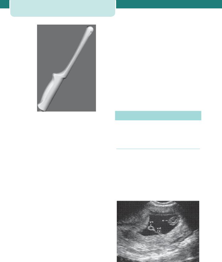
The ultrasound technique uses very high frequency sound waves of between 3.5 and 7.0 mega hertz emitted from a transducer. Transducers can be placed and moved across the maternal abdomen (transabdominal, Figure 6.1) or mounted on a probe which can be inserted into the vagina (transvaginal, Figure 6.2)
Transvaginal ultrasonography is useful in early pregnancy, for examining the cervix later in pregnancy and for identifying the lower edge of the placenta. It is also useful in early pregnancy in women with significant amounts of abdominal adipose tissue through which abdominal ultrasound waves would need to travel and
Figure 6.1 Ultrasound probe; abdominal
hence be attenuated prior to reaching the uterus and its contents, making visualization difficult. In general, however, after 12 weeks gestation, an abdominal transducer, which is a flat or curvilinear probe with a
62 Antenatal imaging and assessment of fetal well-being
frequency being proportional to the velocity of the blood cells. If the red blood cells are moving towards the beam, the reflected signal will be at a higher frequency than the transmitted one and conversely the reflected beam will be at a lower signal if the flow is away from the beam. In this modality, signals from a particular vessel can be isolated and displayed in graphic form, with the velocity plotted against time. The significance of changes observed in waveform patterns obtained from placental and fetal vessels and how these observations can be used in clinical practice will be discussed later in the chapter.
Ultrasound scanning is currently considered to be a safe, non-invasive, accurate and cost-effective investigation in the fetus. This chapter will consider the diagnostic use of these techniques in more detail.
Figure 6.2 Ultrasound probe; trans vaginal
Clinical applications of ultrasound
much wider array, is used. Crystals within the transducer emit a focused ultrasound beam in a series of pulses and then receive the reflected signals from within the uterus between the pulses. The strength of the reflected sound wave depends on the difference in ‘acoustic impedance’ between adjacent structures. The acoustic impedance of a tissue is related to its density; the greater the difference in acoustic impedance between two adjacent tissues the more reflective will be their boundary. The returning signals are transformed into visual signals and generate a continuous picture of the moving fetus. Movements such as fetal heart beat and malformations in the fetus can be assessed and measurements can be made accurately on the images displayed on the screen. Such measurements enable the assessment of gestational age, size and growth in the fetus. Ultrasound images obtained can also be processed with computer software to produce three-dimensional (3D) images and even four-dimensional (moving 3D images) which provide more detail on fetal anatomical structure and the identification of anomalies.
The use of Doppler ultrasound allows the assessment of the velocity of blood within fetal and placental vessels and provides indirect assessment of fetal and placental condition. Doppler ultrasound makes use of the phenomenon of the Doppler frequency shift, where the reflected wave will be at a different frequency from the transmitted one if it interacts with moving structures, such as red blood cells flowing along a blood vessel with the change in
The main uses of ultrasonography in pregnancy are in the areas discussed below.
Diagnosis and confirmation of viability in early pregnancy
The gestational sac can be visualized from as early as 4–5 weeks of gestation and the yolk sac at about 5 weeks (Figure 6.3). The embryo can be observed and measured at 5–6 weeks gestation. A visible heartbeat can be visualized by about 6 weeks.
Transvaginal ultrasound plays a key role in the diagnosis of disorders of early pregnancy, such as incomplete or missed miscarriage, blighted ovum where no fetus is present (Figure 6.4) and ectopic
Figure 6.3 Ultrasound sac showing yolk sac (ys) and embryo (e) with the vitelline duct (vd)
pregnancy. In a missed miscarriage, for example, the fetus can be identified, but with an absent fetal heart and in a blighted ovum, the absence of fetal development results in the presence of a gestational sac which is empty. An ectopic pregnancy is suspected if, in the presence of a positive pregnancy test, an ultrasound scan does not identify a gestation sac within the uterus, there is an adnexal mass with or without a fetal pole, or there is fluid in the pouch of Douglas.
Determination of gestational age and assessment of fetal size and growth
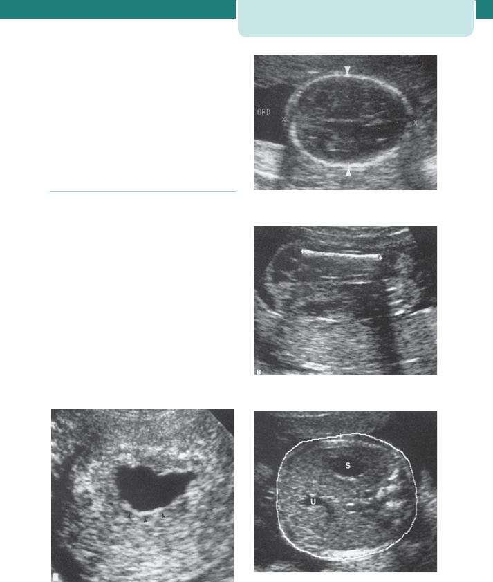
Up to approximately 20 weeks gestation the range of values around the mean for measurements of fetal length, head size and long bone length is narrow and hence assessment of gestation based on these measures is accurate. The crown-rump length (CRL) is used up to 13 weeks 6 days, and the head circumference (HC) from 14 to 20 weeks gestation. The biparietal diameter (BPD) (Figure 6.5) and femur length (FL) (Figure 6.6) can also be used to determine gestational age. Essentially, the earlier the measurement is made, the more accurate the prediction, and measurements made from an early CRL (accuracy of prediction 5 days) will be preferred to a biparietal diameter at 20 weeks (accuracy of prediction 7 days).
In the latter part of pregnancy, measuring fetal abdominal circumference (AC) (Figure 6.7) and HC will allow assessment of the size and growth of the fetus and will assist in the diagnosis and management
Clinical applications of ultrasound |
63 |
Figure 6.5 Biparietal diameter (BPD)
Figure 6.6 Femur length (FL)
Figure 6.4 Ultrasound image showing empty gestation sac in a case of blighted ovum
Figure 6.7 Abdominal circumference (AC) measurement demonstrating the correct section showing the stomach (S) and the umbilical vein (U)
64Antenatal imaging and assessment of fetal well-being
of fetal growth restriction. In addition to AC and HC, BPD and FL, when combined in an equation, provide a more accurate estimate of fetal weight (EFW) than any of the parameters taken singly.
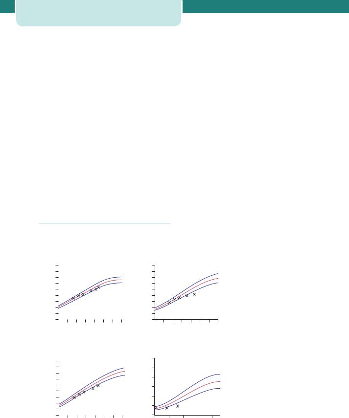
In pregnancies at high risk of fetal growth restriction (FGR), serial measurements are plotted on the normal reference range. Growth patterns are helpful in distinguishing between different types of growth restriction (symmetrical and asymmetrical). Asymmetry between head measures (BPD, HC) and AC can be identified in FGR, where a brain-sparing effect will result in a relatively large HC compared with the AC (Figure 6.8). The opposite would occur in a diabetic pregnancy, where the abdomen is disproportionately large due to the effects of insulin on the fetal liver and fat stores. Cessation of growth is an ominous sign of placental failure.
Gestational age cannot be accurately calculated by ultrasound after 20 weeks gestation because of the wider range of normal values of AC and HC around the mean.
Multiple pregnancy
Ultrasound is now the most common way in which multiple pregnancies are identified (Figure 6.9). In
addition to identifying the presence of more than one fetus, it can also be used to determine the chorionicity of the pregnancy.
Monochorionic twin pregnancies (i.e. those who ‘share’ a placenta) are associated with an increased risk of pregnancy complications and a higher perinatal mortality rate than dichorionic twin pregnancies. It is therefore clinically useful to be able to determine chorionicity early in pregnancy (see Chapter 9, Twins and higher multiple gestations).
The dividing membrane in monochorionic twins is formed by two layers of amnion and in dichorionic twins by two layers of chorion and two of amnion. Dichorionic twins therefore have thicker membranes than monochorionic twins and this can be perceived qualitatively on ultrasound. Ultrasonically, dichorionic twin pregnancies in the first trimester of pregnancy have a thick inter-twin separating membrane (septum), flanked on either side by a very thin amnion. This is in contrast to a monochorionic twin pregnancy, which on twodimensional ultrasound has a very thin inter-twin septum.
Another method of determining chorionicity in the first trimester uses the appearance of the septum at its origin from the placenta. On ultrasound, a
mm
mm
|
|
Head circumference |
|
|
|
|||||
450 |
|
|
|
|
|
|
|
|
|
450 |
|
|
|
|
|
|
|
|
|
||
400 |
|
|
|
|
|
|
95% |
400 |
||
350 |
|
|
|
|
|
|
350 |
|||
|
|
|
|
|
|
|
|
|
||
300 |
|
|
|
|
|
|
5% |
|
300 |
|
250 |
|
|
|
|
|
|
mm |
250 |
||
|
|
|
|
|
|
|
|
|||
200 |
|
|
|
|
|
|
|
|
200 |
|
|
|
|
|
|
|
|
|
|
||
150 |
|
|
|
|
|
|
|
|
|
150 |
100 |
|
|
|
|
|
|
|
|
|
100 |
50 |
|
|
|
|
|
|
|
|
|
50 |
0 |
|
|
|
|
|
|
|
|
|
0 |
|
|
|
|
|
|
|
|
|
||
16 |
20 |
24 |
28 |
32 |
36 |
40 |
|
|||
|
|
|
|
Weeks |
|
|
|
|
|
|
|
|
|
Femur length |
|
|
|
|
|||
90 |
|
|
|
|
|
|
|
|
|
6000 |
|
|
|
|
|
|
|
95% |
|
||
80 |
|
|
|
|
|
|
|
5000 |
||
70 |
|
|
|
|
|
|
|
|
|
4000 |
60 |
|
|
|
|
|
|
|
5% |
||
|
|
|
|
|
|
|
|
|||
50 |
|
|
|
|
|
|
|
|
g |
3000 |
40 |
|
|
|
|
|
|
|
|
|
2000 |
30 |
|
|
|
|
|
|
|
|
|
|
20 |
|
|
|
|
|
|
|
|
|
1000 |
10 |
|
|
|
|
|
|
|
|
|
|
|
|
|
|
|
|
|
|
|
0 |
|
0 |
|
|
|
|
|
|
|
|
|
|
|
|
|
|
|
|
|
|
|||
16 |
20 |
24 |
28 |
32 |
36 |
40 |
|
|||
Weeks
Abdomen circumference
95%
5%
16 20 24 28 32 36 40 Weeks
Estimated fetal weight
|
|
|
|
95% |
|
|
|
|
5% |
24 |
28 |
32 |
36 |
40 |
|
|
Weeks |
|
|
Figure 6.8 Ultrasound plots on reference range for head circumference (HC), abdominal circumference (AC) and estimated fetal weight (EFW) in
a case of early onset fetal growth restriction (FGR). Note that HC remains above 5th centile while the AC falls below 5th centile. This is a case of asymmetric FGR with head sparing
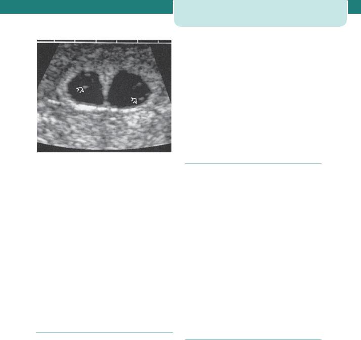
Figure 6.9 Early twin dichorionic pregnancy; note the ‘peaked’ inter twin membrane
tongue of placental tissue is seen within the base of dichorionic membranes and has been termed the ‘twin peak’ or ‘lambda’ sign. The optimal gestation at which to perform such ultrasonic chorionicity determination is 9–10 weeks. Dichorionicity will also be confirmed by the identification of two placental masses and later in pregnancy by the presence of different-sex fetuses.
Ultrasound is also invaluable in the management of twin pregnancy in terms of confirming fetal presentations, which may be difficult on abdominal palpation, evidence of growth restriction, fetal anomaly and the presence of placenta praevia, all of which are more common in this type of pregnancy, and any suggestion of twin-to-twin transfusion syndrome.
Diagnosis of fetal abnormality
Major structural abnormalities occur in 2–3 per cent of pregnancies and many can be diagnosed by an ultrasound scan at around or before 20 weeks gestation. Common examples include spina bifida and hydrocephalus, skeletal abnormalities such as achondroplasia, abdominal wall defects such as exomphalos and gastroschisis, cleft lip/palate and congenital cardiac abnormalities.
Detection rates of between 40 and 90 per cent have been reported. This means that a ‘normal scan’ is not a guarantee of a normal baby. A number of factors can influence the success of detecting an abnormality. Some are very difficult to visualize or to be absolutely certain about. Some conditions, for
Clinical applications of ultrasound |
65 |
example hydrocephalus, may not have been obvious at the time of early scans. The position of the baby in the uterus will influence visualization of organs such as the heart, face and spine. Repeat scans are sometimes required if visualization is a problem in anticipation that the fetus will be in a more accessible position.
First trimester ultrasonic ‘soft’ markers for chromosomal abnormalities such as the absence of fetal nasal bone, an increased fetal nuchal translucency (the area at the back of the neck) are now in common use to enable detection of fetuses at risk of chromosomal anomalies such as Down’s syndrome.
Placental localization
Placenta praevia can cause life-threatening haemorrhage in pregnancy. Ultrasonography has become indispensible in the localization of the site of the placenta and thus ultrasonographic identification of the lower edge of the placenta to exclude or confirm placenta praevia as a cause for antepartum haemorrhage is now a part of routine clinical practice. The transvaginal approach, undertaken with caution, can be helpful in clearly identifying the lower placental edge if not seen clearly with an abdominal probe.
At the 20 weeks scan, it is customary to identify women who have a low-lying placenta. At this stage, the lower uterine segment has not yet formed and most low-lying placentas will appear to ‘migrate’ upwards as the lower segment stretches in the late second and third trimesters. About 5 per cent of women have a low-lying placenta at 20 weeks, and only 5 per cent of this group will eventually be shown to have a placenta praevia.
Amniotic fluid volume assessment
Ultrasound can be used to identify both increased and decreased amniotic fluid volumes. The fetus has a role in the control of the volume of amniotic fluid. It swallows amniotic fluid, absorbs it in the gut and later excretes urine into the amniotic sac. Congenital abnormalities that impair the fetus’s ability to swallow, for example anencephaly or oesophageal atresia, will result in an increase in amniotic fluid. Congenital abnormalities that result in a failure of urine production or passage, for example renal agenesis and posterior urethral valves, will result in reduced or absent amniotic fluid. Fetal growth restriction can be associated with reduced amniotic