

Obstetrics_by_Ten_Teachers_19E_-_Kenny_Louise
.pdf106 Antenatal obstetric complications
Feto-placental |
Maternal |
circulation |
circulation |
d
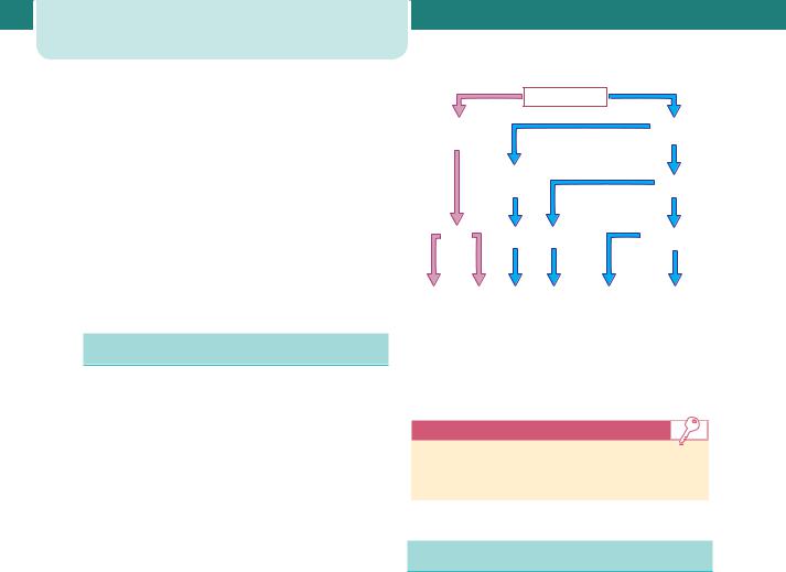
(a) Sensitization
D
D d
D
D |
d |
Maternal antibodies produced against fetal red cells
Fetal red |
Maternal red |
cells with |
cells without |
‘D’ antigen |
‘D’ antigen |
(Rh-positive) |
(Rh-negative) |
(b)
Fetal red cell destruction
Next pregnancy
D |
Maternal anti-D |
D |
antibodies produced |
which pass across placenta |
|
|
causing fetal red cell lysis |
D |
|
Figure 8.12 The mechanism of rhesus sensitization (a) and fetal red cell destruction (b)
given intramuscularly as soon as possible after any potentially sensitizing event. It is normal practice to administer anti-D after any of these events; the exact dose is determined by the gestation at which sensitization has occurred and the size of the fetomaternal haemorrhage.
In the first trimester of pregnancy, because the volume of fetal blood is so small, it is unlikely that sensitization would occur, and a ‘standard’ dose of anti-D (the exact dose varies from country to country) is given; this will more than cover even the largest feto-maternal transfusion. In the second and third trimesters, fetal blood volume is greater and because there is a possibility of a fetomaternal transfusion of several millilitres, a larger dose is given and a Kleihauer test performed.
A Kleihauer is a test of maternal blood to determine the proportion of fetal cells present (relying on their ability to resist denaturation by alcohol or acid); it will allow calculation of the amount of extra anti-D immunoglobulin required should a large transfusion have occurred.
In many countries, Rhesus-negative women are given anti-D at 28 and/or 34 weeks routinely. This is based on the finding that a small number of Rhesusnegative women become sensitized during pregnancy despite the administration of anti-D at delivery and without a clinically obvious sensitizing event. The likelihood is that a small feto-maternal haemorrhage occurs without any obvious clinical signs; therefore, prophylactic anti-D would reduce the risk of iso-immunization from this event.

Signs of fetal anaemia
Note: Clinical and ultrasound features of fetal anaemia do not usually become evident unless fetal haemoglobin is more than 5 g/dL less than the mean for gestation. Usually, features are not obvious unless the fetal haemoglobin is less than 6 g/dL.
•Polyhydramnios.
•Enlarged fetal heart.
•Ascites and pericardial effusions.
•Hyperdynamic fetal circulation (can be detected by Doppler ultrasound by measuring increased velocities in the middle cerebral artery or aorta).
•Reduced fetal movements.
•Abnormal CTG with reduced variability, eventually a ‘sinusoidal’ trace.
The management of Rhesus disease in a sensitized woman
Once a woman who is D Rhesus negative has been sensitized to the D Rhesus antigen, no amount of anti-D will ever turn the clock back. In a subsequent pregnancy, close surveillance is required. Rhesus disease gets worse with successive pregnancies, so it is important to note the severity of the disease in previous pregnancies. The management depends on the clinical scenario.
•The father of the next baby is D Rhesus negative. In this situation, there is no risk that the baby will
be D Rhesus positive and therefore there is no chance of Rhesus disease.
•The father of the next baby is D Rhesus positive. He may be heterozygous and in this situation
determining the paternal phenotype is useful in anticipating the likely fetal phenotype and, thus, the potential for development of HDFN. However, it is important to bear in mind that there are issues regarding paternal testing, and assuming paternity runs the risk of false prediction. Not withstanding this issue, paternal blood grouping is frequently used and often useful.
•In a sensitized woman, if the father is D Rhesus positive or unknown, standard management
involves monitoring antibody levels every 2–4 weeks from booking. Antibody levels or quantity can be described using the titre or by using
Rhesus iso-immunization |
107 |
IU (international units) as a standard quantification method. The titre simply refers to the number of times a sample has been diluted before the amount of antibody becomes undetectable; titre of 2, 4, 8, 16, 32, 64, 128, etc.
Each time a sample is tested, it should be checked in parallel with the previous sample to ensure the detection of significant changes in the antibody level. It has been found that titrations of anti-D do not correlate well with the development of HDFN, and that the standard quantification method (IU/mL) gives more clinically relevant levels.
Anti-D level |
Outcome |
4 IU/mL |
HDFN unlikely |
|
|
4–15 IU/mL |
Moderate risk of HDFN |
|
|
15 IU/mL |
High risk of hydrops |
|
fetalis |
|
|
•If antibody levels rise, the baby should be examined for signs of anaemia. In the past,
the bilirubin concentration of amniotic fluid was determined optically to give an indirect measure of fetal haemolysis. This involved an invasive procedure with the attendant risks of miscarriage/preterm labour and further boosting of the alloimmune response. In the last decade, middle cerebral artery (MCA) Dopplers (peak velocity measurement) have been shown to correlate reliably with fetal
anaemia. In practice, this means that the use of invasive tests to monitor disease progression (once a critical antibody level has been reached) have been replaced by non-invasive assessment using MCA Doppler. There is now substantial data to support the use of peak MCA velocity as a correlate of fetal anaemia. The sensitivity
is reported at 100 per cent with a false positive rate of 12 per cent (see figure 6.17 in Chapter 6, Antenatal imaging and assessment of fetal wellbeing).
•A fetus with raised MCA Dopplers has a high probability of anaemia. These cases are not
common and the treatment should be in, or guided by, tertiary fetal medicine centres.
Treatment options include delivery or fetal blood transfusion. Delivery of the fetus is an option if the fetus is sufficiently mature. However, delivery

108 Antenatal obstetric complications
of an anaemic, rapidly haemolysing premature baby is a significant risk and should not be undertaken lightly. Delivery must take place in a unit where adequate neonatal support and
expertise is available and generally delivery should not be before 36–37 weeks of gestation unless there are specific reasons, such as special difficulty with fetal transfusion.
•Fetal blood transfusion is life saving in a severely anaemic fetus that is too premature for delivery
to be contemplated. The aim is to restore haemoglobin levels, reversing or preventing hydrops or death. A side effect is that transfusion will also suppress fetal erythropoesis, which reduces the concentration of antigen positive cells available for haemolysis. Blood can be transfused into a fetus in various ways depending on the gestation, the site of the cord insertion and the clinical situation.
•Routes of administration:
•into the umbilical vein at the point of the cord insertion (ideally through the placenta and not
through the amniotic sac);
•into the intrahepatic vein;
•into the peritoneal cavity (not as effective but some blood is absorbed and this may
be the only option, for example in low gestations);
•into the fetal heart.
Once a decision has been made that the fetus is severely anaemic and requires a blood transfusion, the invasive procedure aims to first take a sample to confirm the anaemia and then infuse the blood during a single puncture.
•Transfused blood is:
•RhD negative;
•crossmatched with a maternal sample;
•densely packed (Hb usually around 30 g/L) so that small volumes are used;
•white cell depleted and irradiated;
•screened for infection including CMV.
At delivery
If the baby is known to be anaemic or has had multiple transfusions, a neonatologist must be present at delivery should exchange transfusion be required.
Blood must therefore always be ready for the delivery. All babies born to Rhesus-negative women should have cord blood taken at delivery for a blood count, blood group and indirect Coomb’s test.
Key points
D Rhesus disease
•Rhesus disease gets worse with successive pregnancies.
•If the father of the fetus is Rhesus negative, the fetus cannot be Rhesus positive.
•If the father of the fetus is Rhesus positive, he may be a heterozygote (50 per cent likelihood that the baby is D Rhesus positive) or a homozygote (100 per cent likelihood).
•Anti-D is given only as prophylaxis and is useless once sensitization has occurred.
•Prenatal diagnosis for karyotype, or attempts at determining fetal blood group by invasive testing (e.g. chorion villus sampling), may make the antibody levels higher in women who are already sensitized.
ABO
ABO blood group iso-immunization may occur when the mother is blood group O and the baby is blood group A or B. Anti-A and anti-B antibodies are present in the maternal circulation naturally, usually secondary to sensitization against A or B substances in food or bacteria. This means that ABO incompatibility may occur in a first pregnancy. In this situation, anti-A or anti-B antibodies may pass to the fetal circulation, causing fetal haemolysis and anaemia. However, most anti-A and anti-B are mainly IgM and do not cross the placenta. In addition, A and B antigens are not fully developed in the fetus. Therefore ABO incompatibility generally causes only mild haemolytic disease of the baby, but may sometimes explain unexpected jaundice in an otherwise healthy term infant.
Additional reading
James DK, Steer PJ, Weiner CP, Gonik B. High risk pregnancy, 3rd end. London: WB Saunders, 2005.
Lewis, G (ed.). The Confidential Enquiry into Maternal and Child Health (CEMACH). Saving Mothers’
Lives: reviewing maternal deaths to make motherhood safer – 2003–2005. The Seventh Report on Confidential Enquiries into Maternal Deaths in the United Kingdom. London: CEMACH, 2007.

|
|
|
|
|
|
|
|
|
|
|
|
|
|
|
|
C H A P T E R 9 |
T W I N S A N D H I G H E R |
|
|
|
|||||||||
|
|
|
M U L T I P L E G E S T A T I O N S |
|
|
|
||||||||
|
|
|
Griffith Jones |
|
|
|
|
|
|
|
|
|||
|
|
|
|
|
|
|
|
|
|
|||||
|
Definitions .................................................................................................... |
|
|
109 |
|
Complications unique to monoamniotic |
|
|
|
|||||
|
Prevalence ................................................................................................... |
|
|
109 |
|
|
twinning............................................................................................... |
114 |
|
|||||
|
Classification .............................................................................................. |
|
|
110 |
|
Differential diagnosis ............................................................................ |
114 |
|
||||||
|
Aetiology........................................................................................................ |
|
|
110 |
|
Antenatal management....................................................................... |
114 |
|
||||||
|
Other physiology ...................................................................................... |
|
|
111 |
|
Intrapartum management.................................................................. |
116 |
|
||||||
|
Complications relevant to twin pregnancy ............................. |
111 |
|
Higher multiples....................................................................................... |
118 |
|
||||||||
|
Complications unique to monochorionic twinning |
............113 |
|
|
|
|
|
|
|
|
|
|||
|
|
|
|
|
|
|
|
|
|
|
|
|
|
|
O V E R V I E W
In 1–2 per cent of pregnancies, there is more than one fetus. The chances of miscarriage, fetal abnormalities, poor fetal growth, preterm delivery and intrauterine or neonatal death are considerably higher in twin than in singleton pregnancies. In about two-thirds of twins the fetuses are non-identical, or dizygotic, and in one-third they are identical, or monozygotic. In all dizygotic pregnancies there are two functionally separate placentae (dichorionic). In two-thirds of monozygotic pregnancies there are vascular communications within the two placental circulations (monochorionic) and in the other one-third of cases there is dichorionic placentation. Monochorionic, compared to dichorionic, twins have a much higher risk of abnormalities and death. The maternal risks are also increased in multiple gestations, including adverse symptoms such as nausea and vomiting, tiredness and discomfort, and the risk of serious complications, including hypertensive and thromboembolic disease, and antepartum and postpartum haemorrhage.
Definitions
In general terms, multiple pregnancies consist of two or more fetuses. There are rare exceptions to this, such as twin gestations made up of a singleton viable fetus and a complete mole. Twins make up the vast majority (nearly 99 per cent) of multiple gestations. Pregnancies with three or more fetuses are referred to as ‘higher multiples’.
Prevalence
Risk factors for multiple gestations include assisted reproduction techniques (both ovulation induction and in vitro fertilization (IVF)), increasing maternal age, high parity, black race and maternal family history.
In the UK, twins currently account for approximately 1.5 per cent of all pregnancies, up from 1 per cent in 1984.
Since the mid-1980s, the incidence of multiple pregnancy has been increasing. Two related and overlapping trends are contributors to this. Delay in childbearing results in increased maternal age at conception. The increased use of infertility treatments, also often by older women, is another factor.
Traditionally, the expected incidence was calculated using Hellin’s rule. Using this rule, twins were expected in 1 in 80 pregnancies, triplets in 1 in 802 and so on. Based upon the number of births in the UK in 2007, 9555 twins would have been predicted. In fact, 11 573 were delivered, 21 per cent higher than expected. The figures for triplets are similar; 119 sets may have been expected but in actuality 149 were delivered. Now seen in just over 1 in 5000 UK pregnancies, this represents a 25 per cent excess over expected; however, these figures for triplets have actually fallen dramatically since the late 1990s. At that time, the Human Fertilisation and Embryology Authority placed restrictions on fertility centres, limiting the number of embryos that could
110Twins and higher multiple gestations
be routinely transferred per cycle. More recently, they have promoted single embryo transfers for the majority of patients, hoping to cap or even reduce the incidence of iatrogenic twinning.
In contrast, there is scant regulation of fertility services in the USA. Figures from 2006 suggest an incidence of twin births 31 per cent above expected with triplets increased by a remarkable 211 per cent (although this had fallen slightly compared to the previous year). Even more staggering is the 355 quadruplet pregnancies seen in the USA, compared to seven in the UK the same year.
Classification
The classification of multiple pregnancy is based on:
•number of fetuses: twins, triplets, quadruplets, etc.
•number of fertilized eggs: zygosity
•number of placentae: chorionicity
•number of amniotic cavities: amnionicity.
Non-identical or fraternal twins are dizygotic, having resulted from the fertilization of two separate eggs. Although they always have two functionally separate placentae (dichorionic), the placentae can become anatomically fused together and appear to the naked eye as a single placental mass. They always have separate amniotic cavities (diamniotic) and the two cavities are separated by a thick three-layer membrane (fused amnion in the middle with chorion on either side). The fetuses can be either same-sex or differentsex pairings.
Identical twins are monozygotic – they arise from fertilization of a single egg and are always same-sex pairings. They may share a single placenta (monochorionic) or have one each (dichorionic). If dichorionic, the placentae can become anatomically fused together and appear to the naked eye as a single placental mass, as mentioned above. The vast majority of monochorionic twins have two amniotic cavities (diamniotic) but the dividing membrane is thin, as it consists of a single layer of amnion alone. Monochorionic twins may occasionally share a single sac (monoamniotic). Figure 9.1 shows the relative contributions of the different types of twins to a hypothetical random selection of 1000 twin pairs.
|
|
|
1000 Twins |
|
|
700 |
|
|
|
300 |
|
|
|
|
Monozygotic |
||
Dizygotic |
|
|
|
||
|
|
|
|
||
|
|
100 |
|
|
200 |
|
|
Dichorionic |
|
||
|
|
|
Monochorionic |
||
|
|
|
|
|
|
700 |
100 |
190 |
|
10 |
|
DCDA |
DCDA |
MCDA |
|
Monoamniotic |
|
350 |
350 |
100 |
190 |
9 |
1 |
Different |
Same |
Same |
Same |
Non-conjoined |
Conjoined |
sex |
sex |
sex |
sex |
|
|
Figure 9.1 Incidence of monozygotic and dizygotic twin pregnancies. (DCDA, dichorionic diamniotic; MCDA, monochorionic diamniotic; MCMA, monochorionic monoamniotic.)
Key points
•Not all dichorionic pregnancies are dizygotic.
•All monochorionic pregnancies are monozygotic.
Aetiology
Dizygotic twins may arise spontaneously from the release of two eggs at ovulation. This tendency to release more than one egg can be familial or racial in origin and increases with maternal age. Ovulation induction treatments may also cause the release of more than one egg. With assisted conception techniques such as IVF, two or more embryos fertilized in the laboratory may be replaced in the uterus, although single embryo transfer is becoming more frequent.
Monozygotic twins arise from a single fertilized ovum that splits into two identical structures. The type of monozygotic twin depends on how long after conception the split occurs. When the split occurs within 3 days of conception, two placentae and two amniotic cavities result, giving rise to a dichorionic diamniotic (DCDA) pregnancy. When splitting occurs between days 4 and 8, only the chorion has differentiated and a monochorionic diamniotic (MCDA) pregnancy results. Later splitting after the amnion has differentiated leads to both twins developing in a single amniotic cavity, a
monochorionic monoamniotic (MCMA) pregnancy. If splitting is delayed beyond day 12, the embryonic disc has also formed, and conjoined, or ‘Siamese’ twins will result.
The incidence of monozygotic or identical twins is generally accepted to be constant at 1 in 250. It is not influenced by race, family history or parity. There is some evidence of a small increase in monozygotic twinning after IVF, for reasons that are unclear.
Other physiology
Maternal
All the physiological changes of pregnancy (increased cardiac output, volume expansion, relative haemodilution, diaphragmatic splinting, weight gain, lordosis, etc.) are exaggerated in multiple gestations. This results in much greater stresses being placed on maternal reserves. The ‘minor’ symptoms of pregnancy may be exaggerated; however, for women with pre-existing health problems, such as cardiac disease, a multiple pregnancy may substantially increase their risk of morbidity.
Fetal
Monochorionic placentae have the unique ability to develop vascular connections between the two fetal circulations. These anastomoses carry the potential for complications, discussed in the next section.
Complications relevant to twin
pregnancy
Miscarriage and severe preterm delivery
General
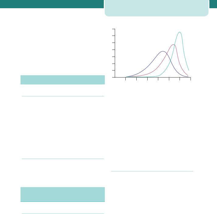
Spontaneous preterm delivery is an ever-present risk in any twin pregnancy (Figure 9.2) where the average gestation at delivery is 37 weeks. Therefore, about half of all twins deliver preterm. With two or more babies resulting from each delivery, multiple gestations account for 20–25 per cent of NICU admissions. Due to their associated high mortality and/or morbidity rates, the births that attract the most interest are births below 32 weeks gestation. In singleton pregnancies,
Complications relevant to twin pregnancy |
111 |
%
20 |
24 |
28 |
32 |
36 |
40 |
44 |
|||
|
|
Gestation (weeks) |
|
|
|
|
|||
|
Singleton |
|
|
Twin |
|
|
|
Triplet |
|
|
|
|
|
|
|
||||
Figure 9.2 Gestational age distribution at delivery of singleton, twin and triplet pregnancies
each of these outcomes has a frequency of about 1 per cent.
Dichorionic/monochorionic differences
In a dichorionic pregnancy, the chance of late miscarriage is 2 per cent. In 15 per cent of cases, delivery will be very preterm. For monochorionic twins, the chance of early birth is increased even further, with 12 per cent born before viability and 25 per cent delivering between 24 and 32 weeks (Table 9.1).
Perinatal mortality in twins
General
The overall infant mortality rate for twins is around 5.5 times higher than for singletons. The biggest contributor to this high rate is complications related to preterm birth. Data from the UK suggest that the survival at any given gestation is identical for singletons and multiples.
Dichorionic/monochorionic differences
UK birth registration data for 2007 show at least one twin is stillborn in 20 per 1000 twin maternities. This is in contrast to a figure of 5 per 1000 singleton births. However, the rate for different-sex pairs (who must be dichorionic) is 12 per 1000, while that for same-sex pairs (roughly one-third of whom will be monochorionic) is 23 per 1000. As two-thirds of these same-sex pairs are dichorionic (and should have a stillbirth rate of 12 per 1000), the true stillbirth rate for monochorionic pairs must be substantially higher.

112 Twins and higher multiple gestations
Table 9.1 Common pregnancy complications in twin pregnancies according to chorionicity, compared with singleton pregnancies
Complication |
|
Twins |
|
|
Singleton (%) |
Dichorionic (%) |
Monochorionic (%) |
Miscarriage at 12–23 weeks |
1 |
2 |
12 |
|
|
|
|
Delivery at 24–32 weeks |
|
15 |
25 |
|
|
|
|
Small for gestation |
10 |
25 |
50 |
|
|
|
|
Fetal defects |
1 |
2 |
4 |
|
|
|
|
Some of this increase in perinatal mortality will be due to the excess of preterm delivery in monochorionic twins, but monozygotic twins also have both additional risks and unique complications that further increase their chance of death and handicap, as discussed below.
Death of one fetus in a twin pregnancy
General
With the more liberal use of early pregnancy scanning, it has been recognized that up to 25 per cent of twins may suffer an early demise and subsequently ‘vanish’ well before they would have previously been detected. After the first trimester, the intrauterine death of one fetus in a twin pregnancy may be associated with a poor outcome for the remaining co-twin. Maternal complications such as disseminated intravascular coagulation have been reported, but the incidence of this appears to be very low.
Dichorionic/monochorionic differences
In dichorionic twins, the second or third trimester intrauterine death of one fetus may be associated with the onset of labour, although in some cases the pregnancy may continue uneventfully and even result in delivery at term. Careful fetal and maternal monitoring is required. By contrast, fetal death of one twin in monochorionic twins may result in immediate complications in the survivor. These include death or brain damage with subsequent neurodevelopmental handicap. Acute hypotensive episodes, secondary to placental vascular anastomoses between the two fetuses, result in haemodynamic volume shifts from the live to the dead fetus. The acute release of vasoactive substances into the survivor’s circulation may also play a role. Death or handicap of the co-twin occurs in up to 30 per cent of cases.
Fetal growth restriction
General
Compared to singletons, the risk of poor growth is higher in each individual twin alone and substantially raised in the pregnancy as a whole.
When a fetus is growth restricted, the main aims of antenatal care become prediction of the severity of impaired fetal oxygenation and selecting the appropriate time for delivery. In singletons, this is a balance between the relative risks of intrauterine death versus the risk of neonatal death or handicap from elective preterm delivery. The situation is much more complicated in twin pregnancies. The potential benefit of expectant management or elective delivery for the small fetus must also be weighed against the risk of the same policy for the normally grown co-twin.
Dichorionic/monochorionic differences
In a dichorionic pregnancy, each fetus runs twice the risk of a low birth weight and there is a 25 per cent chance that at least one of the fetuses will be small for gestational age. The chance of suboptimal fetal growth for monochorionic twins is almost double that for dichorionic twins (see Table 9.1).
In dichorionic twin pregnancies where one fetus has fetal growth restriction (FGR), elective preterm delivery may lead to iatrogenic complications of prematurity in the previously healthy co-twin. In general, delivery should be avoided before 28–30 weeks gestation, even if there is evidence of imminent intrauterine death of the smaller twin; however, this may not be applicable in the management of monochorionic twins. The death of one of a monochorionic twin pair may result in either death or handicap of the co-twin because of acute hypotension secondary to placental vascular anastomoses between the two circulations (see earlier discussion under

Death of one fetus in a twin pregnancy). As the damage potentially happens at the moment of death of the first twin, the timing of delivery may be a very difficult decision. Below 30 weeks gestation, the aim is to prolong the pregnancy as far as possible without risking the death of the growth-restricted twin.
Fetal abnormalities
General
Compared to singletons, twin pregnancies carry at least twice the risk of the birth of a baby with an anomaly. There are, however, important differences in both risk and management, based upon chorionicity.
Dichorionic/monochorionic differences
Each fetus of a dichorionic twin pregnancy has a risk of structural anomalies, such as spina bifida, that is similar to that of a singleton. Therefore, the chance of finding an anomaly within a dichorionic twin pregnancy is twice that of a singleton. In contrast, each fetus in a monochorionic twin pregnancy carries a risk for abnormalities that is four times that of a singleton. This is presumably due to a higher risk of vascular events during embryonic development (see Table 9.1).
Multiple gestations with an abnormality in one fetus can be managed expectantly or by selective fetocide of the affected twin. In cases where the abnormality is non-lethal but may well result in handicap, the parents may need to decide whether the potential burden of a handicapped child outweighs the risk of loss of the normal twin from fetocide-related complications, which occur after 5–10 per cent of procedures. In cases where the abnormality is lethal, it may be best to avoid such risk to the normal fetus, unless the condition itself threatens the survival of the normal twin. Anencephaly is a good example of a lethal abnormality that can threaten the survival of the normal twin. At least 50 per cent of pregnancies affected by anencephaly are complicated by polyhydramnios, which can lead to the spontaneous preterm delivery of both babies.
Fetocide in monochorionic pregnancies carries increased risk and requires a different technique. As there are potential vascular anastomoses between the two fetal circulations, intracardiac injections cannot be employed. Methods have evolved that employ cord occlusion techniques. These require significant instrumentation of the uterus and are therefore associated with a higher complication rate.
Complications unique to monochorionic twinning |
113 |
Chromosomal defects and twinning
General
In twins, as in singletons, the risk for chromosomal abnormalities increases with maternal age. The rate of spontaneous dizygotic twinning also increases with maternal age. Many women undergoing assisted conception techniques (which increase the chance of dizygotic twinning) are also older than the mean maternal age. Chromosomal defects may be more likely in a multiple pregnancy for various reasons, and couples should be counselled accordingly.
Dichorionic/monochorionic differences
Monozygotic twins arise from a single fertilized egg and therefore have the same genetic make up. It is clear that in monozygotic twin pregnancies, chromosomal abnormalities such as Down’s syndrome affect neither fetus or both. The risk is based upon maternal age.
In dizygotic twins, the maternal age-related risk for chromosomal abnormalities for each individual twin remains the same as for a singleton pregnancy. Therefore, at a given maternal age, the chance that at least one of the twin pair is affected by a chromosomal defect is twice as high as for a singleton pregnancy. For example, a 40-year-old woman with a singleton pregnancy has a fetal risk of trisomy 21 of 1 in 100. If she has a dizygotic twin pregnancy, the risk that one fetus would be affected is 1 in 50 (1 in 100 plus 1 in 100).
Complications unique to monochorionic
twinning
In all monochorionic twin pregnancies there are placental vascular anastomoses present, which allow communication between the two fetoplacental circulations. In approximately 15 per cent of monochorionic twin pregnancies, imbalance in the flow of blood across these arteriovenous communications results in twin-to-twin transfusion syndrome (TTTS). One fetus becomes overperfused and the other underperfused. The development of mild, moderate or severe TTTS depends on the degree of imbalance. The growth-restricted donor fetus suffers from hypovolaemia and becomes oliguric. As fetal urine is the major component of amniotic fluid, this fetus develops oligohydramnios. The recipient fetus becomes hypervolaemic, leading to polyuria and

114Twins and higher multiple gestations
polyhydramnios. There is also a risk of myocardial damage and high output cardiac failure. Severe disease may become apparent in the second trimester. The mother often complains of a sudden increase in abdominal girth associated with extreme discomfort. Clinical examination shows tense polyhydramnios and ultrasound confirms the diagnosis.
More than 90 per cent of pregnancies complicated by TTTS end in miscarriage or very preterm delivery. With treatment, one or both babies survive in about 70 per cent of pregnancies.
The long-standing method of treatment has been amniocentesis every 1–2 weeks with the drainage of large volumes of amniotic fluid. Exactly how this treatment improves the underlying pathophysiology is uncertain, but it appears to prolong the pregnancy and improve survival. More recently, a small number of centres have used fetoscopically guided laser coagulation to disrupt the placental blood vessels that connect the circulations of the two fetuses. Initial randomized studies suggest that laser therapy is associated with delivery at later gestational ages, higher survival rates and lower levels of handicap at six months of age. It remains, however, a highly specialized procedure with limited availability.
Complications unique to monoamniotic
twinning
Monoamniotic twins share a single amniotic cavity, with no dividing membrane between the two fetuses. They are at increased risk of cord accidents, predominantly through their almost universal cord entanglement. Many clinicians advocate elective delivery by Caesarean section at 32–34 weeks gestation, as this complication is usually acute, fatal and unpredictable.
Differential diagnosis
The differential diagnosis of a multiple gestation includes all the other causes of a ‘large for dates’ uterus: polyhydramnios, uterine fibroids, urinary retention and ovarian masses.
Antenatal management
Routine antenatal care for all women involves screening for hypertension and gestational diabetes. These
conditions occur more frequently in twin pregnancies and there is also a higher risk of other problems (such as antepartum haemorrhage and thromboembolic disease); however, the management is the same as for a singleton. Due to the increased fetoplacental demand for iron and folic acid, many would recommend routine (as opposed to selective) supplementation in multiple pregnancies. Minor symptoms of pregnancy are more common, but management is again unchanged compared to singletons.
Elements of antenatal care that have specific importance in multiple pregnancies include the following.
Determination of chorionicity
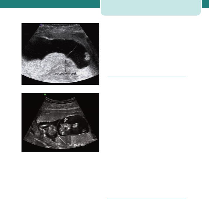
As described, there are important differences in risk and outcome between dichorionic and monochorionic pregnancies. Therefore, determination of chorionicity is critical to ‘good’ management, and this is done most reliably by ultrasound in the late first trimester. In dichorionic twins, there is a V-shaped extension of placental tissue into the base of the inter-twin membrane, referred to as the ‘lambda’ or ‘twin-peak’ sign. In monochorionic twins, this sign is absent and the inter-twin membrane joins the uterine wall in a T shape (Figure 9.3).
Assessment of chorionicity later in pregnancy is less reliable and relies upon the assessment of fetal gender, number of placentae and characteristics of the membrane between the two amniotic sacs. The ‘lambda’ sign becomes less accurate, and membrane thickness must be utilized. Different-sex twins must be dizygotic and therefore dichorionic. In same-sex twins, two separate placentae mean dichorionic, although the babies may still be monozygotic. However, monozygotic dichorionic twins do not carry the additional risks of vascular anastomoses.
Screening for fetal abnormalities
Screening for trisomy 21 using maternal serum biochemistry has been described in twin pregnancies but has not come into widespread use, in part due to its lower sensitivity. Normal ranges for serum alpha-fetoprotein have been established and allow detection of increased risk for neural tube defects. The measurement of nuchal translucency at 12 weeks gestation allows each fetus to have an individualized
(a)
(b)
Figure 9.3 Ultrasound appearance of monochorionic (a) and dichorionic (b) twin pregnancies at 12 weeks gestation. Note that in both types there appears to be a single placental mass, but in the dichorionic type there is an extension of placental tissue into the base of the inter-twin membrane forming the lambda sign
assessment of risk. The NHS Fetal Anomaly Screening Programme has recommended it as the screening test of choice for multiple pregnancies. If prenatal diagnosis is required (see Chapter 7, Prenatal diagnosis), knowledge of chorionicity is essential. Monochorionic twins are monozygotic and therefore only one sample is needed for karyotyping.
Both amniocentesis and chorion villus sampling (CVS) can be performed in twin pregnancies, but in dichorionic pregnancies, it is essential that both fetuses are sampled. As the placentae are often fused together, CVS has special challenges. With amniocentesis, dye-injection techniques have previously been used to prevent sampling the same sac twice; however,
Antenatal management |
115 |
to avoid this, many practitioners now rely on direct puncture of the inter-twin membrane.
Screening for fetal structural anomalies is done using second trimester ultrasound, with optimal detection rates seen at 20 weeks gestation. More time must be allowed for each appointment. As monochorionic twins have a significantly increased risk of fetal anomalies, many argue that they should be screened within specialized fetal medicine units.
Monitoring fetal growth and well-being
Measurement of symphysis–fundal height and maternal reporting of fetal movements are unreliable, as the individual contribution of each twin cannot be assessed. Monitoring for fetal growth and well-being in twins is principally by ultrasound. Each assessment should include fetal measurements, fetal activity, fetal lies and amniotic fluid volumes. In monochorionic twins, features of TTTS should be sought, including discordances between fetal size, fetal activity, bladder volumes, amniotic fluid volumes and cardiac size. In any twin pregnancy, when one or both fetuses are small, additional information about fetal wellbeing can be obtained from Doppler assessment of the fetal circulations and cardiotocography (CTG). Specialized twin monitors should be used to ensure each twin’s heart rate is sampled.
It is reasonable to plan 4- to 6-weekly ultrasound scans in uncomplicated dichorionic twins. However, due to their higher background risk, fortnightly ultrasound is appropriate in monochorionic pregnancies. These are approximate guidelines that should be modified around individual pregnancy circumstances.
Threatened preterm labour
As in singleton pregnancies, neither bed rest nor prophylactic administration of tocolytics is useful in preventing preterm delivery. Despite this, screening for preterm birth may be worthwhile. Antenatal strategies in those identified as at high risk may include maternal steroid therapy to enhance fetal lung maturation, supplementary education as to the signs and symptoms of preterm labour, advance planning regarding intrapartum care, screening for group B streptococcus (GBS) (intrapartum antibiotics reduce neonatal infection) and additional medical and midwifery support.