

Obstetrics_by_Ten_Teachers_19E_-_Kenny_Louise
.pdf186Labour
including both emergency and elective Caesarean sections. Although this is recognized by most as being too high, the reason for the steady rise in Caesarean births and the ideal rate are unclear. Undoubtedly, maternal choice is an important factor. How far maternal wishes should be followed remains a matter of debate. Healthcare professionals have a duty to provide good quality evidence to pregnant women to help them in their decision-making. Conflict should be avoided and a degree of compromise is often required to optimize outcomes.
Before exploring the process of labour in detail, an understanding of the anatomy of the female pelvis and the fetus is crucial if the mechanisms of labour are to be understood.
Fetal and maternal anatomy relevant to
labour
The pelvis
The pelvic brim or inlet
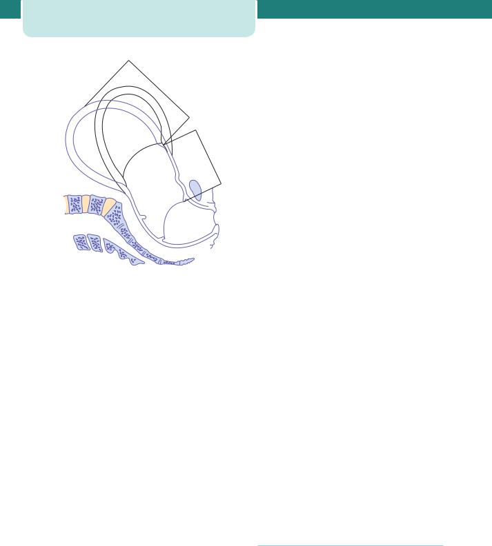
The pelvic brim is the inlet of the pelvis and is bounded in front by the symphysis pubis (the joint separating the two pubic bones), on each side by the upper margin of the pubic bone, the ileopectineal line and the ala of the sacrum, and posteriorly by the promontory of the sacrum (Figure 14.1). The normal transverse diameter in this plane is 13.5 cm and is wider than the anterior–posterior (AP) diameter,
|
Iliac crest |
|
Sacroiliac |
Ilium |
|
joint |
||
|
||
Sacrum |
Anterior |
|
|
superior |
|
|
iliac spine |
|
Coccyx |
Superior and |
|
|
||
Os pubis |
inferior rami |
|
|
of the pubic |
Obturator |
bone |
|
|
foramen |
|
Ischium |
Ischial tuberosity |
|
|
Pubic symphysis |
|
which is normally 11 cm (Figure 14.2). The angle of the inlet is normally 60º to the horizontal in the erect position, but in Afro-Caribbean women this angle may be as much as 90º (Figure 14.3). This increased angle may delay the head entering the pelvis during labour.
The pelvic mid-cavity
The pelvic mid-cavity can be described as an area bounded in front by the middle of the symphysis pubis,
Sacroiliac joint
13.5 cm
11.0 cm
Figure 14.2 The pelvic brim
|
|
|
|
|
60° |
|
|
|
|
|
|
|
|
AP |
|
|
|
AP |
|
|
|
of |
|
|
|
of |
|
|
inlet |
||
|
|
|
mid |
|
|
|
|
|
|
|
|
pelvis |
|
|
|
|
|
|
|
|
|
11 |
|
|
|
|
|
|
12. |
|
. |
|
|
|
|
|
|
cm |
|
AP |
|
|
|
|
|
|
0 |
of |
outlet |
|
0 |
cm |
|
||
|
|
|
|
||||
|
|
|
|
|
|||
|
|
|
|
|
|
||
|
|
|
13. |
|
|
|
|
|
|
|
|
5 |
cm |
|
|
|
|
|
|
|
|
|
|
Figure 14.1 The bony pelvis |
Figure 14.3 Sagittal section of the pelvis demonstrating the |
anterior–posterior (AP) diameters of the inlet and outlet |
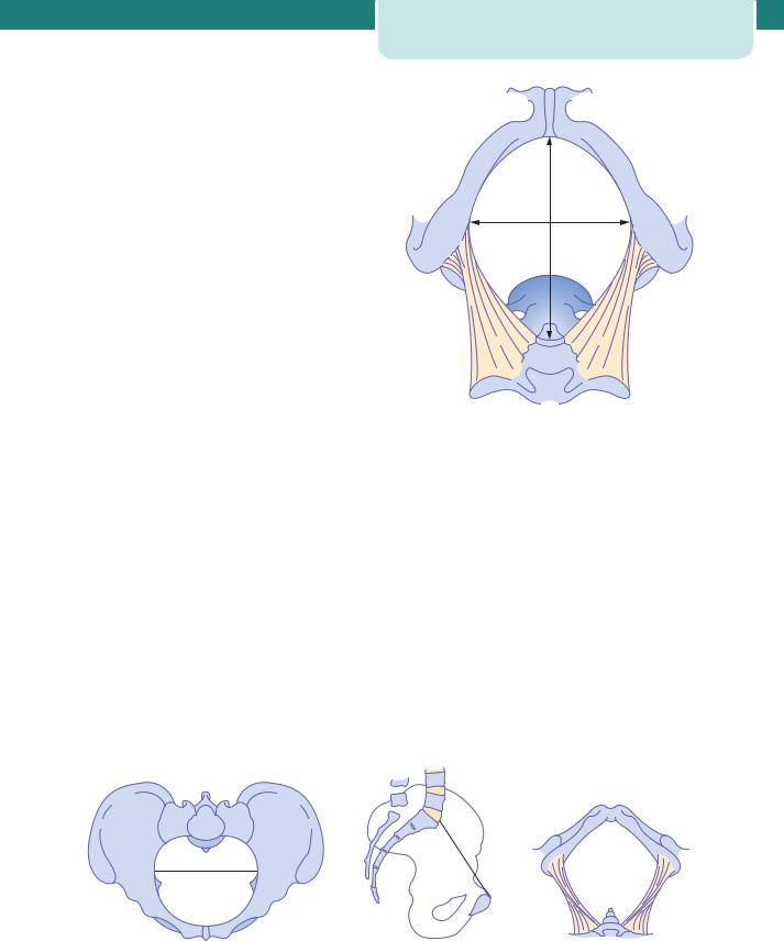
on each side by the pubic bone, the obturator fascia and the inner aspect of the ischial bone and spines, and posteriorly by the junction of the second and third sections of the sacrum. The cavity is almost round, as the transverse and anterior diameters are similar at 12 cm. The ischial spines are palpable vaginally and are used as landmarks to assess the descent of the head on vaginal examination (station). They are also used as landmarks for providing an anaesthetic block to the pudendal nerve. The pudendal nerve passes behind and below the ischial spine on each side. The pelvic axis describes an imaginary curved line, a path that the centre of the fetal head must take during its passage through the pelvis.
The pelvic outlet
The pelvic outlet is bounded in front by the lower margin of the symphysis pubis, on each side by the descending ramus of the pubic bone, the ischial tuberosity and the sacrotuberous ligament, and posteriorly by the last piece of the sacrum. The AP diameter of the pelvic outlet is 13.5 cm and the transverse diameter is 11 cm (Figure 14.4). Therefore, the transverse is the widest diameter at the inlet, but at the outlet it is the AP. Recognizing this is crucial to the understanding of the mechanism of labour.
The pelvic measurements given here are obviously average values and relate to bony points. Maternal stature, previous pelvic fractures and metabolic bone disease, such as rickets, may all be associated with measurements less than these population means. Furthermore, as the pelvic ligaments at the pubic ramus and the sacroiliac joints loosen towards the end of the third trimester, the pelvis often becomes more flexible and these diameters may increase during labour. It is now uncommon to perform x-rays or computed tomography (CT) scans of the pelvis to measure the pelvis because they have, on the whole,
Fetal and maternal anatomy relevant to labour |
187 |
13.5 cm
11.0 cm
Figure 14.4 The pelvic outlet
proven to be of minimal clinical use in predicting the outcome of labour.
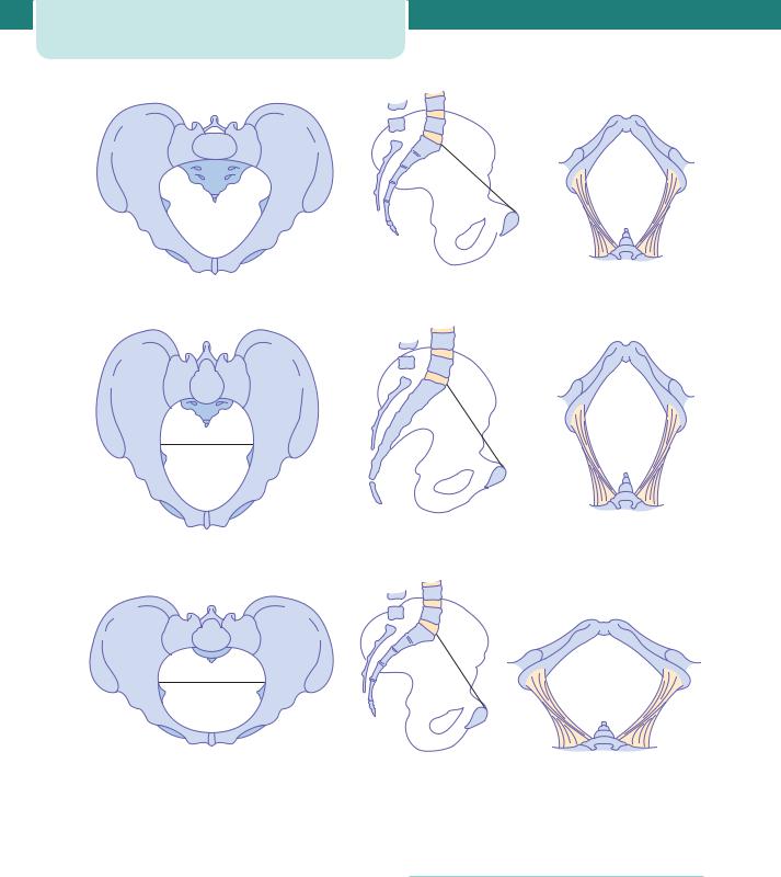
A variety of pelvic shapes has been described, and these may contribute to difficulties encountered in labour. The gynaecoid pelvis is the most favourable for labour, and the most common (Figure 14.5). Other pelvic shapes are shown in Figures 14.6 to 14.8. An android-type pelvis is said to predispose to deep transverse arrest (see Figure 14.22) and the anthropoid shape encourages an occipito-posterior (OP) position (see below). A platypelloid pelvis also is associated with an increased risk of obstructed labour.
The pelvic floor
This is formed by the two levator ani muscles which, with their fascia, form a musculofascial gutter during the second stage of labour (Figure 14.9).
(a) |
(b) |
(c) |
Figure 14.5 The gynaecoid pelvis: (a) brim, (b) lateral view, (c) outlet
188 Labour
(a) |
(b) |
(c) |
Figure 14.6 The android pelvis: (a) brim, (b) lateral view, (c) outlet
(a) |
(b) |
(c) |
Figure 14.7 The anthropoid pelvis: (a) brim, (b) lateral view, (c) outlet
(a) |
(b) |
(c) |
Figure 14.8 The platypelloid pelvis: (a) brim, (b) lateral view, (c) outlet
The perineum
The final obstacle to be negotiated by the fetus during labour is the perineum. The perineal body is a condensation of fibrous and muscular tissue lying between the vagina and the anus (Figure 14.10). It receives attachments of the posterior ends of the bulbo-cavernous muscles, the medial ends of the superficial and deep transverse perineal muscles, and the anterior fibres of the external anal sphincter. It is
always involved in a second-degree perineal tear and an episiotomy.
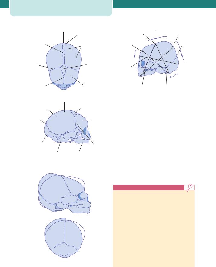
The fetal skull
The bones, sutures and fontanelles
The fetal skull is made up of the vault, the face and the base. The sutures are the lines formed where the individual bony plates of the skull meet one another.
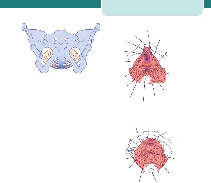
Figure 14.9 The musculofascial gutter of the levator sling
At the time of labour, the sutures joining the bones of the vault are soft, unossified membranes, whereas the sutures of the face and the skull base are firmly united (Figure 14.11).
The vault of the skull is formed by the parietal bones and parts of the occipital, frontal and temporal bones. Between these bones there are four membranous sutures: the sagittal, frontal, coronal and lambdoidal sutures.
Fontanelles are the junctions of the various sutures. The anterior fontanelle, or bregma (diamond shaped), is at the junction of the sagittal, frontal and coronal sutures. The posterior fontanelle (triangular shaped) lies at the junction of the sagittal suture and the lambdoidal sutures between the two parietal bones and the occipital bone. The fact that these sutures are not united is important for labour. It allows these bones to move together and even to overlap. The parietal bones usually tend to slide over the frontal and occipital bones. Furthermore, the bones themselves are compressible. Together, these characteristics of the fetal skull allow a process called ‘moulding’ to occur, which effectively reduces the diameters of the fetal skull and encourages progress through the bony pelvis, without harming the underlying brain (Figure 14.12). However, severe moulding can be a sign of cephalopelvic disproportion (see below under Cephalopelvic disproportion).
The area of the fetal skull bounded by the two parietal eminences and the anterior and posterior fontanelles is termed the ‘vertex’.
The diameters of the skull
The fetal head is ovoid in shape. The attitude of the fetal head refers to the degree of flexion and extension at the upper cervical spine. Different longitudinal diameters are presented to the pelvis in labour
Fetal and maternal anatomy relevant to labour |
189 |
|
|
Ischio-cavernosus |
|
Symphysis |
muscle |
|
pubis |
Bulbo-cavernosus |
|
muscle |
|
|
|
|
|
Clitoris |
Superficial |
|
|
|
|
|
transverse |
|
Urethra |
perineal muscle |
|
|
|
|
Urogenital |
|
|
diaphragm |
|
|
|
Pubo- |
|
Perineal |
coccygeus |
|
body |
muscle |
|
Anus |
Ilio-coccygeus |
|
|
|
|
|
muscle |
|
Coccygeus |
Gluteus maximus |
|
muscle |
muscle |
|
(a) |
Coccyx |
|
|
|
|
|
Symphysis |
|
Obturator |
pubis |
|
|
|
|
foramen |
|
|
Arcus |
Urethra |
|
|
|
|
tendineus |
|
|
|
Vagina |
|
Ischial |
Anal |
|
spine |
canal |
|
Pubo- |
|
|
coccygeus |
Coccygeus |
|
muscle |
|
|
muscle |
|
|
|
|
|
Ilio-coccygeus Coccyx
muscle
(b)
Figure 14.10 The perineum, perineal body and pelvic floor from below, showing superficial (a) and deeper (b) views. The pelvic floor muscles are made up of the levator ani (pubo-coccygeus and ilio-coccygeus)
depending on the attitude of the fetal head (Figures 14.13 and 14.14).
The longitudinal diameter that presents in a well-flexed fetal head (vertex presentation) is the suboccipito-bregmatic diameter. This is usually 9.5 cm and is measured from the suboccipital region to the centre of the anterior fontanelle (bregma). The longitudinal diameter that presents in a less wellflexed head, such as is found in the OP position, is
190 Labour
|
Posterior |
|
|
fontanelle |
Lambdoidal |
|
|
|
|
|
suture |
Sagittal |
|
Parietal bone |
suture |
|
and |
|
|
eminence |
Coronal |
|
Anterior |
suture |
|
fontanelle |
Frontal |
|
Frontal |
suture |
|
bone |
|
Vertex |
Anterior |
Parietal |
|
|
bone |
|
fontanelle |
Posterior
fontanelle Frontal bone
Occipital
bone
Coronal suture
Temporal Mandible bone
Figure 14.11 The fetal skull from superior and lateral views
Figure 14.12 A schematic representation of moulding of the fetal skull
Anterior
fontanelle
Bregma
Suboccipitofrontal diameter
Occipitofrontal diameter
S
|
|
t |
|
u |
|
|
ip |
|
|
c |
|
n |
|
|
i |
|
|
|
10 |
|
|
|
cm |
9 .
5 cm
V |
|
|
||
|
|
e |
|
|
|
|
rt |
|
|
|
|
|
e |
|
|
|
|
|
x |
13 |
cm |
|
|
|
|
|
|
|
|
11 |
cm |
|
||
|
|
|
||
Occipitomental diameter
Posterior fontanelle
|
|
O |
|
|
c |
|
|
c |
u |
p |
i |
|
||
t |
|
|
Submento-bregmatic |
Suboccipito- |
diameter 9.5 cm |
bregmatic diameter |
Figure 14.13 The diameters of the fetal skull
the suboccipito-frontal diameter, and is measured from the suboccipital region to the prominence of the forehead. It measures 10 cm.
With further extension of the head, the occipitofrontal diameter presents. This is measured from the root of the nose to the posterior fontanelle and is 11.5 cm. The greatest longitudinal diameter that may present is the mento-vertical, which is taken from the chin to the furthest point of the vertex and measures 13 cm. This is known as a brow presentation and it is usually too large to pass through the normal pelvis.
Extension of the fetal head beyond this point results in a smaller diameter. The submentobregmatic diameter is measured from below the chin to the anterior fontanelle and is 9.5 cm. This is clinically a face presentation.
Key points
The female pelvis and fetal skull
•The pelvic inlet is wider in the transverse than in the AP diameter.
•The pelvic outlet is wider in the AP than in the transverse diameter.
•Pelvic measurements may widen during labour due to pelvic ligament laxity.
•The soft tissues of the pelvic floor and perineum have a vital role to play in labour.
•Moulding may reduce the absolute measurements of the fetal skull during labour.
•The degree of flexion of the fetal skull at the cervical spine (the attitude) determines the diameter of the fetal skull presenting to the pelvis.
|
|
The process of labour |
191 |
|
Flexed |
|
|
Extended |
|
|
|
|
|
Attitude |
Well flexed |
Less well flexed |
Extended |
Hyperextended |
|
|
(partially extended) |
‘brow |
‘face |
|
|
or deflexed |
presentation’ |
presentation’ |
|
|
|
|
|
Diameter |
Suboccipito- |
Occipito-frontal |
Occipito-mental |
Submento- |
|
bregmatic |
|
|
bregmatic |
|
|
|
|
|
Measurement |
9.5 cm |
11.5 cm |
13.0 cm |
9.5 cm |
|
|
|
|
|
|
|
|
|
|
Figure 14.14 The effect of fetal attitude on the presenting diameter
The process of labour
The onset of labour
The onset of labour can be defined as regular contractions bringing about progressive cervical change. Therefore, a diagnosis of labour is usually made in retrospect. Loss of a ‘show’ (a blood-stained plug of mucus passed from the cervix) or spontaneous rupture of the membranes (SROM) does not define the onset of labour, although they may occur at the same time. Labour can be well established before either of these events occur, and both may precede labour by many days. Although much is understood about the physiology of labour in humans, the initiating biological event is still unclear (see below under Understanding the physiology of labour). It is certainly true however that the uterine body and cervix undergo a number of changes in preparation for labour which start a number of weeks before its onset.
anatomical criteria, and in certain situations this may be a disadvantage, as labour is essentially a physiological process. In normal labour, the division into three stages is of little clinical significance. The important events in normal labour are the diagnosis of labour and the maternal urge to push, which usually corresponds with full dilatation of the cervix and the baby’s head resting on the perineum. Defining the three stages of labour becomes more relevant if the labour does not progress normally. Because the definition of a normal labour can only be made retrospectively, there is difficulty in defining exactly when a normal labour becomes abnormal. Indeed, this definition will be different depending on the gestation, the previous obstetric record and the onset of labour. The average duration of first labours is approximately 8 hours, and that of subsequent labours 5 hours. First labours rarely last more than 18 hours, and second and subsequent labours not usually more than 12 hours.
The stages of labour are as follows.
The stages of labour |
First stage |
|
Labour can be divided into three stages. The definitions of these stages rely predominantly on
This describes the time from the diagnosis of labour to full dilatation of the cervix (10 cm) (Figure 14.15).
192 Labour
Upper uterine segment
Lower uterine segment
cervix
vagina
Figure 14.15 The thick upper segment and the thin lower segment of the uterus at the end of the first stage of labour. The dotted lines indicate the position assumed by the uterus during contraction
The first stage of labour can be divided into two phases. The ‘latent phase’ is the time between the onset of labour and 3–4 cm dilatation. During this time, the cervix becomes ‘fully effaced’. Effacement is a process by which the cervix shortens in length as it becomes included into the lower segment of the uterus. The process of effacement may begin during the weeks preceding the onset of labour, but will be complete by the end of the latent phase. The cervical os cannot usually begin to dilate until effacement is complete. Effacement and dilatation should be thought of as consecutive events in the nulliparous woman, but may occur simultaneously in the multiparous woman. Dilatation is expressed in centimetres between 0 and 10. The duration of the latent phase is variable, and time limits are arbitrary. However, it usually lasts between 3 and 8 hours, being shorter in multiparous women.
The second phase of the first stage of labour is called the ‘active phase’ and describes the time between the end of the latent phase (3–4 cm dilatation) and full dilatation (10 cm). It is also variable in length, usually lasting between 2 and 6 hours. Again, it is usually shorter in multiparous women. Cervical dilatation during the active phase usually occurs at 1 cm/hour or more in a normal labour (again, an arbitrary value),
but is only considered abnormal if it occurs at less than 2 cm/hour.
Second stage
This describes the time from full dilatation of the cervix to delivery of the fetus or fetuses. The second stage of labour may also be subdivided into two phases. The passive phase describes the time between full dilatation and the onset of involuntary expulsive contractions. There is no maternal urge to push and the fetal head is still relatively high in the pelvis. The second phase is rather confusingly called the ‘active second stage’. There is a maternal urge to push because the fetal head is low (often visible), causing a reflex need to ‘bear down’. In a normal labour, second stage is often diagnosed at this point because the maternal urge to push prompts the midwife to perform a vaginal examination. If a woman never reaches a point of involuntary pushing, the active second stage is said to begin when she starts making voluntary active efforts directed by her midwife. Conventionally, a normal active second stage should last no longer than 2 hours in a primiparous woman and 1 hour in those who have delivered vaginally before. Again, these definitions are largely arbitrary, but there is evidence that a second stage of labour lasting more than 3 hours is associated with increased maternal and fetal morbidity.
Use of epidural anaesthesia may influence the length and the management of the second stage of labour.
Third stage
This is the time from delivery of the fetus or fetuses until delivery of the placenta(e). The placenta is usually delivered within a few minutes of the birth of the baby. A third stage lasting more than 30 minutes should be considered abnormal, unless the woman has opted for ‘physiological management’ (see below under Third stage under Management of normal labour) in which case it is reasonable to extend this definition to 60 minutes.
The duration of labour
More than any other objective measurement, the duration of labour determines the impact of childbirth, particularly on mothers but also on babies, and also on those who care for both of them. The morale of most women starts to deteriorate

after 6 hours in labour, and after 12 hours the rate of deterioration significantly accelerates. There is a greater incidence of fetal hypoxia and need for operative delivery associated with longer labours. Shorter labours will also mean that personal attention for each woman in labour is a realistic possibility. An early artificial rupture of membranes (ARM) does shorten the length of labour, but does not necessarily alter the outcome.
It is difficult to define prolonged labour, but it would be reasonable to suggest that labour lasting longer than 12 hours in nulliparous women and 8 hours in multiparous women should be regarded as prolonged.
The mechanism of labour
This refers to the series of changes in position and attitude that the fetus undergoes during its passage through the birth canal. It is described here for the vertex presentation and the gynaecoid pelvis. The relation of the fetal head and body to the maternal pelvis changes as the fetus descends through the pelvis. This is essential so that the optimal diameters of the fetal skull are present at each stage of the descent.
Engagement
The head normally enters the pelvis in the transverse position or some minor variant of this, so taking advantage of the widest diameter. Engagement is said to have occurred when the widest part of the presenting part has passed successfully through the inlet. Engagement has occurred in the vast majority of nulliparous women prior to labour, but not so for the majority of multiparous women.
The number of fifths of the fetal head palpable abdominally is often used to describe whether engagement has taken place. If more than two-fifths of the fetal head is palpable abdominally, the head is not yet engaged.
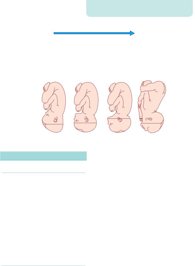
Descent
Descent of the fetal head is needed before flexion, internal rotation and extension can occur (Figure 14.16). During the first stage and first phase of the second stage of labour, descent of the fetus is secondary to uterine action. In the active phase of the second stage of labour, descent of the fetus is helped by voluntary use of abdominal musculature and the Valsalva manoeuvre (‘pushing’).
The process of labour |
193 |
Figure 14.16 Descent and flexion of the head followed by internal rotation and ending in birth of the head by extension
Flexion
The fetal head may not always be completely flexed when it enters the pelvis. As the head descends into the narrower mid-cavity, flexion should occur. This passive movement occurs, in part, due to the surrounding structures and is important in minimizing the presenting diameter of the fetal head.
Internal rotation
If the head is well flexed, the occiput will be the leading point and on reaching the sloping gutter of the levator ani muscles, it will be encouraged to rotate anteriorly so that the sagittal suture now lies in the AP diameter of the pelvic outlet (i.e. the widest diameter). If the fetus has engaged in the OP position, internal rotation can occur from an OP position to an occipito-anterior (OA) position. This long internal rotation may explain the increased duration of labour associated with this malposition. Alternatively, an OP position may persist, resulting in a ‘face to pubes’ delivery. More often, the persistent OP position is associated with extension of the fetal head and a resulting increase in the diameter presented to the pelvic outlet. This may lead to obstructed labour and the need for instrumental delivery or even Caesarean section.
Extension
Following completion of internal rotation, the occiput is underneath the symphysis pubis and the bregma is near the lower border of the sacrum. The soft tissues of the perineum still offer resistance, and may be traumatized in the process. The well-flexed head now extends and the occiput escapes from underneath the
194Labour
symphysis pubis and distends the vulva. This is known as ‘crowning’ of the head. The head extends further and the occiput underneath the symphysis pubis acts as a fulcrum point as the bregma, face and chin appear in succession over the posterior vaginal opening and perineal body. This extension and movement minimize soft-tissue trauma by utilizing the smallest diameters of the head for the birth.
Restitution
When the head is delivering, the occiput is directly anterior. As soon as it escapes from the vulva, the head aligns itself with the shoulders, which have entered the pelvis in the oblique position. The slight rotation of the occiput through one-eighth of the circle is called ‘restitution’.
External rotation
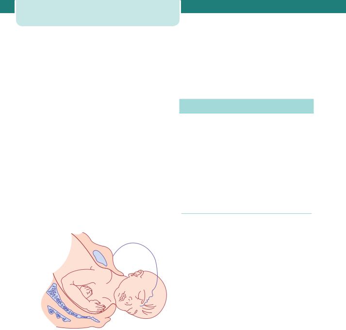
In order to be delivered, the shoulders have to rotate into the direct AP plane (remember the widest diameter at the outlet). When this occurs, the occiput rotates through a further one-eighth of a circle to the transverse position. This is called external rotation (Figure 14.17).
Figure 14.17 External rotation of the head after delivery as the anterior shoulder rotates forward to pass under the subpubic arch
Delivery of the shoulders and fetal body
When restitution and external rotation have occurred, the shoulders will be in the AP position. The anterior shoulder is under the symphysis pubis and delivers first, and the posterior shoulder delivers subsequently. Although this process may occur without assistance, lateral traction is often exerted by gently pulling the fetal head in a downward direction
to help release the anterior shoulder from beneath the pubic symphysis.
Normally the rest of the fetal body is delivered easily, with the posterior shoulder guided over the perineum by traction in the opposite direction, so sweeping the baby on to the maternal abdomen.
Understanding the physiology of labour
The mechanism responsible for initiating human parturition is still unknown and is somewhat different from that in all other animal models that have been studied. There are, however, certain processes that seem to be of particular importance.
The onset of labour occurs when those factors which inhibit contractions and maintain a closed cervix diminish and are succeeded by the actions of factors which do the opposite. Both mother and fetus make contributions toward this.
The myometrium
Myometrial cells contain filaments of actin and myosin, which interact and bring about contraction in response to an increase in intracellular calcium. Prostaglandins and oxytocin increase intracellular free calcium ions, whereas beta-adrenergic compounds and calcium-channel blockers do the opposite. Separation of the actin and myosin filaments brings about relaxation of the myocyte, however, unlike in any other muscle cell of the body, this actin– myosin interaction occurs along the full length of the filaments so that a degree of shortening occurs with each successive interaction. This progressive shortening of the uterine smooth muscle cells is called retraction and occurs in the cells of the upper part of the uterus. The result of this retraction process is the development of the thicker, actively contracting ‘upper segment’. At the same time, the lower segment of the uterus becomes thinner and more stretched. Eventually, this results in the cervix being taken up into the lower segment of the uterus so forming a continuum with the lower uterine segment (see Figure 14.15). The cervix effaces and then dilates, and the fetus descends in response to this directional force.
It is essential that the myocytes of the uterus contract together in a coordinated fashion. Individual myometrial cells are laid down in a mesh of collagen. There is cell-to-cell communication by

means of gap junctions, which facilitate the passage of various products of metabolism and electrical current between cells. These gap junctions are absent for most of the pregnancy but appear in significant numbers at term. Gap junctions increase in size and number with the progress of labour and allow greater coordination of myocyte activity. Prostaglandins stimulate their formation, while beta-adrenergic compounds possibly do the opposite. A uterine pacemaker from which contractions originate probably does exist but has not been demonstrated histologically.
Uterine contractions are involuntary in nature and there is relatively minimal extrauterine neuronal control. The frequency of contractions may vary during labour and with parity. Throughout the majority of labour, they occur at intervals of 2–4 minutes. Their duration also varies during labour, from 30 to 60 seconds, or occasionally longer. The intensity or amplitude of the intrauterine pressure generated with each contraction averages between 30 and 60 mmHg.
The cervix
The cervix contains muscle cells and fibroblasts separated by a ‘ground substance’ made up of extracellular matrix molecules. Interactions between collagen, fibronectin and dermatan sulphate (a proteoglycan) during the earlier stages of pregnancy keep the cervix rigid and closed. Contractions at this point do not bring about effacement or dilatation. Under the influence of prostaglandins, and other humoral mediators, there is an increase in proteolytic activity and reduction in collagen and elastin. Interleukins bring about a pro-inflammatory change with a significant invasion by neutrophils. Dermatan sulphate is replaced by the more hydrophilic hyaluronic acid, which results in an increase in water content of the cervix. This causes cervical softening or ‘ripening’, so that contractions, when they begin, can bring about the processes of effacement and dilatation.
Hormonal factors
Progesterone maintains uterine quiescence by suppressing prostaglandin production, inhibiting communication between myometrial cells and preventing oxytocin release. Oestrogen opposes the action of progesterone. Prior to labour, there is a reduction in progesterone receptors and an increase in the concentration of oestrogen relative to the
Place of birth |
195 |
progesterone. Prostaglandin synthesis by the chorion and the decidua is enhanced, leading to an increase in calcium influx into the myometrial cells. This change in the hormonal milieu also increases gap junction formation between individual myometrial cells, creating a functional syncytium, which is necessary for coordinated uterine activity. The production of corticotrophin-releasing hormone (CRH) by the placenta increases in concentration towards term and potentiates the action of prostaglandins and oxytocin on myometrial contractility. The fetal pituitary secretes oxytocin and the fetal adrenal gland produces cortisol, which stimulates the conversion of progesterone to oestrogen.
Which of these hormonal steps initiates labour is unclear. As labour becomes established, the output of oxytocin increases through the Fergusson reflex. Pressure from the fetal presenting part against the cervix is relayed via a reflex arc involving the spinal cord and results in increased oxytocin release from the maternal posterior pituitary.
Place of birth
The most recent recommendation from the National Institute for Health and Clinical Excellence (NICE) (see Clinical Guideline 55) is that women should be offered the choice of planning birth at home, in a midwife-led unit, or in an obstetric unit. Currently, less than 5 per cent of women deliver at home in most areas of the UK. Not all women have access to a midwifery-run unit. Some of these units are based within a hospital environment, and some are standalone. The evidence base guiding women on the outcomes of birth in the different settings is poor and full of bias. The chances of a normal birth at home, or in a midwifery unit, are higher than in an obstetric unit, but these women self-select and are more likely to be multiparous and without complicating factors. Use of epidural pain relief is restricted to consultant-led hospital labour wards, and this is known to increase the chances of delivery by forceps or ventouse. There is simply too little information to state conclusively where it is safest to give birth, from a maternal or fetal perspective. All women should be informed, however, that unexpected emergencies can occur in labour and that the outcome from these may be better in a hospital setting. It should also be made clear that the need for transfer into hospital, during labour, is