

Obstetrics_by_Ten_Teachers_19E_-_Kenny_Louise
.pdf296Neonatology
65 per cent of those in routine or manual occupations. Only 51 per cent of mothers aged less than 20 initiated breastfeeding. There is still a high drop-out rate, with a fifth giving up within a fortnight, citing painful nipples and the baby rejecting the breast as reasons.
Formula feeding
Modern formula milks are adjusted (humanized) so that the protein content and the whey:casein ratio are nearer those of human milk. Manufacturers do this by adding demineralized whey (from cheese production) and lactose, but differences in the fatty acid and amino acid composition remain, and formula milk cannot contain any of the anti-infective agents. There is no evidence to support the claim that formulae with a higher casein content are more ‘satisfying’ for ‘the hungry and demanding baby’. Additives are required to emulsify and thicken the milk. Water is required to reconstitute milk powder. Some products sold as ‘natural mineral water’ contain unacceptably high levels of sodium and nitrate for babies and are unsuitable for rehydrating dried formula milk.
Unmodified ‘doorstep’ cow’s milk, sheep’s and goat’s milk are completely unsuitable foods for babies less than a year old. The electrolyte composition is vastly different from that of human milk and they are highly allergenic. Soy formulae have no lactose, the carbohydrate being derived from corn syrup and sucrose. Soy protein is nutritionally inferior to human milk protein and infants grow less well on soy milk. The only reason to use soy formula is if the infant has a cow’s milk allergy or requires a lactose-free formula.
Care of the ill term newborn baby
A brief description of a few of the more common and serious illnesses that afflict term newborns follows. For more detail, consult standard texts.
Birth trauma
Birth trauma is fortunately rare in modern neonatal practice, but occasional cases are still encountered.
Brachial plexus palsy (Erb’s palsy)
Brachial plexus palsy is caused by damage to the brachial plexus, which does not always occur during
Figure 19.9 Brachial plexus palsy.
birth; cases have been described in babies born by Caesarean section. However, it is more common in large babies, particularly those whose delivery is complicated by shoulder dystocia. A brachial plexus lesion is revealed by lack of movement in the arm; initially the arm is flaccid. After 48 hours, an upper palsy can be distinguished from a complete palsy. In an upper root palsy (C5, C6, sometimes C7) the arm is internally rotated and pronated, there is no active abduction or elbow flexion (the waiter’s tip position; Figure 19.9). In a complete palsy of upper and lower roots, the arm is flail; there may be a ptosis and a Horner’s syndrome due to damage to the stellate ganglion adjacent to C8 and T1. Phrenic nerve palsy should be considered in these cases. While the prognosis of brachial plexus lesions is generally good, with most series reporting an initial recovery rate of 75–95 per cent, a recent study of the long-term effects revealed a surprisingly high incidence of later problems in childhood. The results of surgical nerve repair have improved markedly since the early days, and babies who have no recovery in biceps function by three months should be referred to a specialist.
Subgaleal (subaponeurotic) haemorrhage
The subaponeurotic space is potentially very large, lying as it does outside the skull and below the scalp. Babies who bleed into this space can become shocked, and there is a mortality of 20 per cent. The

condition is fortunately rare after normal vertex vaginal delivery, but is reported in as many as 6 per 1000 babies delivered by the ventouse. Increasing use of the ventouse apparatus means that early recognition of subgaleal haemorrhage has become more important. The clue to the diagnosis is a boggy swelling of the scalp that crosses suture lines. The baby’s head circumference will have increased at least 1 cm from the birth measurement if there is a sizeable subaponeurotic collection. When appropriately recognized and treated with blood transfusion, the long-term prognosis is good.
Transient tachypnoea of the newborn
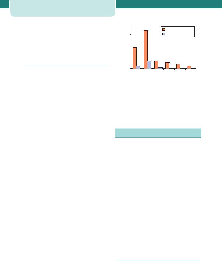
Transient tachypnoea of the newborn (TTN) is the most common respiratory disease of term infants, occurring in 4 per 1000. The disease is due to delayed clearance of lung liquid and is much more common after Caesarean section delivery, particularly without labour. At term, the incidence falls between 37 and 40 weeks (Figure 19.10), and this finding has implications for the timing of elective Caesarean section at term. Fortunately, the disease is usually mild, but sometimes requires intubation and ventilation, with the associated risk of complications.
Meconium aspiration syndrome
Meconium aspiration syndrome (MAS) is a disease of post-term pregnancies, with an incidence of about 1:1000 total births in Europe and 2–6 per 1000 in the United States. Meconium can be aspirated before or after birth. The coexistence of asphyxia is the main determining factor in MAS; asphyxia exerts its own detrimental effect on lung function and is associated with the development of persistent pulmonary hypertension, which complicates the treatment of MAS still further. These problems, together with a pre-existing aspiration of meconium into the airway that is not amenable to even the most aggressive suctioning at delivery, combine to make MAS a very serious neonatal illness.
The fact that some cases cannot be prevented by tracheal toilet should not discourage attempts at preventing meconium entering the airway at birth. Suctioning of the baby’s oropharynx when only the head has been delivered is no longer recommended, but the airways should be cleared of meconium after birth. Meconium in the airway creates a ball-valve effect in which air can be sucked past the obstruction
Care of the ill term newborn baby |
297 |
|
80 |
1000) |
60 |
(per |
|
morbidity |
40 |
Respiratory |
20 |
|
|
|
0 |
|
37 0.6 |
38 0.6 |
39 0.6 |
40 0.6 |
41 0.6 |
Weeks of gestation
Delivery by Cesarean section before labour Cesarean section during labour
Vaginal delivery
Figure 19.10 Respiratory morbidity (transient tachypnoea of the newborn plus respiratory distress syndrome) at term in infants admitted to the Neonatal Intensive Care Unit, Rosie Maternity Hospital, Cambridge, by each week of gestation and mode of delivery. (From Morrison JJ, Rennie JM,
Milton P, 1995. Neonatal respiratory morbidity and mode of delivery at term: influence of timing of elective caesarean section. British Journal of Obstetrics and Gynaecology 102: 101–6, with permission.)
but not exhaled past it, and the substance acts as a chemical irritant to the airways.
Persistent pulmonary hypertension
The term persistent pulmonary hypertension of the newborn (PPHN) is preferable to that of persistent fetal circulation, because the placenta is no longer in the circuit. In this disorder, the baby is cyanosed because there is a failure of the usual rapid postnatal fall in pulmonary vascular resistance. There is no parenchymal lung disease, but the pulmonary capillaries are structurally abnormal, possessing excess smooth muscle that persists into smaller branches than usual. PPHN can occur as a primary disorder or as a complication of asphyxia, infection (such as GBS) or pulmonary hypoplasia. The diagnosis should be suspected in a baby who remains hypoxic in 100 per cent oxygen and whose chest x-ray is normal. Echocardiography confirms the right-to-left shunt at
298Neonatology
atrial and/or ductal level and excludes congenital heart disease from the differential diagnosis. Nitric oxide has recently been confirmed as effective treatment for PPHN and is now the therapy of choice if warmth, artificial ventilation, oxygen and/or alkali therapy do not succeed in correcting the acidosis.
Hypoxic–ischaemic encephalopathy
Seizures are the hallmark of this condition, and hypoxic–ischaemic encephalopathy (HIE) is the most common cause of early-onset seizures in a term baby. There are many other causes of neonatal seizure, for example meningitis, stroke and hypoglycaemia. A diagnosis of HIE should be considered when there is a combination of:
•fetal distress;
•birth depression (low Apgar score requiring resuscitation);
•metabolic acidosis on cord pH or an early neonatal sample;
•seizures;
•renal impairment (blood in the urine and a low urine output);
•alteration of central nervous system state – the baby is not normally conscious between seizures,
but is irritable or lethargic with abnormal primitive reflexes.
The diagnosis should be supported by checking serum creatinine, calcium and glucose, performing a lumbar puncture to exclude meningitis, and carrying out a cranial ultrasound scan. This scan may be normal or show a loss of the gyral pattern with obliterated ventricles, suggesting cerebral oedema. Early electroencephalography (EEG) often confirms electrical seizure activity, and the background pattern can help in prognosis: a normal background, even in the presence of frequent seizures, is reassuring, whereas a very depressed or deteriorating background is an indication of a poor prognosis. An MRI scan, if available, is another investigation that confirms the diagnosis and helps in prognostication. Babies who are depressed at birth and who remain encephalopathic can be referred to a specialist centre for consideration of therapeutic hypothermia. Hypothermia has to be initiated within 6 hours to be effective, and is not yet a standard of care – results of several large trials are awaited.
|
50 |
45 |
|
|
Extremely preterm |
||
|
|
|
|
||||
|
40 |
|
|
|
Classmates |
|
|
(%) |
30 |
|
|
|
|
|
|
Children |
25 |
|
|
|
|
|
|
|
|
|
|
|
|
||
20 |
|
|
|
|
|
|
|
|
9 |
9 |
|
|
|
|
|
|
10 |
|
7 |
5 |
|
||
|
3 |
|
|
3 |
|||
|
|
|
1 |
|
|||
|
|
|
|
|
|||
|
0 |
|
|
|
|
|
|
|
|
|
|
|
|
|
|
One Small group |
PsychologistPhysio/occupational |
|
Outreath |
teacher |
|
-support |
support |
therapist |
|
and |
|
|
|
therapist |
|
||
-to |
|
Speech |
|
|
|
|
language |
|
|
|
|
|
|
|
|
|
|
Type of SEN provision (%)
Figure 19.11 Special educational needs of the EPICURE cohort (courtesy of Professor Neil Marlow). (From Johnson S et al. 2009. Archives of Disease in Childhood. Fetal and Neonatal edition 94: F283–9, with permission.)
Management of the preterm infant
The prognosis for preterm infants has improved dramatically over the last 30 years, with survival rates for infants delivered beyond 30 weeks now over 95 per cent. Neurological handicap, predominantly cerebral palsy, is a serious problem in 10 per cent of survivors below 30 weeks. Cognitive impairment or behavioural difficulties, such as attention deficit hyperactivity disorder, is more common; around 10 per cent have a DQ less than 55, and a further 30 per cent have less severe learning difficulties. Less than a third of the EPICURE cohort of children born below 26 weeks gestation in 1995 had an IQ above 85 at the age of 11. The children were particularly poor at mathematics, and of those in mainstream school 55 per cent had special educational needs (Figure 19.11).
The management of many of the complications of prematurity (Table 19.8) is beyond the scope of this chapter, and the reader is directed to standard textbooks. A few of the major conditions are discussed below.
Respiratory distress syndrome, chronic lung disease
The incidence of respiratory distress syndrome (RDS) is strongly related to gestational age, occurring
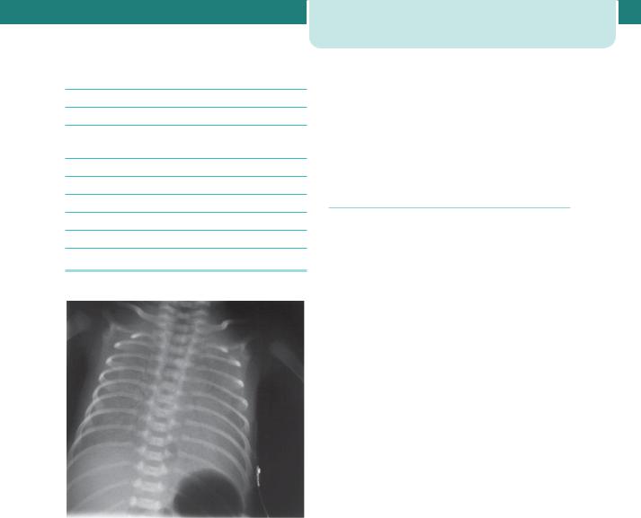
Table 19.8 Main problems of prematurity
Respiratory distress syndrome
Chronic lung disease
Intraventricular haemorrhage, parenchymal cerebral haemorrhage
Periventricular leukomalacia
Infection
Hypoglycaemia
Necrotizing enterocolitis
Patent ductus arteriosus
Jaundice
Figure 19.12 Chest x-ray in respiratory distress syndrome
in virtually 100 per cent of infants delivered at 26 weeks gestation, 40–50 per cent at 30–31 weeks and about 5 per cent at 35 weeks. RDS is a condition of increasing respiratory distress, commencing at, or shortly after, birth and increasing in severity until progressive resolution occurs among the survivors, usually between the 2nd and 4th day. It is due, at least in part, to insufficiency of pulmonary surfactant. RDS is manifest by respiratory distress (cyanosis, tachypnoea, grunting and recession), and respiratory failure is diagnosed by blood-gas analysis. Diagnosis can be confirmed by an x-ray film showing a groundglass appearance and air bronchograms, although these radiological features are not pathognomonic of RDS (Figure 19.12). Antenatal steroids and post-natal surfactant are beneficial. Artificial ventilation remains
Management of the preterm infant |
299 |
the mainstay of management, although the modern trend is for ‘gentle ventilation’, aiming to reduce barotrauma and minimize the risk of chronic lung disease. Chronic lung disease still afflicts as many as 50 per cent of babies weighing 1kg at birth, and these infants spend many months in oxygen, sometimes only to succumb later to winter viral infections or cor pulmonale.
Preterm brain injury
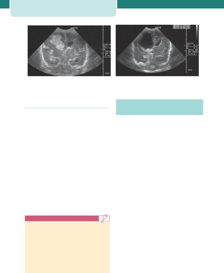
The neonatal brain is vulnerable to injury, and both intracranial parenchymal haemorrhage and periventricular leukomalacia (PVL) are associated with handicap in childhood. Intracranial haemorrhage is common in preterm infants, and occurs in the germinal matrix region. The germinal matrix is situated in the floor of the lateral ventricle. Bleeding into the germinal matrix often extends into the lateral ventricle of the brain (germinal matrix-intraventricular haemorrhage, GMH-IVH). GMH-IVH can resolve, but is sometimes complicated by persisting enlargement of the lateral ventricles or even progressive hydrocephalus. In these cases, the risk of handicap is more than 50 per cent. GMH-IVH can be diagnosed with ultrasound imaging via the fontanelle (Figure 19.13). Uncomplicated GMH-IVH – that is, bleeding not followed by ventricular dilatation or accompanied by a parenchymal lesion – carries a good prognosis. Only about 4 per cent of ex-preterm infants with no GMHIVH or an uncomplicated GMH-IVH will develop major neurodevelopmental sequelae. Ventricular enlargement is often a sign of periventricular myelin loss and brain shrinkage, rather than raised intracranial pressure hydrocephalus. Brain growth is an important differentiating feature. The presence of progressive hydrocephalus requiring treatment increases the risk of serious sequelae in preterm infants to about 75 per cent.
Bleeding into the substance of the brain is usually followed by breakdown of tissue into a porencephalic cyst (Figure 19.13). The outlook for infants with such a cyst can be surprisingly good, but many have a hemiplegia. ‘Periventricular leukomalacia’ is the term used to describe multiple small cysts that are visualized within the periventricular white matter. MRI scanning later in childhood shows a paucity of myelin in such cases, and the lesion is a very reliable predictor of later cerebral palsy. Cerebral palsy is almost universal in cases with bilateral occipital PVL. Factors that predispose to PVL include prolonged rupture of membranes, chorioamnionitis and neonatal hypocarbia.
300 Neonatology
(a) |
(b) |
Figure 19.13 Evolution of a right-sided parenchymal lesion (a) into a porencephalic cyst (b) seen on a coronal ultrasound scan. The time interval between the scans was two months
Necrotizing enterocolitis
Necrotizing enterocolitis is a serious gastrointestinal disease affecting 2–5 per cent of preterm infants. Babies who have reversed end-diastolic flow in the umbilical artery and who are growth restricted are at particular risk. The characteristic clinical presentation is of a preterm infant less than 7 days old in whom enteral formula feeding has been commenced. Feeding is accompanied by abdominal distension, increased volume of gastric aspirate, which may be bile-stained or blood-stained, and a tender abdomen. Abdominal x-ray may reveal the characteristic signs of intramural gas, a sentinel loop or even gas in the portal tract. Treatment involves omission of enteral feeds and surgery for perforation or failure to respond to medical management. Mortality is about 10–20 per cent and is highest in very preterm infants who develop necrotizing enterocolitis in the first week of life. Longterm complications include stoma requirement, short bowel syndrome and nocturnal diarrhoea.
Key points
•Ten per cent of all babies require admission to neonatal units, but only 1 per cent require full intensive care.
•All those who attend deliveries should be able to initiate resuscitation with a bag and mask system.
•Survival rates for babies born after 29 weeks gestation are now over 95 per cent and most will grow and develop normally, but the outcome for those born at less than
26 weeks gestation includes cognitive impairment sufficient to require special educational resource in over half.
Additional reading and website
resources
American Academy of Pediatrics and American College of Obstetricians and Gynecologists. Guidelines for perinatal care, 5th edn. Washington DC: AAP and ACOG, 2002.
Department of Health. Report of DH Expert Working Group on Neonatal Intensive Care Services. London: DOH, 2003.
Department of Health. National Service Framework for Children, Young People and Maternity Services. London: DOH, 2004.
Rennie JM (ed.). Roberton’s textbook of neonatology, 4th edn. London: Churchill Livingstone, 2004.
Rennie JM, Roberton NRC. A manual of neonatal intensive care, 4th edn. London: Arnold, 2002.
Royal College of Paediatrics and Child Health/Royal College of Obstetricians and Gynaecologists. Resuscitation of babies at birth. London: BMJ Publishing Group, 1997.
Website resources
AIDS/HIV treatment information: www.hivatis.org.
Baby Life Support charity (support for parents): www.bliss. org.uk.
British Association of Perinatal Medicine: www.bapm.org.
Center for Disease Control and Prevention (USA): www. cdc.gov.
Confidential Enquiry into Maternal and Child Health: www.cemach.org.uk.
Contact a Family (information on specific disorders): www.cafamily.org.uk.
Department of Health (UK Government): www.doh.gov.uk.
Group B streptococcus support: www.gbss.org.uk.
Health Protection Agency (public health, UK): www.phls. co.uk.

National Institutes of Health (US Government): www.nih.gov.
National Institute for Health and Clinical Excellence: www. nice.org.uk.
National Neonatal audit programme (UK): www.rcpch. ac.uk/nnap.
Newborn Physical Examination: http://nipe.screening.nhs.uk.
OMIM – genetic diagnosis search site: www.ncbi.nlm.nih. gov/Omim.
Additional reading and website resources |
301 |
Resuscitation Council (UK): www.resus.org.uk.
Statistics (UK Government): www.statistics.gov.uk.
Stillbirth and Neonatal Death Society: www.uk-sands.org.
Screening – UK national programme: www.nsc.nhs.uk; www.screening.nhs.uk.
UNICEF, world statistics: www.unicef.org.

|
|
|
|
|
|
|
|
|
|
|
|
|
|
|
|
C H A P T E R 2 0 |
E T H I C A L A N D M E D I C O L E G A L |
|
|
||||||||||
|
|
I S S U E S I N O B S T E T R I C P R A C T I C E |
||||||||||||
|
|
Philip N Baker |
|
|
|
|
|
|
|
|
|
|||
|
|
|
|
|
|
|
||||||||
|
Ethics in obstetric practice................................................................ |
302 |
|
Medicolegal issues................................................................................. |
305 |
|
||||||||
|
Consent.......................................................................................................... |
304 |
|
Additional reading ................................................................................... |
308 |
|
||||||||
|
|
|
|
|
|
|
|
|
|
|
|
|
|
|
O V E R V I E W
Obstetrics is a unique speciality, characterized by rapidly evolving clinical situations and full of ethical dilemmas. The importance of both ethical and medicolegal issues is arguably greater than in any other branch of medicine.
Ethics in obstetric practice
Declare the past, diagnose the present, foretell the future; practice these acts. As to disease, make a habit of two things – to help or at least do no harm.
Hippocrates
Obstetrics is full of ethical dilemmas; from genetic diagnosis to fetal therapy, there are issues of extreme complexity. Physicians need a sound knowledge of ethical principles, in order to ensure that their decisions are defensible to their peers, their patients and in a Court of Law. Obstetrics is unique in that the practitioner is often dealing with two patients, both inextricably linked, and whose interests usually but not invariably, coincide.
Obstetricians are bound by the duties of a doctor, as laid down by the General Medical Council, as for all other aspects of clinical practice. These principles of good practice bear repetition here:
•Make the care of your patient your first concern.
•Treat every patient politely and with consideration.
•Respect patients’ dignity and privacy.
•Listen to patients and respect their views.
•Give patients information in a way they can understand.
•Respect the right of patients to be fully involved in decisions about their care.
•Keep your professional knowledge and skills up to date.
•Recognize the limits of your professional competence.
•Be honest and trustworthy.
•Respect and protect confidential information.
•Make sure that your personal beliefs do not prejudice your patients’ care.
•Act quickly to protect patients from risk if you have good reason to believe that you or a
colleague may not be fit to practice.
•Avoid abusing your position as a doctor.
•Work with colleagues in the ways that best serve patients’ interests.
The principle of ‘beneficence’ is fundamental to the doctor–patient relationship. This requires the practitioner to objectively assess the different diagnostic and therapeutic options, and then to implement those that protect the interests of the patients by securing the greatest clinical benefits over harm. For centuries, this principle of beneficence guided clinical decisionmaking. However, over the past 100 years, it became increasingly apparent that beneficence alone was not enough; too often it led to paternalism, or the practitioner overriding a patient’s wishes. Beneficence must be balanced by a respect for ‘autonomy’, which accepts that patients have their own perspectives on health-related interests, and thus have the right to make decisions based on their values and beliefs. In most situations, beneficence-based and autonomybased obligations coincide. If there is conflict, patient autonomy should prevail, unless in the opinion of the practitioner, the course of action requested by

the patient offends the practitioner’s conscience. In such circumstances, the practitioner must refuse to accede to the patient’s request. Private conscience does not justify the practitioner being judgemental or denying transfer of a patient to a colleague whose conscience is not affected by the issue.
The care of pregnant women involves an additional ethical issue peculiar to obstetrics, namely the status of the fetus. The fetus is not a person and has no rights in law. It could thus be argued that the fetus does not have moral status, however, this concept is increasingly being challenged, especially after 24 weeks gestation when the fetus is independently viable, albeit with technical support. Bioethicists contend that the independently viable fetus should be afforded moral status, i.e. that both the practitioner and the pregnant woman have beneficence-based moral obligations to the fetus. This means that obstetricians and midwives should regard the viable fetus as their patient. On rare occasions, there are conflicts between the autonomybased decision of the mother regarding her viable fetus, and the professional judgement of the practitioner.
C A S E H I S T O R Y
A 32-year-old woman attended for a midtrimester detailed ultrasound scan in her third pregnancy (her previous pregnancies were uncomplicated and culminated in the vaginal delivery of term infants). The fetus was diagnosed as having a lumbosacral spina bifida. After extensive counselling, including advice from a paediatric neurosurgeon, the woman requested a termination of pregnancy. The obstetrician responsible for her care had strong religious beliefs that a termination of pregnancy was wrong.
Abortion is legal in the United Kingdom if two doctors decide in good faith that a particular pregnancy is associated with factors that satisfy one or more of five grounds specified in the Regulations of the Abortion Act and Section 37 of the Human Fertilisation and Embryology Act:
A.The continuance of the pregnancy would involve risk to the life of the pregnant woman greater than if the pregnancy were terminated.
B.The termination is necessary to prevent grave permanent injury to the physical or mental health of the pregnant woman.
C.The pregnancy has not exceeded its 24th week and the continuance of the pregnancy would involve risk, greater than if the pregnancy
Ethics in obstetric practice |
303 |
were terminated, of injury to the physical or mental health of the pregnant woman.
D.The pregnancy has not exceeded its 24th week and the continuance of the pregnancy would involve risk, greater than if the pregnancy were terminated, of injury to the physical or mental health of the existing child(ren) of the family of the pregnant woman.
E.There is a substantial risk that if the child were born it would suffer from such physical or mental abnormalities as to be seriously handicapped.
The Regulations also permit abortion to be performed in an emergency on the basis of the signature of the doctor performing the procedure, which may be provided up to 24 hours after the termination. The emergency grounds are:
F.To save the life of the pregnant woman.
G.To prevent grave permanent injury to the physical or mental health of the pregnant woman.
Spina bifida is associated with a high risk of infant death or serious handicap. Although most abortions are undertaken on grounds C or D, the woman had the right to ask for termination of pregnancy under Section E of the Abortion Act (only 1 per cent of terminations are performed under these grounds). Furthermore, as the fetus was less than 24 weeks gestation, it was considered pre-viable and did not have the moral status of being a patient unless the woman conferred that status, something she was free to withhold. A decision to terminate the pregnancy was justified in respect of her autonomy-based decision.
The obstetrician declined to carry out the procedure as a matter of personal conscience. However, the woman was referred to another practitioner who had no objection to performing the termination of pregnancy.
C A S E H I S T O R Y
A 23-year-old woman in her second pregnancy underwent an ultrasound scan at 34 weeks gestation in order to assess fetal size; her first pregnancy had been complicated by fetal growth restriction. Ultrasound biometry was within the normal range, however, the fetus was found to have the ‘double bubble’ sign of duodenal atresia. A fetal blood sample was obtained by cordocentesis (see Chapter 7, Prenatal diagnosis) and rapid karyotyping revealed that the fetus had trisomy 21. Both the woman and her partner requested a termination of pregnancy.

304 Ethical and medicolegal issues in obstetric practice
A fetus at 34 weeks gestation has no legal rights. However, the fetus is viable and has therefore acquired the moral status of being a fetal patient. Duodenal atresia, a rare condition which occurs in about one in 10 000 births, is usually cured by surgery; onethird of children with duodenal atresia have Down’s syndrome. Although Down’s syndrome is associated with a low IQ, the child usually has a normal but dependent life.
Under Section E of the Abortion Act, there is no definition of ‘serious abnormality’. To some obstetricians, this case would fit the description of ‘serious abnormality’; they would recommend that a termination of pregnancy would be permitted under the Abortion Act and justified on the basis of maternal autonomy. However, Down’s syndrome does not necessarily result in a life not worth living, and on ethical grounds, other practitioners would find it difficult to agree to a termination of pregnancy. Beneficence-based obligations to the fetus would not justify causing its death. On this basis, it would be ethically reasonable to deny maternal autonomy in this case.
The obstetricians responsible for the care of this case declined the woman’s request for a termination of pregnancy. The pregnancy proceeded to a term delivery of a male infant with Down’s syndrome and with duodenal atresia, but no other apparent abnormalities.
Consent
An adult patient who suffers from no mental incapacity has an absolute right to choose whether to consent to medical treatment, to refuse it or to choose one rather than another of the treatments being offered. . . . This right of choice
is not limited to decisions which others might regard as sensible. It exists not withstanding that the reasons for making the choice are rational, irrational, unknown or even non-existent.
Lord Donaldson in the Court of Appeal
The law requires that the practitioner must obtain valid consent from the patient; all patients undergoing treatment should be given appropriate information on the nature and purpose of the treatment. Patients must be both capable and competent. The emphasis should be on consent as a process, not merely obtaining the patient’s signature on a consent form. All too often clinicians equate consent with the signing of a form or consider consent primarily as protection
against litigation. The signatures on a form are not a substitute for a proper discussion of the proposed intervention and engaging the patient in decisionmaking about her own care.
The law of consent is complex and is evolving in favour of providing patients with comprehensive information about the benefits and risk of treatments and procedures, including alternative management and the consequences of no treatment. Particular care must be taken when the benefits and risk of a treatment are unclear.
C A S E H I S T O R Y
A 34-year-old nulliparous women was admitted at 39 weeks gestation after the community midwife recorded that her blood pressure was 144/94 mmHg in the presence of
proteinuria. The fetal lie was longitudinal with a cephalic presenting part noted to be 4/5th palpable on abdominal examination. On vaginal examination, the cervix was found to be long and closed. When a cardiotocography (CTG) was
commenced, the baseline fetal heart rate was normal; however, variability was reduced and there were unprovoked decelerations. The attending obstetric registrar made a diagnosis of preeclampsia (see Chapter 10, Pre-eclampsia and other disorders of placentation) and recommended Caesarean section on the basis of the pathological fetal heart rate allied to a cervix which was unfavourable for induction of labour. However, the woman and her partner refused to accept this advice. The obstetrician had to determine whether the woman was competent to give consent, i.e. that she was able to:
•comprehend and retain the treatment information;
•weigh that information in the balance to arrive at a choice.
A small minority of the population lack the necessary mental capacity to give consent, due to mental illness or retarded development. However, if she was deemed to be competent and the obstetrician proceeded with the Caesarean section, the operation would be performed against the express wishes of the woman, with the possible accusation that an assault on her body was being carried out. A competent adult has the right to refuse treatment, even if others, including health carers, believe that the refusal is neither in his or her best interests.
In this case, the solution was to persuade the prospective parents of the wisdom of performing the Caesarean section for the safety of the woman

and her unborn child. In addition to benefits for the baby, delivery by Caesarean section will reduce the likelihood of maternal complications from preeclampsia. Persuasion should not be strident or threatening, but respectful and carefully reasoned.
Key points
•Valid consent requires the practitioner to give the patient detailed information about any proposed procedure.
•Any conflicts between medical advice and the patient’s wishes should be carefully documented.
•In difficult cases, advice from health care and legal colleagues should be sought.
Medicolegal issues
The nature of obstetrics is characterized by rapidly evolving clinical situations; risks to the mother and fetus are considerable and the potential for misjudgement or mismanagement by the attending doctors and midwives is ever present. Sometimes, patients suffer harm, physical or psychological, from care that was intended to heal them. In some cases, this is due to human error or to defects in the organization and delivery of care. In other cases, the harm is attributable to substandard care associated with technical incompetence, poor decision-making or departure from accepted clinical practice. Whatever the underlying cause, litigation may follow.
Some mistakes in clinical practice are obvious – such as leaving a swab inside the abdomen at Caesarean section. In contrast, most medicolegal issues are much more contentious. The most significant claims arise from brain damage and cerebral palsy; these are based on the allegation that negligent management resulted in fetal asphyxia and this resulted in brain damage.
As with all medicolegal cases, for the claimant to be successful it needs to be established that:
•There was a breach of duty. This means that the attending staff owed the patient a duty of care and
that the standard of care afforded to the patient was below a standard which she could reasonably have expected. To determine this, the court relies on the evidence of expert witnesses. In turn, expert witnesses will take account of national and local evidence-based guidelines and conventional practice when advising on the standard of care
Medicolegal issues |
305 |
provided. The courts will apply the principle which states that a doctor is not negligent if he/she acts in accordance with accepted medical practice at the time, even though there may be doctors who hold a contrary opinion (the Bolam test), but the court must be satisfied that exponents of that practice could demonstrate that their opinion had a logical basis (the Bolitho test).
•There was causation, i.e. that the injury sustained was caused by the substandard care. A breach of
duty, while regrettable and unacceptable, will not in itself be enough to establish a case of clinical negligence. The claimant has to show that the breach caused an injury; in other words, it must be shown that but for the breach of duty the injury would not have occurred (or would not have been as severe). If causation is established, the court will grant compensation for losses which the claimant has suffered as a result of the injury, provided that such losses are recognized by the court as deserving of compensation. The compensation comprises a sum for the ‘pain, suffering and loss of amenity’ caused by the injury and another sum covering the financial losses and extra expenses caused by the injury.
The litigation pathway
Obstetricians working under a contract of employment with the National Health Service (NHS) in the United Kingdom, unlike those working in the private sector or in countries like the United States, cannot be sued in their personal capacity. This is because they are indemnified by their employer for any alleged negligence in the course of their employment. This indemnity has implications for pattern of care because clinicians working under the fear of litigation are often accused of practising ‘defensive medicine’ – that is, practising an interventionist style of medicine in a bid to avert litigation.
In the United Kingdom, claims against the NHS are handled by the NHS Litigation Authority (NHSLA). Between 2001 and 2007, the NHSLA received 5691 new obstetric claims and the total amount paid out on obstetric claims was £1592 million. Apart from handling claims, the NHSLA has a statutory duty to help improve the quality of patient care by assisting NHS bodies with risk management. It does this largely through the Clinical Negligence Scheme for Trusts (CNST). This scheme, funded by member trusts, provides an indemnity to members