

Obstetrics_by_Ten_Teachers_19E_-_Kenny_Louise
.pdf86 Antenatal obstetric complications
Gastrointestinal symptoms
Constipation
Constipation is common in pregnancy and usually results from a combination of hormonal and mechanical factors that slow gut motility. Concomitantly administered iron tablets may exacerbate the condition. Women should be given clear explanations, reassurance and advice regarding the adoption of a high-fibre diet. Medications are best avoided but if necessary, mild (non-stimulant) laxatives, such as lactulose, may be suggested.
Hyperemesis gravidarum
Nausea and vomiting in pregnancy are extremely common; 70–80 per cent of women experience these symptoms early in their pregnancy and approximately 35 per cent of all pregnant patients are absent from work on at least one occasion through nausea and vomiting. Although the symptoms are often most pronounced in the first trimester, they by no means are confined to it. Similarly, despite common usage of the term ‘morning sickness’, in only a minority of cases are the symptoms solely confined to the morning. Nausea and vomiting in pregnancy tends to be mild and self-limited and is not associated with adverse pregnancy outcome.
Hyperemesis gravidarum, however, is a severe, intractable form of nausea and vomiting that affects 0.3–2.0 per cent of pregnancies. It causes imbalances of fluid and electrolytes, disturbs nutritional intake and metabolism, causes physical and psychological debilitation and is associated with adverse pregnancy outcome, including an increased risk of preterm birth and low birthweight babies. The aetiology is unknown and various putative mechanisms have been proposed including an association with high levels of serum human chorionic gonadotrophin (hCG), oestrogen and thyroxine. The likely cause is multifactorial. Severe cases of hyperemesis gravidarum cause malnutrition and vitamin deficiencies, including Wernicke’s encephalopathy, and intractable retching predisposes to oesophageal trauma and Mallory– Weiss tears. Treatment includes fluid replacement and thiamine supplementation. Antiemetics such as phenothiazines are safe and are commonly prescribed. Other proposed treatments including the administration of corticosteroids have not yet been adequately proven and remain empirical.
Gastroesophageal reflux
This is very common. Altered structure and function of the normal physiological barriers to reflux, namely the weight effect of the pregnant uterus and hormonally induced relaxation of the oesophageal sphincter, explain the extremely high incidence in the pregnant population. For the majority of patients, lifestyle modifications such as smoking cessation, frequent light meals and lying with the head propped up at night are helpful. When these prove insufficient to control symptoms, medications can be added in a stepwise fashion starting with simple antacids. Histamine-2 receptor antagonists and proton pump inhibitors can be used if more simple measures fail although their safety record in pregnancy is less certain. Severe, refractory dyspeptic symptoms warrant gastroenterology referral just in case a stomach ulcer or hiatus hernia is being overlooked.
Haemorrhoids
Several factors conspire to render haemorrhoids more common during pregnancy including the effects of circulating progesterone on the vasculature, pressure on the superior rectal veins by the gravid uterus and increased circulating volume. A conservative approach is usually advocated including local anaesthetic/antiirritant creams and a high-fibre diet. Never overlook the ‘warning’ symptoms of tenesmus, mucus, blood mixed with stool and back passage discomfort that may suggest rectal carcinoma; a rectal digital examination should be carried out if these symptoms are suggested.
Varicose veins
Varicose veins may appear for the first time in pregnancy or pre-existing veins may become worse. They are thought to be due to the relaxant effect of progesterone on vascular smooth muscle and the dependent venous stasis caused by the weight of the pregnant uterus on the inferior vena cava.
Varicose veins of the legs may be symptomatically improved with support stockings, avoidance of standing for prolonged periods and simple analgesia. Thrombophlebitis may occur in a large varicose vein, more commonly after delivery. A large superficial varicose vein may bleed profusely if traumatized; the leg must be elevated and direct pressure applied. Vulval and vaginal varicosities are uncommon but

symptomatically troublesome; trauma at the time of delivery (episiotomy, tear, instrumental delivery) may also cause considerable bleeding.
Oedema
This is common, occurring to some degree in approximately 80 per cent of all pregnancies. There is generalized soft-tissue swelling and increased capillary permeability, which allows intravascular fluid to leak into the extravascular compartment. The fingers, toes and ankles are usually worst affected and the symptoms are aggravated by hot weather. Oedema is best dealt with by frequent periods of rest with leg elevation; occasionally, support stockings are indicated. Excessively swollen fingers may necessitate removal of rings and jewellery before they get stuck. It is important to remember that generalized (rather than lower limb) oedema may be a feature of preeclampsia, so remember to check the woman’s blood pressure and urine for protein. More rarely, severe oedema may suggest underlying cardiac impairment or nephrotic syndrome.
Other common ‘minor’ disorders:
•Itching
•Urinary incontinence
•Nose bleeds
•Thrush (vaginal candidiasis)
•Headache
•Fainting
•Breast soreness
•Tiredness
•Altered taste sensation
•Insomnia
•Leg cramps
•Striae gravidarum and chloasma.
Problems due to abnormalities of the
pelvic organs
Fibroids (leiomyomata)
Fibroids are compact masses of smooth muscle that lie in the cavity of the uterus (submucous), within the uterine muscle (intramural) or on the outside
Problems due to abnormalities of the |
87 |
pelvic organs |
|
surface of the uterus (subserous). They may enlarge in pregnancy, and in so doing present problems later on in pregnancy or at delivery (Figure 8.1). A large fibroid at the cervix or in the lower uterine segment may prevent descent of the presenting part and obstruct vaginal delivery.
Red degeneration is one of the most common complications of fibroids in pregnancy. As it grows, the fibroid may become ischaemic; this manifests clinically as acute pain, tenderness over the fibroid and frequent vomiting. If these symptoms are severe, uterine contractions may be precipitated, causing miscarriage or preterm labour. Red fibroid degeneration requires treatment in hospital, with potent analgesics (usually opiates and intravenous fluids). The symptoms usually settle within a few days. The differential diagnosis of red degeneration includes acute appendicitis, pyelonephritis/urinary tract infection, ovarian cyst accident and placental abruption.
A subserous pedunculated fibroid may tort in the same way that a large ovarian cyst can. When this happens, acute abdominal pain and tenderness may make the two difficult to distinguish from one another. In this scenario, a pertinent history followed by an ultrasound scan (transvaginal in the first trimester, transabdominal in the second and third) will aid the diagnosis.
Figure 8.1 Fibroids complicating pregnancy. The tumour in the anterior wall of the uterus has been drawn up out of the pelvis as the lower segment was formed, but the
fibroid arising from the cervix remains in the pelvis and will obstruct labour

88 Antenatal obstetric complications
Retroversion of the uterus
Fifteen per cent of women have a retroverted uterus. In pregnancy, the uterus grows and a retroverted uterus will normally ‘flip’ out of the pelvis and begin to fill the abdominal cavity, as an anteverted uterus would. In a small proportion of cases, the uterus remains in retroversion and eventually fills up the entire pelvic cavity; as it does so, the base of the bladder and the urethra are stretched. Retention of urine may occur, classically at 12–14 weeks, and this is not only very painful but may also cause long-term bladder damage if the bladder becomes over-distended. In this situation, catheterization is essential until the position of the uterus has changed.
Congenital uterine anomalies
The shape of the uterus is embryologically determined by the fusion of the Mullerian ducts. Abnormalities of fusion may give rise to anything from a subseptate uterus through to a bicornuate uterus and even (very rarely) to a double uterus with two cervices. These findings are often discovered incidentally at the time of a pelvic operation such as a laparoscopy, or an ultrasound scan.
The problems associated with bicornuate uterus are:
•miscarriage;
•preterm labour;
•preterm prelabour rupture of membranes (PPROM);
•abnormalities of lie and presentation;
•higher Caesarean section rate.
Ovarian cysts in pregnancy
Ovarian cysts are common in pregnant women; fortunately, the incidence of malignancy is uncommon in women of childbearing age. The most common types of pathological ovarian cyst are serous cysts and benign teratomas. Physiological cysts of the corpus luteum may grow to several centimetres but rarely require treatment, therefore asymptomatic cysts may be followed up by clinical and ultrasound examination, but large cysts (for example, dermoids) may require surgery in pregnancy.
Surgery is usually postponed until the late second or early third trimester, when there is the potential that if the baby were delivered, it would be able to survive.
The major problems are of large ( 8 cm) ovarian cysts in pregnancy, which may undergo torsion, haemorrhage or rupture, causing acute abdominal pain. The resulting pain and inflammation may lead to a miscarriage or preterm labour. Symptomatic cysts, most commonly due to torsion, will require an emergency laparotomy and ovarian cystectomy or even oophorectomy if the cyst is torted. A full assessment must include a family history of ovarian or breast malignancy, tumour markers (although these are of limited value in pregnancy) and detailed ultrasound investigation of both ovaries. Surgery in the late second and third trimester of pregnancy is normally performed through a midline or paramedian incision; a low transverse suprapubic incision would not allow access to the ovary, as it is drawn upwards in later pregnancy.
Cervical cancer
Cervical abnormalities are much more difficult to deal with in pregnancy, partly because the cervix itself is more difficult to visualize at colposcopy, and also because biopsy may cause considerable bleeding. Cervical carcinoma most commonly arises in poor attenders for cervical screening. The disease is commonly asymptomatic in early stages, but later stage presentation includes vaginal bleeding (especially postcoital). Examination may reveal a friable or ulcerated lesion with bleeding and purulent discharge. The prospect of cervical carcinoma in pregnancy leads to complex ethical and moral dilemmas concerning whether the pregnancy must be terminated (depending on the stage it has reached) to facilitate either surgical treatment (radical hysterectomy) or chemotherapy. Cervical cancer is dealt with in greater detail in Chapter 14, premalignant and malignant disease of the cervix in Gynaecology by Ten Teachers, 19th edition.
Urinary tract infection
Urinary tract infections (UTIs) are common in pregnancy. Eight per cent of women have asymptomatic bacteriuria; if this is untreated, it may progress to UTI or even pyelonephritis, with the attendant associations of low birth weight and preterm delivery.
The predisposing factors are:
•history of recurrent cystitis;
•renal tract abnormalities: duplex system, scarred kidneys, ureteric damage and stones;

•diabetes;
•bladder emptying problems, for example multiple sclerosis.
The symptoms of UTI may be different in pregnancy; it occasionally presents as low back pain and general malaise with flu-like symptoms. The classic presentation of frequency, dysuria and haematuria is not often seen. On examination, tachycardia, pyrexia, dehydration and loin tenderness may be present. Investigations should include a full blood count and midstream specimen of urine (MSU) sent for urgent microscopy, culture and sensitivities. If there is a strong clinical suspicion of UTI, treatment with antibiotics should start immediately. The woman should drink plenty of clear fluids and take a simple analgesic, such as paracetamol.
The most common organism for UTI is Escherichia coli; less commonly implicated are streptococci, Proteus, Pseudomonas and Klebsiella. If more than 105 organisms are present at culture, this confirms a diagnosis of UTI. The commonly reported ‘heavy mixed growth’ is often associated with UTI symptoms and may be treated, or the MSU repeated after a week, depending on the clinical scenario. The first-line antibiotic for UTI is amoxycillin or oral cephalosporins.
Pyelonephritis is characterized by dehydration, a very high temperature ( 38.5ºC), systemic disturbance and occasionally shock. This requires urgent and aggressive treatment including intravenous fluids, opiate analgesia and intravenous antibiotics (such as cephalosporins or gentamicin). In addition, renal function should be determined, with at least baseline urea and electrolytes, and the baby must be monitored with cardiotocography (CTG). Recurrent UTIs in pregnancy require MSU specimens to be sent to the microbiology laboratory at each antenatal visit, and low-dose prophylactic oral antibiotics may be prescribed. Investigation should take place after delivery, unless frank haematuria or other symptoms suggest that an urgent diagnosis is essential. Investigations might include a renal ultrasound scan, renal DMSA function scan, creatinine clearance, intravenous urogram and cystoscopy.
Abdominal pain in pregnancy
Abdominal pain is one of the most common minor disorders of pregnancy; the problem is in
Abdominal pain in pregnancy |
89 |
distinguishing pathological from ‘physiological’ pains. There are many possibilities to exclude. Furthermore, the anatomical and physiological changes of pregnancy may alter ‘classical’ presenting symptoms and signs making clinical diagnosis challenging. The causes listed in Table 8.1 are not exhaustive, but cover most possible diagnoses. The crucial point is that certain conditions are potentially
Table 8.1 Causes of abdominal pain in pregnancy
Pregnancy-caused (obstetric) conditions
Early pregnancy ( 24 weeks)
Ligament stretching Miscarriage Ectopic pregnancy
Acute urinary retention due to retroverted gravid uterus
Later pregnancy ( 24 weeks)
Labour
Placental abruption
HELLP syndrome
Uterine rupture
Chorioamnioitis
Pregnancy-unrelated conditions
Uterine/ovarian causes
Torsion or degeneration of fibroid Ovarian cyst accident
Urinary tract disorders
Urinary tract infection (acute cystitis and acute pyelonephritis)
Renal colic
Gastrointestinal disorders
Medical gastric/duodenal ulcer Acute appendicitis
Acute pancreatitis Acute gastroenteritis
Intestinal obstruction or perforation
Medical causes
Sickle cell disease (abdominal crisis) Diabetic ketoacidosis
Acute intermittent porphyria Pneumonia (especially lower lobe) Pulmonary embolus
Malaria

90Antenatal obstetric complications
dangerous or debilitating (e.g. acute appendicitis) and may be masked by the altered anatomy and physiology of pregnancy. Obstetricians may therefore have to perform x-rays and arrange invasive assessments to make a diagnosis.
Venous thromboembolism
Venous thromboembolic disease (VTE) is the most common cause of direct maternal death in the UK. In the most recent triennium, there were 41 fatalities, giving a maternal mortality rate of 1.94 per 100 000 – more than twice that of the next most common cause (pre-eclampsia).
Pregnancy is a hypercoagulable state because of an alteration in the thrombotic and fibrinolytic systems. There is an increase in clotting factors VIII, IX, X and fibrinogen levels, and a reduction in protein S and anti-thrombin (AT) III concentrations (see Chapter 3, Physiological changes in pregnancy). The net result of these changes is thought to be an evolutionary response to reduce the likelihood of haemorrhage following delivery.
These physiological changes predispose a woman to thromboembolism and this is further exacerbated by venous stasis in the lower limbs due to the weight of the gravid uterus placing pressure on the inferior vena cava in late pregnancy and immobility, particularly in the puerperium.
Pregnancy is associated with a 6–10-fold increase in the risk of venous thromboembolic disease compared to the non-pregnant situation. Without thromboprophylaxis, the incidence of non-fatal pulmonary embolism (PE) and deep vein thrombosis (DVT) in pregnancy is about 0.1 per cent in developed countries, this increases following delivery to around 1–2 per cent and is further increased following emergency Caesarean section.
Thrombophilia
Some women are predisposed to thrombosis through changes in the coagulation/fibrinolytic system that may be inherited or acquired. There is growing evidence that both heritable and acquired thrombophilias are associated with a range of adverse pregnancy outcomes particularly recurrent fetal loss. The major hereditary forms of thrombophilia
Risk factors for thromboembolic disease
Pre-existing
•Maternal age ( 35 years)
•Thrombophilia
•Obesity ( 80 kg)
•Previous thromboembolism
•Severe varicose veins
•Smoking
•Malignancy
Specific to pregnancy
•Multiple gestation
•Pre-eclampsia
•Grand multiparity
•Caesarean section, especially if emergency
•Damage to the pelvic veins
•Sepsis
•Prolonged bed rest
currently recognized include: deficiencies of the endogenous anticoagulants protein C, protein S and AT III; abnormalities of procoagulant factors, factor V Leiden (caused by a mutation in the factor V gene) and the prothrombin mutation G20210A. It seems probable that there are still some thrombophilias not yet discovered or described. Heritable thrombophilias are present in at least 15 per cent of Western populations.
Acquired thrombophilia is most commonly associated with antiphospholipid syndrome (APS). APS is the combination of lupus anticoagulant with or without anti-cardiolipin antibodies, with a history of recurrent miscarriage and/or thrombosis. It may (or, more commonly, may not) be associated with other autoantibody disorders, such as systemic lupus erythematosus (SLE).
If thrombophilic disorders are taken together, more than 50 per cent of women with pregnancyrelated VTE will have a thrombophilia. It is therefore vital that women with a history of thrombotic events are screened for thrombophilia. The presence of thrombophilia, with a history of thrombotic episode(s), means that prophylaxis should be considered for pregnancy.

Diagnosis of acute venous thromboembolism
Clinical diagnosis of VTE is unreliable, therefore women who are suspected of having a DVT or PE should be investigated promptly.
Deep vein thrombosis
The most common symptoms are pain in the calf with varying degrees of redness or swelling. Women’s legs are often swollen during pregnancy; therefore unilateral symptoms should ring alarm bells. The signs are few, except that often the calf is tender to gentle touch. It is mandatory to ask about symptoms of PE (see next section), as a woman with PE might present initially with a DVT.
Compression ultrasound has a high sensitivity and specificity in diagnosing proximal thrombosis in the non-pregnant woman and should be the first investigation used in a suspected DVT. Calf veins are often poorly visualized, however, it is known that a thrombus confined purely to the calf veins with no extension is very unlikely to give rise to a PE.
Venography is invasive, requiring the injection of contrast medium and the use of x-rays. It does, however, allow excellent visualization of veins both below and above the knee.
Pulmonary embolus
It is crucial to recognize PE, as missing the diagnosis could have fatal implications. The most common presentation is of mild breathlessness, or inspiratory chest pain, in a woman who is not cyanosed but may be slightly tachycardic ( 90 bpm) with a mild pyrexia (37.5ºC). Rarely, massive PE may present with sudden cardiorespiratory collapse (see Chapter 16, Obstetric emergencies).
If PE is suspected, initial electrocardiogram (ECG), chest x-ray and arterial blood gases should be performed to exclude other respiratory diagnoses. However, these investigations are insufficient on their own to exclude or diagnose PE and it may be sensible to investigate the lower limbs for evidence of DVT by ultrasound and if positive treat with a presumptive diagnosis of PE. If all the tests are normal but a high clinical suspicion of PE remains, a ventilation perfusion (V/Q) scan or computed tomography pulmonary angiogram (CTPA) should be performed. In both cases the radiation to the fetus is below the threshold considered safe.
D-dimer is now commonly used as a screening test for thromboembolic disease in non-pregnant women,
Venous thromboembolism |
91 |
in whom it has a high negative predictive value. Outwith pregnancy, a low level of D-dimer suggests the absence of a DVT or PE, and no further objective tests are necessary, while an increased level of D-dimer suggests that thrombosis may be present and an objective diagnostic test for DVT and/or PE should be performed. In pregnancy, however, D-dimer can be elevated due to the physiological changes in the coagulation system, limiting its clinical usefulness as a screening test in this situation.
Treatment of VTE
Warfarin is given orally and prolongs the prothrombin time (PT). Warfarin is rarely recommended for use in pregnancy (exceptions include women with mechanical heart valves) as it crosses the placenta and can cause limb and facial defects in the first trimester and fetal intracerebral haemorrhage in the second and third trimesters.
Low molecular weight heparins (LMWHs) are now the treatment of choice. They do not cross the placenta and have been shown to be at least as safe and effective as unfractionated heparin (UFH) in the treatment of VTE with lower and fewer haemorrhagic complications in the initial treatment of non-pregnant subjects. In addition, LMWH is safe and easy to administer. Women are taught to inject themselves and can continue on this treatment for the duration of their pregnancy.
Following delivery, women can choose to convert to warfarin (with the need for stabilization of the doses initially and frequent checks of the international normalized ratio (INR) or remain on LMWH. Both warfarin and LMWH are safe in women who are breastfeeding.
Graduated elastic stockings should be used for the initial treatment of DVT and should be worn for two years following a DVT to prevent post phlebitic syndrome.
Prevention of VTE in pregnancy and postpartum
The Royal College of Obstetricians and Gynaecologists have recently released updated guidelines on the prevention of thrombosis and embolism in pregnancy and the puerperium (Greentop Guideline No. 37, November 2009) and these are summarized in Figure 8.2.
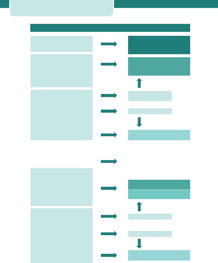
92 Antenatal obstetric complications
Antenatal thromboprophylaxis risk assessment and management
Single previous VTE+
•Thrombophilia or family history
•Unprovoked/oestrogen-related previous recurrent VTE (>1)
Single previous VTE with no family history or thrombophilia
Thrombophilia + no VTE
Medical cormorbidities, e.g. heart or lung disease, SLE, cancer, inflammatory conditions, nephrotic syndrome, sickle cell disease, intravenous drug user
Surgical procedure, e.g. appendicectomy
Age >35 years
Obesity (BMI >30 kg/m2)
Parity ≥3
Smoker
Gross varicose veins
Current systemic infection
Immobility, e.g. paraplegia, SPD, long-distance travel
Pre-eclampsia
Dehydration/hyperemesis/OHSS
Multiple pregnancy or ART
High risk
Requires antenatal prophylaxis with LMWH
Refer to trust-nominated thrombosis in pregnancy expert/team
Intermediate risk
Consider antenatal prophylaxis with LMWH
Seek trust-nominated thrombosis in pregnancy expert/team advice
3 or more risk factors
2 or more if admitted
<3 risk factors
Lower risk
Mobilization and avoidance of dehydration
Postnatal thromboprophylaxis risk assessment and management |
|||
|
|
|
|
|
|
||
Any previous VTE+ |
|
|
|
High risk |
|||
|
|
||
Anyone requiring antenatal LMWH |
|
At least 6 weeks postnatal prophylactic LMWH |
|
|
|
||
|
|
|
|
Caesarean section in labour
Asymptomatic thrombophilia (inherited or acquired)
BMI >40 kg/m2
Prolonged hospital admission
Medical cormorbidities, e.g. heart or lung disease, SLE, cancer, inflammatory conditions, nephrotic syndrome, sickle cell disease, intravenous drug user
Age >35 years
Obesity (BMI >30 kg/m2) Parity ≥3
Smoker
Elective Caesarean section
Any surgical elective in puerperium Gross varicose veins
Current systemic infection
Immobility, e.g. paraplegia, SPD, long-distance travel
Pre-eclampsia
Mid-cavity rotational operative delivery Prolonged labour (>24hours)
PPH >1 litre or blood transfusion
Intermediate risk
At least 7 days postnatal prophylactic LMWH
Note: if persisting or >3 risk factors, consider extending thrombophylaxis with LMWH
2 or more risk factors
<2 risk factors
Lower risk
Mobilization and avoidance of dehydration
Figure 8.2 Obstetric thromboprophylaxis – risk assessment and management.

Key points
•Screening for thrombophilias should be carried
out in those with a strong family or personal history of VTE.
•Rapid treatment of suspected VTE in pregnancy should be commenced while awaiting diagnosis.
•LMWHs are now the treatment of choice.
•Graduated compression stockings should be fitted and worn for two years to reduce the incidence of post thrombotic syndrome.
Substance abuse in pregnancy
Approximately one-third of adults who access drug services are women of reproductive age. There are approximately 6000 births to problem drug users in the UK each year (about 1 per cent of all deliveries). Multidisciplinary care is often necessary to optimize outcomes because the financial, psychological, social and domestic problems associated with drug misuse are often greater than the physical and medical concerns.
Opioids, especially heroin, remain the most commonly used drugs in the UK, although many drug users take combinations of drugs that often include cocaine or crack cocaine. Most problem drug users smoke tobacco and are heavy users of alcohol and cannabis. Taking drugs in combination greatly increases the unpredictability of their effect on the user. Intravenous injection of drugs also puts drug users at greater risk of infection with blood-borne viruses (hepatitis B and C and HIV). Many drug users live in disadvantaged communities in conditions of poverty and social exclusion. Many have had poor parenting experiences, poor education and significant mental health problems. The aims of management are to stabilize the mother’s drug-taking habits and ensure contact with social/care workers and psychiatric/drug liaison services as appropriate.
It is important not to try to reduce the opiate dose too rapidly in pregnancy, as this can easily precipitate acute withdrawal in both the mother and fetus; the principle is to administer the lowest effective dose of methadone liquid in three divided doses every day.
Screening for infections such as hepatitis B and human immunodeficiency virus (HIV) is routinely offered in the UK. In many cases,
Substance abuse in pregnancy |
93 |
Problems frequently encountered amongst
drug addicts
•Social problems: housing, crime, other children in care or abused.
•Co-existent addictions: alcohol and smoking.
•Malnutrition: especially iron, vitamins B and C.
•Risk of viral infections, e.g. human immunodeficiency virus (HIV) or hepatitis B.
•Specific fetal and neonatal risks (Table 8.2).
Table 8.2 The effects of some drugs of abuse on the fetus and neonate
Drug |
Fetal and neonatal risk |
Tobacco |
Fetal growth restriction |
|
|
Placental abruption |
Fetal growth restriction |
Alcohol |
Fetal alcohol syndrome |
|
(FAS) |
|
|
Opiates |
Pre-term labour |
|
Neonatal withdrawal |
|
syndrome |
Cocaine and |
Placental abruption |
derivatives |
Fetal growth restriction |
|
Pre-term labour |
|
|
multidisciplinary case conferences should be held to make arrangements and decisions for when the baby is delivered.
Alcohol
There is much debate about what is a ‘safe’ dose of alcohol during pregnancy. What is likely is that an intake of less than 100 g per week (approximately two drinks per day, e.g. two medium glasses of wine or one pint of beer) is not associated with any adverse effects. Doses greater than this have been related to fetal growth restriction (FGR). Massive doses, in excess of 2 g/kg of body weight (17 drinks per day), have been associated with fetal alcohol syndrome. However, the syndrome is not seen consistently in infants born to women who are heavy consumers of alcohol, and occurs only in approximately 30–33 per cent of children born to women who drink about 2 g/kg of

94Antenatal obstetric complications
body weight per day (equivalent to approximately 18 units of alcoholic drink per day). The differing susceptibility of fetuses to the syndrome is thought to be multifactorial and reflects the interplay of genetic factors, social deprivation, nutritional deficiencies, tobacco and other drug abuse, along with alcohol consumption.
If alcohol abuse is suspected, it may be necessary to involve social workers and arrange for formal psychiatric/addiction assessment. It is extremely difficult to ‘test’ for alcohol abuse, as even markers such as mean corpuscular volume and gamma GT are not reliable in pregnancy. Malnutrition is very likely in heavy alcohol abuse and requires (in addition to a change in basic diet) B vitamin supplements and iron; the problem is that the majority of such patients not only do not take their medicines but also default antenatal appointments.
Smoking and pregnancy
Smoking acutely reduces placental perfusion. Overall perinatal mortality is increased, babies are smaller at delivery and there is a higher risk of antepartum haemorrhage in smokers compared with nonsmokers. It is estimated that a baby will weigh less than its target weight by a multiple of 15 g times the average number of cigarettes a woman smokes per day; smoking fewer than five cigarettes per day has a barely discernible obstetric effect and quitting by 15 weeks gestation reduces the risk as much as quitting before pregnancy. Consequently, all women should be counselled regarding smoking cessation at their booking visit.
Oligohydramnios and polyhydramnios
Amniotic fluid is produced almost exclusively from fetal urine from the second trimester onwards. It serves a vital function in protecting the developing baby from pressure or trauma, allowing limb movement, hence normal postural development, and permitting the fetal lungs to expand and develop through breathing.
Oligohydramnios
centile for gestation. The amniotic fluid index (AFI) is an ultrasound estimation of amniotic fluid derived by adding together the deepest vertical pool in four quadrants of the abdomen. The AFI (in cm) is therefore associated with some degree of error. In general, however, it is possible to differentiate subjectively on ultrasound between ‘too much’, ‘too little’ and ‘normal looking’.
Oligohydramnios may be suspected antenatally following a history of clear fluid leaking from the vagina; this may represent PPROM (see Chapter 11, Late miscarriage and early birth). Clinically, on abdominal palpation, the fetal poles may be very obviously felt and ‘hard’, with a small for dates uterus. The possible causes of oligohydramnios and anhydramnios (no amniotic fluid) are described in the box.
The fetal prognosis depends on the cause of oligohydramnios, but both pulmonary hypoplasia and limb deformities (contractures, talipes) are common to severe early-onset ( 24 weeks) oligohydramnnios. Renal agenesis and bilateral multicystic kidneys carry a lethal prognosis, as life after birth is impossible without functioning kidneys. In this situation, the fetal lungs would probably be hypoplastic; this may also be true of severe urinary tract obstruction. Oligohydramnios due to FGR/uteroplacental insufficiency is usually of a less severe degree and less commonly causes limb and lung problems.
Polyhydramnios
Polyhydramnios is the term given to an excess of amniotic fluid, i.e. AFI 95th centile for gestation on ultrasound estimation. It may present as severe abdominal swelling and discomfort. On examination, the abdomen will appear distended out of proportion to the woman’s gestation (increased SFH). Furthermore, the abdomen may be tense and tender and the fetal poles will be hard to palpate. The condition may be caused by maternal, placental or fetal conditions.
Maternal
• Diabetes
Placental
Too little amniotic fluid (oligohydramnios) is |
• |
Chorioangioma |
commonly defined as amniotic fluid index 5th |
• |
Arterio-venous fistula |

|
|
|
|
|
|
|
|
|
|
Fetal malpresentation at term |
95 |
||
|
|
|
|
|
||
|
|
Possible causes of oligohydramnios and anhydramnios |
|
|
||
|
|
|
|
|
|
|
|
|
Too little production |
Diagnosed by |
|
||
|
|
|
|
|
|
|
|
|
Renal agenesis |
Ultrasound: no renal tissue, no bladder |
|
||
|
|
|
|
|
|
|
|
|
Multicystic kidneys |
Ultrasound: enlarged kidneys with multiple cysts, no visible |
|
||
|
|
|
bladder |
|
|
|
|
|
Urinary tract abnormality/ |
Ultrasound: kidneys may be present, but urinary tract |
|
||
|
|
obstruction |
dilatation |
|
||
|
|
|
|
|
|
|
|
|
FGR and placental insufficiency |
Clinical: reduced SFH, reduced fetal movements, possibly |
|
||
|
|
|
abnormal CTG |
|
||
|
|
|
Ultrasound: FGR, abnormal fetal Dopplers |
|
||
|
|
|
|
|
|
|
|
|
Maternal drugs (e.g NSAIDs) |
Withholding NSAIDs may allow amniotic fluid to |
|
||
|
|
|
re-accumulate |
|
||
|
|
|
|
|
|
|
|
|
Post-dates pregnancy |
|
|
|
|
|
|
Leakage |
Diagnosed by |
|
||
|
|
PPROM |
Speculum examination: pool of amniotic fluid on posterior blade |
|
|
|
NSAIDs, non-steroidal anti-inflammatory drugs; SFH, symphysis–fundal height.
Fetal
•Multiple gestation (in monochorionic twins, it may be twin-to-twin transfusion syndrome)
•Idiopathic
•Oesophageal atresia/tracheo-oesophageal fistula
•Duodenal atresia
•Neuromuscular fetal condition (preventing swallowing)
•Anencephaly.
The management of polyhydramnios is directed towards establishing the cause (hence determining fetal prognosis), relieving the discomfort of the mother (if necessary by amniodrainage), and assessing the risk of preterm labour due to uterine over-distension. The lastmentioned may require assessment of cervical length by ultrasound. If prior to 24 weeks, following amniotic fluid drainage, the cervical length is less than 25 mm, consideration might be given to cervical suture insertion.
Polyhydramnios due to maternal diabetes needs urgent investigation, as it often suggests high maternal blood glucose levels. In this context, polyhydramnios should correct itself when the mother’s glycaemic control is optimized.
Twin-to-twin transfusion syndrome is a rare cause of acute polyhydramnios in the recipient
sac of monochorionic twins. It is associated with oligohydramnios and a small baby in the other sac. The condition may be rapidly fatal for both twins; amniodrainage and removal by laser of the placental vascular connections are two therapeutic modalities employed in dealing with this condition. This is discussed further in Chapter 9, Twins and higher multiple gestations.
Fetal malpresentation at term
Malpresentation is a presentation that is not cephalic. Breech presentation is the most commonly encountered malpresentation and occurs in 3–4 per cent of term pregnancies, but is more common at earlier gestations. Similarly, oblique and transverse positions are not uncommon antenatally. They only become a problem if the baby (or first presenting baby in a multiple gestation) is not cephalic by 37 weeks.
Breech presentation
There are three types of breech: the most common is extended (frank) breech (Figure 8.3a); less common is a flexed (complete) breech (Figure 8.3b); and least common is footling breech, in which a foot presents at the cervix (Figure 8.3c). Cord and foot prolapse are risks in this situation.