

Obstetrics_by_Ten_Teachers_19E_-_Kenny_Louise
.pdf166Medical diseases complicating pregnancy
diet. The risks to the mother are from hypercalcaemic crises and complications such as acute pancreatitis, while fetal risks include increased rates of miscarriage, intrauterine death, preterm labour and neonatal tetany in untreated cases.
Hypoparathyroidism may be caused by autoimmune disease, but is more commonly a complication of thyroid surgery. It is diagnosed by finding low serum calcium and low PTH levels. Untreated, it is associated with increased risks of second trimester miscarriage and fetal hypocalcaemia, resulting in bone demineralization and neonatal rickets. The aim of treatment is to maintain normocalcaemia through vitamin D and oral calcium supplements, with regular monitoring of calcium and albumin levels during pregnancy.
Pituitary tumours in pregnancy
Hyperprolactinaemia is an important cause of infertility and amenorrhoea, and is most often due to a benign pituitary microadenoma. The diagnosis is confirmed with a combination of measurement of the prolactin level and computed tomography (CT) or magnetic resonance imaging (MRI) of the pituitary fossa. In 80 per cent of cases, it is treated with the dopamine agonists bromocriptine or cabergoline, which cause the tumour to reduce in size. Larger tumours may require surgery or radiotherapy, which is best undertaken before pregnancy.
The pituitary gland enlarges by 50 per cent during pregnancy, but it is rare for microadenomas to cause problems. Serial prolactin levels are unhelpful to monitor tumour growth in pregnancy. Bromocriptine and cabergoline are usually stopped in pregnancy, and visual fields and relevant symptoms, such as frontal headache, are monitored. If there is evidence of tumour growth during pregnancy, bromocriptine or cabergoline should be recommenced, and appropriate neuroimaging arranged. In women with macroadenomas ( 1 cm), it is advisable to continue with dopamine agonists because of the risk of the tumour enlarging under oestrogen stimulation.
Adrenal disease
Cushing’s syndrome
Cushing’s syndrome is rare in pregnancy as most affected women are infertile. It is characterized by
increased glucocorticoid production, usually due to hypersecretion of adrenocorticotrophic hormone (ACTH) from a pituitary tumour. However, in pregnancy, adrenal causes (tumours) are more common. Diagnosis is difficult because many of the symptoms – striae, weight gain, weakness, glucose intolerance and hypertension – mimic normal pregnancy changes. If suspected, plasma cortisol levels should be measured (although levels increase in pregnancy) and adrenal imaging with ultrasound, CT or MRI should be used. There is a high incidence of pre-eclampsia, preterm delivery and stillbirth.
Addison’s disease
Addison’s disease (adrenal insufficiency) is an autoimmune process, associated with clinical symptoms of exhaustion, nausea, hypotension, hypoglycaemia and weight loss. The diagnosis is difficult to make in pregnancy because the cortisol levels, instead of being characteristically decreased, may be in the low–normal range due to the physiological increase in cortisol-binding globulin in pregnancy. Occasionally, the disease may present as a crisis, and treatment consists of glucocorticoid and fluid replacement. In diagnosed and adequately treated patients, the pregnancy usually continues normally. Replacement steroids should be continued in pregnancy and increased at times of stress such as hyperemesis and delivery.
Phaeochromocytoma
Phaeochromocytoma is a rare catecholamineproducing tumour, reported in 1 in 50 000 pregnancies. The tumours arise from the adrenal medulla in 90 per cent of cases. In pregnancy, it may present as a hypertensive crisis and the symptoms may be similar to those of pre-eclampsia. A characteristic feature is paroxysmal hypertension, whereas the other symptoms of headaches, blurred vision, anxiety and convulsions may occur in pre-eclampsia. The diagnosis is confirmed by measurement of catecholamines in a 24-hour urine collection and in plasma, as well as by adrenal imaging. Treatment is by alpha-blockade with prazosin or phenoxybenzamine, but surgical removal is the only cure. Caesarean section is the preferred mode of delivery, as it minimizes the likelihood of sudden increases in catecholamines associated with vaginal delivery. Maternal and perinatal mortality is greatly increased, especially if the diagnosis is not made before pregnancy.

Skin disease
Pre-existing skin disease
Some pre-existing skin conditions, such as eczema or acne, worsen in pregnancy. Atopic eczema is a common pruritic skin condition affecting 1–5 per cent of the general population and causes the commonest pregnancy rash. It can be treated with emollients and bath additives. Hand and nipple eczema are common postpartum. Acne usually improves in pregnancy, but can flare in the third trimester and acne rosacea often worsens. Oral or topical erythromycin can be used, but retinoids are contraindicated. Psoriasis affects 2 per cent of the population and, during pregnancy, it remains unchanged in around 40 per cent of patients, improves in another 40 per cent and worsens in around 15 per cent. Topical steroids can still be used, while methotrexate is contraindicated.
Specific dermatoses of pregnancy
Pemphigoid gestationis
Pemphigoid gestationis (PG) is a rare pruritic autoimmune bullous disorder, with an incidence of around 1 in 60 000 pregnancies. It most commonly presents in the late second or third trimester with lesions beginning on the abdomen 50 per cent of the time and progressing to widespread clustered blisters, sparing the face. Diagnosis is made by the clinical appearance and by direct immunofluorescence. Once established, the disease runs a complex course with exacerbations and remissions, and flares postpartum in 75 per cent of cases. Management aims to relieve pruritus and prevent new blister formation, and is achieved through the use of potent topical steroids and/or oral prednisolone. There is some association with preterm delivery and small for gestational age births, but no increase in pregnancy loss has been reported. PG recurs in most subsequent pregnancies.
Polymorphic eruption of pregnancy
Polymorphic eruption of pregnancy (PEP) is a selflimiting pruritic inflammatory disorder that usually presents in the third trimester and/or immediately postpartum. The estimated incidence is 1 in 160 pregnancies and 75 per cent of affected pregnancies are primagravida. PEP often begins on the lower
Skin disease |
167 |
abdomen involving pregnancy striae, and extends to thighs, buttocks, legs and arms, while sparing the umbilicus and rarely involving face, hands and feet. In 70 per cent of patients the lesions become confluent and widespread, resembling a toxic erythema. Symptomatic treatment is sufficient and pregnancies appear to be otherwise unaffected, with no tendency to recur.
Prurigo of pregnancy
Prurigo of pregnancy is a common pruritic disorder, which occurs in 1 in 300 pregnancies, and presents as excoriated papules on extensor limbs, abdomen and shoulders. It is more common in women with a history of atopy. Prurigo usually starts at around 25–30 weeks of pregnancy and resolves after delivery, with no effect on the mother or baby. Treatment is symptomatic with topical steroids and emollients.
Pruritic folliculitis of pregnancy
Pruritic folliculitis is a pruritic follicular eruption, with papules and pustules that mainly affect the trunk, but can involve the limbs. It is similar in appearance to acne lesions and is sometimes considered a type of hormonally induced acne. Its onset is usually in the second and third trimesters, and it resolves weeks after delivery. Topical steroid treatment is effective.
Key points
•Women with medical conditions that adversely affect pregnancy outcome should be offered pre-pregnancy counselling by appropriately experienced health-care professionals.
•Women with medical problems that preclude safe pregnancy should be offered safe, effective and appropriate contraception.
•Asthma is the commonest chronic disease encountered in pregnancy.
•Pulmonary hypertension is associated with a risk of maternal mortality of up to 50 per cent in pregnancy.
•Women found to be hypertensive in the first half of pregnancy require investigation for possible underlying causes.
•Women with pre-existing hypertension are at increased risk of superimposed pre-eclampsia, FGR and placental abruption.

168Medical diseases complicating pregnancy
•Women who become pregnant with serum creatinine values above 125 mol/L have an increased risk of accelerated decline in renal function and poor outcome of pregnancy.
•Pre-existing diabetes increases maternal and fetal obstetric morbidity.
•The incidence of fetal macrosomia in diabetes can be reduced through good blood glucose control.
•The risk of perinatal and maternal morbidity is increased in pregnancies complicated by sickle cell disease.
•The main issue for pregnant women with epilepsy relates to the teratogenic risk of anticonvulsant medication drugs.
•Obstetric cholestasis is associated with an increased risk of term stillbirth.
scans from 28 weeks gestation showed fetal growth accelerating beyond the 95th centile. At 36 weeks, she attended complaining of decreased fetal movement. An ultrasound scan showed a singleton fetus with a cephalic presentation and the presence
of marked polyhydramnios (AFI 34 cm). A CTG was performed and showed reduced baseline variability with unprovoked decelerations. An urgent Caesarean section was performed and a 4.9 kg male infant was delivered. Within minutes of delivery, the baby was found to have signs of respiratory distress and he was admitted to the neonatal unit. The baby required ventilation for 48 hours. Additionally, the baby became hypoglycaemic within an hour of admission and in total spent 9 days on the neonatal unit while blood glucose levels stabilized and feeding became established.
Mrs M had an oral glucose tolerance 6 weeks following delivery. The 2-hour glucose level was within normal limits.
C A S E H I S T O R Y
Mrs M is a 30-year-old woman in her second pregnancy. She has a body mass index of 38. Her first pregnancy was complicated by gestational diabetes, which was detected following screening at 28 weeks gestation. She was poorly compliant with dietary intervention and required insulin
treatment from 32 weeks gestation. At 36 weeks, she developed polyhydramnios (amniotic fluid index of 28 cm) and her baby was found to be in a breech position. She subsequently had an
elective Caesarean section at 39 weeks gestation, which resulted in the birth of a 4.4 kg male infant.
In this pregnancy, she booked for antenatal care at 18 weeks gestation. Her HbA1c at booking was found to be 9.2 per cent and she was therefore commenced on insulin. An anomaly scan and fetal echocardiography at 22 weeks were normal. Despite weekly attendance at the combined obstetric diabetic clinic, her blood sugar control was poor and serial ultrasound
Additional reading
de Swiet M. Medical disorders in obstetric practice. Oxford: Blackwell Science, 2002.
Kaaja RJ, Greer IA. Manifestations of chronic disease during pregnancy. JAMA 2005; 294: 2751–7.
Lombaard H, Pattinson RC. Underlying medical conditions. Best Practice and Research. Clinical Obstetrics and Gynaecology 2008; 22: 847–864.
Taylor R, Davison JM. Type 1 diabetes and pregnancy. BMJ 2007; 334: 742–5.
William D, Davison JM. Chronic kidney disease in pregnancy. BMJ 2008; 336: 211–15.
Williams J, Mozurkewich E, Chilimigras J, Van De Ven C. Critical care in obstetrics: pregnancy-specific conditions.
Best Practice and Research. Clinical Obstetrics and Gynaecology 2008; 22: 825–46.

C H A P T E R 1 3
PERINATAL INFECTIONS
Sarah Vause
.........................Infections causing congenital abnormalities |
169 |
Infections acquired around the time of delivery |
|
Other congenital infections associated with |
|
with serious neonatal consequences .............................. |
176 |
pregnancy loss and preterm birth ...................................... |
174 |
Perinatal infections causing long-term disease.................. |
180 |
|
|
|
|
O V E R V I E W
Infectious diseases contracted during pregnancy can have a serious impact on both the mother and the fetus. Some infections cause congenital abnormalities in the fetus (rubella, syphilis, toxoplasmosis, cytomegalovirus, chickenpox). Other infections can be transmitted from mother to baby during pregnancy (parvovirus) or around the time of delivery (human immunodeficiency virus (HIV), hepatitis B, hepatitis C, herpes, group B streptococcus).
For several infectious diseases, screening programmes and effective interventions can improve the outcome for the fetus. For other diseases, early diagnosis and appropriate treatment can be of benefit to both mother and baby.
This chapter will describe some of these infections.
Infections causing congenital
abnormalities
Rubella
Infective organism
Rubella virus is a togavirus spread by droplet transmission.
Prevalence
Since the introduction of the measles, mumps and rubella vaccine (MMR), an average of three births affected by congenital rubella a year and four rubellaassociated terminations were registered from 1996 to 2000 in the UK. In the 1990s there was reduced uptake of the MMR vaccine for infants due to concern about a possible link with autism. It is too soon to say whether this will eventually lead to an increase in the prevalence of rubella in the community and an increase in incidence of congenital rubella when this cohort of non-immunized infants reach childbearing age.
In England and Wales susceptibility is slightly higher in nulliparous women (2 per cent) than in parous women (1.2 per cent). Certain ethnic groups
also appear to have higher susceptibility, such as Asian and black women (5 per cent) and Oriental women (8 per cent), compared with less than 2 per cent in white women, with an overall susceptibility of about 2.5 per cent reported for pregnant women in the UK.
Screening
In the UK, NICE recommendations are that all women should be offered rubella susceptibility screening early in their pregnancy to identify women at risk of contracting rubella infection and to enable vaccination in the post-natal period for the protection of future pregnancies.
This is an unusual antenatal screening test as there is no effective intervention which can be implemented during the index pregnancy to reduce the risk of harm to that fetus, nor does it attempt to identify currently affected pregnancies. The aim of screening for rubella in pregnancy is to identify susceptible women so that postpartum vaccination may protect future pregnancies against rubella infection and its consequences.
For pregnant women who are screened and rubella antibody is not detected, rubella vaccination after pregnancy should be advised. Vaccination during pregnancy is contraindicated because of a theoretical
170Perinatal infections
risk that the vaccine itself could be teratogenic, as it is a live vaccine. No cases of congenital rubella syndrome resulting from vaccination during pregnancy have been reported. However, women who are vaccinated postpartum should be advised to use contraception for three months.
Clinical features
Rubella infection is characterized by a febrile rash but may be asymptomatic in the mother in 20–50 per cent of cases.
Features of congenital rubella syndrome can include sensorineural deafness, congenital cataracts, blindness, encephalitis and endocrine problems.
The risk of congenital rubella infection reduces with gestation. If infection of the fetus does occur the defects caused are also less severe with more advanced gestations. Congenital infection in the first 12 weeks of pregnancy among mothers with symptoms is over 80 per cent and reduces to 25 per cent at the end of the second trimester. One hundred per cent of infants infected during the first 11 weeks of pregnancy have rubella defects, whereas primary rubella contracted between 16 and 20 weeks of gestation carries only a minimal risk of deafness. Rubella infection prior to the estimated date of conception or after 20 weeks gestation carries no documented risk to the fetus.
Management
If infection during pregnancy is confirmed, the risk of congenital rubella syndrome should be assessed depending on the gestation when infection occurred. If infection occurred prior to 16 weeks gestation, termination of pregnancy should be offered. If the infection occurs later in pregnancy the woman should be given appropriate information and reassured.
Syphilis
Infective organism
Syphilis is a sexually acquired infection caused by
Treponema pallidum.
Prevalence
The incidence of infectious syphilis in England and Wales is low, but has increased markedly in the last ten years. The increase has mainly been seen in rates of infection in homosexual men, but part of the increase is due to immigration to the UK from countries where prevalence is higher, for example Eastern Europe.
The prevalence of syphilis in pregnant women has been estimated at approximately 68 per million with regional variation, highest in north-east London. This equates to approximately 50–60 cases per year in the UK.
Clinical features
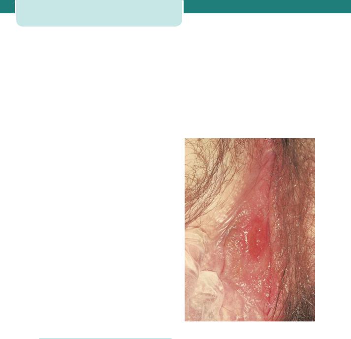
Primary syphilis may present as a painless genital ulcer 3–6 weeks after the infection is acquired (condylomata lata) (Figure 13.1); however, this may be on the cervix and go unnoticed.
Figure 13.1 Primary syphilitic chancre. (Courtesy of Dr Raymond Maw, Royal Victoria Hospital, Belfast.)
Secondary manifestations occur 6 weeks to six months after infection and present as a maculopapular rash or lesions affecting the mucous membranes. Ultimately, 20 per cent of untreated patients will develop symptomatic cardiovascular tertiary syphilis and 5–10 per cent will develop symptomatic neurosyphilis.
In pregnant women with early untreated (primary or secondary) syphilis, 70–100 per cent of infants will be infected and approximately 25 per cent will be

stillborn. Mother-to-child transmission of syphilis in pregnancy is associated with fetal growth restriction (FGR), fetal hydrops, congenital syphilis (which may cause long-term disability), stillbirth, preterm birth and neonatal death. The risk of congenital transmission declines with increasing duration of maternal syphilis prior to pregnancy. Adequate treatment with benzathine penicillin markedly improves the outcome for the fetus.
Screening
Because treatment is so effective, routine antenatal screening for all pregnant women is recommended.
The body’s immune response to syphilis is the production of non-specific and specific treponemal antibodies. These can be detected by serological tests. Non-treponemal tests detect non-specific treponemal antibodies and include the Venereal Diseases Research Laboratory (VDRL) and rapid plasma reagin (RPR) tests. Treponemal tests detect specific treponemal antibodies and include enzyme immunoassays (EIA), T. pallidum haemagglutination assay (TPHA) and the fluorescent treponemal antibody-absorbed test (FTA-abs). EIA tests that detect immunoglobulin (Ig) G or IgG and IgM are rapidly replacing the VDRL and TPHA combination for syphilis screening in the UK. EIAs are over 98 per cent sensitive and over 99 per cent specific. Non-treponemal tests, on the other hand, may result in false negatives, particularly in very early or late syphilis, in patients with reinfection or those who are HIV positive. The VDRL may be falsely positive in women with lupus. Therefore, positive results should be interpreted with caution and the pregnant woman should be referred for expert assessment and diagnosis in a genitourinary medicine clinic.
None of these serological tests will detect syphilis in its incubation stage, which may last for an average of 25 days.
Management
The initial step is to confirm the diagnosis and to test for any other sexually transmitted diseases. Once a diagnosis of syphilis is confirmed the genitourinary medicine clinic will institute appropriate contact tracing of sexual partners. Older children may also need to be screened for congenital infection.
Parenteral penicillin has a 98 per cent success rate for preventing congenital syphilis. A Jarish– Herxheimer reaction may occur with treatment as a result of release of pro-inflammatory cytokines
Infections causing congenital abnormalities |
171 |
in response to dying organisms. This presents as a worsening of symptoms, and fever for 12–24 hours after commencement of treatment. It may be associated with uterine contractions and fetal distress. Many clinicians therefore admit women at the time of commencement of treatment for monitoring.
If a woman is not treated during pregnancy her baby should be treated after delivery. An infected baby may be born without signs or symptoms of disease but if not treated immediately, may develop serious problems within a few weeks. Untreated babies often develop developmental delay, have seizures or die.
Toxoplasmosis
Infective organism
Toxoplasma gondii is a protozoan parasite found in cat faeces, soil or uncooked meat. Infection occurs by ingestion of the parasite from undercooked meat or from unwashed hands.
Prevalence
The prevalence varies in different countries. It is 16 per 1000 in France but in the UK it is estimated to be less than two per 1000 pregnancies.
Screening
Only about ten severely affected babies are diagnosed per year in the UK and for this reason the UK National Screening Committee recently reported that screening for toxoplasmosis should not be offered routinely. There is a lack of evidence that antenatal screening and treatment reduces mother-to-child transmission or the complications associated with toxoplasma infection. In the UK women should be advised about appropriate preventative measure such as avoiding eating rare or raw meat, avoiding handling cats and cat litter, and wearing gloves and washing hands when gardening or handling soil.
Even in France where more women acquire infection during pregnancy, and women are screened antenatally, the benefits of such a programme appear to be limited.
Clinical features
The initial infection may be relatively asymptomatic, or may be a glandular fever-like illness. Parasitaemia usually occurs within 3 weeks of infection. Therefore, congenital infection is only a significant risk if the

172Perinatal infections
mother acquires the infection during or immediately before pregnancy.
Infection during the first trimester of pregnancy is most likely to cause severe fetal damage (85 per cent), but only 10 per cent of infections are transmitted to the fetus at this gestation. In the third trimester 85 per cent of infections are transmitted, but the risk of fetal damage decreases to around 10 per cent.
Severely infected infants may have ventriculomegaly or microcephaly, chorioretinitis and cerebral calcification. These features may be detected on ultrasound scan. The majority of infected infants are asymptomatic at birth but develop sequelae several years later.
Management
The diagnosis of primary infection with toxoplasmosis during pregnancy is made by the Sabin-Feldman dye test. Enzyme-linked immunosorbant assays are available for IgM antibody. However, IgM may persist for months or even years, so often serial testing for rising titres is necessary. If suspicion of congenital toxoplasmosis has arisen because of an abnormal ultrasound scan of the fetus, an amniocentesis can be performed. Polymerase chain reaction (PCR) analysis of amniotic fluid is highly accurate for the identification of T. gondii.
Spiramycin treatment can be used in pregnancy (a 3-week course of 2–3 g per day). This reduces the incidence of transplacental infection but has not been shown to definitively reduce the incidence of clinical congenital disease.
If toxoplasmosis is found to be the cause of abnormalities detected on ultrasound scan of the fetus, then termination of pregnancy can be offered.
Cytomegalovirus
Infective organism
Cytomegalovirus (CMV) is a DNA herpes virus. It is transmitted by respiratory droplet transmission and is excreted in the urine.
Prevalence
It is a common virus in the UK and about 60 per cent of women are already immune when they become pregnant and, consequently, approximately 40 per cent are susceptible to infection. The incidence of infection in pregnancy is estimated to be around 1–2 per cent of pregnancies. Of those infected, approximately 30 per cent will transmit the viral
infection to the fetus and of these approximately 30 per cent of the fetuses will be affected by the virus.
Clinical features
Primary infection often produces no symptoms or mild non-specific flu-like symptoms in the mother.
The diagnosis is often made after abnormalities are seen in the fetus on ultrasound scan. The main features seen in an affected fetus are FGR, microcephaly, ventriculomegaly, ascites or hydrops.
Some fetuses which are infected may not show any features on ultrasound, but may later be found to have neurological damage such as blindness, deafness or developmental delay. The neonate can also be anaemic and thrombocytopenic, with hepatosplenomegaly, jaundice and a purpural rash.
Management
A serological diagnosis can be made by demonstrating the development of CMV antibodies in a seronegative woman, who initially develops CMV IgM antibody, and subsequently IgG antibody. Virology labs usually keep the initial sample taken at booking, so if infection is suspected a sample taken at the time of presentation can be compared with the initial booking sample to determine whether seroconversion has occurred. Since IgM can be secreted for several months, it is not sufficient to simply demonstrate IgM in a sample at the time of presentation; it has to be a new finding in a woman who was negative for IgM at the time of booking.
If there is a suspicion that the fetus may be infected, amniotic fluid can be tested for the virus by PCR. Since the virus is excreted in fetal urine it can be found in amniotic fluid.
If abnormalities are detected on ultrasound and these are felt to be due to congenital CMV infection, termination of pregnancy should be offered. A much more difficult situation is when CMV infection is known to have occurred, but the fetus appears normal on ultrasound, as there is approximately a 20 per cent chance of neurological abnormality in the fetus.
Like other herpes viruses, CMV has the capacity to establish latency and be reactivated. After infection the virus is excreted for weeks or months by adults and for years by infants. It persists in the lymphocytes throughout life and can be transmitted by blood transfusion or transplantation. Reactivation occurs intermittently, with shedding in the genital urinary

or respiratory tract. Reactivation during pregnancy very rarely causes congenital CMV infection.
Chickenpox
Infective organism
Chickenpox is caused by the varicella zoster virus (VZV), a herpes virus which is transmitted by droplet spread.
Prevalence
In the UK 90 per cent of adults are immune to chickenpox. However, contact with chickenpox is common in pregnancy and approximately one in 200 women will contract chickenpox during their pregnancy.
Clinical features
Non-immune pregnant women are more vulnerable to chickenpox and may develop a serious pneumonia, hepatitis or encephalitis. It can occasionally be fatal with the mortality rate being approximately five times higher in pregnant women than in non-pregnant adults. Pneumonia occurs in about 10 per cent of women with chickenpox and seems more severe at later gestations. It may also cause the fetal varicella syndrome (FVS) or varicella infection of the newborn.
Management
Women should be asked whether they have had chickenpox at the initial booking visit. If they have not had chickenpox, they should be advised to avoid contact with it during pregnancy, and if they accidentally come into contact with it should advise their doctor or midwife about the exposure as soon as possible.
When contact occurs with chickenpox, a careful history must be taken to confirm the significance of the contact (length of exposure and closeness of contact) and the susceptibility of the patient. Significant contact is defined as being in the same room as someone for 15 minutes or more, or face-to-face contact.
Testing for immunity
If a women reports that she has been in contact with chickenpox, she should have a blood test for confirmation of VZV immunity, by testing for VZV IgG. This can usually be performed within 24–48 hours and the virology laboratory may be able to use serum stored from booking antenatal bloods. At least
Infections causing congenital abnormalities |
173 |
80–90 per cent of women tested will have VZ IgG and can be reassured.
Management of the non-immune woman exposed to chickenpox
If the pregnant woman is not immune to VZV and she has had a significant exposure, she should be given varicella zoster immunoglobulin (VZIG) as soon as possible. VZIG is effective when given up to 10 days after contact and may prevent or attenuate the disease. Women who have had exposure to chickenpox (regardless of whether or not they have received VZIG) should be asked to notify their doctor or midwife early if a rash develops.
Management of chickenpox in pregnancy
Women with chickenpox should avoid contact with susceptible individuals; that is, other pregnant women and neonates, until the lesions have crusted over. This is usually about 5 days after the onset of the rash. Symptomatic treatment and hygiene is advised to prevent secondary bacterial infection of the lesions.
The UK Advisory Group on Chickenpox recommends that oral aciclovir 800 mg five times per day for 7 days be prescribed for pregnant women with chickenpox if they present within 24 hours of the onset of the rash and if they are more than 20 weeks gestation. Aciclovir should be used cautiously before 20 weeks gestation. VZIG has no therapeutic benefit once chickenpox has developed. If the woman smokes cigarettes, has chronic lung disease, is taking corticosteroids or is in the latter half of pregnancy, a hospital assessment should be considered, even in the absence of complications.
Women hospitalized with varicella should be nursed in isolation from babies or potentially susceptible pregnant women or non-immune staff. Delivery during the viraemic period may be extremely hazardous. The maternal risks are bleeding, thrombocytopenia, disseminated intravascular coagulopathy and hepatitis. There is a high risk of varicella infection of the newborn with significant morbidity and mortality. Supportive treatment and intravenous aciclovir is therefore desirable, allowing resolution of the rash and transfer of protective antibodies from the mother to the fetus. However, delivery may be required in women to facilitate assisted ventilation in cases where varicella pneumonia is complicated by respiratory failure.

174Perinatal infections
The fetus
Spontaneous miscarriage does not appear to be increased if chickenpox occurs in the first trimester.
FVS is characterized by one or more of the following:
•skin scarring in a dermatomal distribution;
•eye defects (microphthalmia, chorioretinitis, cataracts);
•hypoplasia of the limbs;
•neurological abnormalities (microcephaly, cortical atrophy, developmental delay and
dysfunction of bowel and bladder sphincters).
This only occurs in a minority of infected fetuses (approximately 1 per cent). FVS has been reported to complicate maternal chickenpox that occurs as early as 3 weeks and up to 28 weeks of gestation. The risk appears to be lower in the first trimester (0.55 per cent). No case of FVS has been reported when maternal infection has occurred after 28 weeks.
If the mother has contracted chickenpox during pregnancy, referral to a fetal medicine specialist should be considered at 16–20 weeks or 5 weeks after infection for discussion and detailed ultrasound examination, when findings such as limb deformity, microcephaly, hydrocephalus, soft-tissue calcification and fetal growth restriction can be detected. A time lag of at least 5 weeks after the primary infection is advised as it takes several weeks for these features to manifest.
Maternal infection around the time of delivery
If maternal infection occurs at term, there is a significant risk of varicella of the newborn. Elective delivery should normally be avoided until 5–7 days after the onset of maternal rash to allow for the passive transfer of antibodies from mother to child. Neonatal ophthalmic examination should be organized after birth.
If birth occurs within the 7-day period following the onset of the maternal rash, or if the mother develops the chickenpox rash within the 7-day period after birth, the neonate should be given VZIG. The infant should be monitored for signs of infection until 28 days after the onset of maternal infection.
Neonatal infection should be treated with aciclovir following discussion with a neonatologist and virologist. VZIG is of no benefit once neonatal chickenpox has developed.
Contact with shingles
Following the primary infection, the virus remains dormant in sensory nerve root ganglia but can be reactivated to cause a vesicular erythematous skin rash in a dermatomal distribution known as herpes zoster or shingles. The risk of a pregnant woman acquiring infection from an individual with herpes zoster in non-exposed sites (for example, thoracolumbar) is remote.
Other congenital infections associated
with pregnancy loss and preterm birth
Parvovirus
Infective organism
Parvovirus is a relatively common infection in pregnancy, and is spread by droplet infection.
Incidence
Fifty per cent of women at childbearing age are immune to PVB19 infection and therefore 50 per cent are susceptible to infection during pregnancy. It occurs most commonly in those pregnant women who work with young children, for example teachers. In some years the incidence of parvovirus is higher than others.
Clinical features
In adults, symptoms are a mild flu-like illness. In children it may cause a characteristic rash (slapped cheek syndrome). In the fetus it can cause an aplastic anaemia. The anaemic fetus may then become hydropic due to high output cardiac failure and liver congestion. This is the most common presentation during pregnancy and is seen on ultrasound scan. If a fetus is hydropic, the velocity of blood flow in the fetal middle cerebral artery can be measured. If the velocity is high, it is suggestive of anaemia, and parvovirus would be one of several differential diagnoses.
Management
The diagnosis is made by demonstrating seroconversion of the mother, who develops IgM antibodies to parvovirus, having previously tested negative. Viral DNA amplifications using PCR in maternal and fetal serum or amniotic fluid, is the most sensitive and accurate diagnostic test.

A hydropic fetus may recover spontaneously as the mother and fetus recover from the virus, or may require treatment by in utero transfusion.
Infection in the first 20 weeks of pregnancy can lead to hydrops fetalis and intrauterine death, as treatment by intrauterine transfusion is not possible at early gestations. Prior to 20 weeks the fetal loss rate is approximately 10 per cent.
If the anaemia is treated by in utero transfusion, the fetus can make a complete recovery. Parvovirus does not cause neurological damage, and if the fetus survives the anaemia, there can be a completely normal outcome. After 20 weeks gestation the fetal loss rate is estimated to be approximately 1 per cent.
Listeria
Infective organism
Listeria monocytogenes is an aerobic and facultatively anaerobic motile Gram-positive bacillus. It has an unusual life cycle with obligate intracellular replication. People with reduced cell-mediated immunity, e.g. pregnant women and the elderly, are therefore most at risk.
Incidence
The incidence of listeria infection in pregnant women is estimated at 12 per 100 000 compared to 0.7 per 100 000 in the general population. Contaminated food is the usual source of infection. Usual sources include unpasteurized milk, ripened soft cheeses and pâté. It is therefore recommended that pregnant women should be offered information on how to reduce the risk of listeriosis by dietary modification. Listeria is not transmitted in hot cooked foods and does not multiply in the freezer; however, it survives and multiplies at refrigerator temperatures and hence can be transmitted in chilled foods, e.g. milk, soft cheese, prawns and pâté.
Clinical features
Pregnant women with listeriosis most commonly suffer from a flu-like illness with fever and general malaise. About a third of women may be asymptomatic. Transmission to the fetus may either occur via the ascending route through the cervix, or transplacentally secondary to maternal bacteraemia. Approximately 20 per cent of affected pregnancies result in miscarriage or stillbirth. Premature delivery may occur in over 50 per cent. Neonates may have respiratory distress, fever, sepsis or neurological
Other congenital infections associated with |
175 |
pregnancy loss and preterm birth |
|
symptoms and the overall neonatal mortality rate has been estimated at 38 per cent.
Management
The diagnosis of listeria depends on clinical suspicion and isolation of the organism from blood, vaginal swabs or the placenta. Meconium staining of the amniotic fluid in a preterm fetus may increase clinical suspicion for listeriosis.
For women with listeriosis during pregnancy, intravenous antibiotic treatment (ampicillin 2 g given every 6 hours) is indicated.
Malaria
Infective organisms
Although malaria can be caused by four species of malarial parasite (Plasmodium falciparum, P. vivax,
P. ovale and P. malariae) the species which carries the worst prognosis for the mother and fetus, and the organism of greatest importance on a worldwide scale, is P. falciparum. This is a protozoan parasite transmitted by the female anopheline mosquito.
Incidence
Incidence varies depending on geographical location, but is endemic in sub-Saharan Africa, South Asia and some parts of South America. It is estimated that one billion people worldwide carry parasites at any time, and 0.5–3 million people per year die from malaria. Pregnant women have an increased risk of malarial infection compared to their non-pregnant adult counterparts. The incidence of parasitaemia has been found to be higher in primiparous women (66 per cent) than multiparous women (21–29 per cent).
In endemic areas where many women are semiimmune, malarial parasites may often be found in large numbers sequestrated in the placenta, even when blood films are negative. This may lead to the diagnosis being missed, unless a high index of suspicion is maintained.
Clinical features
Maternal effects include a cyclical spiking pyrexia, which may be associated with miscarriage and preterm labour. Severe anaemia may develop rapidly, but many women from endemic areas may also have other risk factors for severe anaemia. Hypoglycaemia is common and may be severe in pregnancy. Pulmonary oedema, due to abnormal capillary permeability,