

Obstetrics_by_Ten_Teachers_19E_-_Kenny_Louise
.pdf96 Antenatal obstetric complications
(a) |
(b) |
(d)
(c)
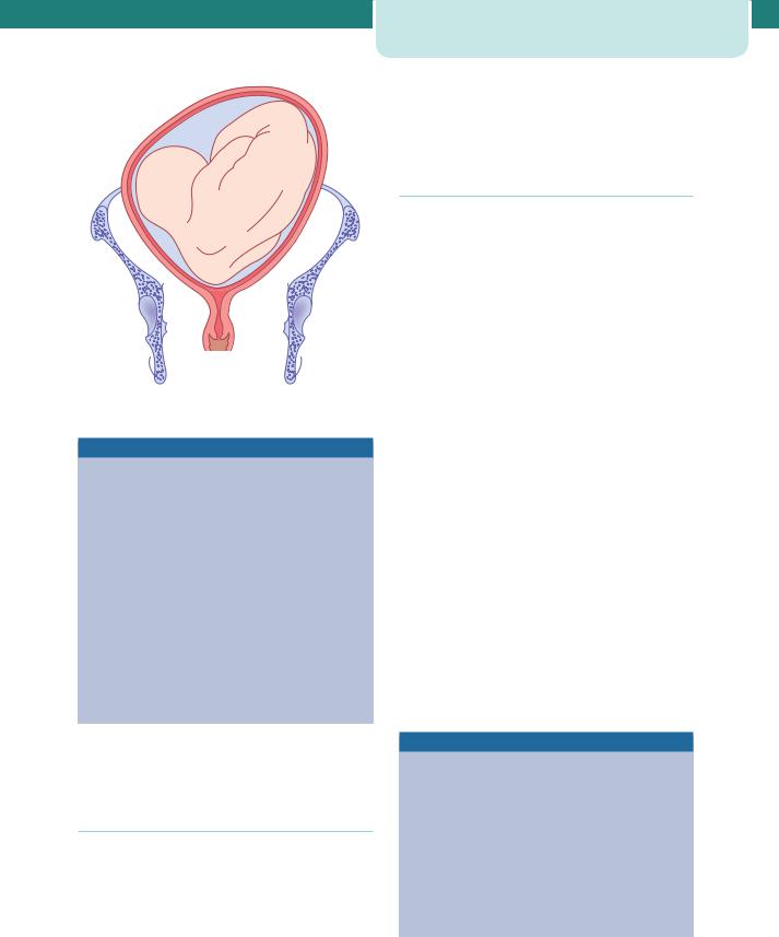
Figure 8.3 (a) Frank breech (also known as extended breech) presentation with extension of the legs. (b) Breech presentation with flexion of the legs. (c) Footling breech presentation. (d) Transverse lie. (e) Oblique lie
(e)
Figure 8.3 (continued )
Predisposing factors for breech presentation
Maternal
•Fibroids
•Congenital uterine abnormalities, e.g. bicornuate uterus
•Uterine surgery
Fetal/placental
•Multiple gestation
•Prematurity
•Placenta praevia
•Abnormality, e.g. anencephaly or hydrocephalus
•Fetal neuromuscular condition
•Oligohydramnios
•Polyhydramnios
Antenatal management of breech presentation
If a breech presentation is clinically suspected at or after 36 weeks, this should be confirmed by ultrasound scan. The scan should document fetal biometry, amniotic fluid volume, the placental site and the position of the fetal legs. The scan should also look for any anomalies previously undetected.
Fetal malpresentation at term |
97 |
The three management options available at this point should be discussed with the woman. These are external cephalic version (ECV), vaginal breech delivery and elective Caesarean section.
External cephalic version
ECV is a relatively straightforward and safe technique and has been shown to reduce the number of Caesarean sections due to breech presentations. Success rates vary according to the experience of the operator but in most units are around 50 per cent (and are higher in multiparous women who tend to have more lax abdominal musculature).
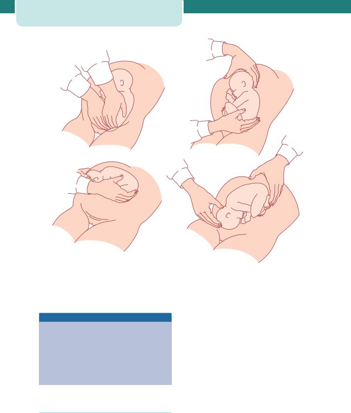
The procedure is performed at or after 37 completed weeks by an experienced obstetrician at or near delivery facilities. ECV should be performed with a tocolytic (e.g. nifedipine) as this has been shown to improve the success rate. The woman is laid flat with a left lateral tilt having ensured that she has emptied her bladder and is comfortable. With ultrasound guidance, the breech is elevated from the pelvis and one hand is used to manipulate this upward in the direction of a forward role, while the other hand applies gentle pressure to flex the fetal head and bring it down to the maternal pelvis (Figure 8.4).
The procedure can be mildly uncomfortable for the mother and should last no more than 10 minutes. If the procedure fails, or becomes difficult, it is abandoned. A fetal heart rate trace must be performed before and after the procedure and it is important to administer anti-D if the woman is Rhesus-negative.
Contraindications to ECV
•Fetal abnormality (e.g. hydrocephalus)
•Placenta praevia
•Oligohydramnios or polyhydramnios
•History of antepartum haemorrhage
•Previous Caesarean or myomectomy scar on the uterus
•Multiple gestation
•Pre-eclampsia or hypertension
•Plan to deliver by Caesarean section anyway
98 Antenatal obstetric complications
(a) |
(b) |
|
(c) |
(d) |
Figures 8.4 External cephalic version. (a) The breech is disengaged from the pelvic inlet. (b) Version is usually performed in the direction that increases flexion of the fetus and makes it do a forward somersault. (c) On completion of version, the head is often not engaged for a time. (d) The fetal heart rate should be checked after the external version has been completed
Risks of ECV
•Placental abruption
•Premature rupture of the membranes
•Cord accident
•Transplacental haemorrhage (remember anti-D administration to Rhesus-negative women)
•Fetal bradycardia
Mode of delivery
If ECV fails, or is contraindicated, and Caesarean section is not indicated for other reasons, then women
should be counselled regarding elective Caesarean section and planned vaginally delivery. A recent large multicentre trial (the Term Breech Trial) confirmed that planned vaginal delivery of a breech presentation is associated with a 3 per cent increased risk of death or serious morbidity to the baby. Although this trial did not evaluate long-term outcomes for child or mother, it has led to the recommendation that the best method of delivering a term breech singleton is by planned Caesarean section. Despite this, either by choice or as a result of precipitous labour, a small proportion of women with breech presentations will deliver vaginally. It therefore remains important that clinicians and hospitals are prepared for vaginal breech delivery.

Prerequisites for vaginal breech delivery
Feto-maternal:
•The presentation should be either extended (hips flexed, knees extended) or flexed (hips
flexed, knees flexed but feet not below the fetal buttocks).
•There should be no evidence of feto-pelvic disproportion with a pelvis clinically thought
to be adequate and an estimated fetal weight of3500 g (ultrasound or clinical measurement).
•There should be no evidence of hyperextension of the fetal head, and fetal abnormalities that
would preclude safe vaginal delivery (e.g. severe hydrocephalus) should be excluded.
Management of labour:
•Fetal well-being and progress of labour should be carefully monitored.
•An epidural analgesia is not essential but may be advantageous; it can prevent pushing before full
dilatation.
•Fetal blood sampling from the buttocks provides an accurate assessment of the acid-base status
(when the fetal heart rate trace is suspect).
•There should be an operator experienced in delivering breech babies available in the
hospital.
Although much emphasis is placed on adequate case selection prior to labour, a survey of outcome of the undiagnosed breech in labour managed by experienced medical staff showed that safe vaginal delivery can be achieved.
Technique
Breech delivery epitomizes the position of ‘masterly inactivity’ (hands-off). Problems are more likely to arise when the obstetrician tries to speed up the process (by pulling on the baby).
Delivery of the buttocks
In most circumstances, full dilatation and descent of the breech will have occurred naturally. When the buttocks become visible and begin to distend the perineum, preparations for the delivery are made. The buttocks will lie in the anterior–posterior diameter. Once the anterior buttock is delivered and the anus is
Fetal malpresentation at term |
99 |
seen over the fourchette (and no sooner than this), an episiotomy can be cut.
Delivery of the legs and lower body
If the legs are flexed, they will deliver spontaneously. If extended, they may need to be delivered using Pinard’s manoeuvre. This entails using a finger to flex the leg at the knee and then extend at the hip, first anteriorly then posteriorly. With contractions and maternal effort, the lower body will be delivered. Usually a loop of cord is drawn down to ensure that it is not too short.
Delivery of the shoulders
The baby will be lying with the shoulders in the transverse diameter of the pelvic mid-cavity. As the anterior shoulder rotates into the anterior–posterior diameter, the spine or the scapula will become visible. At this point, a finger gently placed above the shoulder will help to deliver the arm. As the posterior arm/ shoulder reaches the pelvic floor, it too will rotate anteriorly (in the opposite direction). Once the spine becomes visible, delivery of the second arm will follow. This can be imagined as a ‘rocking boat’ with one side moving upwards and then the other. Loveset’s manoeuvre essentially copies these natural movements (Figure 8.5). However, it is unnecessary and meddlesome to do routinely (one risks pulling the shoulders down but leaving the arms higher up, alongside the head).
Delivery of the head
The head is delivered using the Mauriceau–Smellie– Veit manoeuvre: the baby lies on the obstetrician’s arm with downward traction being levelled on the head via a finger in the mouth and one on each maxilla (Figure 8.6). Delivery occurs with first downward and then upward movement (as with instrumental deliveries). If this manoeuvre proves difficult, forceps need to be applied. An assistant holds the baby’s body aloft while the forceps are applied in the usual manner (Figure 8.7).
Complications
The greatest fear with a vaginal breech is that the baby will get ‘stuck’. Interference in the natural process by the inappropriate use of oxytocic agents or by trying to pull the baby out (breech extraction) will (paradoxically) increase the risk of obstruction occurring. When delay occurs, particularly with

100 Antenatal obstetric complications
(a) |
(b) |
(c) |
(d) |
Figure 8.5 Loveset’s manoeuvre. |
Figure 8.6 Mauriceau–Smellie–Veit manoeuvre for delivery of the head

Figure 8.7 Delivery of the aftercoming head with forceps
delivery of the shoulders or head, the presence of an experienced obstetrician will reduce the risk of death or serious injury.
Key points
Breech presentation
•ECV should be offered at 36–37 weeks in selected women.
•Elective Caesarean section is safer than vaginal delivery for a baby presenting by the breech at or close to term.
•Planned or unexpected vaginal breech deliveries should be attended by experienced clinicians.
Other fetal malpresentations
A transverse lie occurs when the fetal long axis lies perpendicular to that of the maternal long axis and classically results in a shoulder presentation (see Figure 8.3d). An oblique lie occurs when the long axis of the fetal body crosses the long axis of the maternal body at an angle close to 45 degrees (see Figure 8.3e).
Any woman presenting at term with a transverse or oblique lie is at potential risk of cord prolapse following spontaneous rupture of the membranes, and prolapse of the hand, shoulder or foot once in labour. In most cases, the woman is multiparous with a lax uterus and
Post-term pregnancy |
101 |
abdominal wall musculature, and gentle version of the baby’s head in the clinic or on the ward will restore the presentation to cephalic. If this does not occur, or the lie is unstable (alternating between transverse, oblique and longitudinal), it is important to think of possible uterine or fetal causes of this (see the box describing predisposing factors for breech presentation).
The diagnosis of transverse or oblique lie might be suspected by abdominal inspection: the abdomen often appears asymmetrical. The SFH may be less than expected, and on palpation the fetal head or buttocks may be in the iliac fossa. Palpation over the pelvic brim will reveal an ‘empty’ pelvis.
It goes without saying that a woman in labour with the baby’s lie anything other than longitudinal will not be able to deliver vaginally; this is one situation in which if Caesarean section is not performed both the mother and baby are at considerable risk of morbidity and mortality. The only exception to this is for exceptionally preterm or small babies, where vaginal delivery may occur irrespective of lie or presentation.
A woman with an unstable lie at term should be admitted to the antenatal ward. The normal plan would be to deliver by Caesarean section if the presentation is not cephalic in early labour or if spontaneous rupture of the membranes occurs. In a multiparous woman, an unstable lie will often correct itself in early labour (as long as the membranes are intact).
Post-term pregnancy
A pregnancy that has extended to or beyond 42 weeks gestation is defined as a prolonged or post-term pregnancy. Accurate dating remains essential for the correct diagnosis and should ideally involve a firsttrimester ultrasound estimation of crown–rump length.
Post-term pregnancy affects approximately 10 per cent of all pregnancies and the aetiology is unknown. Post-term pregnancy is associated with increased risks to both the fetus and the mother an increased risk of stillbirth and perinatal death, an increased risk of prolonged labour and an increased risk of Caesarean section.
Fetal surveillance and induction of labour are two strategies employed that may reduce the risk of adverse outcome. Unfortunately, there are no

102Antenatal obstetric complications
known tests that can accurately predict fetal outcome post-term; an ultrasound scan may give temporary reassurance if the amniotic fluid and fetal growth are normal. Similarly, a CTG should be performed at and after 42 weeks.
Immediate induction of labour or delivery postdates should take place if:
•there is reduced amniotic fluid on scan;
•fetal growth is reduced;
•there are reduced fetal movements;
•the CTG is not perfect;
•the mother is hypertensive or suffers a significant medical condition.
Induction of labour is discussed further in Chapter 14, Labour.
When counselling the parents regarding waiting for labour to start naturally after 42 weeks, it is important that the woman is aware that no test can guarantee the safety of her baby, and that perinatal mortality is increased (at least two-fold) beyond 42 weeks. A labour induced post-term is more likely to require Caesarean section; this may partly be due to the reluctance of the uterus to contract properly, and the possible compromise of the baby leading to abnormal CTG.
Antepartum haemorrhage
This is defined as vaginal bleeding from 24 weeks to delivery of the baby. The causes are placental or local.
Placental causes
•Placental abruption
•Placenta praevia
•Vasa praevia
Local causes
•Cervicitis
•Cervical ectropion
•Cervical carcinoma
•Vaginal trauma
•Vaginal infection
The incidence of antepartum haemorrhage is 3 per cent. It is estimated that 1 per cent is attributable to placenta praevia, 1 per cent is attributable to placental abruption and the remaining 1 per cent is from other causes. Placental causes are obviously the most worrying, as potentially the mother’s and/or fetus’ life is in danger. However, any antepartum haemorrhage must always be taken seriously, and any woman presenting with a history of fresh vaginal bleeding must be investigated promptly and properly. The key question is whether the bleeding is placental, and is compromising the mother and/or fetus, or whether it has a less significant cause. A pale, tachycardic woman looking anxious with a painful, firm abdomen, underwear soaked in fresh blood and reduced fetal movements needs emergency assessment and management for a possible placental abruption (see Chapter 16, Obstetric emergencies). A woman having had a small postcoital bleed with no systemic signs or symptoms represents a different end of the spectrum.
History
•How much bleeding?
•Triggering factors (e.g. postcoital bleed).
•Associated with pain or contractions?
•Is the baby moving?
•Last cervical smear (date/normal or abnormal)?
Examination
•Pulse, blood pressure.
•Is the uterus soft or tender and firm?
•Fetal heart auscultation/CTG.
•Speculum vaginal examination, with particular importance placed on visualizing the cervix
(having established that placenta is not a praevia, preferably using a portable ultrasound machine).
Investigations
•Depending on the degree of bleeding,
full blood count, clotting and, if suspected praevia/abruption, crossmatch six units of blood.
•Ultrasound (fetal size, presentation, amniotic fluid, placental position and
morphology).
Placental abruption
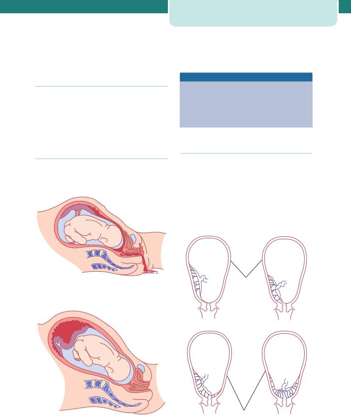
The premature separation of the placenta is termed ‘abruption’. The bleeding is maternal and/or fetal and abruption is acutely dangerous for both the mother and fetus (Figures 8.8 and 8.9; see Chapter 10, Preeclampsia and other disorders of placentation and Chapter 16, Obstetric emergencies).
Placenta praevia
A placenta covering or encroaching on the cervical os may be associated with bleeding, either provoked or
Figure 8.8 Placental abruption with revealed haemorrhage
Figure 8.9 Placental abruption with concealed haemorrhage
Antepartum haemorrhage |
103 |
spontaneous. The bleeding is from the maternal not fetal circulation and is more likely to compromise the mother than the fetus (Figure 8.10).
Risk factors for placenta praevia
•Multiple gestation
•Previous Caesarean section
•Uterine structural anomaly
•Assisted conception
Vasa praevia
Vasa praevia is present when fetal vessels traverse the fetal membranes over the internal cervical os. These vessels may be from either a velamentous insertion of the umbilical cord or may be joining an accessory (succenturiate) placental lobe to the main disk of the placenta. The diagnosis is usually suspected when either spontaneous or artificial rupture of the membranes is accompanied by painless fresh vaginal bleeding
Minor
(a) Type I |
(b) Type II |
|
Major |
(c) Type III |
(d) Type IV |
Figure 8.10 Classification of placental praevia

104Antenatal obstetric complications
from rupture of the fetal vessels. This condition is associated with a very high perinatal mortality from fetal exsanguination. If the baby is still alive once the diagnosis is suspected, the immediate course of action is delivery by emergency Caesarean section.
Key points
•Placenta praevia is most dangerous for the mother.
•Placental abruption is more dangerous for the fetus than the mother.
•Vasa praevia is not dangerous for the mother but is nearly always fatal for the baby.
Further management
If there is minimal bleeding and the cause is clearly local vaginal bleeding, symptomatic management may be given (for example, antifungal preparations for candidiasis), as long as there is reasonable certainty that cervical carcinoma is excluded by smear history and direct visualization of the cervix. Placental causes of bleeding are a major concern. A large-gauge intravenous cannula is sited, blood sent for full blood count, clotting and crossmatch, and appropriate fetal and maternal monitoring instituted. If there is major fetal or maternal compromise, decisions may have to be made about immediate delivery irrespective of gestation. Emergency management is described in Chapter 16, Obstetric emergencies. If bleeding settles, the ongoing management depends on the underlying cause. If the cause was a suspected placental abruption, the woman must be admitted for 48 hours as the risk of rebleeding is high within this time frame. Steroids should be administered if the gestation is less than 34 weeks. Rhesus status is important: if the mother is Rhesus negative, send a Kleihauer test (to determine whether any, or how much, fetal blood has leaked into the maternal circulation) and administer anti-D. The ongoing management of placenta praevia is more contentious. Many clinicians favour retaining major placenta praevias in hospital until delivery.
Rhesus iso-immunization
Blood group is defined in two ways. First, there is the ABO group, allowing four different permutations of
blood group (O, A, B, AB). Second, there is the rhesus system, which consists of C, D and E antigens. The importance of these blood group systems is that a mismatch between the fetus and mother can mean that when fetal red cells pass across to the maternal circulation, as they do to a greater or lesser extent during pregnancy, sensitization of the maternal immune system to these fetal ‘foreign’ red blood cells may occur and subsequently give rise to haemolytic disease of the fetus and newborn (HDFN).
The Rhesus system is the one most commonly associated with severe haemolytic disease.
The aetiology of Rhesus disease
The Rhesus system is coded on two adjacent genes, which sit within chromosome one. One gene codes for antigen polypeptides C/c and E/e while the other codes for the D polypeptide (Rhesus antigen). Note that the d (little d) antigen has not been identified so it may be that women who are D negative lack the antigen altogether, as opposed to those with c (little c) or e (little e), where c is the allelic antigen of C and e is the allelic antigen of E. Antigen expression is usually dominant, whereas those who have a negative phenotype are either homozygous for the recessive allele or have a deletion of that gene (Figure 8.11). In practice, only anti-D and anti-c regularly cause HDFN. Anti-D is much more common than anti-c and is therefore the focus of this discussion.
Occurrence of HDFN as a result of Rhesus isoimmunization involves three key stages (see Figure 8.12). First, a Rhesus negative mother must conceive a baby who has inherited the Rhesus positive phenotype from the father. Second, fetal cells must gain access to the maternal circulation in a sufficient volume to provoke a maternal antibody response. Finally, maternal antibodies must gain transplacental access and cause immune destruction of red cells in the fetus. Rhesus disease does not affect a first pregnancy as the primary response is usually weak and consists primarily of IgM antibodies that do not cross the placenta. Thereafter IgG antibodies are produced and these can cross the placenta. Rhesus antigens are well expressed by the fetus from as early as 30 days gestation so in a subsequent pregnancy, when maternal resensitization occurs (Rhesuspositive red cells pass from the baby to the maternal circulation; Figure 8.12), IgG antibodies cross from the mother to the fetal circulation. If these antibodies

Rhesus locus on chromosome 1
1p34
D gene |
CE gene |
Homozygous rhesus D positive |
|
D gene |
CE gene |
D gene |
CE gene |
Heterozygous rhesus D positive |
|
D gene |
CE gene |
|
CE gene |
|
Rhesus D negative |
CcEe gene
CcEe gene
Figure 8.11 The parental genotype determinants of Rhesus group
Rhesus iso-immunization |
105 |
Rh-neg |
Rh-neg father |
mother |
(homozygote) |
dd |
dd |
=> All offspring Rh-neg
dd |
dd |
dd |
dd |
Rh-neg |
Rh-pos father |
||
mother |
(heterozygote) |
||
|
dd |
|
Dd |
=> Half offspring Rh-pos
dd |
Dd |
dd |
Dd |
Rh-neg |
Rh-neg father |
||
mother |
(homozygote) |
||
|
dd |
|
Dd |
=> All offspring Rh-pos
Dd |
Dd |
Dd |
Dd |
are present in sufficient quantities, fetal haemolysis may occur, leading to such severe anaemia that the fetus may die unless a transfusion is performed. It is for this reason that Rhesus-negative women have frequent antibody checks in pregnancy; an increasing titre of atypical antibodies may suggest an impending problem.
Potential sensitizing events for Rhesus disease
•Miscarriage
•Termination of pregnancy
•Antepartum haemorrhage
•Invasive prenatal testing (chorion villus sampling, amniocentesis and cordocentesis)
•Delivery
Prevalence of Rhesus disease
The prevalence of D Rhesus negativity is 15 per cent in the Caucasian population, but lower in all other ethnic groups. It is very uncommon in Orientals. Rhesus disease is most common in countries where anti-D prophylaxis is not widespread, such as the Middle East and Russia.
Preventing Rhesus iso-immunization
The process of iso-immunization can be ‘nipped in the bud’ by the intramuscular administration of anti-D immunoglobulins to a mother, preferably within 72 hours of exposure to fetal red cells. Anti-D immunoglobulins ‘mop up’ any circulating rhesus-positive cells before an immune response is excited in the mother. The practical implications of this are that anti-D immunoglobulin must be