

Obstetrics_by_Ten_Teachers_19E_-_Kenny_Louise
.pdf246 Obstetric emergencies
Minor placenta praevia |
Major placenta praevia |
Figure 16.3 Placenta praevia
common in older (often multiparous) women and in women with previous uterine surgery. In women who have had a previous caesearean section, there is a risk that the placenta implants into, and thus invades, into the previous scar. This is called a ‘morbidly adherent placenta’ and there are three types:
1.Placenta accreta. Placenta is abnormally adherent to the uterine wall.
2.Placenta increta. Placenta is abnormally invading into the uterine wall.
3.Placenta percreta. Placenta is invading through the uterine wall.
The risk of a morbidly adherent placenta increases with increasing numbers of previous Caesarean sections.
Diagnosis
The mother will present with painless bleeding, often recurrent in the third trimester, and ultrasound scans will demonstrate the abnormal location of the placenta. The bleeding occurs due to separation of the placenta as the lower segment develops in the third trimester. Contractions can also precipitate bleeding by a similar mechanism. On abdominal palpation, the uterus will be soft and non-tender and the presenting part will be free as it cannot
enter the pelvis due to obstruction by the placenta. A digital examination is contraindicated as this can precipitate bleeding. Approximately 10 per cent of cases of placenta praevia can also be complicated by placental abruption (see below under Placental abruption).
Management
The patient should be initially resuscitated using the structured approach of ABC. If the bleeding is relatively minor and the fetus uncompromised, the patient should be admitted for observation and not allowed home until at least 24 hours has passed without further bleeding. Women with major placenta praevia who have had recurrent bleeding should be admitted as inpatients from 34 weeks, and those who have not bled need a careful risk assessment before being managed at home. Major bleeding will require fluid resuscitation and delivery of the fetus by Caesearean section by a senior obstetrician. The risk to the fetus is mainly prematurity due to early Caesarean section. There is considerable risk of serious maternal haemorrhage, either as APH or during Caesarean section when the placental bed may not contract, or due to morbid adherence. This may lead to massive postpartum haemorrhage (PPH). The indications for delivery are reaching 37–38 weeks

gestation, a massive ( 1500 mL) bleed, or continuing significant bleeding of lesser severity. Cases of minor placenta praevia can be considered for a vaginal delivery if the placenta is a minimum of 2 cm away from the cervical os.
Placental abruption
Aetiology and epidemiology
A placental abruption is separation of a normally sited placenta from the uterine wall. In most cases, the separation reaches the edge of the placenta, tracks down to the cervix and is revealed as vaginal bleeding. The remaining cases are concealed, and present as uterine pain and potentially maternal shock or fetal distress without obvious bleeding. The fetus is at risk because of hypoxia following placental separation and premature delivery. The mother is at risk of hypovolaemic shock, clotting disorders and consequent more widespread organ damage. The aetiology and pathophysiological consequences of placental abruption are discussed in further detail in Chapter 10, Pre-eclampsia and other disorders of placentation.
Diagnosis
Placental abruption typically presents as vaginal bleeding associated with pain. The pain can be constant, or as frequent short-lasting contractions caused by the irritable effect of blood within the uterus. The patient may report reduced fetal movements and the cardiotocograph may demonstrate a non-reassuring fetal heart rate pattern. Constant pain associated with a uterus that is very hard on palpation is known as a Couvelaire uterus and is due to a large volume of blood within the myometrium.
Management
The patient should be initially resuscitated using the structured approach of ABC. Management depends on recognition of the problem, realization that true blood loss may be far greater than the blood loss seen, and rapid institution of major haemorrhage management (see below under Postpartum haemorrhage). In very severe cases, the fetus will be dead and vaginal delivery can be accelerated by artificial rupture of the membranes once the mother is reasonably stable. If the fetus is alive, delivery without compromising the mother’s resuscitation is urgent and this will usually be by Caesarean section.
Management of specific obstetric emergencies |
247 |
Other causes of antepartum haemorrhage
Other causes of APH include cervical bleeding (ectropion, post-coital), genital tract infection, genital tract tumours, a show and vasa praevia. With the exception of vasa previa, these generally cause insignificant amounts of blood loss. Vasa praevia is rupture of fetal vessels running within the membranes, often near to the cervical os and damaged when the membranes rupture. It is a rare condition, but it is catastrophic for the fetus as it is fetal blood that is lost. Risk factors include placenta praevia, a velamentous placental insertion and multiple pregnancy. Although relatively small amounts of bleeding are seen, this can represent a large proportion of the total fetal blood volume. Hence, the fetus can rapidly exsanguinate and there is a high risk of fetal death. The cardiotocograph will rapidly become abnormal with a fetal tachycardia, followed by deep decelerations. Although tests for fetal haemoglobin are possible (rarely used in UK practice), the best solution is a high index of suspicion and rapid Caesarean section.
Postpartum haemorrhage
Postpartum haemorrhage (PPH) is probably one of the most common obstetric emergencies. In the UK Confidential Enquiry 2003–5, haemorrhage was the third most common cause of death. It is defined as:
•Primary PPH. Loss of 500 mL blood from the genital tract within 24 hours of delivery;
•Secondary PPH. Loss of 500 mL blood from the genital tract between 24 hours and 12 weeks post
delivery.
It is considered to be minor if the blood loss is between 500 and 1000 mL and major if it is greater than 1000 mL. In practice, blood losses between 500 and 1000 mL are relatively common, and can usually be tolerated well by the woman. Thus, it has been suggested that losses over 1000 mL should trigger emergency PPH protocols. However, it should be remembered that estimation of blood loss is notoriously inaccurate, and if a woman demonstrates evidence of cardiovascular compromise, such as tachycardia, or if there is continued bleeding, then protocols should be instituted even if estimated losses are less than 1000 mL. In common with other

248 Obstetric emergencies
Table 16.3 Risk factors for postpartum haemorrhage
Maternal |
|
Fetal |
Pre-existing |
Raised |
Large baby |
|
maternal age |
|
|
|
|
|
Primiparity |
Multiple |
|
|
pregnancy |
|
Grand |
Polyhydramnios |
|
multiparity |
|
|
|
|
|
Uterine |
Shoulder |
|
fibroids |
dystocia |
|
|
|
|
Previous |
|
|
caesarean |
|
|
|
|
|
Bleeding |
|
|
disorders |
|
|
|
|
|
Obesity |
|
|
|
|
|
Antepartum |
|
|
haemorrhage |
|
|
|
|
|
Previous PPH |
|
Intrapartum |
Prolonged |
|
|
labour |
|
|
|
|
|
Caesarean |
|
|
section |
|
|
|
|
|
Instrumental |
|
|
delivery |
|
|
Pyrexia in |
|
|
labour |
|
|
|
|
|
Episiotomy |
|
|
|
|
obstetric emergencies, PPH can often be predicted and preventative measures undertaken if significant risk factors are present (Table 16.3).
Aetiology and epidemiology
The causes of PPH can be remembered as the four ‘Ts’:
Tone |
Uterine atony |
|
|
Tissue |
Retained placenta and/or |
|
membranes |
|
|
Trauma |
Injury to vagina, perineum |
|
and uterine tears at |
|
Caesarean section |
Thrombin |
Clotting disorders |
|
|
Uterine atony, or failure of the uterus to contract after the delivery of the placenta (‘tone’), is the most common cause of PPH and can cause torrential loss of blood immediately following delivery. It can be predicted, and therefore steps taken to prevent it, by the use of oxytocic infusions and active management of the third stage of labour. A retained placenta (‘tissue’) can also prevent a uterus from contracting efficiently until the tissue is removed. Occasionally, parts of the placenta or membranes can be retained, and this can be identified by careful examination of the placenta following delivery. Almost all types of delivery can cause some degree of genital tract trauma in the form of perineal and vaginal tears, although this is most common following a forceps delivery. Rarely, the cervix can be torn if delivery has occurred before the cervix is fully dilated. More rarely, abnormal blood clotting (‘thrombin’) can contribute to an excessive blood loss. This can occur in women with an underlying disorder such as Von Willebrand’s disease, or platelet disorders. It more commonly arises in women who have developed a consumptive coagulopathy as a result of another obstetric complication, such as a massive placental abruption, an unidentified dead fetus, amniotic fluid embolus or massive haemorrhage.
Diagnosis
Early recognition of blood loss and rapid action is vital in the management of PPH. Appreciation of risk factors, accurate estimation of blood loss and recognition of the maternal signs of cardiovascular compromise are vital. These include a tachycardia, low blood pressure, symptoms of nausea, vomiting and feeling faint, pallor and slow capillary refill (greater than 2 seconds). It is important to recognize that young, fit women have the capacity to tolerate large amounts of blood loss without demonstrating many clinical symptoms. The earliest symptom will be a tachycardia and often blood pressure does not fall until massive haemorrhage has occurred (often 1200–1500 mL of blood).
Management
In practice, diagnosis and management of PPH occur simultaneously. The structured ABC approach outlined above should be instituted. This management is summarized in Table 16.4. Rapid fluid resuscitation should occur at the same time as assessing and treating the cause. Since uterine atony is the most common cause, the uterus should be massaged to

Table 16.4 Management of severe postpartum haemorrhage
Summon help from: |
senior obstetrician |
|
anaesthetist |
|
senior midwife |
|
porter |
|
|
Oxygen by mask initially |
|
2 14-gauge intravenous lines
Full blood count and clotting studies
Test for renal function and liver function tests
Cross-match at least 6 units of blood
Fluid resusication intravenously
Notify blood bank and consult haematologist
Foley catheter into the bladder and fluid balance chart
Transfuse blood as soon as possible – uncrossmatched same group as mother or, in extreme cases, O negative
Central venous pressure and arterial lines
May need fresh frozen plasma, platelets and cryoprecipitate (consult haematologist)
Eliminate the cause – deliver the baby and placenta, manage postpartum haemorrhage
encourage contraction and oxytocics given. In the first instance, this would be a further bolus of the drug used to manage the third stage (oxytocin or Syntometrine) and an infusion of oxytocin (40 IU in 500 mL saline over 4 hours). Bimanual compression and more potent drugs can also be used. These include ergometrine, prostaglandin F2α or misoprostol. The bladder should be catheterized as an empty bladder aids uterine contraction. A vaginal examination should be conducted to expel clots which will prevent contraction of the uterus and assess for genital tract trauma. Any identified tears will need prompt compression to limit blood loss followed by repair. The placenta should be delivered if retained and inspected. If bleeding continues, the patient should be transferred to theatre to allow a further thorough examination under anaesthetic. This will also allow the use of further measures, including uterine tamponade using uterine balloons, radiological occlusion of the uterine vessels, laparotomy for bilateral iliac artery ligation, uterine compression sutures, and, as a last resort, hysterectomy. Massive PPH will require
Management of specific obstetric emergencies |
249 |
correction of clotting factors using fresh frozen plasma, platelets and cryoprecipitate.
Secondary PPH is a rare cause of massive bleeding. It is usually the result of retained products of conception and/or uterine infection. Although bleeding can be life threatening, it is usually slight or moderate.
Key points
Massive haemorrhage
•Summon senior multidisciplinary help
•Resuscitate
•Replace and maintain fluid volume
•Investigate status and cause of bleeding
•Arrest blood loss
Hypertensive disorders
Pre-eclampsia is a disease of pregnancy characterized by a blood pressure of 140/90 mmHg or more on two separate occasions after the 20th week of pregnancy in a previously normotensive woman. This is accompanied by significant proteinuria (300 mg in 24 hours). Eclampsia is the same condition that has proceeded to the presence of convulsions. Imminent eclampsia (the development of seizures), or fulminating pre-eclampsia, is the transitional condition characterized by increasing symptoms and signs. Pre-eclampsia is discussed in detail in Chapter 10, Pre-eclampsia and other disorders of placentation. The management of severe or fulminating preeclampsia and eclampsia is included in this chapter.
Aetiology and epidemiology
Eclampsia is relatively rare in the UK, occurring in approximately 1:2000 pregnancies. It may occur antepartum (40 per cent), intrapartum (20 per cent) or postpartum (40 per cent). Severe pre-eclampsia is more common than eclampsia, occurring in 5:1000 pregnancies. Other risk factors are discussed in Chapter 13, Perinatal infections.
Diagnosis
Severe pre-eclampsia is identified by a blood pressure of 160/110 mmHg or more and the presence of proteinuria on ‘dipstick’ testing. A 24-hour urine collection for quantification of proteinuria may be started, but in practice there may not be time to

250 Obstetric emergencies
wait for its completion before effecting delivery. The symptoms and signs of severe pre-eclampsia are:
Symptoms
•Frontal headache
•Visual disturbance (blurred vision and flashing lights)
•Epigastric pain
•General malaise and nausea
•Restlessness
Signs
•Agitation
•Hyper-reflexia and clonus
•Facial (especially periorbital) oedema
•Right upper quadrant tenderness
•Poor urine output
•Papilloedema
In addition, the fetus may appear small, with oligohydramnios and reduced fetal movements. The cardiotocograph may demonstrate signs of hypoxia with a fetal tachycardia, reduced variability and decelerations. Eclampsia is obvious as a grand mal convulsion. However, other causes of fits such as epilepsy have to be considered. Preceding preeclampsia suggests eclampsia, but in approximately one-third of cases the eclamptic fit precedes other signs. After the convulsion, the blood pressure is frequently normal for a while, but proteinuria will usually still be present. Any convulsion in pregnancy should be considered to be eclamptic until proved otherwise.
Management
In the same way as other emergencies, the structured ABC approach outlined above should be used. Call for help from senior obstetric and anaesthetic colleagues. Depending upon the results of clotting studies and the severity of the disease, a consultant haematologist may need to be informed.
In a woman with severe pre-eclampsia, the airway and breathing are likely to be secure. However, if a seizure has occurred, these will need assessment and treatment. Seizures occurring due to eclampsia are usually short-lasting and self-limiting. However, the patient should be moved to the side (recovery
position) and oxygen applied. Large bore intravenous cannulae should be sited and blood taken for full blood count, clotting studies, renal and liver function tests and cross-matching. The mother’s condition needs to be stabilized urgently, before considering delivery in antenatal cases. Stabilizing the mother’s condition will involve blood pressure control, prevention and treatment of fits and management of fluid balance.
Intravenous magnesium sulphate is used to treat and prevent fits. The MAGPIE trial showed that administration of magnesium sulphate halved the risk of seizures. An initial loading dose of 4 g is given, followed by an infusion of 1 g/hour. Magnesium sulphate can also lower blood pressure and cause some maternal side effects, such as flushing. It is important to recognize that overdose of magnesium sulphate can cause respiratory and cardiac depression. This can be reversed using calcium gluconate.
The blood pressure should be reduced to safe levels. In common with previous reports, the UK Confidential Enquiry 2003–5 found that deaths in association with preeclampsia and eclampsia occur as result of intracranial haemorrhage, largely due to poor blood pressure control. In particular, systolic hypertension greater than 160 mmHg must be treated. Antihypertensives can be either labetalol (can be given orally while intravenous access is obtained), oral nifedipine or intravenous hydrallazine. If given intravenously, a bolus is initially used followed by an infusion that can be titrated to obtain a safe blood pressure. Maternal observations should be conducted frequently until the mother has stabilized (every 5–15 minutes depending on condition) and continuous fetal monitoring used. A gradual reduction of the blood pressure is optimal to avoid precipitating fetal distress (in the form of a bradycardia) secondary to sudden drops in maternal blood pressure that reduce uterine blood flow.
Management of fluid balance can be problematic. In pre-eclampsia, there is intense peripheral vasoconstriction accompanied by a decrease in the plasma volume, together with redistribution of the extracellular fluid. The urine output falls, and overenthusiastic efforts to provide a fluid challenge may cause pulmonary and cerebral oedema. A strict input/output balance (this needs catheterization) must be maintained, using either blood products or crystalloids, as appropriate. In the absence of bleeding, no more than 80 mL/hour of fluids (oral and intravenous) should be given. Renal failure is uncommon and if it occurs is usually reversible.

Timing of delivery depends upon the gestation, the presence of other complicating factors, the severity of the disease and the stability of the patient’s condition. In general, if the patient has disease severe enough to warrant antihypertensive and anticonvulsant therapy, delivery should follow stabilization of the maternal condition.
When it is clear that early delivery is likely, if the gestation is less than 34 weeks, steroids should be given to improve lung maturity and decrease neonatal complications. Steroid administration must not delay delivery that is necessary for the control of severe maternal problems, although recent evidence indicates that steroids also benefit the mother with pre-eclampsia.
Delivery is often by Caesarean section, although if labour is well established, vaginal delivery is possible. If at all possible, clotting disorders must be corrected before delivery (by whatever means) is attempted. Postpartum, both fulminating pre-eclampsia and eclampsia may occur. Management is as for the antenatal case except that delivery has already taken place.
HELLP syndrome
HELLP syndrome – a combination of haemolysis, elevated liver enzymes and low platelets – is seen in 5–10 per cent of cases of severe pre-eclampsia. It is more common in multiparous women. It may be associated with disseminated intravascular coagulation, placental abruption and fetal death.
Key points
Hypertensive disorders
•Fulminating pre-eclampsia and eclampsia are dangerous.
•Recognize women at risk.
•Manage minor hypertensive problems to prevent progression.
•In the serious case:
•call for help
•prevent or control convulsions with magnesium sulphate
•control the blood pressure
•tightly control fluid balance
•minimize or avoid organ damage
•control coagulopathy
•deliver a healthy baby safely.
Management of specific obstetric emergencies |
251 |
Uterine causes of obstetric emergency
Uterine rupture
Aetiology and epidemiology
Uterine rupture, or a tear in the uterus, usually happens due to a previous uterine injury. It is rare, occurring with an incidence of 0.03–0.3 per cent. It occurs mainly in association with a previous Caesarean section. This is because scar tissue does not have the same inherent strength as myometrium. However, scarring can also exist as a result of previous uterine surgery, such as a surgical evacuation of retained products of conception resulting in a perforation. The vast majority of cases occur during labour, usually during late first stage or the active second stage. The risk factors for uterine rupture are given in Table 16.5.
Diagnosis
The patient may complain of abdominal pain (‘scar tenderness’, often not masked epidural analgesia) and vaginal bleeding. Haematuria may be present if the uterus has ruptured into the bladder. Typically, contractions stop and decelerations are present on the cardiotocograph. If the rupture occurs in the late second stage of labour, it may not be recognized immediately. In this scenario, the fetus has usually delivered by ventouse or forceps for an abnormal cardiotocograph (CTG). In the immediate postnatal period, the mother bleeds internally and shows signs of circulatory collapse while complaining of abdominal discomfort.
Management
Immediate resuscitation of ABC is required as previously outlined. Immediate laparotomy to deliver
Table 16.5 Risk factors for uterine rupture
Previous Caesarean section
Previous uterine surgery
Induction and augmentation of labour
High parity
Macrosomic fetus
Placenta percreta
Fetal version, e.g. breech extraction
Congenital uterine anomaly, e.g. unicornuate uterus
252 Obstetric emergencies
the baby and repair the uterine is required. Frequently, the only safe treatment is hysterectomy.
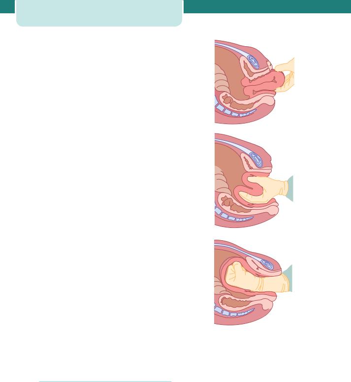
Uterine inversion
Aetiology and epidemiology
Uterine inversion is a rare complication occurring during the third stage of labour. It has a reported incidence of between 1:2000 and 1:6000. The uterine fundus descends either the uterine cavity, through the cervix, and very rarely beyond the introitus (Figure 16.4). It is caused by traction on the umbilical cord before the placenta has separated and can occur after vaginal deliveries or Caesarean section. Associated factors are a fundal placenta, a short cord and a morbidly adherent placenta.
Diagnosis
The prolapsed uterus stretching the cervix causes vagal stimulation, thus the woman will demonstrate signs of cardiovascular collapse and shock. Although haemorrhage is commonly present, the symptoms will be out of proportion to the estimated blood loss. The inverted uterus may be obvious at the introitus, but other signs include the lack of a palpable uterus in the abdomen or the feeling of a ‘dimple’ in the uterine fundus on abdominal examination.
Management
Resuscitate the patient using the structured ABC approach. It is very important not to remove the placenta if it is still attached as this will increase the bleeding. Immediately replace the uterus through the cervix by manual compression. If that fails, hydrostatic pressure can be applied by pouring warmed saline into the vagina, usually via a silc cup ventouse. Tocolysis may be helpful to relax the cervical ring. Surgery, to reposition the uterus from above, should be used as a last resort. After replacement, uterine contraction is maintained with an oxytocic.
Sudden maternal collapse
Sudden collapse, as in non-pregnant adults, occurs due to a multitude of reasons. Some will be benign, such as a vasaovagal attack (simple faint) or an epileptic fit in a known epileptic, but other causes are life threatening and are outlined in Table 16.2.
(a)
(b)
(c)
Figure 16.4 Uterine inversion
The management approach should be the same, structured ABC approach. The causes of particular relevance to pregnant women are discussed in more detail below.
Pulmonary embolism
Aetiology and epidemiology
Thrombosis is consistently the most common cause of maternal death in the UK, and the UK Confidential

Enquiry 2003–5 reported 33 deaths caused by pulmonary embolism (PE). The most recent data (Table 16.1) suggest an incidence of between 10.6 and 16.1 per 100 000 maternities in the UK. It is important to recognize that although PE is more common in the puerperium, it can occur at any time in the antenatal and post-natal period. The diagnosis and management of thromboembolic events is discussed in detail in Chapter 8, Antenatal obstetric complications, thus this chapter will focus on the emergency situation.
Diagnosis and management
PE can be a cause of sudden cardiorespiratory collapse. In this situation, diagnosis and management should occur simultaneously. Urgent resuscitation using the structured ABC approach is needed. If PE is suspected, anticoagulation should be instituted.
Amniotic fluid embolism
Aetiology and epidemiology
Amniotic fluid embolism is a rare cause of maternal collapse specific to pregnancy, believed to be caused by amniotic fluid entering the maternal circulation. This causes acute cardiorespiratory compromise and severe disseminated intravascular coagulation. In some cases, there may be an abnormal maternal reaction to amniotic fluid as the primary event. It is difficult to diagnose in life, and is typically diagnosed at postmortem, with the presence of fetal cells (squames or hair) in the maternal pulmonary capillaries. It caused 18 deaths in the 2003–5 maternal mortality report.
Diagnosis and management
In the case of sudden collapse, management should be the structured ABC approach. Symptoms occurring just before the collapse may be helpful in diagnosis. The 2003–5 maternal mortality report suggested that women may report the following symptoms:
•breathlessness
•chest pain
•feeling cold
•lightheadedness
•restlessness, distress and panic
•pins and needles in the fingers
•nausea and vomiting.
Management of specific obstetric emergencies |
253 |
The prognosis is poor, with around 30 per cent of patients dying in the first hour and only 10 per cent surviving overall. Management is supportive, requiring intensive care and there are no specific therapies available.
Fetal emergencies
The fetus may be severely affected by any of the preceding maternal emergencies that occur before delivery. However, there are some emergencies that directly affect the fetus without major immediate physical compromise of the mother. Major abnormalities of the fetal heart rate, in particular prolonged fetal bradycardia, call for immediate delivery, usually by Caesarean section. This is discussed further in Chapter 14, Labour. This chapter will discuss two specific causes of fetal emergency: cord prolapse and shoulder dystocia.
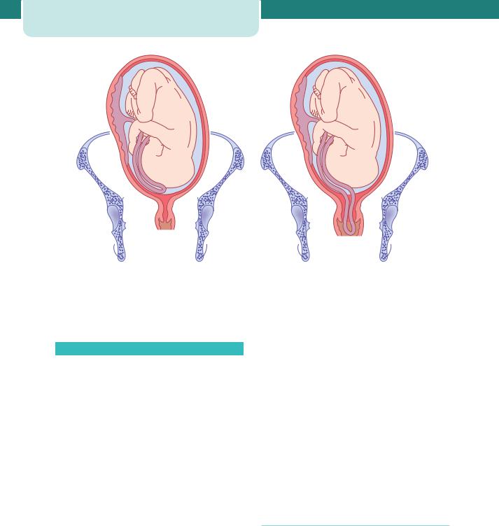
Umbilical cord accidents (cord prolapse)
Aetiology and epidemiology
A cord presentation is defined as the presence of umbilical cord below the fetal presenting part when the membranes are intact. Cord prolapse is the presence of the cord below the presenting part when the membranes are ruptured (Figure 16.5). It has an incidence of 1:500 deliveries and occurs when the fetal presenting part does not fit well into the maternal pelvis, giving ‘space’ for the cord to prolapse when the membranes rupture. It is associated with risk factors outlined in Table 16.6.
Diagnosis
Most commonly, it is diagnosed by seeing the cord at the introitus, or feeling it during a vaginal examination. However, an abnormal fetal heart rate pattern may suggest it, as compression of the umbilical vein between the presenting part and the pelvis, reduces or stops the flow of oxygenated blood to the fetus, causing deep variable decelerations, then bradycardia if the situation is not relieved.
Management
Immediate management aims to minimize the pressure of the fetal presenting part on the cord,
254 Obstetric emergencies
(a) |
(b) |
Figure 16.5 |
Cord prolapse. (a) Cord presentation. The cord is below the presenting part (head in this case but commonly |
a malpresentation) with the membranes intact. (b) Cord prolapse. The membranes have ruptured and the cord is below the presenting part. In this case, it has prolapsed into the vagina
Table 16.6 Risk factors for cord prolapse
Maternal causes |
Fetal causes |
Pelvic tumours |
Prematurity |
(e.g. fibroids in the |
|
lower segment) |
|
|
|
Narrow pelvis |
Malpresentation, e.g. |
|
breech, transverse lie |
|
Multiple pregnancy |
|
Polyhydramnios |
|
Placenta praevia |
|
Large baby |
|
|
while plans are made to deliver the baby. This is achieved by moving the woman on to all fours with the head down, applying pressure vaginally to push the presenting part out of the pelvis, or by filling the bladder with 500 mL of saline. There should be minimal handling of the cord, as this causes spasm which will worsen blood flow. However, if the cord is beyond the introitus it should be replaced into the vagina to keep it warmer. Emergency Caesarean
section is required unless the cervix is fully dilated and an assisted vaginal delivery can be safely and easily performed. Fetal outcome depends upon the gestation, other complicating factors such as intrauterine growth restriction, and for how long the cord has been compressed. With a term baby and a prompt diagnosis in hospital, the prognosis is usually excellent. If the cord prolapse occurs outside hospital, the fetus is likely to be dead by the time of admission. Total cord compression for longer than 10 minutes will cause cerebral damage and, if continued for around 20 minutes, death. These times will be shorter in a fetus that is already compromised for reasons such as prematurity or fetal growth restriction.
Shoulder dystocia
Aetiology and epidemiology
Shoulder dystocia is defined by the Royal College of Obstetricians and Gynaecologists (RCOG) as the need for ‘additional obstetric manoevres to release the shoulders after gentle downward traction has failed’.
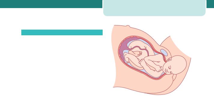
Table 16.7 Risk factors for shoulder dystocia
Maternal |
Fetal |
Intrapartum |
Diabetes |
Macrosomia |
Long first stage |
|
|
of labour |
|
|
|
Short |
Postmaturity |
Long second |
stature |
|
stage of labour |
|
|
|
Previous |
|
Instrumental |
shoulder |
|
delivery |
dystocia |
|
|
|
|
|
Obesity |
|
Induction of |
|
|
labour |
|
|
|
|
|
Use of |
|
|
oxytocin |
|
|
|
The incidence is approximately 0.6 per cent in the UK and it results in excessive morbidity for both mother and fetus. There is a risk of fetal hypoxia, death and fetal trauma, in the form of fractures (usually long bones of the arm or clavicle), or brachial plexus injury. For the mother, there is an increased risk of bleeding and perineal trauma, including third and fourth degree tears. It is very difficult to predict shoulder dystocia, although the well-recognized risk factors are given in Table 16.7. Unfortunately, none of these risk factors are clinically useful predictors. Approximately half of shoulder dystocias occur in infants of ‘normal’ weight (i.e. less than 4 kg).
During vaginal delivery, the fetal head and shoulders rotate to make use of the widest pelvic diameters. After delivery of the head, restitution occurs and the shoulders rotate into the anteroposterior (AP) diameter. This makes use of the widest AP diameter of the pelvic outlet. However, if the shoulders have not entered the pelvic inlet, the anterior shoulder may become caught above the symphysis pubis. Occasionally, both shoulders (or rarely, the posterior shoulder) may remain above the pelvic brim (see Figure 16.6).
Diagnosis
Diagnosis is usually obvious when the shoulders fail to deliver during the next contraction after delivery of the head. It is sometimes preceded by the ‘turtle sign’,
Management of specific obstetric emergencies |
255 |
Figure 16.6 Shoulder dystocia. After delivery of the head, shoulder dystocia occurs due to the shoulders being unable to pass under the maternal symphysis pubis
which is the head appearing to be pulled back on to the perineum at delivery.
Management
Shoulder dystocia is very alarming and its management should be practised in ‘drill-type’ training. The high-risk of litigation necessitates that meticulous notes are kept, and no more than gentle traction is ever applied to the fetal head. Thus, on recognition, the emergency buzzer should be activated and help summoned. This should include senior obstetricians, midwifery staff, a paediatrician and an individual to document the times at which the specific manoeuvres are used. The vessels in the fetal neck are occluded after delivery of the head, and cerebral damage will occur if delivery is delayed significantly. The fetus may already be compromised because of the prolongation of labour. After 5 minutes, there is the risk of cerebral damage. However, inappropriate traction, particularly downward traction on the head causing lateral flexion of the head on the neck, will cause stretching of the brachial plexus and nerve damage. Erb’s palsy is a likely consequence.
Shoulder dystocia is managed by a sequence of manoeuvres designed to facilitate delivery while minimizing the risk of fetal damage. The basic