

Obstetrics_by_Ten_Teachers_19E_-_Kenny_Louise
.pdf216 Labour
Resuscitating the fetus in labour
•Maternal dehydration and ketosis can be corrected with intravenous fluids.
•Maternal hypotension secondary to an epidural can be reversed by a fluid bolus, although a vasoconstrictor such as ephedrine is occasionally necessary.
•Uterine hyperstimulation from excess oxytocin can be treated by turning off the infusion temporarily and using tocolytic drugs, such as terbutaline.
•Venocaval compression and reduced uterine blood flow can be eased by turning the woman into a left lateral position.
Fresh, thick meconium in the presence of a reassuring CTG is still a cause for concern, and although the labour should be allowed to continue, the threshold for intervention will be lowered and a paediatrician should be present at delivery.
Fetal blood sampling
•Explanation is given and consent obtained from the woman. She is asked to lie in the left lateral position.
•An amnioscope is inserted into the vagina and its distal end is placed at right angles on to the fetal head. The scalp is cleaned and a small cut is made using a blade with a guard. The resulting blood is collected into a microtube. The amount of blood required is approximately 25 mL.
•A normal pH value is above 7.25. A pH below 7.20 is confirmation of fetal compromise. Values between 7.20 and 7.25 are ‘borderline’.
•The base deficit can also be useful in interpretation of the fetal scalp pH. A base excess of more than 10 demonstrates a significant metabolic acidosis, with increasing risk of fetal neurological injury beyond this level.
•More than one fetal scalp sample may be necessary over the course of the labour. A downward trend in the fetal scalp pH values is to be expected and should be assessed together with how the labour is progressing.
•If an abnormal CTG persists in labour, then, despite normal values, fetal scalp sampling should be repeated every 60 minutes, or sooner if the CTG deteriorates. If the result is borderline, it should be repeated no more than 30 minutes later.
C A S E H I S T O R Y
Mrs S is a 30-year-old gravida 2 para 1. She is 153 cm tall. Her previous baby weighed 3.2 kg at term and was delivered by
forceps after a prolonged second stage. The father of this second baby is a new partner.
She has been admitted in spontaneous labour 10 days past her due date. Her hand-held records show concern from her midwife that this is a significantly larger baby than last time. Indeed, a scan at 36 weeks gestation placed the abdominal and head circumference measurements on the 97th centiles.
A subsequent glucose tolerance test was normal.
Despite good progress in the earlier part of the labour, dilatation has arrested at 7 cm. On examination, the head is found to be 3/5 palpable per abdomen; the position is left occipito-transverse and the contractions are poor. There is meconium-stained amniotic fluid.
What risks do Mrs S and her baby face?
The large fetal size and the short maternal stature raise the possibility of CPD. The previous need for a forceps delivery of only an average-size baby may indicate small pelvic diameters. There is a risk of obstructed labour, poor progress and the need for a Caesarean section. The meconium may simply be a sign of fetal maturity, but it may also indicate a degree of fetal compromise. At the very least, it poses the risk of meconium aspiration at delivery.
What care should Mrs S receive?
She should be informed of the concerns and provided with adequate pain relief. Fetal monitoring with a continuous CTG should begin (fortunately, this proves to be reassuring). There is no need for an FBS at this point.
She should then be assessed by an obstetric consultant or an experienced registrar, who must make a diagnosis as to the cause of this secondary arrest in first stage. Has the delay in labour occurred simply because of poor uterine contractions? Is there CPD, exacerbated by fetal malposition?
If there is no caput or moulding, and the contractions do indeed prove to be poor and infrequent, any maternal dehydration should be corrected and a cautious trial of oxytocin can be considered. However, this woman has significant risk factors for CPD, and
augmenting the labour with oxytocin risks uterine rupture if the labour is truly obstructed. An alternative option is to offer a Caesarean section at this point, or reassess 2 hours later without augmentation.
What subsequent assessment should be undertaken?
If oxytocin is used, a repeat examination should be performed by the same doctor 2 hours later. If the augmentation has been unsuccessful and the cervix is no further dilated, a Caesarean section is indicated. Even if full dilatation is reached, a vaginal delivery cannot be guaranteed. The obstetrician must be
confident that the head has descended appropriately, without the development of excessive moulding or caput.

Labour in special circumstances
Women with a uterine scar
Some women will have a pre-existing uterine scar, usually because of a previous Caesarean section. Approximately 20 per cent of all deliveries in developed countries are by Caesarean section and 99 per cent are performed through the lower segment of the uterus because blood loss is less, healing is better and the risk of subsequent rupture is lower than that following an upper segment or ‘classical’ Caesarean section. There are still a few indications for upper segment Caesarean section and it is important that these women are counselled appropriately. It is estimated that uterine rupture or dehiscence (partial rupture) occurs in approximately 1 in 200 women who labour spontaneously with a pre-existing lower segment uterine scar. The risk is two to three times higher than this in women with a previous upper segment incision.
Signs of uterine rupture include severe lower abdominal pain, vaginal bleeding, haematuria, cessation of contractions, maternal tachycardia and fetal compromise (often a bradycardia, see Figure 14.32). Uterine rupture carries serious maternal risks (shock, need for blood transfusion and operative repair, possibly a hysterectomy) and also serious fetal risks (including hypoxia, permanent neurological injury and intrapartum death).
Rupture of the uterus is particularly likely to occur:
•late in the first stage of labour,
•with induced or accelerated labour,
•in association with a large baby.
Labour after a previous Caesarean section is known as ‘vaginal birth after Caesarean’ (VBAC). Approximately 70–80 per cent of women who attempt a VBAC will give birth vaginally and the remainder will need repeat Caesarean delivery. The chances of a successful vaginal birth depend on a number of factors, including a previous history of vaginal birth, size of the baby and the original indication for the first Caesarean.
Relative contraindications to VBAC include:
•two or more previous Caesarean section scars,
•the need for induction of labour (IOL)
Labour in special circumstances |
217 |
•a previous labour progress and outcome suggestive of CPD.
A previous classical Caesarean section is an absolute contraindication to attempting vaginal birth.
If a woman with a previous history of a Caesarean section delivery is admitted in labour, intense surveillance is required to identify early signs of uterine rupture. Continuous CTG monitoring is strongly recommended and there should be a low threshold for urgent delivery by repeat Caesarean section.
Some women will have scars on the uterus as a result of a previous myomectomy. In general, there is minimal danger of rupture of a myomectomy scar unless the uterine cavity was extensively opened during the procedure.
Malpresentations
Breech presentation
The antenatal management of breech presentation and the mechanics of the delivery are discussed in Chapter 8, Antenatal obstetric complications. For this discussion, it is sufficient to list some of the complications of a breech labour, which include the following:
•Increased risk of prolapsed cord: this is particularly so with footling breech presentations,
and to a lesser extent with the flexed breech. This can cause rapid and severe hypoxia in the fetus (see Chapter 16, Obstetric emergencies).
•Increased risk of CTG abnormalities: cord compression is common during a breech vaginal
delivery.
•Mechanical difficulties with the delivery of the shoulders and/or after-coming head: damage
to the visceral organs or the brachial plexus can occur if traction is exerted on the breech. Delay in the delivery of the head may occur with the larger fetus, leading to prolonged compression of the umbilical cord and asphyxia. An uncontrolled rapid delivery of the head may occur with a smaller fetus and this predisposes to tentorial tears and intracranial bleeding.
The majority of persistent breech presentations recognized antenatally are delivered by Caesarean section to avoid these risks. Although this is evidence based, and it is probably safer for breech babies to be

218Labour
delivered this way, the most frequent cause of harm in breech labours is the failure to recognize and respond to CTG abnormalities, rather than mechanical problems at the very end of the labour. There is still a place for the vaginal delivery of a breech presentation. Maternal choice and the failure to detect breech presentation until very late in labour will mean that there will continue to be a need for obstetricians to be expert in the skills of breech vaginal delivery.
Poor progress in a breech labour is taken by most to be an indication for Caesarean section. However, a few would support the use of augmentation with oxytocin if contractions are infrequent.
Face presentation
This malpresentation occurs in about 1:500 labours and is due to complete extension of the fetal head (Figure 14.14). In the majority of cases, the cause for the extension is unknown, although it is frequently attributed to excessive tone of the extensor muscles of the fetal neck. Rarely, extension may be due to a fetal anomaly such as a thyroid tumour. The presenting diameter is the submento-bregmatic, which measures 9.5 cm, and is approximately the same in dimension as the suboccipito-bregmatic (vertex) presentation. Despite this, engagement of the fetal head is late and progress in labour is frequently slow, possibly because the facial bones do not mould. It is diagnosed in labour by palpating the nose, mouth and eyes on vaginal examination (Figure 14.25). If progress in labour is excellent, and the chin remains mentoanterior, vaginal delivery is possible, the head being delivered by flexion (Figure 14.26). If the chin is posterior (mento-posterior position), delivery is impossible, as extension over the perineum cannot occur. In this circumstance, Caesarean section is performed. Oxytocin should not be used, and if there is any concern about fetal condition, Caesarean section should be carried out. Forceps delivery is permitted for low mento-anterior face presentations.
Brow presentation
This arises when there is less extreme extension of the fetal neck than that with a face presentation. It can be considered a midway position between vertex and face. It is the least common malpresentation, occurring in 1:2000 labours. The causes of this are similar to those of face presentation, although some brow presentations arise as a result of exaggerated
extension associated with OP position. The presenting diameter is the mento-vertical (measuring 13.5 cm) (Figures 14.14 and 14.27). This is incompatible with a vaginal delivery. It is diagnosed in labour by palpating the anterior fontanelle, supra-orbital ridges and nose on vaginal examination (Figure 14.28). If this presentation persists, delivery will only be achieved by Caesarean section.
Shoulder presentation
This is reported as occurring in 1:300 pregnancies at term, but few of these women will go into labour. Shoulder presentation occurs as the result of a transverse or oblique lie of the fetus and the causes of this abnormal presentation include placenta praevia, high parity, pelvic tumour and uterine anomaly (see Chapter 8, Antenatal obstetric complications). Delivery should be by Caesarean section. Delay in making the diagnosis risks cord prolapse and uterine rupture.
Multiple gestations
About 1:80 pregnancies at term are multi-fetal. Highorder multiples, such as triplets and quadruplets, are now invariably delivered by Caesarean section, because of the difficulties in monitoring more than two fetuses and the elevated risks of fetal compromise. The second twin is at greater risk of sustaining intrapartum injury than the presenting twin, or a singleton. Currently, the evidence base does not strongly support recommending elective Caesarean delivery for all twins and this is the subject of a large on-going multinational randomized controlled trial. Nevertheless, elective (i.e. planned) Caesarean section is frequently performed in twin pregnancies for a variety of justifiable reasons.
Indications for elective Caesarean section in
twin pregnancy
•Malpresentation of the first twin
•Second twin larger than the first
•Evidence of IUGR in one or both twins
•Monoamniotic twins
•Placenta praevia
•Maternal request
|
|
Induction of labour |
219 |
the term ‘presumed fetal compromise’ should be used instead.
• Augmentation of labour with oxytocin will often correct poor uterine contractions and may help to resolve fetal malposition.
• Augmentation of labour with oxytocin can be dangerous in multiparous women, in those with a uterine scar, and in cases of malpresentation.
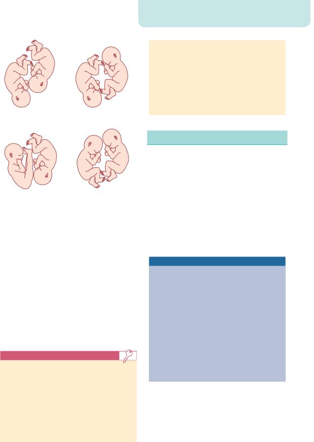
Cephalic/Cephalic (60%) |
Cephalic/Breech (20%) |
Breech/Cephalic (10%) |
Breech/Breech (10%) |
Figure 14.37 The four most common patterns of fetal presentation in a twin pregnancy
In 70–80 per cent of twin gestations the presenting twin is cephalic, with the majority of the remainder being breech (see Figure 14.37). Essentially, the lie and the presentation of the second twin are not crucially important. However, planned Caesarean section will usually be performed if the first twin presents by the breech, and certainly if it is transverse.
The mechanics of the delivery of twins is discussed in greater detail in Chapter 9, Twins and higher multiple gestations. Suffice to say that CTG abnormalities, fetal compromise, malpresentations, cord prolapse, need for emergency Caesarean section and postpartum haemorrhage all occur more commonly than in singleton labours.
Key points
Labour
•Most labours are uncomplicated and the outcomes are good.
•Labour can be a hazardous journey for the baby.
•Abnormalities of the uterine contractions (the ‘powers’), the fetus (the ‘passenger’) and the pelvis and lower genital tract (the ‘passages’) can cause abnormal labour.
•The term ‘fetal distress’ is unhelpful and often misleading. If there are concerns regarding fetal well-being in labour,
Induction of labour
Induction of labour is the planned initiation of labour prior to its spontaneous onset. Approximately one in five deliveries in the United Kingdom occur following induction of labour. The reasons for IOL are listed below. Broadly speaking, an IOL is performed when the risks to the fetus and/or the mother of the pregnancy continuing outweigh those of bringing the pregnancy to an end. It should only be performed if there is a reasonable chance of success and if the risks of the process to the mother and/or fetus are acceptable. If either of these is not the case, a planned Caesarean section should be performed instead.
Common indications for induction of labour
•Prolonged pregnancy
•Fetal growth restriction
•Pre-eclampsia and other maternal hypertensive disorders
•Deteriorating maternal illnesses
•Prelabour rupture of membranes
•Unexplained antepartum haemorrhage
•Diabetes mellitus
•Twin pregnancy continuing beyond 38 weeks
•Intrahepatic cholestasis of pregnancy
•Maternal iso-immunization against red cell antigens
•‘Social’ reasons
The most common reason for IOL is prolonged pregnancy (previously described as ‘post-term’ or ‘post-dates’). There is evidence that pregnancies extending beyond 42 weeks gestation are associated with a higher risk of stillbirth, fetal compromise in labour, meconium aspiration and mechanical

220Labour
problems at delivery. Because of this, women are usually recommended IOL between 41 and 42 weeks gestation. Induction for prolonged pregnancy does not increase the rate of Caesarean section. The evidence is good quality, and the recommendation a strong one, however 300–400 pregnancies need to be induced to prevent the one perinatal death which would have occurred if the pregnancies had been managed expectantly beyond 42 weeks gestation. Women who choose not to be induced for this reason are offered more intensive serial fetal monitoring (see Chapter 8, Antenatal obstetric complications).
Prelabour rupture of membranes (PROM) is another common indication for IOL. It is not uncommon for the membranes to rupture and the subsequent onset of labour to be significantly delayed. The longer the time delay between membrane rupture and the delivery of the baby, the greater the risk of ascending infection (chorioamnionitis) and neonatal infectious morbidity. At term (beyond 37 weeks), good quality evidence supports IOL approximately 24 hours following membrane rupture. This policy, endorsed in the NICE guideline on IOL, reduces rates of chorioamnionitis, endometritis and admissions to the neonatal unit. The evidence is less clear at present when PROM occurs preterm. Before 34 weeks, some other additional indication is needed to justify IOL if the membranes rupture (e.g. maternal infection, fetal compromise, growth restriction). Between 34 and 37 weeks, in an otherwise straightforward pregnancy, the risks and benefits of IOL need to be assessed on an individual basis.
Pre-eclampsia and other maternal hypertensive disorders often indicate earlier delivery. Pre-eclampsia at term is normally managed with IOL, however at very preterm gestations ( 34 weeks), or where there is rapid deterioration or significant fetal compromise, Caesarean delivery may be a better option. Maternal diabetes, twin gestation and intrahepatic cholestasis of pregnancy are all common reasons for IOL at 38 weeks gestation, and sometimes earlier. The evidence base could be better, and a number of randomized controlled trials are underway to address these issues.
Suspected fetal macrosomia, in the absence of maternal diabetes, and isolated oligohydramnios at term are not evidence-based indications for IOL. Indeed, a number of trials have demonstrated an increased risk of Caesarean section, without improved perinatal morbidity and mortality, when IOL is performed because of concerns regarding large fetal size.
‘Social’ induction of labour is controversial and is performed to satisfy the domestic and organizational needs of the woman and her family. It is mostly discouraged, and there must be careful counselling as to the potential risks involved. These are determined essentially by the parity and the cervical condition. If the situation is favourable for vaginal birth, with higher parity and a favourable cervix (see below under The Bishop score), ‘soft’ indications are more acceptable. In any circumstance, an induced labour cannot be considered ‘normal’ and should be carefully supervised.
There are a number of absolute contraindications to IOL, including placenta praevia and severe fetal compromise. Deteriorating maternal condition with major antepartum haemorrhage, pre-eclampsia or cardiac disease may favour Caesarean delivery. Breech presentation is a relative contraindication to IOL, and women with a previous history of caesarean birth need to be informed of the greater risk of uterine rupture. Preterm gestation is not an absolute contraindication, but induction at 34 weeks is associated with a much higher risk of failure and the need for subsequent Caesarean section.
The Bishop score
As the time of spontaneous labour approaches, the cervix becomes softer, shortened, moves forward and starts to dilate. This reflects the natural preparation for labour. If labour is induced before this process is nearly complete, the induction process will tend to take longer. Bishop produced a scoring system (Table 14.1) to quantify how far this process had progressed prior to the IOL. High scores (a ‘favourable’ cervix) are associated with an easier, shorter induction that is less likely to fail. Low scores (an ‘unfavourable’ cervix) point to a longer IOL that is more likely to fail and result in Caesarean section.
Methods
Induction of labour was traditionally performed by artificial rupture of membranes. In the mid-1950s, synthetic oxytocin (Syntocinon) became available and was then used as an intravenous adjunct after rupture of the membranes. In unfavourable cases, it was often unsuccessful and sometimes it was impossible to rupture the membranes. In the late 1960s, prostaglandin became available. Various routes and various preparations have been used,

|
|
|
|
|
|
|
|
|
|
|
|
Induction of labour |
221 |
|
Table 14.1 Modified Bishop score |
|
|
|
|
|
|
Score |
0 |
1 |
2 |
3 |
|
|
Dilatation of cervix (cm) |
0 |
1 or 2 |
3 or 4 |
5 or more |
|
|
Consistency of cervix |
Firm |
Medium |
Soft |
– |
|
|
Length of cervical canal (cm) |
2 |
2–1 |
1–0.5 |
0.5 |
|
|
Position of cervix |
Posterior |
Central |
Anterior |
– |
|
|
Station of presenting part |
3 |
2 |
1 or 0 |
Below spines |
|
|
|
|
|
|
|
|
|
|
|
|
|
|
|
but the most common formulation in current use is PGE2, inserted vaginally into the posterior fornix as a tablet or gel. Two doses are often required, given at least 6 hours apart. A controlled-release pessary is also available and this is left in place for up to 24 hours. Prostaglandins are recommended even when the cervix is favourable. Labour may ensue following the administration of prostaglandin, but ARM and oxytocin are often also necessary, particularly in primiparous women. Oxytocin is given intravenously, as a dilute solution. The response to oxytocin is highly variable and a strict protocol exists for its use. The starting infusion rate is low and defined increments follow every 30 minutes until 3–5 contractions are achieved every 10 minutes.
Mifepristone (an anti-progesterone) and misoprostol (another prostaglandin) can be used to induce labour, but complication rates seem higher and this drug combination is currently used in the UK only to induce labour following intrauterine fetal death.
‘Membrane sweeping’ describes the insertion of a gloved finger through the cervix and its rotation against the wall of the uterus. This safe technique strips off the chorionic membrane from the underlying decidua and releases natural prostaglandins. It can be uncomfortable for the woman, and is only possible if the cervix is beginning to dilate and efface. It can be performed more than once and evidence shows that it reduces the need for formal induction. It is usually only performed at term, and placenta praevia must be excluded before it is offered. It should be considered an adjunct to the normal processes of induction.
Complications of induction of labour
It is generally agreed that a woman is likely to experience more pain with an induced labour and the use of epidural analgesia is more common. The rates of instrumental delivery and Caesarean section are higher following
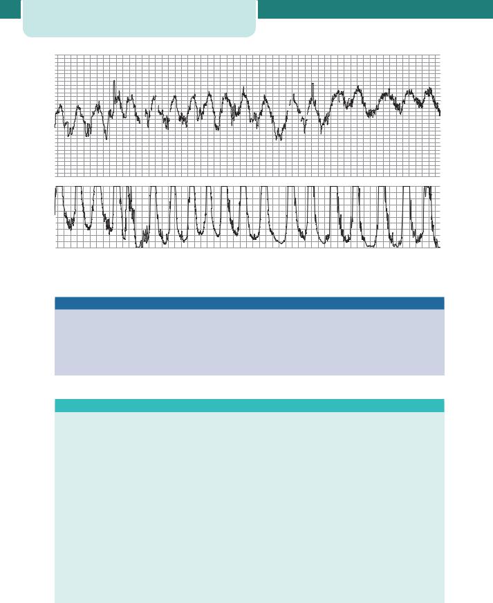
induction, and not all of this increase can be explained by the underlying problems which necessitated the induction in the first place. Long labours augmented with oxytocin predispose to postpartum haemorrhage secondary to uterine atony. Fetal compromise is more common during induced labours and this, in part at least, is due to uterine hyperstimulation caused by the injudicious use of prostaglandins and oxytocin (Figure 14.38). A contraction frequency of 5 per 10 minutes should be treated by stopping the oxytocin and the administration of a tocolytic drug, most commonly the subcutaneous injection of the β2-agonist terbutaline. Uterine hyperstimulation may precipitate a fetal bradycardia and the need for emergency Caesarean section if the fetal heart rate fails to resolve promptly. If ARM is performed while the fetal head is high then cord prolapse may occur, again precipitating the need for emergency delivery by Caesarean section. Women with a previous Caesarean section scar, or some other form of old uterine injury, are at greater risk of uterine rupture if they are induced. The risk of scar rupture increases from one in 200 in a spontaneous labour, to as high as one in 70 if induction of labour is performed using prostaglandins.
Induction of labour may fail and this is said to have occurred if an ARM is still impossible after the maximum number of doses of prostaglandin have been given, or if the cervix remains uneffaced and less than 3 cm dilated after an ARM has been performed and oxytocin has been running for 6–8 hours with regular contractions. When an induction fails, the options include attempting induction again at some point in the future, or performing a Caesarean section. Delaying delivery further is only acceptable if there is no major threat to fetal or maternal condition. This may be the case with a failed social induction, for example. Failed induction in the setting of preeclampsia or fetal growth restriction will usually necessitate a Caesarean delivery.
222 |
Labour |
|
|
|
|
|
|
|
|
|
200 |
|
200 |
|
200 |
|
|
|
|
|
—US2 |
|
|
|
|
|
180 |
|
180 |
|
180 |
|
|
|
160 |
|
160 |
|
160 |
|
|
|
140 |
|
140 |
|
140 |
|
|
|
120 |
|
120 |
|
120 |
|
|
|
100 |
|
100 |
|
100 |
|
|
|
80 |
|
80 |
|
80 |
|
|
|
60 |
|
60 |
|
60 |
|
|
|
22:50 22.09.06 1cm/min |
|
23:00 22.09.06 1cm/min |
|
23:10 22.09.06 1cm/min |
|
|
12 |
100 |
12 |
100 |
12 |
100 |
|
|
80 |
80 |
80 |
|||
|
|
10 |
10 |
10 |
|||
|
|
|
—TOCOext |
|
|||
|
|
8 |
60 |
8 |
60 |
8 |
60 |
|
|
6 |
40 |
6 |
40 |
6 |
40 |
|
|
4 |
4 |
4 |
|||
|
|
20 |
20 |
20 |
|||
|
|
2 |
2 |
2 |
|||
|
|
|
|
|
|||
|
|
0 |
0 |
0 |
0 |
0 |
0 |
|
|
kPa |
|
kPa |
|
kPa |
|
|
|
101 |
|
102 |
|
103 |
|
|
Figure 14.38 A pathological cardiotocograph (CTG) secondary to uterine hyperstimulation |
|
|
||||
|
Risks of induction of labour |
|
|
|
|
||
|
• Greater pain in labour |
|
• |
Failure |
|
|
|
|
• |
Uterine hyperstimulation |
|
• Increased need for Caesarean or instrumental |
|
||
|
• |
Cord prolapse |
|
|
delivery |
|
|
|
• Greater risk of uterine rupture during VBAC |
• |
Fetal compromise |
|
|
||
C A S E H I S T O R Y
Mrs B is a 33-year-old woman in her first pregnancy. She is
41 weeks gestation and a report of reduced fetal movements has prompted an ultrasound scan. Although the growth parameters of the fetus are on the 50th centile, there is oligohydramnios (reduced amniotic fluid). A CTG is normal.
An IOL has been discussed and a vaginal examination performed. The Bishop score is 2.
What risks do Mrs B and her baby face?
Oligohydramnios at term, in isolation, is a poor indicator of placental function. Reduced fetal movements is a worrying but insensitive marker of fetal compromise. However, this is a
prolonged pregnancy and this is sufficient on its own to recommend IOL. The other features support this conclusion. Continuation
of the pregnancy will increase the risks of intrauterine hypoxia, acidosis or even fetal death. Labour will carry a higher risk of compromising the fetus and there is a greater risk of meconium
passage and aspiration by the newborn. The reactive normal CTG gives no guarantee of future fetal well-being or its ability to tolerate contractions.
The unfavourable cervix increases the chances of long induction, which may ultimately be unsuccessful. Whether the labour is induced or spontaneous, this scenario carries a greater risk of assisted delivery, either instrumental or by Caesarean section. Women and their partners should be advised of these risks before embarking on the procedure, and the alternative policy of expectant management should be discussed objectively, even if the clinician does not favour it.
What care should Mrs B receive?
She should be given full psychological support from the midwifery and medical team and counselled against delaying the delivery of the fetus any further. Despite the concerns, there is a good chance of a normal birth and IOL should be recommended. Mrs B should

receive vaginal prostaglandin, and is likely to need two doses, 6 hours apart. Electronic fetal heart rate monitoring should be instituted when significant contractions begin. ARM should be
performed and an oxytocin infusion used if labour fails to establish or progress following the ARM. Pain relief should be offered as usual, although epidural anaesthesia might be encouraged in these circumstances, as the labour process may be long.
What ongoing care should she receive?
Attention should be paid to the need for continuity of care despite staff shift changes. A senior doctor should be involved and should
Clinical risk management |
223 |
review the situation at intervals to determine if it is safe to continue or whether emergency Caesarean section should be performed. If labour is prolonged and difficult, an H2-blocker and an anti-emetic should be given orally to suppress gastric acid secretion and reduce the risks in the event of a general anaesthetic being necessary. Caesarean section should be performed if induction fails, the labour establishes but fails to progress, or if fetal monitoring suggests compromise.
Clinical risk management
Risk management is an approach to healthcare provision that aims to limit harm occurring to patients. Clinical risk management (CRM) can be applied to all areas of medicine, but the labour ward provides one of the best illustrations of its importance to modern health care.
Labour and delivery carry a serious risk of harm. Maternal trauma (both physical and psychological) and infant neurological injuries are examples of poor outcomes following birth that can potentially be avoided in many cases. Legal action is frequently taken after outcomes such as these, and this is expensive for the National Health Service in litigation payments, and distressing for the staff involved. The aim of CRM is to improve standards of care and subsequently reduce the harm occurring to women and their babies. This in turn should reduce the number of complaints made against hospital trusts and the financial costs of litigation.
Shoulder dystocia, for example, can result in Erb’s palsy, intrapartum asphyxial damage and serious maternal perineal trauma. In the majority of cases, these poor outcomes following shoulder dystocia can be avoided by appropriate management. Regular staff education, and the performance of shoulder dystocia ‘drills’, should limit these outcomes. In these drills, the manoeuvres used to safely overcome shoulder dystocia are rehearsed to aid with recollection in the event of a real emergency. The use of guidelines and protocols drawn from evidence-based medicine is another tool of CRM. These help to reduce errors and to prevent bizarre or unusual decision-making.
Medical and midwifery staff are encouraged to report when things ‘go wrong’. Once a ‘near-miss’ or a
poor outcome has occurred, careful documentation is vital if claims of negligence are to be defended. Good communication between the staff and the patient involved may help to clear up misunderstandings and minimize the chances of a formal complaint or legal action. ‘Root-cause analysis’ is a technique that serves to examine in detail a poor outcome, or near-miss, so that every step of the patient-journey is scrutinized to see if the outcome could have been prevented. In this way, lessons are learned for the future and unit policies and guidelines can be adjusted accordingly. It is usually the case that a whole series of failings or errors need to occur together for a poor outcome to result. Institutional systems as a whole often contribute, and one single individual is rarely solely responsible for a poor outcome.
Audit of labour ward outcomes is an important tool in risk management. If guidelines are not being followed, and certain standards are not being met, this will be detected by audit and steps taken to address the problem. A repeat audit should show that improvements have occurred as a result of the actions taken.
Key points
Induction of labour and clinical risk management
•Every induction of labour should have a valid indication.
•The most common indication for induction of labour is prolonged pregnancy.
•A high Bishop score predicts an easier induction of labour.
•Clinical risk management aims to improve standards of intrapartum care and to reduce the number and severity of poor obstetric outcomes.

|
|
|
|
|
|
|
|
|
|
|
|
|
|
|
C H A P T E R 1 5 |
O P E R A T I V E I N T E R V E N T I O N |
|
|
|||||||||
|
|
I N O B S T E T R I C S |
|
|
|||||||||
|
|
Philip N Baker |
|
|
|
|
|
|
|
|
|||
|
|
|
|
|
|
|
|||||||
|
Introduction ................................................................................................. |
224 |
|
Perineal repair ........................................................................................... |
226 |
|
|||||||
|
Episiotomy.................................................................................................... |
224 |
|
Operative vaginal delivery ................................................................. |
227 |
|
|||||||
|
Perineal trauma ........................................................................................ |
225 |
|
Caesarean section .................................................................................. |
234 |
|
|||||||
|
|
|
|
|
|
|
|
|
|
|
|
|
|
O V E R V I E W
For any practitioner, the best outcome of pregnancy is a healthy mother and baby. Ideally, this outcome should follow a normal vaginal delivery with an intact perineum, however, 40–50 per cent of deliveries in the United Kingdom are associated with an episiotomy, or are effected by forceps, ventouse or Caesarean section. The indications and prerequisites for these procedures merit constant scrutiny, as do the optimal techniques to minimize complications.
Introduction
The anatomy of the birth canal and the fetal head must be understood as a prerequisite to becoming skilled in the safe use of the forceps or vacuum extractor, just as knowledge of the abdominal and pelvic anatomy must precede Caesarean section. The Royal College of Obstetricians and Gynaecologists recommends that obstetricians achieve experience in spontaneous vaginal delivery prior to commencing training in operative deliveries.
The goal of operative delivery is to expedite delivery with a minimum of maternal or neonatal morbidity. There has been an increasing awareness of the potential for morbidity for both the mother and the baby. The risk of traumatic delivery in relation to forceps, particularly rotational procedures, has been long established. In 1998, the US Food and Drug Administration issued a warning about the potential dangers of delivery with the ventouse or vacuum extractor; this followed several reports of infant fatality secondary to intracranial haemorrhage. In addition, there has been a growing recognition of the shortand long-term morbidity of pelvic floor injury following operative vaginal delivery. Caesarean section, particularly in the second stage of labour, also carries significant morbidity and implications for future births. It is not surprising, therefore, that there has been an increase in litigation relating to obstetric
delivery. If we are to offer women the option of safe operative deliveries, we need to improve our approach to clinical care. The goal should be to minimize the risk of morbidity and, where morbidity occurs, to minimize the likelihood of litigation, without limiting maternal choice.
Episiotomy
Definition
An episiotomy is an incision through the perineum made to enlarge the diameter of the vulval outlet and assist childbirth.
Prevalence
Although episiotomies were first described almost 300 years ago, widespread use of the procedure increased during the twentieth century. By the early 1970s, rates were as high as 90 per cent and it was often advocated that there were two reasons for episiotomy; one was a primigravida and the other a previous episiotomy. In other words, every vaginal delivery should be accompanied by episiotomy. It was argued that this reduced the risk of tears and subsequent problems from prolonged bearing down, such as prolapse. The evidence for the latter was tenuous.
The uncritical liberal use of episiotomy was opposed by consumer groups including the National Childbirth Trust and these very high rates of episiotomy have been reversed. In the UK, rates approximate to the World Heath Organization (WHO) recommendation of 10 per cent of normal deliveries, however, there is considerable international variation (rates are 50 per cent in the United States and 99 per cent in Eastern Europe).
Technique
The question of informed consent needs to be addressed during antenatal care; when the fetal head is crowning, it is not possible to obtain true informed consent.
•An episiotomy is performed in the second stage, usually when the perineum is being stretched and
it is deemed necessary.
•If there is not a good epidural, the perineum should be infiltrated with local
anaesthetic.
•If an effective epidural anaesthetic is in place it should be topped up for delivery with the
patient upright to get best coverage of the perineal area.
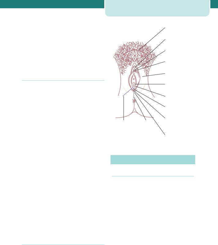
•The incision can be midline or at an angle from the posterior end of the vulva (a mediolateral
episiotomy).
•A mediolateral episiotomy is usually recommended; a midline episiotomy is an
incision in a comparatively avascular area and results in less bleeding, quicker healing and less pain, however, there is an increased risk of extension to involve the anal sphincter (third/ fourth-degree tear).
•A mediolateral episiotomy should start at the posterior part of the fourchette, move backwards
and then turn medially well before the border of the anal sphincter, so that any extension will miss the sphincter (Figure 15.1).
Complications
Complications include haemorrhage, infection (prophylactic antibiotics may be indicated if contamination is suspected), extension to the anal sphincter (third/fourth-degree tears) and dyspareunia.
|
|
Perineal trauma |
225 |
|
|
Mons pubis |
|
|
|
Prepuce of |
|
|
|
clitoris |
|
|
|
Glans of |
|
|
|
clitoris |
|
|
|
Frenulum of |
|
|
|
prepuce |
|
|
|
Labium majus |
|
|
|
(pulled aside) |
|
|
|
Hymen of |
|
|
|
vagina |
|
|
|
Duct of greater |
|
|
|
vestibular gland |
|
|
|
Vestibular fossa |
|
|
|
(fossa navicularis) |
|
|
|
Frenulum |
|
Mediolateral |
Midline |
labiorum |
|
|
|
Union of labia majora (commissura posterior)
Figure 15.1 A right mediolateral episiotomy
Perineal trauma
Definitions
1.First-degree trauma corresponds to lacerations of the skin/vaginal epithelium alone.
2.Second-degree tears involve perineal muscles and therefore include episotomies.
3.Third-degree extensions involve any part of the anal sphincter complex (external and internal sphincters):
iLess than 50 per cent of the external anal sphincter is torn.
iiMore than 50 per cent of the external anal sphincter is torn.
iiiTear involves the internal anal sphincter (usually there is complete disruption of the external sphincter).
4.Fourth-degree tears involve injury to the anal sphincter complex extending into the rectal mucosa.