

Obstetrics_by_Ten_Teachers_19E_-_Kenny_Louise
.pdf236 Operative intervention in obstetrics
Morbidity and mortality
Although Caesarean section is becoming increasingly safe and evidence is mounting regarding the risks of labour and vaginal delivery, pregnant women, their midwives and doctors need to understand and appreciate the maternal risks associated with the different modes of delivery. Confidential Enquiries into Maternal Deaths have enabled the risks associated with different methods of delivery to be analysed; case fatality rate for all Caesarean sections is five times that for vaginal delivery, although for elective Caesarean section the difference does not reach statistical significance. Some maternal deaths following Caesarean section are not attributable to the procedure itself, but rather to medical or obstetric disorders that lead to the decision to deliver using this approach. Many women who deliver vaginally encounter the same problems.
Repeat Caesarean section
In many units, Caesarean section rates for primigravidae of 24 per cent are seen. Consequently, the problem of management of a woman with a scarred uterus in subsequent pregnancies is a common antenatal problem. It is a vital part of antenatal care that women be given a clear understanding of the plan of management from early on in their pregnancy, with the caveat that this may need to be adapted if the pregnancy presents unexpected problems. The management in pregnancy following a Caesarean section should be to assess the available options and to select the appropriate choice for an individual woman. The dictum ‘once a Caesarean section, always a Caesarean section’ is not true; up to 70 per cent of women with a previous Caesarean section can achieve a vaginal delivery. Patient choice cannot and should not be ignored in decisions regarding management, and it is important to discuss the risks and benefits of elective Caesarean section as compared to trial of vaginal delivery.
From a maternal perspective, elective Caesarean section avoids labour with its risk of perineal trauma (urinary and fetal problems), the need to undergo emergency Caesarean section, and scar dehiscence/ rupture with subsequent morbidity and mortality. However, elective Caesarean section carries maternal risks: increased bleeding, thromboembolism, febrile morbidity, prolonged recovery, long-term bladder dysfunction and increased risks of placenta praevia in subsequent pregnancies. From a fetal perspective,
an elective Caesarean section reduces the risk of scar rupture, but increases the risk of transient tachypnoea/respiratory distress syndrome. There is remarkably little evidence to inform practice with regard to management of previous Caesarean section: there are no randomized trials and much of the data relate to observational studies.
Consideration of the risk of scar rupture is probably the most important consideration when determining whether delivery should be by elective Caesarean section or by trial of vaginal delivery. Most published studies do not differentiate between scar dehiscence and rupture, however, analysis of observational and comparative studies indicates that the excess risk of uterine rupture following trial of labour compared with women undergoing repeat elective Caesarean section is considerably lower than 1 per cent; indeed, some studies do not demonstrate any increased risk.
Providing the first operation was carried out for non-recurrent cause, and providing the obstetric situation close to term in the succeeding pregnancy is favourable, then it is appropriate to offer a trial of labour to any woman with a previous uncomplicated lower uterine segment Caesarean section and no other adverse obstetric feature. The factors to be weighed when determining the recommended mode of delivery depend on the balance between the desires of the mother, the risks of a repeat operation, the risks to her child of labour, and the risk of labour on the strength of the old scar.
Procedure
Informed consent
Full informed consent must always be obtained prior to operation. The level of information discussed must be commensurate with the urgency of the procedure, and a common sense approach is needed. However, although it is often difficult to impart complete and thorough information when Caesarean sections are performed as emergency procedures, mothers must understand what is being planned and why. Where possible, all women must be educated in pregnancy about Caesarean section and the occasions under which it may be urgently needed. It is important to remember that no other adult may give consent for another (although it is good practice to keep relatives fully informed). Where there is incapacity to consent
(as may occur with conditions such as eclampsia), the doctor is expected to act in the patient’s best interests.
The national consent forms require both the risks and benefits to be discussed with patients and recorded on the consent form. Common medical practice is to highlight risks but not benefits. It is important to remember that the operation is being offered because of perceived benefits, both maternal and fetal in many cases.
Surgical basics
The bladder should be emptied before the procedure. A left lateral tilt minimizes compression of the maternal inferior vena cava and reduces the incidence of hypotension (with its consequent reductions in placental perfusion).
The Pfannenstiel incision
The skin and subcutaneous tissues are incised using a transverse curvilinear incision two fingerbreadths above the symphysis pubis extending from and to points lateral to the lateral margins of the abdominal rectus muscles. Subcutaneous tissues are separated by blunt dissection and the rectus sheath is incised transversely along the middle 2 cm. This incision is then extended with scissors before the fascial sheath is separated from the underlying muscle by further blunt dissection. Separation is performed cephalad to permit adequate exposure of the peritoneum in a longitudinal plane. The recti are separated, the peritoneum incised and the abdominal cavity entered. The transverse Pfannensteil incision has the advantages of improved cosmetic results, decreased analgesic requirements and superior wound strength.
The infra-umbilical incision
A vertical skin incision is indicated in cases of extreme maternal obesity, suspicion of other intra-abdominal pathology necessitating surgical intervention, or where access to the uterine fundus may be required (classical Caesarean section). The lower midline incision is made from the lower border of the umbilicus to the symphysis pubis, and may be extended caudally toward the xiphisternum. Sharp dissection to the anterior rectus sheath is performed and is then freed of subcutaneous fat. The rectus sheath is then incised taking care to avoid damage to any underlying bowel, and extended inferiorly to the vesical peritoneal reflection and superiorly to the upper limit of the abdominal incision. The vertical incision provides
Caesarean section |
237 |
greater ease of access to the pelvic and intra-abdominal organs and may be enlarged more easily; however, the incidence of wound dehiscence is increased.
Uterine incision
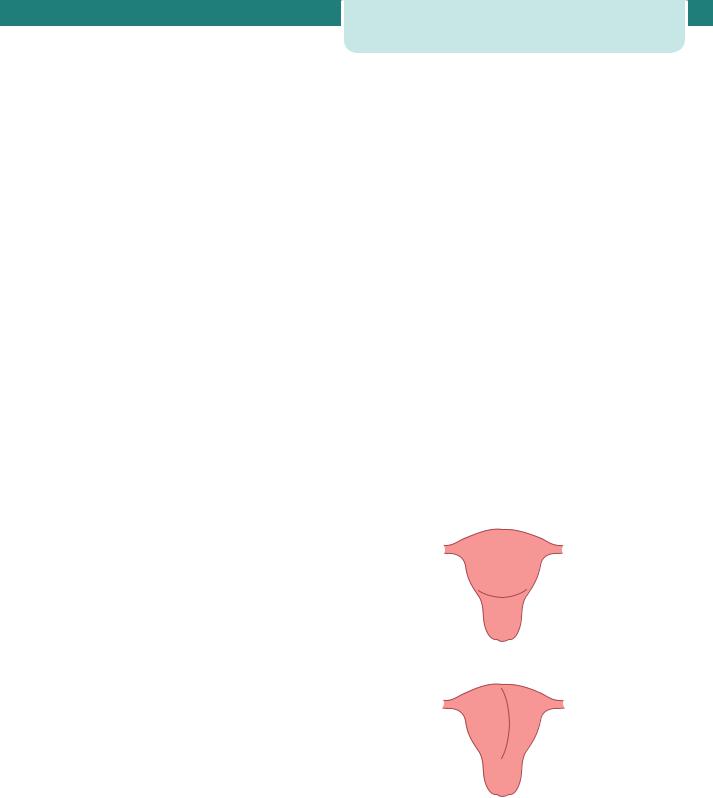
A lower uterine segment incision is used in over 95 per cent of Caesarean deliveries due to ease of repair, reduced blood loss and low incidence of dehiscence or rupture in subsequent pregnancies (Figure 15.7). The loose reflection of vesico-uterine serosa overlying the uterus is divided laterally, the underlying lower uterine segment is reflected with blunt dissection, the developed bladder flap is retracted and the lower uterine segment is opened in a transverse plane for a distance of 1–2 cm; the incision is extended laterally to allow delivery of the fetus without extension into the broad ligament or uterine vessels. There are relatively few absolute indications for classical section (which incorporates the upper uterine segment, Figure 15.7). These include a lower uterine segment containing fibroids or a lower segment covered with dense adhesions, both of which may make entry difficult. Other indications include placenta praevia, transverse lie with the back down, fetal abnormality (e.g. conjoined twins), or Caesarean section in the presence of a carcinoma of the cervix (so as to avoid damage to the cervix and its vascular and lymphatic supply).
(a)Transverse lower segment incision
(b) Classical caesarean section incision
Figure 15.7 Uterine incisions for Caesarean section.
(a) Transverse lower segment incision. (b) classical Caesarean section incision

238 Operative intervention in obstetrics
Once the uterus is incised, the membranes are ruptured if still intact, and the accoucheur’s hand is positioned below the presenting part. If cephalic, the head is flexed and delivered by elevation though the uterine incision either manually, or with forceps. Fundal pressure is applied to aid delivery; this should not commence until the presenting part is located within the incision – for fear of converting the lie from longitudinal to transverse. Once the fetus is delivered, an oxytocic (5 IU Syntocinon i.v.) is administered to aid uterine contraction and placental separation. The placenta is delivered by combined cord traction; manual removal significantly increases the intraoperative blood loss and postoperative infectious morbidity.
Closure of the uterus should be performed in either single or double layers with continuous or interrupted sutures. The initial suture should be placed just lateral to the incision angle, and the closure continued to a point just lateral to the angle on the opposite side. A running stitch is often employed and this may be locked to improve haemostasis. If a second layer is used, an inverting suture or horizontal suture should overlap the myometrium. Once repaired, the incision is assessed for haemostasis and ‘figure-of- eight’ sutures can be employed to control bleeding. Peritoneal closure is unnecessary. Abdominal closure is performed in the anatomical planes with high strength, low reactivity materials, such as polyglycolic acid or polyglactin.
Complications
Caesarean section is a major abdominal surgical procedure and carries significant risks.
Intraoperative complications
Bowel damage
Bowel damage may occur during a repeat procedure or if adhesions are present from previous surgery.
Caesarean hysterectomy
The most common indication for Caesarean hysterectomy is uncontrollable maternal haemorrhage; life-threatening haemorrhage requiring immediate treatment after 1 in 1000 deliveries. The most important risk factor for emergency postpartum hysterectomy is a previous Caesarean section – especially when the placenta overlies the old scar, increasing the risks of placenta accreta (see Chapter 16, Obstetric emergencies).
Other indications for hysterectomy are atony, uterine rupture, extension of a transverse uterine incision and fibroids preventing uterine closure and haemostasis. This operation, while a major undertaking, should not be left too late, as the risk of operative complications, maternal morbidity and mortality increase with increasing haemorrhage.
Haemorrhage
Haemorrhage may be a consequence of damage to the uterine vessels, or may be incidental as a consequence of uterine atony or placenta praevia. In patients with an anticipated high risk of haemorrhage, e.g. known cases of placenta praevia, blood should be routinely crossmatched. There are many manoeuvres to manage haemorrhage; these range from bimanual compression, oxytocin infusion, administration of prostaglandins, conservative surgical procedures, such as uterine compression sutures to the more radical, but life saving, hysterectomy.
Placenta praevia
The proportion of patients with a placenta praevia increases almost linearly after each previous Caesarean section, and as the risks of such a complication increases with increasing parity, future reproductive intentions are very relevant to any individual decision for operative delivery.
Urinary tract damage
The risk of bladder injury is increased after prolonged labours where the bladder is displaced caudally, after previous Caesarean section where scarring obliterates the vesicouterine space, or where a vertical extension to the uterine incision has occurred. If damage is suspected, then transurethral instillation of methylene blue-coloured saline will help to delineate the defect. When such an injury is observed, repair with 2-0 Vicryl as a single continuous or interrupted layer is appropriate. The urinary catheter should remain in situ for 7–10 days. Damage to the ureters is uncommon as reflection of the bladder displaces them rostrally.
Post-operative complications
Infection and endometritis
Women undergoing Caesarean section have a 5–20-fold greater risk of an infectious complication when compared with a vaginal delivery. Complications include fever, wound infection, endometritis, bacteraemia

and urinary tract infection. Other common causes of postoperative fever include haematoma, atelectasis and deep vein thrombosis. Labour, its duration and the presence of ruptured membranes appear to be the most important risk factors, with obesity playing a particularly important role in the occurrence of wound infections. The most important source of microorganisms responsible for post-Caesarean section infection is the genital tract, particularly if the membranes are ruptured preoperatively. Even in the presence of intact membranes, microbial invasion of the intrauterine cavity is common, especially with preterm labour. Infections are commonly polymicrobial and pathogens isolated from infected wounds and the endometrium include Escherichia coli, other aerobic Gram-negative rods, and Group B streptococcus. General principles for the prevention of any surgical infection include careful surgical technique, skin antisepsis; prophylactic antibiotics should be administered to reduce the incidence of postoperative endometritis.
Pulmonary emboli and deep vein thrombosis
Deaths from pulmonary embolism remain the leading direct cause of maternal death, and Caesarean section is a major risk factor. The signs and symptoms of pulmonary emboli and deep vein thrombosis are as detailed in Chapter 8, Antenatal obstetric complications. The incidence of such complications can undoubtedly be reduced by the peri-operative
C A S E H I S T O R Y
Caesarean section |
239 |
administration of prophylactic heparin and the prompt initiation of treatment when required (see Chapter 8, Antenatal obstetric complications).
Psychological
All difficult deliveries carry increased maternal psychological and physical morbidity. The compromised postpartum psychological functioning in women delivered by Caesarean section may be secondary to delayed contact with the baby; a factor that in most cases should be amenable to remedy.
Key points
•The increasing rate of delivery by Caesarean section continues to be an issue of great concern to many midwives, obstetricians, politicians and society as a whole.
•Maternal satisfaction is an important part of childbirth and must be taken into consideration when implementing any changes in childbirth policy. There is a need for national debate on whether maternal choice is a valid indication for Caesarean section.
•It is appropriate to offer a trial of labour to any woman with a previous uncomplicated lower uterine segment Caesarean section and no other adverse obstetric feature.
•Although Caesarean section continues to be associated with considerable morbidity and mortality, thromboembolism prophylaxis and prophylactic antibiotics have reduced the risks of thrombotic and infective complications.
Mrs B, a 26-year-old primigravid woman, went into spontaneous labour at 40 weeks gestation. There was no relevant past medical history and findings on examination were unremarkable; the symphseal fundal height was deemed to be equivalent to the gestational age. Delay in the first stage of labour led to artificial rupture of the membranes (clear liquor drained) and then to
the instigation of an oxytocin infusion. Two successive vaginal examinations, 4 hours apart, identified a cervical dilatation of 9 cm. The cardiotocograph (CTG) tracing was adjudged to show a normal fetal heart rate pattern. An epidural was in situ and was effective. The management plan was for a further examination after an interval of 2 hours; if the cervix was not fully dilated, delivery was to be by Caesarean section.
When the examination was repeated, the cervix was fully dilated, with the fetal head at the level of the ischial spines and in
the right occipitio-transverse position. There was marked caput and moulding. Clear liquor drained and there was a normal, reactive fetal heart rate pattern.
Were the prerequisites for an operative vaginal delivery met?
The findings on abdominal examination are crucial to any consideration of this question. On examination, 1/5th of the fetal head was palpable. The prerequisites for a forceps/ventouse delivery were thus not met. The appropriate management plan was for transfer to theatre for probable Caesarean section delivery.
Following an epidural ‘top up’ and transfer to theatre, the abdominal and vaginal examinations were repeated. On abdominal examination, the fetal head was no longer palpable and on vaginal examination the fetal head was below the level of the ischial spines and in the occipito-anterior position.
continued

240 Operative intervention in obstetrics
C A S E H I S T O R Y continued
How should the delivery be effected? |
What is the appropriate management plan? |
On the assumption that the delivery was to be performed by an appropriately trained and experienced obstetrician, and that
informed consent was elicited, the prerequisites for an operative delivery were now met. Such a delivery, following delay in the first stage of labour, and in the presence of marked caput and moulding should optimally be performed in theatre.
Use of either forceps or the ventouse would be reasonable. Although the ventouse is associated with less perineal trauma, the obstetrician opted to use forceps as the presence of caput and moulding indicated that the ventouse was more likely to fail.
What type of forceps should be used?
The fetal head had rotated to the occipito-anterior position prior to the application of forceps. Non-rotational forceps (for example, Neville Barnes or Simpsons forceps), with a pelvic curve, were applied.
The forceps were positioned and ‘locked’. Traction was applied with a contraction, and with maternal pushing. There was no descent of the fetal head. Following traction with another contraction, there was no descent of the fetal head.
There has been a failed trial of an operative vaginal delivery, with no descent of the fetal head. Delivery must be by Caesarean section.
A lower segment Caesarean section was performed, through a Pfannenstiel incision. Although there was difficulty delivering a deeply impacted fetal head, there was no significant extension of the uterine incision. The infant birth weight was 4.35 kg. Closure of the uterus and abdomen was unremarkable.
What complications should be anticipated after delivery?
There was a significant risk of postpartum haemorrhage due to uterine atony. In addition to prophylactic antibiotic and anticoagulant therapy (to reduce the likelihood of infective and thrombotic complications), the bolus dose of oxytocin at delivery was followed by an intravenous infusion of oxytocin over 4 hours.
No postoperative complications ensued and Mrs B was discharged home, with her baby, 4 days after the Caesarean section. She was given a hospital follow-up appointment to discuss the events of the labour and delivery.

C H A P T E R 1 6
OBSTETRIC EMERGENCIES
Clare Tower
.......................................................................................................Definition |
241 |
......................................................................................................Summary |
256 |
The structured approach to obstetric emergencies ......... |
241 |
Additional reading................................................................................... |
257 |
Management of specific obstetric emergencies................. |
245 |
|
|
|
|
|
|
O V E R V I E W
Obstetric emergencies occur not infrequently and can have catastrophic consequences for the mother, baby and their family. In the United Kingdom, CMACE (Centre for Maternal and Child Enquiries) produces regular confidential reports on all maternal deaths and perinatal deaths. Unfortunately, each report includes a substantial number of cases in which there has been substandard care. The maternal mortality report which reviewed all deaths between 2003 and 2005 found evidence of substandard care in 64 per cent of maternal deaths occurring directly as a result of obstetric complications. In order to improve the care of women and their babies, there has been an increasing use of a structured approach to obstetric emergencies. In other words, by using the same principles in each emergency situation, it is hoped that outcomes improve and staff can cope better with a stressful and traumatic situation.
In the UK, the maternal mortality rate is currently 14 per 100 000 maternities, a rate which has stayed constant over recent years. It is expected that this rate may increase in the future due to rising obesity, maternal age and more women, particularly from overseas, with complex medical problems. The most common reasons for maternal death in the UK are thrombosis, pre-eclampsia and eclampsia, amniotic fluid embolus and haemorrhage, as shown in Table 16.1.
Definition
An emergency is defined as a serious situation or occurrence that happens unexpectedly and demands immediate action. Although the definition implies that it is unforeseen, preparation and prevention should always be used to reduce the risks of emergencies occurring. For example, an eclamptic fit is an obstetric emergency that may be prevented by the administration of magnesium sulphate to women with severe preeclampsia. In obstetrics, emergencies can be classified as maternal (occurring antenatally and post-natally) and fetal. The causes are summarized in Table 16.2.
Table 16.1 Causes of maternal death 2003–2005
|
No. |
Rate per |
|
|
100 000 |
|
|
maternities |
Direct deaths |
|
|
Thrombosis |
|
1.94 |
41 |
||
(cerebral and |
|
|
pulmonary) |
|
|
|
|
|
Pre-eclampsia/ |
18 |
0.85 |
eclampsia |
|
|
|
|
|
Haemorrhage |
14 |
0.66 |
|
|
|
Amniotic fluid |
17 |
0.80 |
embolus |
|
|
|
|
|
Early pregnancy |
14 |
0.66 |
deaths |
|
|
Genital tract |
18 |
0.85 |
sepsis |
|
|
|
|
|
Other direct |
4 |
0.19 |
deaths |
|
|
|
|
|
Anaesthetic |
6 |
0.28 |
deaths |
|
|
|
|
|
|
|
continued |

242Obstetric emergencies
Table 16.1 Continued
|
No. |
Rate per |
|
|
100 000 |
|
|
maternities |
Cardiac |
|
2.27 |
48 |
||
|
|
|
Psychiatric |
18 |
0.85 |
|
|
|
Malignancies |
10 |
0.47 |
|
|
|
Others |
87 |
4.12 |
Coincidental |
55 |
2.60 |
|
|
|
Direct deaths are defined as those resulting from obstetric complications, Indirect deaths as those resulting from pre-existing disease that has been worsened by pregnancy.
Source. Lewis G (ed.). The Confidential Enquiry into Maternal and Child Health (CEMACH). Saving Mothers Lives: reviewing maternal deaths to make motherhood safer, 2003–2006. The Seventh Report on Confidential Enquiries into Maternal Deaths in the United Kingdom. London: CEMACH, 2007.
The structured approach to obstetric
emergencies
Developing a structured approach to emergencies that can be practised within a ‘drill-type’ scenario provides staff with an ordered sequence of actions that can help in a stressful and sometimes chaotic situation. While many of the fetal obstetric emergencies require the use of specific drill, the maternal emergencies can make use of the common ABC approach used for all adult emergencies. When called to any emergency situation, the first action should be to call for help. After this, a systematic evaluation and resuscitation should be conducted in the following order:
1.Airway
2.Breathing and ventilation
3.Circulation with volume replacement and control of bleeding
4.Disability
5.Environment and exposure.
|
|
|
|
|
Prepare and prevent |
|
|
|
|
|
|
|
|
|
|
|
|
|
|
|
|
|
|
|
|
|
|
|
|
|
|
|
|
|
|
|
|
|
|
|
|
|
|
|
|
|
|
|
|
|
|
|
Call for help |
|
|
|
|
|
|
|
|
|
|
|
|
|
|
|
|
|
|
|
|
|
|
|
|
|
|
|
|
|
|
|
|
|
|
|
|
|
|
|
|
|
|
|
|
|
|
|
Talk to the patient |
|
|
|
|
|
|
|
|
|
|
|
|
|
|
|
|
|
|
|
|
|
|
|
|
|
|
|
|
|
|
|
|
|
|
|
|
|
|
|
|
|
|
|
|
|
|
|
Turn into left lateral tilt |
|
|
|
|
||
|
|
|
|
|
|
|
|
|
|
|
|
|
|
|
|
|
|
|
|
|
|
||
|
|
|
|
|
|
|
|
|
|
|
|
|
|
|
|
|
Assess the airway |
|
|
|
|
|
|
Administer high flow |
|
|
|
|
|
|
|
|
|
||
|
|
|
|
|
|
|
|
|
|
|
|
oxygen if airway |
|
|
|
|
|
|
|
|
|
|
Insert two large bore |
|
|
|
|
|
|
|
|
|
|
||
open and breathing |
|
|
|
|
|
|
|
|
|
|
cannulae |
|
Assess breathing and circulation |
||||||||||
|
|
|
|
|
|||||||
|
|
|
|
|
|||||||
Consider and treat |
|
|
|
|
|
|
|
|
|
|
|
the cause |
|
|
|
|
|
|
|
|
|
|
|
|
|
|
|
|
|
|
|
|
|
||
|
|
|
|
Immediately commence chest |
|
|
|
||||
|
|
|
|
compressions 100/minute |
|
|
|||||
|
|
|
|
|
|
|
|
||||
|
|
|
|
|
|
|
|
||||
|
|
|
|
|
|
|
|
|
|
|
|
|
|
|
|
Provide chest compressions to |
|
|
|
||||
|
|
|
|
|
ventilation breaths 30:2 |
|
|
||||
|
|
|
|
|
|
|
|
|
|
|
|
Figure 16.1 The structured approach to managing obstetric emergencies

Conducting a structured assessment
The stuctured assessment should be conducted according to Figure 16.1.
Approach
On approaching the patient, first speak to them to find out whether they are able to respond. In addition to being an important form of communication, this also reveals important information. If the patient responds appropriately, they must have an open airway and be able to move sufficient air to speak. Furthermore, it also gives useful information about neurological
Table 16.2 Causes of obstetric emergencies
The structured approach to obstetric emergencies |
243 |
status. In contrast, if the patient is unable to respond, it should trigger the immediate call for help and assessment of airway, breathing and circulation. In pregnant patients (20 weeks or more) and in the immediate postpartum period, the enlarged uterus compresses the inferior vena cava, thus reducing venous return to the heart when the patient is lying flat on their back. It is therefore extremely important to tilt the patient to the left to aid resuscitation (left lateral tilt, Figure 16.2). This will lift the heavy uterus away from the abdominal vessels, improving the venous return to the heart and aiding the resuscitation attempt.
Maternal |
|
|
|
Fetal |
Haemorrhage |
Antepartum |
|
Placenta previa |
Bradycardia |
|
||||
|
|
|
|
|
|
|
|
Abruption |
Abruption |
|
Postpartum |
|
Uterine atony |
Cord prolapse |
|
|
|
|
|
|
|
|
Trauma |
Shoulder dystocia |
|
|
|
|
|
|
|
|
Retained placenta |
Vasa previa |
|
|
|
|
|
|
|
|
Disseminated |
|
|
|
|
Intravascular |
|
|
|
|
coagulaopathy (DIC) |
|
|
|
|
|
|
Hypertensive |
Pre-eclampsia |
|
|
|
disorders |
|
|
|
|
|
|
|
|
|
|
Eclampsia |
|
|
|
|
|
|
|
|
Uterine causes |
Inversion |
|
|
|
|
Rupture |
|
|
|
|
|
|
|
|
Sudden maternal |
Amniotic fluid embolus |
|
|
|
collapse |
|
|
|
|
|
|
|
|
|
|
Pulmonary embolus |
|
|
|
|
Shockincluding |
|
|
|
|
sepsis, haemorrhage, |
|
|
|
|
anaphylaxis, etc. |
|
|
|
|
|
|
|
|
|
Cardiac causes, e.g. |
|
|
|
|
myocardial infarct |
|
|
|
|
|
|
|
|
|
Intracranial events – |
|
|
|
|
bleeds, thrombosis |
|
|
|
|
|
|
|
|
|
Biochemical causes – |
|
|
|
|
e.g. hypoglycaemia |
|
|
|
|
|
|
|
|
|
Anaesthetic events |
|
|
|
|
|
|
|
|

244 Obstetric emergencies
Figure 16.2 Left lateral tilt. The pregnant woman is tilted to the left to move the pregnant uterus of the abdominal vessels, thus improving cardiac output. There are various methods of achieving this; many hospitals have special wedge-shaped cushions (‘the Cardiff wedge’), but pillows, blankets or even an individual kneeling can also be used.
Assessing the airway (A)
First, check in the mouth for any obstructing material, such as blood or vomit, and remove using suction. Such obstruction is uncommon in obstetric patients. Next, open the airway by using either the head tilt and chin lift, or a jaw thrust. The head tilt and chin lift is carried out by placing a hand on the forehead and gently tilting back, and two fingers of the other hand under the chin and gently lifting. The alternative manoeuvre, the jaw thrust, involves placing the fingers of each hand behind the two angles of the mandible and pushing upwards.
Assessing breathing (B) and circulation (C)
Having opened the airway, the breathing should be assessed for 10 seconds by looking for chest movement and listening and feeling for signs of air movement. Although experienced clinicians may also feel the carotid pulse at this stage, the current resuscitation guidelines advise that lack of breathing also indicates a lack of circulation. If the airway is open and the patient breathing, high flow oxygen should be administered via a face mask.
If there is no circulation, or there is some uncertainty, cardiopulmonary resuscitation (CPR) should be commenced immediately. This begins immediately with 30 chest compressions followed by two ventilation breaths. Administering chest compressions should be conducted with the patient in the left lateral position. Both hands should be placed on top of one another with straight arms, in the middle of the lower half of the sternum in the midline
and the sternum be depressed by 4–5 cm at a rate of 100 per minute. Providing chest compressions is tiring, and in the hospital situation, help is readily available. Therefore, it is recommended that the person doing chest compression should change every 2 minutes.
Ventilation breaths, usually provided via a bag and mask in hospital with high flow oxygen, should last 1 second and the chest should be seen to rise with each breath. This can be continued until the arrival of skilled help, when more definitive airway management in the form of intubation can be performed. After intubation, ventilation should be at a rate of ten breaths per minute and does not need to be synchronized with chest compressions.
If a circulation is present but no breathing (respiratory arrest), then ventilation breaths with high flow oxygen should be given at a rate of ten breaths per minute, with a regular check on the circulation via the carotid pulse every ten breaths. If the patient begins to breathe spontaneously, they should be placed in the recovery position and closely monitored.
Two large bore cannulae (16or 14-gauge, usually grey or orange) should be inserted into both antecubital fosse to allow blood to be taken for full blood count, cross-matching, and biochemistry, fluid resuscitation and drug administration.
As soon as possible, the defibrillator pads should be applied to assess the cardiac rhythm, treat accordingly and specific treatment initiated. The reversible causes of cardiac arrest

can be remembered as the ‘four Hs and the four Ts’ and are given below (those in italics signify those most likely in pregnancy):
Four Hs |
Four Ts |
Hypovolaemia due |
Thromboembolism |
to haemorrhage or |
|
sepsis |
|
|
|
Hypoxia |
Toxicity due to |
|
drugs, e.g. anaesthetic |
|
|
Hyperkalemia and |
Tension |
other metabolic |
pneumothorax |
disorders |
|
|
|
Hypothermia |
Cardiac tamponade |
|
|
A detailed description of advanced resuscitation is beyond the scope of this book, but can be found in the suggested further reading.
Difficulties in resuscitation due to pregnancy
In addition to compression of the large abdominal blood vessels described above, some other physiological changes of pregnancy can also make resuscitation of the collapsed pregnant woman more difficult. The pregnant uterus presses on the diaphragm, therefore reducing the lung functional residual capacity and making the lungs more difficult to ventilate. Larger breasts compound this problem. Furthermore, pregnancy causes the oesophageal sphincter to become more relaxed, therefore increasing the likelihood of aspiration of the stomach contents into the lungs. It is important that the airway is secured early to prevent this.
The fetus
The presence of a fetus within the uterus makes resuscitation of the mother more difficult due to aortocaval compression, obstruction to ventilation and increased oxygen requirements. In the emergency situation, it is always the welfare of the mother that takes precedence. If resuscitation has not been successful by 4 minutes, an immediate Caesarean section should be conducted, with the aim of having the baby delivered by 5 minutes. The aim of this is primarily to increase the likelihood of successfully resuscitating the mother, and, for the sake of speed, does not require the patient to be in an operating theatre.
Management of specific obstetric emergencies |
245 |
Cardiorespiratory arrest in obstetrics is rare. However, many obstetric emergencies occur that have not yet progressed to this dire stage. The principles of management (summarized in Figure 16.1) remain the same, and if successful will prevent the most serious sequelae.
Management of specific obstetric
emergencies
Haemorrhage
Obstetric haemorrhage can occur antenatally or post-natally, and both can present as obstetric emergencies.
Antepartum haemorrhage
Antepartum haemorrhage (APH) is any bleeding occurring in the antenatal period after 20 weeks gestation. It complicates 2–5 per cent of pregnancies. Most cases involve relatively small quantities of blood loss, but they often signify that the pregnancy is at increased risk of subsequent complications, including postpartum haemorrhage. At term, APH can be difficult to distinguish from a ‘show’ which is the release of the cervical mucus in the early stages of labour. The causes of APH are placental abruption (one third), placenta praevia (one third) and other causes (one third). Thus, placental bleeding is responsible for approximately two thirds of APHs. When assessing patients presenting with an APH, a digital examination should not be conducted until an ultrasound scan has identified the location of the placenta (see below under Diagnosis).
Placenta praevia
Aetiology and epidemiology
Placenta praevia is defined as a placenta that has implanted into the lower segment of the uterus. It is now classified as either major, in which the placenta is covering the internal cervical os, or minor, when the placenta is sited within the lower segment of the uterus, but does not cover the cervical os (Figure 16.3). This has replaced the older I–IV classification system. The incidence in the UK is approximately 5 per 1000 and is increasing due to the rising Caesarean section rate and increasing maternal age. It is more