

Obstetrics_by_Ten_Teachers_19E_-_Kenny_Louise
.pdf226 Operative intervention in obstetrics
An increased risk of perineal trauma is associated with:
• larger infants |
|
|
• prolonged labour |
|
|
• instrumental delivery. |
|
|
Prevalence |
|
|
Eighty-five per cent of women who have a vaginal |
|
|
delivery will have some degree of perineal trauma |
|
|
and 60–70 per cent will require suturing. Internal |
|
|
anal sphincter incompetence results in insensible |
|
|
faecal incontinence, whereas external anal sphincter |
|
|
incompetence causes faecal urgency. Third-degree |
(a) |
(b) |
tears are reported in approximately 2.8 per cent of |
|
|
primigravidae and 0.4 per cent of multigravidae. |
|
|
Perineal repair
The following is recommended as a routine for perineal repair (Figure 15.2):
•Ensure adequate analgesia. This may be achieved by topping up an epidural or by infiltration with
local anaesthetic.
•It is often useful to place a pad high in the vagina to prevent blood from the uterus from obscuring
the view.
•Check the extent of cuts and lacerations. Sometimes, the anatomy is not clear and it
becomes more apparent as the wound is repaired. If a tear is complex, a more experienced operator may be required.
•First repair the vaginal mucosa using rapidly absorbed suture material on a large, round body
needle. Start above the apex of the cut or tear (as severed vessels retract slightly) and use a continuous stitch to close the vaginal mucosa.
•Interrupted sutures are then placed to close the muscle layer.
•Closure of the skin follows. Interrupted sutures can be used; however, a continuous subcuticular
stitch produces more comfortable results.
•Perform a gentle vaginal examination to check for any missed tears or inappropriate apposition of
anatomy. Remove the pad that was placed at the top of the vagina and check that no swabs have been left in the vagina.
(c) |
(d) |
Figure 15.2 Repair of an episiotomy/second degree perineal tear. (a) The perineum prior to the repair; (b) continuous repair of the vaginal mucosa and interrupted repair of the muscle; (c) subcutaneous suture of the skin; (d) completed repair
•Finally, put a finger in the rectum to check that no sutures have passed through into the rectal mucosa
and that the sphincter is intact. If sutures are felt in the rectum they must be removed and replaced.
Repair of thirdand fourth-degree trauma should be performed or directly supervised by a trained practitioner. There must be adequate analgesia. In practice, this means either a regional or general anaesthetic, as local infiltration does not allow relaxation of the sphincter enough to allow a satisfactory repair. The lighting must be adequate and an assistant is usually needed.

Repair of the rectal mucosa should be performed first. The torn external sphincter is then repaired. It is important to ensure that the muscle is correctly approximated with long-acting sutures so that the muscle is given adequate time to heal. Some surgeons opt for an end-to-end repair, while others use an overlap technique; current evidence suggests that the outcome is similar with both methods. The remainder of the perineal repair is as for second-degree trauma.
Lactulose and a bulk agent, such as Fybogel, are recommended for 5–10 days. It is common sense to give a broad-spectrum antibiotic that will cover possible anaerobic contamination, such as metronidazole (this should be prescribed orally rather than per rectum). Adequate oral analgesia should also be prescribed.
All women who have sustained a thirdor fourthdegree tear should be offered follow up by someone interested in this field. A team approach is best; physiotherapy should include augmented biofeedback as this has been shown to improve continence.
At 6–12 months, a full evaluation of the degree of symptoms should take place. This must include careful questioning with regard to faecal and urinary symptoms. Symptomatic women should be offered investigation including endoanal ultrasound and manometry (see Chapter 17, The puerperium).
Key points
•If an episiotomy is performed – a mediolateral incision reduces risk of extension to the anal sphincter.
•Following the repair of an episiotomy or perineal tear, a rectal examination should be performed, to ensure that the sphincter is intact and that sutures have not passed through into the rectal mucosa.
•Women who suffer thirdor fourth-degree tears should undergo careful multidisciplinary follow up.
Operative vaginal delivery
Although the use of instruments to facilitate a birth was initially reserved for the extraction of dead infants via destructive techniques, from as early as 1500BC there exist reports of successful deliveries of live infants in obstructed labours. In the sixteenth and seventeenth centuries, the male midwife started to appear, and was frequently required to deal with obstructed labours.
Operative vaginal delivery |
227 |
The development of the modern obstetric forceps by the Chamberlens, a Huguenot family practising in England, dramatically changed the aim of intrapartum intervention in favour of delivery of a live infant. The Chamberlens kept their secret for more than a century, however, it is thought that Peter Chamberlen the Elder was the pioneer in the development of forceps. Hugh Chamberlen, son of Peter, unsuccessfully tried to persuade the great French obstetrician François Mauriceau to adopt obstetric forceps. Chamberlen’s visit to Paris failed miserably when Mauriceau mischievously challenged him to deliver a baby in obstructed labour due to a rachitic pelvis; Chamberlen’s futile efforts had disastrous effects for both mother and baby. That instrumental intervention in the process of labour has become more widely accepted is at least partially due to the work of William Smellie, a Scottish doctor practising in the poorer parts of London. Smellie was a great teacher of both midwives and physicians, and described the use of forceps in his Treatise on the theory and practice of midwifery published in 1752.
Definition
Delivery of a baby vaginally using an instrument for assistance.
Prevalence
In the UK, approximately 12 per cent of deliveries are assisted with forceps/ventouse. The incidence of instrumental intervention varies widely both within and between countries and may be performed as infrequently as 1.5 per cent, or as often as 26 per cent. These differences are often related to variations in labour ward management.
Many different strategies have been suggested and employed to help lower the rates of assisted delivery. A few of these are evidence based:
•provision of a caregiver in labour;
•active management of the second stage with syntocinon in nulliparous women with epidural
analgesia;
•delayed pushing in nulliparous women with epidural analgesia.
Other techniques that are commonly used but have no evidence to support their usage are upright positions in labour and allowing epidural analgesia to wear off.

228 Operative intervention in obstetrics
Indications for assisted vaginal delivery
Fetal
•The most common fetal indications are those concerning malpositions of the fetal head
(occipito-transverse and occipito-posterior). Such positions occur more frequently with regional anaesthesia as a consequence of alterations in the tone of the pelvic floor that impede spontaneous rotation to the optimal occipito-anterior position. Epidural analgesia has been shown to be associated with longer first and second stages of labour, increased incidence of fetal malposition, increased use of oxytocin and increased incidence of instrumental vaginal deliveries. It is possible that the increasing incidence of instrumental deliveries may reflect the rising demand for regional anaesthesia.
•Fetal distress is a commonly cited indication for instrumental intervention, although it is
infrequently the fetus that is actually distressed. ‘Presumed fetal compromise’ is a better term, especially when employed in conjunction with a precise description of the situation surrounding the intervention.
•The use of elective instrumental intervention for infants of reduced weight is more
controversial. In infants of less than 1.5 kg, delivery with forceps offers no advantage over spontaneous delivery and may increase the incidence of intracranial haemorrhage. Ventouse carries the same risks, but in addition should be avoided in infants of less than 35 completed weeks of gestation.
Maternal
•The most common maternal indications for intervention are those of maternal distress,
exhaustion or undue prolongation of the second stage of labour. Labour may be deemed to be prolonged if the second stage lasts 2 hours in a primigravida (3 hours if an epidural is in situ), or 1 hour in a multipara (2 hours if an epidural is in situ).
•Less common indications include medically significant conditions, such as aortic valve
disease with significant outflow obstruction or myasthenia gravis.
The reasons cited for intervention are often imprecise as one or more factors may interact, e.g. delay in the second stage as a consequence of poor maternal effort and transitional malpositions.
Contraindications
The ventouse should not be used:
•in gestations of less than 35 completed weeks because of the risk of cephalohaematoma and
intracranial haemorrhage.
•face or breech presentation.
There is minimal risk of fetal haemorrhage if the vacuum extractor is employed following fetal blood sampling or application of a spiral scalp electrode; no excess bleeding was reported in two randomized trials comparing deliveries performed with forceps or ventouse.
Forceps and vacuum extractor deliveries before full dilatation of the cervix are contraindicated, although possible exceptions occurs with the vacuum delivery of a second twin where the cervix has contracted or with a prolapsed cord at 9 cm if rapid delivery is anticipated.
Instrument choice
The Royal College of Obstetricians and Gynaecologists has issued clinical guidelines regarding the use of instruments to aid vaginal delivery, and has stated that obstetricians should be competent and confident in the use of both forceps and the ventouse; practitioners should use the most appropriate instrument for individual circumstances. The choice of instrument employed by the accoucheur should be based on a combination of indication, experience and training, and the last two of these issues are particularly pertinent, in the context of changes in ‘junior doctors’ hours’ and working practices. It is certainly the case that only adequately trained or supervised practitioners should undertake any vacuum or forceps delivery.
The ventouse, when compared to the forceps is significantly more likely to:
•fail to achieve a vaginal delivery;
•be associated with a cephalohaematoma (subperiosteal bleed);
•be associated with retinal haemorrhage;

•be associated with maternal worries about the baby;
and is significantly less likely to be associated with:
•use of maternal regional/general anaesthesia;
•significant maternal perineal and vaginal trauma;
•severe perineal pain at 24 hours;
and is equally likely to be associated with:
•delivery by Caesarean section;
•low 5 minute Apgar scores.
The incidence of maternal injuries in deliveries performed with the ventouse is significantly reduced when compared with forceps; anal sphincter injury in particular is twice as common with forceps delivery. There is a paucity of long-term follow-up data, however, women enrolled in one of the largest randomized controlled trials showed no difference in the groups delivered by forceps or ventouse when assessed at approximately five years. Although the degree of rotation required is a significant indicator of a potentially difficult delivery, the data currently available from the published trials cannot be analysed separately to compare the ventouse and forceps (e.g. Kiellands) in their use for rotational deliveries.
Rotational instrumental vaginal delivery versus Caesarean section
Often, the decision is whether to perform a rotational vaginal delivery or Caesarean section. Caesarean section has been viewed as the less harmful of the two interventions, however, there are limited data comparing the morbidity of second-stage Caesarean section with instrumental vaginal delivery. Women who are delivered by Caesarean section in the second stage of labour are more likely to have a major haemorrhage of more than a litre and to need a hospital stay of more than 5 days. On the other hand, babies delivered by Caesarean section are less likely to have trauma than babies delivered by forceps but are more likely to require admission for intensive care. It is important to note that the experience of the operator is directly related to the chance of major haemorrhage whatever the mode of delivery. It should therefore be the aim at this stage in labour to deliver women vaginally, unless there are clear signs
Operative vaginal delivery |
229 |
of cephalopelvic disproportion. It is undoubtedly the case that skilled obstetricians should supervise complex operative deliveries, whatever the time of day they occur. Although the psychological consequences of transferring a patient to an operating theatre in the second stage should not be underestimated, most mid-cavity procedures, which by their nature have a higher rate of complications than outlet or low deliveries, should be performed in theatre.
Prerequisites for any instrumental delivery
•Confirmed rupture of the membranes.
•The cervix must be fully dilated (except second twin and rare other situations).
•Vertex presentation with identification of the position.
•For occipitoanterior and transverse positions, no part of the fetal head should be palpable abdominally. For
occipito-posterior positions, it is acceptable that one-fifth of the head may be palpable. The presenting part should be at 1 or more below the ischial spines.
•Adequate analgesia/anaesthesia.
•Empty bladder/no obstruction below the fetal head (contracted pelvis/pelvic kidney/ovarian cyst, etc.).
•A knowledgeable and experienced operator with adequate preparation to proceed with an alternative approach if necessary.
•An adequately informed and consented patient (consent must be sought though not necessarily written).
For forceps, all the prerequisites above apply, but in addition it is essential that the operator check the pair of forceps to ensure that a matching pair has been provided and that the blades lock with ease (both before and after application).
Basic rules
It has been suggested that failure rates of less than 1 per cent should be achieved with well-maintained apparatus and the use of the correct technique. However, this is probably an unrealistic target; most studies suggest failure rates of 10–15 per cent. Several factors contribute to delivery failure:
•inadequate initial case assessment – high head, misdiagnosis of the position and attitude
of the head;

230Operative intervention in obstetrics
•failure due to traction in the wrong plane;
•poor maternal effort with inadequate use of Syntocinon to aid expulsive efforts in the
second stage;
•failure to select the correct ventouse cup type and/ or incorrect cup position.
Evaluation
A careful pelvic examination is essential to determine whether there are any ‘architectural’ contraindications to performing an instrumental vaginal delivery. If, for example, a contracted pelvis is the cause of failure to progress in the second stage, then due consideration must be paid to determining the type of instrument to be employed, or whether it may be more prudent to perform a Caesarean section. The shape of the subpubic arch, the curve of the sacral hollow, the presence of flat or prominent ischial spines, all contribute to the decision as to whether a vaginal delivery may be safely performed. Anthropoid (narrow), android (male/ funnel-shaped) or platypelloid (squashed) pelvises all make instrumental deliveries more difficult and may preclude the use of rotational forceps.
With any difficult instrumental delivery, the risk of shoulder dystocia occurring after successful delivery of the fetal head should always be remembered, as should the subsequent and probable postpartum haemorrhage. As a consequence, the accoucheur must develop the skills necessary to anticipate such events and to manage the consequences in a logical and calm manner.
Analgesia
Analgesic requirements are greater for forceps than ventouse delivery. Where rotational forceps are needed, regional analgesia is preferred. For a rigid cup ventouse delivery, a pudendal block with perineal infiltration may be all that is needed and if a soft cup is used, analgesic requirements may be minimal. A requirement for haste should not preclude the use of analgesia. No operator would consider performing a Caesarean section without the appropriate anaesthesia and the same should be true for vaginal delivery.
Positioning
Instrumental deliveries are traditionally performed with the patient in the lithotomy position and using as
aseptic a technique as is possible. The angle of traction needed requires that the foot of the bed be removed. In patients with limited abduction (such as those with symphysis pubis dysfunction), it may be necessary to limit abduction of the thighs to a minimum. It is the accoucheur’s duty to ensure that the bladder is emptied.
Instrument types
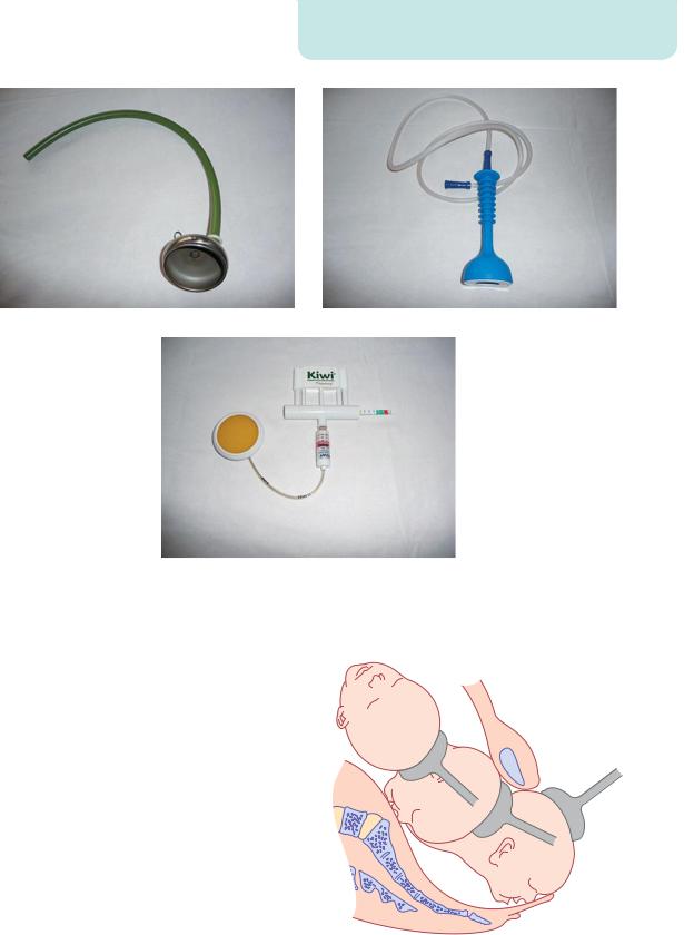
Ventouse/vacuum extractors
The basic premise of such instruments is that a suction cup, of a silastic or rigid construction, is connected, via tubing, to a vacuum source (Figure 15.3). Either directly through the tubing or via a connecting ‘chain’, direct traction can then be applied to the presenting part to expedite delivery. Recent developments have removed the need for cumbersome external suction generators and have incorporated the vacuum mechanism into ‘hand-held’ pumps, e.g. OmniCup™. Such devices appear to be more acceptable to patients than standard equipment and have no obvious effects on instrumental delivery success or on the incidence of maternal or fetal complications, but large trials have yet to be performed.
Technique
Soft cups are significantly more likely to fail to achieve vaginal delivery than rigid cups, however, they are associated with less scalp injury. There appears to be no difference in terms of maternal injury. The soft cups are appropriate for straightforward deliveries with an occipitoanterior position; metal cups appear to be more suitable for ‘occipitoposterior’, transverse and difficult ‘occipitoanterior’ position deliveries where the infant is larger or there is a marked caput.
For successful use of the ventouse, determination of the flexion point is vital. This is located at the vertex, which, in an average term infant, is on the saggital suture 3 cm anterior to the posterior fontanelle and thus 6 cm posterior to the anterior fontanelle. The centre of the cup should be positioned directly over this, as failure to do so will lead to a progressive deflexion of the fetal head during traction, and an inability to deliver the baby.
The operating vacuum pressure for nearly all ventouse is between 0.6 and 0.8 kg/cm2. It is prudent
|
|
Operative vaginal delivery |
231 |
(a) |
(b) |
(c)
Figure 15.3 Ventouse/vacuum extractor cups: (a) Metal venouse cup; (b) silicone rubber cup; (c) OmniCup™
to increase the suction to 0.2 kg/cm2 first and then to |
|
recheck that no maternal tissue is caught under the |
|
cup edge. When this is confirmed the suction can then |
|
be increased. |
|
Traction must occur in the plane of least resistance |
|
along the axis of the pelvis – the traction plane |
|
(Figure 15.4). This will usually be at exactly 90º to |
|
the cup and the operator should keep a thumb and |
|
forefinger on the cup at the traction insertion to |
|
ensure that the traction direction is correct and to feel |
|
for slippage. Safe and gentle traction is then applied |
|
in concert with uterine contractions and voluntary |
|
expulsive efforts. With the ventouse, the operator |
|
should allow no more than two episodes of breaking |
|
the suction in any vacuum delivery, and the maximum |
|
time from application to delivery should ideally be less |
|
than 15 minutes. Rotation is achieved by the natural |
|
progression of the head through the pelvis. |
Figure 15.4 The traction plane |
232 Operative intervention in obstetrics
It is not acceptable to use a ventouse when:
•The position of the fetal head is unknown.
•There is a significant degree of caput that may either preclude correct placement of the cup or,
more sinisterly, indicate a substantial degree of cephalopelvic disproportion.
•The operator is inexperienced in the use of the instrument.
Forceps
Types of forceps
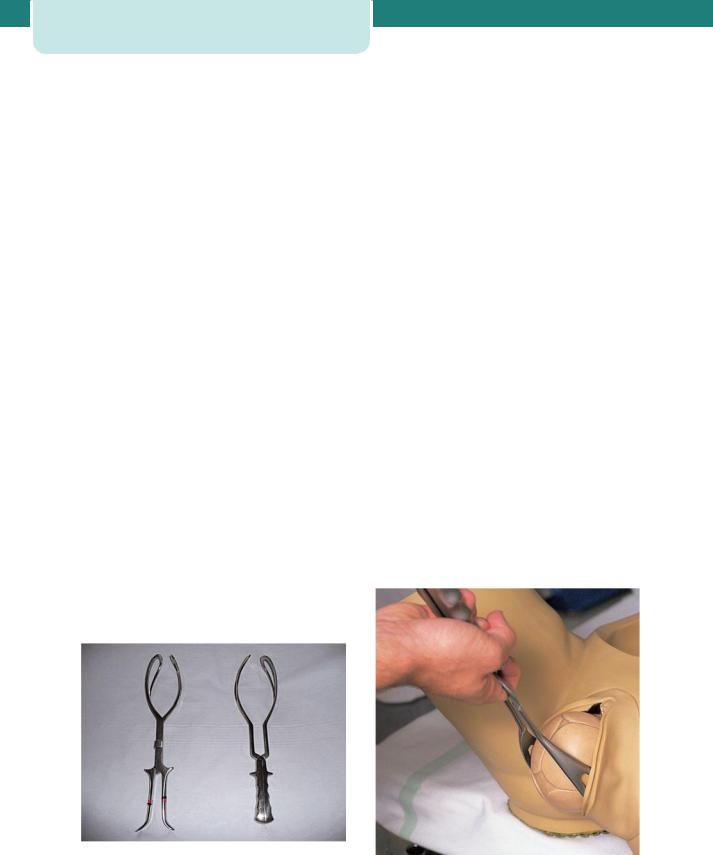
The basic forceps design has not radically changed over many years and, all types in use today consist of two blades with shanks, joined together at a lock, with handles to provide a point for traction. The specific details of construction vary between the instruments. The blades may be fenestrated – open, pseudofenestrated (open with a protruding ridge), or solid. Likewise, the length of the shanks, the design of the lock (convergent, divergent or sliding) and the fashioning of the handles are instrument specific.
Non-rotational forceps are used when the head is occipitoanterior 15º. Examples such as Neville Barnes or Simpson forceps (Figure 15.5) have a pelvic curve and an English or non-sliding lock. If the head is positioned 15º from the vertical, rotation must be accomplished before traction. Forceps designed for rotation, such as Kielland forceps, have minimal pelvic curve to allow rotation around a fixed axis; the sliding lock of the Kielland forceps (Figure 15.5) facilitates correction of asynclitism.
Technique
By convention, the left blade is inserted before the right with the accoucheur’s hand protecting the vaginal wall from direct trauma. With proper placement of the forceps blades, they come to lie parallel to the axis of the fetal head and between the fetal head and the pelvic wall. The operator then articulates and locks the blades, checking their application before applying traction (Figure 15.6).
Traction should be applied intermittently in concert with uterine contractions and maternal expulsive efforts. The axis of traction changes during the delivery and is guided along the ‘J’-shaped curve of the pelvis. As the head begins to crown, the blades are directed to the vertical, and the head is delivered.
(Specific techniques are required for rotational forceps deliveries and only those who have been properly trained in their use should employ them.)
It has been recommended that an episiotomy be cut whenever an instrumental vaginal delivery is performed. This is not based on any robust evidence. It is recognized in spontaneous vaginal delivery that episiotomy does not protect against thirdand fourth-degree tears, but does reduce the incidence of anterior vaginal trauma. Perineal trauma will occur in most nulliparous women undergoing instrumental vaginal delivery and episiotomy should be considered in these women in order to limit multiple lacerations. In multiparae, particularly those requiring only a simple ventouse, an episiotomy may not be needed.
Figure 15.5 Kielland rotational forceps and Simpson |
Figure 15.6 Application of forceps |
non-rotational forceps |

Special considerations
Failure of the chosen instrument
Failures can occur when the choice of instrument is wrong (e.g. a silastic cup ventouse for a rotational delivery), when the positioning of the ventouse cup is wrong or when the position has been wrongly defined, leading to inappropriately large diameters presenting to the pelvis. Failure is also more common if the fetus is large or maternal effort is poor.
There have been no randomized studies assessing the best approach following a failure to deliver with the first choice of instrument. Observational studies show that the outcomes for babies are worse than if the instrument of first choice is successful, but this is hardly surprising. In addition, the rates of thirdand fourth-degree tears are higher when a second instrument is used.
Unfortunately, there are no easy answers; the head may have descended to a point at which Caesarean section becomes hazardous. A policy of delivering all such women by Caesarean section will undoubtedly lead to increased maternal morbidity.
Where the first instrument fails, the following scenarios may pertain:
•The reason for failure was cup detachment and the head is occipitoanterior and on the perineum. A full
reassessment should be made. If it is confirmed that this is still the case (i.e. occipitoanterior, head on the perineum), a simple lift out forceps is acceptable. If there are any concerns, senior help must be sought.
•The instrument failed because there was little or no descent with the first pull. Where there has
been little or no descent, delivery must be by Caesarean section.
•The instrument failed because the position was wrongly defined. In this case, the next option
will either be a rotational forceps or Caesarean section. Already the fetus will have been subjected to traction with probably minimal descent.
Only after a full assessment by a senior person in theatre could further instrumentation be considered. In many cases, delivery by Caesarean section will be the safer option for the fetus.
Complications
Assisted deliveries with both vacuum and forceps can be associated with significant maternal and fetal complications.
Operative vaginal delivery |
233 |
Maternal deaths have been reported with vacuum deliveries, associated with cervical tears in women delivered before full dilatation. Traumatic vaginal delivery is considered the most important risk factor for faecal incontinence in women and may occur not only after recognized third-degree perineal tears, but also after apparently non-traumatic vaginal delivery.
Postpartum haemorrhage is more common in women needing instrumental vaginal delivery compared to women who deliver spontaneously, but less common than in women delivered by Caesarean section in the second stage. Measures to limit this include:
•prophylactic syntocinon infusion post delivery;
•prompt suturing;
•careful identification of high tears.
Underestimation of blood loss at instrumental vaginal delivery is common. Where possible, loss should be measured through the weighing of swabs and towels.
Fetal complications are no less important; the incidence of cephalhaematoma is increased with the use of the ventouse, and there are rare reports of severe intracranial injuries. Risks of perinatal trauma correlate with:
•the duration of the operative delivery;
•the station of the fetal head at the commencement of the delivery;
•the difficulty of the delivery.
•the condition of the baby at the time of commencement of the procedure.
It is important to remember that the risks of such damage significantly increase among babies who are exposed to multiple attempts at both vacuum and forceps delivery.
Clinical risk management in obstetrics
Instrumental vaginal delivery has never been free from criticism, and is certainly not without risks, although most instrumental deliveries have normal outcomes and give no reason for complaint. However, in today’s litigious society, the risks of litigation against an accoucheur or the hospital in which they practise are increased by a bad outcome.
Common allegations against practitioners that are cited in lawsuits include (among others): inadequate indication, failure to exclude cephalopelvic disproportion, improper use of instruments with excessive use of

234Operative intervention in obstetrics
force resulting in fetal or maternal injury, lack of informed consent (although this may not be fully possible in an emergency situation), and inadequate supervision. The fear of litigation must not dictate good medical practice, and assisted vaginal deliveries remain an appropriate intervention when practised with the appropriate safeguards.
Key points
•Careful assessment of each patient combining history and examination must be performed before any intervention is undertaken.
•The prerequisites for an instrumental delivery must all be met before either forceps or the ventouse are applied.
•Instruments should only be used by those trained to do so.
•Failure rates are higher with soft than rigid cups and with ventouse than forceps.
•Use of a second instrument increases the risks of fetal and maternal damage.
•Caesarean section in the second stage is associated with higher rates of haemorrhage than instrumental delivery.
Caesarean section
Definition
A Caesarean section, also known as C-section or Caesar, is a surgical procedure in which incisions are made through a mother’s abdomen (laparotomy) and uterus (hysterotomy) to deliver one or more babies.
There are three theories about the origin of the name. The name is said to derive from a Roman legal code called Lex Caesarea, which allegedly contained a law prescribing that the baby be cut out of its mother’s womb in the case that she dies before giving birth. The derivation of the name is also often attributed to an ancient story, told in the first century AD by Pliny the Elder, who claimed that an ancestor of Caesar was delivered in this manner. An alternative etymology suggests that the procedure’s name derives from the Latin verb caedere, to cut, in which case the term ‘Caesarean section’ is redundant.
Caesar’s mother, Aurelia, lived through childbirth and successfully gave birth to her son, ruling out the possibility that the Roman dictator and general was born by Caesarean section. However, the Catalan
saint, Raymond Nonnatus (1204–40), received his surname (from the Latin non natus, not born) because he was born by Caesarean section; his mother died while giving birth to him. In 1316, the future Robert II of Scotland was delivered by Caesarean section and his mother, Marjorie Bruce, died (this may have been the inspiration for Macduff in Shakespeare’s play Macbeth). The first recorded incidence of a woman surviving a Caesarean section was in 1500, in Siegershausen, Switzerland: Jakob Nufer, a pig gelder, is supposed to have performed the operation on his wife after a prolonged labour. For most of the time since the sixteenth century, the procedure had a high mortality. A Caesarean section was considered an extreme measure, performed only when the mother was already dead or considered to be beyond help. In Great Britain and Ireland, the mortality rate in 1865 was 85 per cent. Key steps in reducing mortality were:
•adherence to principles of asepsis;
•the introduction of uterine suturing by Max Sänger in 1882;
•extraperitoneal Caesarean section and then moving to low transverse incision;
•anaesthesia advances;
•blood transfusion;
•antibiotics.
On March 5, 2000, Inés Ramírez performed a Caesarean section on herself and survived, as did her son. She is believed to be the only woman to have performed a successful Caesarean section on herself.
Birth by Caesarean section has become a commonplace intervention on the modern labour ward. According to some, the Caesarean section rate has reached epidemic proportions and requires a dramatic rethink of obstetric management.
Prevalence
In the UK, more than 21 per cent of all babies are now delivered by Caesarean section. The principal aims must be to ensure that those women and babies who need delivery by Caesarean section are so delivered and that those who do not are saved from unnecessary intervention. In 1985, concern regarding the increasing frequency of Caesarean section led the World Health Organization to hold a consensus

conference. This conference concluded that there were no health benefits above a Caesarean section rate of 10–15 per cent. The Scandinavian countries have managed to hold Caesarean section rates at this level with outcomes comparable or better than those countries with higher Caesarean section rates.
Factors that may contribute to an increase in
the rates of Caesarean section
•Inaccurate dating of the pregnancy. Many practitioners combine the information obtained from a carefully taken history with that from a dating ultrasound scan, particularly when the date of the last menstrual period is uncertain. Such accurate dating reduces the anxiety experienced by many women when they pass their ‘expected date of delivery’ and also reduces the requests for ‘early’ induction of labour. In units that do not date pregnancy by ultrasound scanning before 20 weeks, an opportunity for accurate dating may be missed. This may be of particular importance in units that have a policy of offering routine induction of labour to women who are 41 weeks gestation or more.
•Fetal monitoring. Following its introduction in the 1970s, electronic fetal monitoring (EFM) was universally implemented without the appropriate trials. This has resulted in an increase in the incidence of Caesarean section without demonstrable improvement in perinatal outcome. Current recommendations are for intermittent auscultation to be performed in all ‘low risk pregnancies’, with continuous EFM in those pregnancies deemed to be ‘high risk’.
•Macrosomia. Maternal concern about fetal size is a common problem that frequently engenders anxiety among obstetricians and midwives. Although there is evidence
to suggest that birthweights are rising in developed countries, the amount (30 g over 12 years) is unlikely to be of any biological significance. Unfortunately, both
clinical and ultrasonographic estimations of fetal size are prone to inaccuracy (especially in large term infants), and unnecessary inductions of labour and Caesarean deliveries are performed as a consequence.
•Maternal request. Traditionally, Caesarean sections have been reserved for situations guided by standard clinical indications. However, the request for delivery by an elective Caesarean section where there is not a compelling obstetric indication is becoming more common. There
is huge heterogeneity in studies examining the effect of maternal request on Caesarean section rates; studies range from 2–28 per cent in the contribution of request as the primary reason, due in major part to the differing definitions of ‘request’.
Caesarean section |
235 |
•The arguments surrounding this area are complex and combine ethical dilemmas, the fetal and maternal risks of vaginal and surgical deliveries and the financial consequences of permitting such preferences. During the antenatal period, the dialogue between patients and their
doctors has increased over recent years. Policy documents such as Changing childbirth enshrined in practice the principle of total involvement of the pregnant woman in her own care. Implicit in this is the consideration of her wishes relating to delivery. Therefore, discussions must be accompanied by the careful imparting of information, counselling and advice, but should the patient’s opinions differ from those of her healthcare providers, these cannot simply be ignored. The risks of Caesarean section and labour are different, and the risks of recurrent Caesarean deliveries are additive (increasing risk of placenta praevia, accreta, operative complications, etc.). However, many practitioners believe that if these risks are fully explained to the woman, she should be allowed to choose to accept one set of risks over the other.
Indications
There are many different reasons for performing a delivery by Caesarean section. The four major indications accounting for greater than 70 per cent of operations are:
1.previous Caesarean section
2.dystocia
3.malpresentation
4.suspected acute fetal compromise.
Other indications, such as multifetal pregnancy, abruptio placenta, placenta praevia, fetal disease and maternal disease are less common. No list can be truly comprehensive and whatever the indication, the overriding principle is that whenever the risk to the mother and/or the fetus from vaginal delivery exceeds that from operative intervention, a Caesarean section should be undertaken.
Absolute indications for recommending delivery by Caesarean section are few, almost all indications are relative and there will be circumstances where Caesarean section may be best for one woman but not another. Lack of consent in a woman with capacity to give consent will prohibit Caesarean section regardless of the clinical need.