

Obstetrics_by_Ten_Teachers_19E_-_Kenny_Louise
.pdf26Physiological changes in pregnancy
the release of oxygen from red cells at relatively lower levels of haemoglobin saturation (i.e. shifts the oxygen–haemoglobin dissociation curve to the right). This increases the availability of oxygen within the tissues. The fetus is also adapted to take advantage of this alteration in maternal physiology. Fetal haemoglobin differs from adult haemoglobin in that the two beta-chains are replaced by gammachains. Binding of 2,3-DPG to haemoglobin occurs preferentially to beta-chains, with the result that in the fetus the oxygen–haemoglobin dissociation curve is shifted to the left relative to the maternal state. In this way oxygen transfer from mother to fetus is facilitated.
Oxygen consumption increases by about 45 mL/min during the course of pregnancy, which represents an increase of about 20 per cent from oxygen consumption at rest (300 mL/min). Around one-third of the increase is necessary for the metabolic demands of placenta and fetus, and the remainder is needed for the extra metabolic work of the maternal organs. The increased oxygen consumption coupled with the decreased FRC decreases maternal oxygen reserve and predisposes the pregnant woman to hypoxaemia and hypocapnia during periods of respiratory depression or apnoea.
Arterial gases
Progesterone has respiratory stimulant properties and the greatly increased levels in pregnancy are thought to be responsible for increasing alveolar ventilation through increasing tidal volume. In consequence, there is a marked decrease (15–20 per cent) in the partial pressure of carbon dioxide (pCO2). There is usually a slight increase in the partial pressure of oxygen (pO2). These changes facilitate gas transfer to and from the fetus. Arterial blood gas values differ in pregnant compared to non-pregnant women, but normal ranges of arterial gases at term are not well established, and measured arterial pO2 has been reported to be normal, low and high. Normal oxygen saturation measured non-invasively should be in excess of 95 per cent during pregnancy.
The reduction in pCO2 has implications for acid–base balance, as carbon dioxide forms carbonic acid in the presence of water. Reductions in pCO2 activate compensatory buffering mechanisms so that potentially hazardous alkalosis is prevented. This involves the activity of the enzyme carbonic anhydrase, which converts carbonic acid to bicarbonate, thus releasing hydrogen ions to restore pH. The kidney
then excretes the bicarbonate formed. In pregnancy, renal excretion of bicarbonate increases significantly and maternal arterial pH changes very little, being maintained at 7.40 to 7.45. An increase in the concentration of carbonic anhydrase within maternal erythrocytes has been reported, but whether this is a primary event or a secondary adaptation is uncertain.
Key points
Ventilatory changes
•Thoracic anatomy changes.
•↑ Minute ventilation.
•↑ Tidal volume.
•↓ Residual volume.
•↓ Functional residual capacity.
•Vital capacity unchanged or slightly increased.
Blood gas and acid–base changes
•↓ pCO2.
•↑ pO2.
•pH alters little.
•↑ Bicarbonate excretion.
•↑ Oxygen availability to tissues and placenta.
Cardiovascular system
Signs and symptoms of pregnancy mimic those of heart disease. Elevation of the diaphragm, adjustments of lung volumes and increases in minute ventilation give rise to breathlessness. Oedema in the extremities is a common finding, and results from an increase in total body sodium and water, as well as venous compression by the gravid uterus. The latter can also cause decreased venous return to the heart, leading to light-headedness and syncope. Palpitations are common and usually represent sinus tachycardia, which is normal in pregnancy. They may also occur as increased awareness of the heart beating either regularly or in association with extrasystoles, presumably related to changes in cardiac output. Premature atrial and ventricular ectopic beats are common in pregnancy and the peripheral pulse is increased in volume. Increases in plasma volume cause the jugular veins to fill and pulsate dynamically in pregnancy, but the mean right atrial pressure is unchanged and the height of the jugular

venous pressure is also unchanged. Changes in the left ventricular size and volume mean the apex beat is more forceful.
In normal pregnancy, cardiac output increases as early as 5 weeks gestation (3 weeks after conception) and rises to around 40 per cent above the prepregnancy baseline by 24 weeks, i.e. from about 5.0 to 7.0 L/min when at rest (Table 3.2). The increase in cardiac output is caused partly by an increase in heart rate, which is detected first as early as 5 weeks, and partly by an increase in stroke volume. A progressive increase in heart rate continues until the third trimester of pregnancy, when rates are typically 10–15 beats per minute greater than those found in the non-pregnant state. There is also a progressive amplification of stroke volume (10–20 mL) during the first half of pregnancy, probably related to the changes in plasma volume at this time (Figure 3.3).
Table 3.2 Changes in cardiac output during pregnancy
|
Cardiac output |
Non pregnant adult |
4.5 L/min |
female |
|
|
|
Pregnancy (20 weeks) |
↑ by 40 per cent to |
|
6.3 L/min |
Early labour |
↑ by 17 per cent to |
|
7.3 L/min |
|
|
Active labour |
↑ by 23 per cent to |
|
7.7 L/min |
2nd stage of labour |
↓ by 34 per cent to |
|
8.4 L/min |
90 |
|
8 |
Heart |
|
Cardiac |
|
output |
|
rate |
|
(L/min) |
(bpm) |
|
4 |
|
|
|
Stroke |
|
|
volume |
|
|
(mL) |
|
|
60 |
|
0 |
0 |
20 |
40 |
Weeks of pregnancy
Figure 3.3 There is a marked increase in maternal cardiac output during pregnancy. Increases in both heart rate and stroke volume contribute, but changes in these components are not synchronous
Cardiovascular system |
27 |
140 |
|
|
|
|
120 |
|
|
|
|
100 |
|
|
|
|
80 |
|
|
|
|
60 |
|
|
|
|
40 |
|
|
|
|
20 |
|
|
|
|
0 |
|
|
|
|
0 |
10 |
20 |
30 |
40 |

 systolic BP
  diastolic BP
Figure 3.4 Systolic and diastolic blood pressure in normal pregnancy. After Chamberlain and Broughton Pipkin, Clinical physiology in obstetrics, 1998
Decreases in diastolic blood pressure (10– 15 mmHg) are more marked during the antenatal period than the decrease in systolic pressure (5–10 mmHg). Thus, early pregnancy is associated with a relative increase in pulse pressure. An 11 per cent fall in mean arterial blood pressure at rest has been shown by 15 weeks gestation. Later, diastolic blood pressure increases to levels that are at least equivalent to those found in the non-pregnant state (Figure 3.4). A mean diastolic blood pressure of 66.6 mmHg has been reported in the first trimester, falling to 66.3 mmHg in the second and rising to 68.4 mmHg by 40 weeks. Accurate determination of diastolic pressure is therefore very important, and the best measurements are obtained when the fifth Korotkoff sound (disappearance of sounds) is used. As blood pressure does not rise in pregnancy, and usually falls, the increase in cardiac output is also associated with a fall in peripheral vascular resistance. A 70 per cent reduction in peripheral resistance has been demonstrated by 8 weeks gestation. The mechanisms responsible are uncertain, but alterations have been reported in production of vasoconstrictor and vasodilator agents that control peripheral arterial tone.
The cardiac output is elevated at the onset of labour to over 7.0 L/min, rising further within labour, with a 30 per cent increase in demand in the final stages (Table 3.2). This increase is due to the uterine contractions each of which squeezes 300–500 mL of blood into the maternal circulation. At delivery, a shift of blood from the empty uterus into the maternal circulation – called autotransfusion – causes an increase of 10–20 per cent in the cardiac output. Stroke volume, heart rate and cardiac output remain

28Physiological changes in pregnancy
elevated for the first 2 days postpartum. Within the first 2 weeks after delivery the cardiac output falls rapidly, at 6 weeks postpartum it is halfway between pregnant and non-pregnant values and at 24 weeks after delivery the cardiac output falls to below 5.0 L/min.
Ausculatory changes in pregnancy are well documented. The first heart sound is loud and sometimes split, while a third heart sound is audible in 84 per cent of pregnant women by 20 weeks gestation. An ejection systolic murmur can be heard in 96 per cent of apparently normal pregnant women; this murmur is widely conducted and it disappears after delivery. The third heart sound is frequently misinterpreted as a diastolic murmur, but a true diastolic murmur occurs transiently in only 20 per cent of pregnant women, whereas 10 per cent develop continuous or systolic murmurs due to increased mammary blood flow.
Key points
Cardiovascular changes
•↑ Heart rate (10–20 per cent).
•↑ Stroke volume (10 per cent).
•↑ Cardiac output (30–50 per cent).
•↓ Mean arterial pressure (10 per cent).
•↓ Pulse pressure.
•↓ Peripheral resistance (35 per cent).
Gastrointestinal changes
Oral
The physiological changes of pregnancy include effects on mucous membranes, pigmentation and glandular function. Pregnancy gingivitis is the term used for inflammation and hyperplasia of the gingival mucosa occurring during gestation and from 30 to 75 per cent of pregnant women develop erythema, oedema, hyperplasia and increased bleeding of the gingival tissue. Elevated circulating oestrogen and progesterone levels are implicated in increasing vascular permeability and decreasing immune resistance, thereby increasing susceptibility to gingivitis. The hormone levels of pregnancy also affect the response of the periodontal tissues to bacterial
colonization, creating a more favourable environment for anaerobic infection. The main salivary changes in pregnancy include variations in pH and composition, with a reduction of sodium concentration that leads to a decrease in pH and increased concentration of protein. Salivary oestrogen levels are increased, resulting in an increased proliferation and desquamation of the oral mucosa. While teeth usually retain their structure, salivary changes late in pregnancy, as well as oestrogenenhanced changes in the mucosa, predispose to dental caries. Increased tooth mobility, especially of the upper incisors, has been detected in pregnant women, even those with normal periodontal tissues.
Gut
As gestation advances, the uterus displaces the stomach and intestines upwards, which can hinder diagnosis of intra-abdominal surgical events as well as confound the routine abdominal examination. Elevated progesterone levels reduce lower oesophageal sphincter tone and increase the placental production of gastrin, increasing gastric acidity. These changes combine to increase the incidence of reflux oesophagitis and heartburn, which affect up to 80 per cent of pregnant women. Mechanical factors, the enlarging uterus and progesterone levels all contribute to delayed gastric emptying and increased stomach volume. Gastric motility decreases further during labour and emptying remains delayed during the puerperium. As a result of all of these changes, the pregnant woman is at increased risk of aspiration of gastric contents when sedated or anaesthetized after 16 weeks gestation. Delayed gastric motility and prolonged gastrointestinal transit time may also lead to constipation and alter the bioavailability of medications.
Liver
The liver, normally palpated 2 cm below the right costal margin, may become more difficult to examine because of the expanding uterus within the abdominal cavity. Physical findings such as telangiectasia and palmar erythema, otherwise suggestive of liver disease in non-pregnant women, appear in up to 60 per cent of normal pregnancies because of the hyperoestrogenic state of pregnancy, as the liver cannot easily metabolize the large quantity of placental oestrogen and progesterone. During pregnancy, absolute hepatic blood flow remains largely unaltered and hepatic

function remains normal. Portal vein pressure is increased in late pregnancy, and venous pressure increases in the oesophagus. Although hepatic protein production increases, serum albumin levels decline in pregnancy due to the increase in maternal plasma volume. In contrast, an increase in serum alkaline phosphatase secondary to fetal and placental production is observed in pregnancy and persists postpartum, rendering it unhelpful diagnosing cholestasis during the third trimester (Table 3.1). Probably the most important hepatic changes in pregnancy are the increased production and plasma levels of fibrinogen and the clotting factors VII, VIII, X and XII. Finally, hypercholesterolaemia is well described in pregnancy – plasma cholesterol levels rise by around 50 per cent in the third trimester and triglycerides may rise to two or three times normal levels. Levels fall after delivery, returning to normal faster in lactating women.
The kidneys and urinary tract
Anatomic changes
The kidneys increase in size in normal pregnancy, with a 1–2 cm change in length. The calyces, renal pelvis and ureters dilate, giving the usually incorrect impression of obstruction. By the third trimester, over 80 per cent of women have some evidence of stasis or hydronephrosis, which is more marked on the right side due to uterine dextrorotation. This physical change, together with alterations in the composition of the urine itself, predisposes pregnant women to ascending urinary tract infection, a
The kidneys and urinary tract |
29 |
common complication of pregnancy (1–2 per cent of all pregnancies). Renal parenchymal volumes also increase during pregnancy, most likely due to increases in intrarenal fluid. By 6 weeks postpartum, renal dimensions return to pre-pregnancy values.
Functional changes
Glomerular filtration rate (GFR) rises immediately after conception and increases by about 50 per cent overall, reaching its maximum at the end of the first trimester. GFR then falls by about 20 per cent in the third trimester, returning to pre-pregnancy levels within 12 weeks of delivery (Table 3.3). Renal blood flow increases by up to 80 per cent in the second trimester, due to the combination of increased cardiac output and increased renal vasodilatation, but this is followed by a 25 per cent fall towards term (Figure 3.5). Despite chronic vasodilatation,
50
45
40
35
30
%25
20
15
10
5
0
0 |
|
10 |
|
20 |
30 |
40 |
|
|
|
|
|
|
|
|
|
|
GFR |
|
Plasma volume |
|
|
|
|
|
|
||
|
|
|
|
|
|
|
Figure 3.5 Changes in GFR and plasma volume during normal pregnancy
Table 3.3 Changes in indices of renal function during pregnancy (mean values); data from de Swiet, Medical disorders in obstetric practice, 2002
|
Non-pregnant |
1st Trimester |
2nd Trimester |
3rd Trimester |
RPF mL/min |
480 |
841 |
891 |
771 |
GFR mL/min |
105 |
162 |
174 |
165 |
24 h creatinine clearance |
98 |
151 |
154 |
129 |
Plasma creatinine mol/L |
73 |
60 |
54 |
64 |
Plasma urea mmol/L |
4.3 |
3.5 |
3.3 |
3.1 |
Plasma urate mol/L |
246 |
189 |
214 |
269 |
|
|
|
|
|
|
|
|
|
|

30Physiological changes in pregnancy
the kidney is still able to autoregulate blood flow and GFR over a range of blood pressure, mainly through variations in arteriolar tone. Creatinine clearance increases by 25 per cent at 4 weeks gestation and by 45 per cent at 9 weeks, but over the course of the pregnancy a decrease back to non-pregnant values occurs. The renin-angiotensin system is also modified in normal pregnancy, with increases in plasma renin activity and plasma angiotensin-II.
As GFR increases without alterations in the production of creatinine and urea, plasma levels of these substances decrease. Creatinine levels change from a non-pregnant value of 73 mmol/L to 60, 54 and 64 mmol/L in successive trimesters. The average plasma urea level of 3.1 mmol/L in the third trimester also contrasts with the non-pregnant value of 4.3 mmol/L. Plasma urate concentrations decrease by over 25 per cent from 8 weeks gestation, but due to changes in renal absorption and excretion increase again in the third trimester to levels close to nonpregnant values (Table 3.3). There is also increased renal excretion of various nutrients, calcium and protein, mainly due to changes in renal tubular function. Sodium balance is maintained, despite the GFR increase and increases in filtration, by increasing sodium reabsorption both in proximal tubules (under the influence of oncotic pressure in the renal interstitial space) and in distal portions (under the influence of hormonal factors). Potassium metabolism remains unchanged, despite cumulative retention of potassium, necessary for fetal-placental development and expansion of maternal red blood cell mass, and increased aldosterone levels. Early morning urine is more alkaline than in non-pregnant women, but acid excretion ability of the kidney is unchanged (Table 3.1).
Glycosuria, which is rare in the absence of diabetes in the non-pregnant state, is very common during pregnancy and does not relate reliably to disorders of carbohydrate metabolism. The increase in GFR may be partially responsible, and a reabsorptive mechanism in the proximal renal tubule may become saturated so that the ‘renal threshold’ is exceeded, explaining the increased amount of glucose in the urine. Glucose reabsorption occurs secondarily to the absorption of sodium and therefore other factors contributing to volume homeostasis and sodium retention may also be involved in the physiological glycosuria of pregnancy.
Key points
Renal changes
•↑ Kidney size (1 cm).
•Dilatation of renal pelvis and ureters.
•↑ Blood flow (60–75 per cent).
•↑ Glomerular filtration (50 per cent).
•↑ Renal plasma flow (50–80 per cent).
•↑ Clearance of most substances.
•↓ Plasma creatinine, urea and urate.
•Glycosuria is normal.
Reproductive organs
Uterus
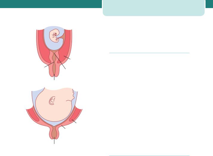
Uterine blood flow increases 40-fold to approximately 700 mL/min at term, with 80 per cent of the blood distributed to the intervillous spaces of the placentae, and 20 per cent to the uterine myometrium. Oestrogen mediates the adaptation of the uterine smooth muscle to pregnancy. High levels of maternal oestradiol and progesterone induce both hyperplasia and hypertrophy of the myometrium, increasing the weight of the uterus from 50–60 g prior to pregnancy to 1000 g by term. The growing size of the uterine contents is an important stimulus, with individual muscle fibres increasing in length by up to 15-fold. The uterine arteries also undergo hypertrophy in the first half of pregnancy, although in the second half increasing uterine distension is matched by arterial stretching. Progesterone helps maintain lower myogenic tone in the uterine vessels despite the increased blood flow. Maternal cortisol also regulates local uterine blood flow, through effects on vascular endothelium and smooth muscle. By the third trimester, the uterus is described in lower and upper segments. The lower segment is the part of the uterus and upper cervix which lies between the attachment of the peritoneum of the uterovesical pouch superiorly and the level of the internal cervical os inferiorly. It is thinner, contains less muscle and fewer blood vessels and is the site of incision for the majority of Caesarean sections (Figure 3.6).
1st trimester
UM
H
A
E
3rd trimester
UM
IL
E
A Anatomical internal os
H Histological internal os
EExternal os
UM |
Uterine muscle |
IL |
Intermediate layer |
Figure 3.6 Formation of the lower uterine segment. With increasing gestation, uterine stretch occurs. This has the effect of drawing the anatomical internal cervical os (A) further from the histological internal cervical os (H). The retraction of the thick intermediate layer (IL) of muscle with increasing gestation thins the lower segment
As well as changes in the size and number of myometrial cells, specialized cellular connections also develop with increasing gestation. These intercellular gap junctions allow changes in membrane potential to spread rapidly from one cell to another, facilitating the spread of membrane depolarization, and subsequent myometrial contraction. Steroid hormones also have an effect on signalling pathways. As these junctions mature, uterine contractions become more frequent. These are apparent initially as Braxton Hicks, painless contractions that are noticed in the second half of
Reproductive organs |
31 |
pregnancy. Subsequently, these allow the pacemaker activity of the uterine fundus to promote the coordinated, fundal-dominant contractions necessary for labour.
Cervix
The cervix is described as looking bluer during pregnancy, which is due to its increased vascularity. It becomes swollen and softer during pregnancy under the influence of progesterone and oestradiol; the latter also stimulates growth of the columnar epithelium of the cervical canal. This becomes visible on the ectocervix and is called an ectropion, which is prone to contact bleeding. In addition, the mucous glands of the cervix become distended and increase in complexity. Prostaglandins induce a remodelling of cervical collagen in late gestation, while collagenase released from leukocytes locally also aids in softening the cervix. Under the influence of oestrogens, the vaginal epithelium becomes more vascular during pregnancy, and there is increased desquamation resulting in increased vaginal discharge. This discharge has a more acid pH than non-pregnant vaginal secretions (4.5–5.0) and may protect against ascending infection.
Breasts and lactation
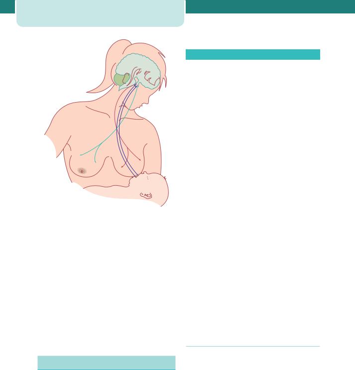
The cyclical changes seen in breast tissue with the menstrual cycle are accentuated during pregnancy. Deposition of fat around glandular tissue occurs, and the number of glandular ducts is increased by oestrogen, while progesterone and human placental lactogen (hPL) increase the number of gland alveoli. Prolactin is essential for the stimulation of milk secretion and during pregnancy prepares the alveoli for milk production. Although prolactin concentration increases throughout pregnancy, it does not then result in lactation since it is antagonized at an alveolar receptor level by oestrogen. The rapid fall in oestrogen concentration over the first 48 hours after delivery removes this inhibition and allows lactation to begin. Towards the end of pregnancy, and in the early puerperium, the breasts produce colostrum, a thick yellow secretion rich in immunoglobulins. Lactation is initiated by early suckling, which stimulates the anterior and posterior pituitary to release prolactin and oxytocin, respectively. During the first 2–3 days
32 Physiological changes in pregnancy
Suckling causes:
Afferent signals to posterior pituitary increasing oxytocin release, inducing myoepithelial cells to contract and express milk. Afferent signals to anterior pituitary increasing prolactin release, thus increasing milk synthesis.
Figure 3.7 Schematic representation of lactation. Suckling induces afferent signals to the anterior and posterior pituitary. This results in the release of prolactin and oxytocin. Prolactin induces milk production by the glandular tissue of the breast. Oxytocin causes contraction of the myoepithelial cells surrounding the glandular ducts, squeezing milk towards the nipple
Table 3.4 Hormones produced within the pregnant uterus
Type |
Name |
Pregnancy |
Human chorionic |
specific |
gonadotrophin (hCG) |
|
Human placental lactogen |
|
(hPL) |
|
|
Hypothalamus |
Gonadotrophin-releasing |
|
hormone (GnRH) |
|
Corticotrophin-releasing |
|
hormone (CRH) |
|
|
Pituitary |
Prolactin |
|
Human growth hormone |
|
(hGH) |
|
Adrenocorticotrophic |
|
hormone (ACTH) |
Steroids |
Oestradiol |
|
Progesterone |
|
|
Other peptides |
Insulin-like growth factor-I |
|
and II |
|
1,25 Dihydroxycholecalciferol |
|
Parathyroid hormone-related |
|
peptide |
|
Renin |
|
Angiotensin-II |
|
|
of the puerperium, prolactin promotes breast engorgement and the alveoli become distended with milk. Oxytocin released from the posterior pituitary causes contraction in myoepithelial cells surrounding the alveoli and small ducts, squeezing milk towards the nipple. Released during labour as well, oxytocin facilitates the rapid onset of maternal–infant bonding as well as an altered emotional state (Figure 3.7).
Endocrinology
Complex endocrinological changes occur in pregnancy. Many physiological adaptations to pregnancy are organized by the maternal brain, predominantly through changes in neuroendocrine systems, and the hormones of pregnancy primarily drive these changes. Other hormones exert their actions indirectly, by interacting with cytokines and
chemokines. The production and activity of these substances are also significantly altered in pregnancy. Finally, during pregnancy, both the placenta and the fetus produce some hormones otherwise produced by the endocrine glands in the non-pregnant state (Table 3.4).
Pituitary gland
The pituitary gland enlarges during normal pregnancy and concentrations of prolactin reach levels during pregnancy that are 15-fold higher than in the non-pregnant state. Oestrogen has a stimulatory role in this process and hPL is inhibitory. The endocrinological mechanisms that regulate prolactin production in the non-pregnant state, such as sleep, which increases and dopamine agonists, which reduce prolactin concentration, remain effective during pregnancy. Therefore prolactin production by the

anterior pituitary gland continues despite intrauterine production from cells within the decidua. Receptors for prolactin are also present on trophoblast cells and within the amniotic fluid. Increased prolactin production is essential for lactation (as already discussed) but also acts in the brain to reduce responses to stress. Further, prolactin may play a role in the regulation of insulin secretion and glucose homeostasis in the post-natal period. In contrast to prolactin, there is evidence that human growth hormone (hGH) production by the anterior pituitary gland is suppressed during pregnancy, and circulating concentrations are also reduced. It is likely that hPL is also involved in suppressing hGH release.
Thyroid function
Human chorionic gonadotrophin (hCG) has thyrotrophic activity owing to subunit homology with thyroid-stimulating hormone (TSH) and maternal TSH production is suppressed during the first trimester of pregnancy, when hCG levels are highest. The TSH response to thyrotrophin-releasing hormone (TRH) is reduced during the first trimester but returns to normal after this. Thyroid binding globulin increases in the first 2 weeks of pregnancy and reaches a plateau by 20 weeks. This leads to increased production of total T3 (tri-iodothyronine) and T4 (thyroxine). The increased GFR of pregnancy results in an increased renal loss of iodide, which is essential for thyroid hormone synthesis, so the thyroid compensates by increasing the proportion of iodide it takes up from the circulation. Where there is relative background iodide deficiency, these changes may result in enlargement of the thyroid gland during pregnancy.
The hypermetabolic state of normal pregnancy makes clinical assessment of thyroid function more difficult and therefore thyroid function often needs to be checked biochemically. However the physiological changes of pregnancy, including the 50 per cent plasma volume expansion, increased thyroid binding globulin production and relative iodine deficiency, mean that thyroid hormone reference ranges for non-pregnant women are not appropriate in pregnancy. Free T4 (fT4), free T3 and TSH should be analysed when assessing thyroid function in pregnancy, and total T3 and T4 not used. There is a fall in TSH and a rise in fT4 concentrations in the first
Endocrinology 33
trimester of normal pregnancy. This is followed by a fall in fT4 concentration with advancing gestation, with the most marked effect in the third trimester (Table 3.1).
Uterus and placenta
Many pregnancy-specific peptides are produced within the uterus, but not all have been shown to have definite endocrine roles. The best known is hCG, produced by trophoblast cells. The β-subunit is pregnancy specific and used as a sensitive pregnancy test, being detectable within the maternal circulation in small quantities within days of implantation. Production of hCG is influenced both by the cytokine leukaemia inhibitory factor (LIF) and by an isoform of gonadotrophin-releasing hormone (GnRH), which is also produced within the placenta. Human chorionic gonadotrophin has a major role during early pregnancy in maintaining the function of the corpus luteum, which produces progesterone, but circulating hCG values fall off after 12 weeks, as the placental production of progesterone becomes dominant. During normal pregnancy, hCG also suppresses the secretion of FSH and LH by the anterior pituitary gland, perhaps by interaction at the hypothalamic level.
Sex steroid hormones are produced in large quantities by the placenta and fetus. Concentrations of oestrogens and progesterone increase substantially from early pregnancy, and then plateau for the remainder of the pregnancy. Both oestrogen and progesterone have effects on the myometrium, where oestrogen encourages cellular hypertrophy while progesterone discourages contraction and, together with prolactin, on the tissues of the breast. They also have effects on many other tissues during pregnancy, such as the smooth muscle of the vascular tree and of the urinary and gastrointestinal tracts.
Corticosteroids
Trophoblast cells produce adrenocorticotrophic hormone (ACTH), which has a role in regulating the activity of the fetal adrenal glands and the myometrium and possibly also the mother’s adrenal glands. A progressive increase in maternal circulating concentrations of cortisol throughout normal pregnancy has been noticed from as early as

34Physiological changes in pregnancy
11 weeks. Cortisol reaches two-to three-fold higher concentrations than in the non-pregnant, despite a decrease in the concentration of ACTH in later gestation. Much of the cortisol is bound to cortisolbinding globulin (CBG), which doubles in concentration during pregnancy, but there is also a slight increase in unbound cortisol. The lack of diurnal variation of cortisol and the attenuated response to dexamethasone suppression suggest that placental ACTH may have a role in regulating maternal cortisol levels. Cortisol responses to stressors are reduced in pregnant women.
Circulating concentrations of the antinatriuretic hormones aldosterone and deoxycorticosterone increase ten-fold in pregnancy. Plasma aldosterone levels rise seven to eight-fold during the first trimester, but the diurnal rhythm of levels is preserved throughout pregnancy. Progesterone has natriuretic properties, and other factors that may influence aldosterone production, such as atrial natriuretic peptide and angiotensins, are produced in greater amounts during pregnancy. The increased production of angiotensins, including the vasoactive angiotensin-II, is the result of an augmented production of the enzyme renin and its substrate angiotensinogen.
Corticotrophin-releasing hormone (CRH) is produced by the placenta in the second half of pregnancy. One function of placental CRH expression is to stimulate the fetal adrenals to synthesize and release dihydroepiandrosterone (DHEA), which the placenta then converts to oestrogens. Another function is to stimulate the fetal adrenal gland (both directly and through stimulation of the fetal pituitary) to synthesize and release cortisol. Fetal cortisol drives the placental-fetal adrenal axis through positive feedback that results in increasing oestrogen production over gestation, and also works by maturing other fetal organs.
Plasma CRH, ACTH and cortisol concentrations increase several-fold with the onset of labour and delivery, with peak CRH levels 48 hours before delivery. Maternal plasma ACTH levels are ten-fold higher in labour than when not pregnant. Thus the progression of gestation is linked to the timing of fetal development, and infants are born with appropriately mature organs. The placental regulation of its own metabolism through effects on the fetus, with subsequent effects on maternal uterine physiology, and possibly the onset of labour, has been called the ‘placental clock’ theory.
Key points
Endocrine changes
•↑ Prolactin concentration.
•Human growth hormone is suppressed.
•↑ Corticosteroid concentrations.
•↓ TSH in early pregnancy.
•↓ fT4 in late pregnancy.
•hCG is produced.
•Insulin resistance develops.
Metabolism
Energy requirements and weight gain
Energy requirements during pregnancy are defined as the level of energy intake from food needed to balance energy expenditure, presuming the woman has a body size and level of physical activity consistent with good health. The energy cost of pregnancy includes energy deposited in maternal and fetal tissues, and the increase in energy expenditure attributed to maintenance and physical activity. As a result of the increased tissue mass the energy cost of maintenance, as well as physical activity, rises during pregnancy. In healthy well-nourished women, the increases in basal metabolic rate (BMR) range from 124 to 210 MJ, with an average increase of 157 MJ for the whole pregnancy (corresponding to an average gestational weight gain of 12.5 kg).
Weight gain during pregnancy consists of the products of conception (fetus, placenta, amniotic fluid), the increase of various maternal tissues (uterus, breasts, blood, extracellular fluid), and the increase in maternal fat stores. The increase in weight is largely fluid, with today body water increasing by around 8 L. Rates of weight gain vary across the trimesters of pregnancy, with estimates of 1.6 kg gained in the first trimester, 0.45 kg per week in the second and 0.4 kg per week in the third trimester reported. The appropriate gestational weight gain for optimal pregnancy outcome is the subject of much debate, and is modified by pre-pregnancy body mass index (BMI) measurements. Ranges of weight gain recommended for women with low pre-pregnancy BMI ( 20) are 12.5–18.0 kg, compared to 11.5–16.0 kg for those

with normal pre-pregnancy BMI (20–26). However, women with a lower BMI must gain more weight to produce infants with birthweights comparable to women with a normal BMI. Women with high BMI not only deliver infants with higher birthweights, but also can do so with lower gestational weight gains.
Carbohydrate metabolism
Changes in carbohydrate and lipid metabolism occur during pregnancy to ensure a continuous supply of nutrients to the growing fetus. In the first half of pregnancy, fasting plasma glucose concentrations are reduced with little change in insulin levels. An oral glucose tolerance test at this time shows an enhanced response compared to the non-pregnant state, with a normal pattern of insulin release but reduced blood glucose values. This pattern changes during the second half of pregnancy, where an increase in glucose values throughout the test despite significant increases in plasma insulin concentrations suggests relative insulin resistance. Insulin action in late normal pregnancy is 50–70 per cent lower than in non-pregnant women and insulin resistance is thought to allow shunting of nutrients to the fetus. This change may involve hPL or other growth-related hormones, such as prolactin or cortisol, which reduce peripheral insulin sensitivity. Pregnancy is also associated with alterations in insulin receptor binding, similar to those described in nonpregnant women who have non-insulin-dependent diabetes mellitus. Whether this results from increased transplacental transfer of glucose or from the growthpromoting characteristics of insulin is unclear. During lactation, glucose levels fall and insulin resistance returns to normal, as glucose homeostasis is reset.
Lipid metabolism
Changes in hepatic and adipose metabolism alter circulating concentrations of triacylglycerols, fatty acids, cholesterol and phospholipids, which all increase after the eighth week of pregnancy. Both the higher concentration of oestrogen and insulin resistance are thought to be responsible for the hypertriglyceridaemia of pregnancy. HDL-cholesterol increases by 12 weeks in response to oestrogen and remains elevated throughout pregnancy, while total and LDL-cholesterol concentrations decrease initially, but then increase in the second and third trimesters. Changes in lipid metabolism influenced by increased
Metabolism 35
oestrogen, progesterone and insulin promote the accumulation of maternal fat stores in early pregnancy and inhibit lipolysis. In late pregnancy, fat mobilization is enhanced to allow pregnant women to use stored lipid for energy needs and minimize protein catabolism, preserving glucose and amino acids for the fetus.
Calcium metabolism
Around 40 per cent of circulating calcium is bound to albumin. Since plasma albumin concentrations decrease during pregnancy, total plasma calcium concentrations also decrease, which is not evidence of true hypocalcaemia. There is little change in the circulating concentration of unbound ionized calcium. In pregnancy, a number of adaptive changes must occur to facilitate positive calcium balance in favour of the developing fetus, which needs to equilibrate its own calcium level and ensure skeletal development. The fetal demand for calcium is substantial and transplacental flux rates of about 6.5 mmol per day have been estimated. There are three potential methods of maternal adaptation: increasing gut absorption, mobilizing skeletal calcium reserves or restricting renal losses. The pregnant woman usually increases the rate or efficiency of gut absorption and slightly decreases excretion, thereby coping with little overall change in transfer rates into and out of bone stores. The increase in gut calcium absorption is a result of increased production of a metabolite of vitamin D3, 1,25-dihydroxycholecalciferol (1,25- (OH)2D3). Production of 1,25-(OH)2D3 is under the influence of parathyroid hormone (PTH), which increases by about one-third during pregnancy. No consistent changes have been reported in circulating concentrations of other agents involved in calcium metabolism, for example calcitonin and other metabolites of vitamin D3. However, in situations of abundant calcium, maternal PTH levels are suppressed and renal calcium retention is overridden to allow excretion. Observational studies have shown either no change or a small decline in maternal storage forms of vitamin D during pregnancy.
Omega-3 fatty acids
Omega-3 fatty acids are essential and can only be obtained from the diet. Requirements in pregnancy have not been established but are presumed to be