

118D. Stamenovic´
and Stamenovi´c, 2003). In the case of suspended cells, this model provides very good correspondence to experimental data. For example, the model of the spectrin lattice successfully describes the behavior of red blood cells during micropipette aspiration measurements (Discher et al., 1998). However, in the case of adherent cells, the model of the actin cortical lattice has enjoyed only moderate success. While it provides a reasonably good correspondence to data from cell poking measurements, it exhibits only some qualitative features of the cell response to twisting and pulling of magnetic beads bound to integrin receptors (Coughlin and Stamenovi´c, 2003). Taken together, the above results show that the two-dimensional cable net model is incomplete to describe mechanical behavior of adherent cells; however the results also show that prestress is a key determinant of the model response.
Cable-and-strut model
This is a cable net model in which the prestress in the cables is balanced by internal compression-supporting struts rather than by inflating pressure. At each free node, one strut meets several cables (see Fig. 6-1). Cables carry initial tension that is balanced by compression of the strut. Together, cables and struts form a self-equilibrated and stable form in the space. This structure may also be attached to the substrate (Fig. 6-1). In this case, the anchoring forces of the substrate also contribute to the balance of tension in the cables. The main difference between these structures and the cable nets is that in the former, the struts directly contribute to the structure’s resistance to shape distortion, whereas in the latter this contribution does not exist.
The shear modulus (G) of the cable and strut model can be also obtained using the affine network approach, as in the case of the tensed cable net model. It was found (Stamenovi´c, 2005) that
G = 0.8(P − PQ ) + 0.2(BP + BQ PQ ) |
(6.4) |
where P is the prestress carried by the cables and PQ is the portion of P balanced by the struts, B ≡ (dF/dL)/(F/L) is the nondimensional cable stiffness and BQ ≡ (dQ/dl )/(Q/l ) is the nondimensional strut stiffness. The difference P − PQ represents the portion of P transmitted to and balanced by the substrate and is denoted by PS (see Fig. 6-2b). It is this PS that can be directly measured using the traction microscopy technique (Wang et al., 2002).
It was shown in the section on tensed cable nets that B = 2.4. The quantity BQ is determined based on the buckling behavior of microtubules (Stamenovi´c et al., 2002a). It is found that BQ ≈ 0.54. By substituting this value and B = 2.4 into Eq. 6.4 and taking into account that in well-spread smooth muscle cells, microtubules balance on average 14 percent of PS , that is, PQ = 0.14PS (Stamenovi´c et al., 2002a), it is obtained that G = 1.19P, which is close to the experimental data of G = 1.04P (Fig. 6-4).
It is noteworthy that if PQ = 0, for example, in a case where microtubules are disrupted, Eq. 6.4 reduces to Eq. 6.3. If disruption of microtubules would not affect P, then according to Eqs. 6.3 and 6.4, for a given P, the shear modulus G would be8 percent lower in the case of intact microtubules than in the case of disrupted microtubules. In reality, such conditions in cells are hard to achieve. An experimental
Models of cytoskeletal mechanics based on tensegrity |
119 |
|
(a) |
(b) |
|
Fig. 6-7. Six-strut tensegrity model in the round (a) and spread (b) configurations anchored to the substrate. Anchoring nodes A1, A2 and A3 (round) and A1, A2, A3, B1, B2, and B3 (spread) are indicated by solid triangles. Pulling force F (thick arrow) is applied at node D1. Reprinted with permission from Coughlin and Stamenovi´c, 1998.
condition that comes close to this occurs in airway smooth muscle cells stimulated by a saturated dose of histamine (10 µM). In those cells the level of prestress was maintained constant prior to and after disruption of microtubules by colchicine (Wang et al., 2001; 2002). It was found that disruption of microtubules causes a small ( 10 percent), but not significant, increase in cell stiffness (Stamenovi´c et al., 2002b), which is close to the predicted value of 8 percent. On the other hand, in nonstimulated endothelial cells, disruption of microtubules causes a significant ( 20 percent) decrease in cell stiffness (Wang et al., 1993; Wang, 1998), which is opposite from the model prediction. A possible reason for this decrease in stiffness in endothelial cells is that in the absence of compression-supporting microtubules, cytoskeletal prestress in those cells decreased, and consequently the cytoskeletal lattice became more compliant.
Most of the criticism for the cable net model also applies to the cable-and-strut model. However, the ability of the model to predict the G vs. P relationship as well as the mechanical role of cytoskeleton-based microtubules such that they are consistent with corresponding experimental data, suggests that the model has captured the basic mechanisms by which the cytoskeleton resists shape distortion.
Consider next an application of a so-called six-strut tensegrity model to study the effect of cell spreading on cell deformability (Coughlin and Stamenovi´c, 1998). This particular model has been frequently used in studies of cytoskeletal mechanics (Ingber, 1993; Stamenovi´c et al., 1996; Coughlin and Stamenovi´c, 1998; Volokh et al., 2000; Wang and Stamenovi´c, 2000; Wendling et al., 1999). It is comprised of six struts interconnected with twenty-four cables (see Fig. 6-7). Although this model represents a gross oversimplification of cytoskeletal architecture, surprisingly it has provided good predictions and simulations of various mechanical behaviors observed in living cells, suggesting that it embodies key mechanisms that determine cytoskeleton mechanics.
In the six-strut tensegrity model, the struts are viewed as slender bars that support no lateral load. Initially, the cables are under tension balanced entirely by compression
120 D. Stamenovic´
(a) |
(b) |
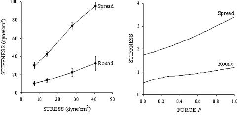
Fig. 6-8. (a) Data for stiffness vs. applied stress in round and spread cultured endothelial cells measured by magnetic twisting cytometry; points means ± SE (n = 3 wells, 20,000 cells/well). Both configurations exhibit stress-hardening behavior with greater hardening in the spread than in the round configuration. (Adapted with permission from Wang and Ingber (1994).) (b) Simulations of stiffness vs. applied force (F) in spread and round configurations of the six-strut model (Fig. 6-7) are qualitatively consistent with the data in panel (a). The force is given in the unit of force and the stiffness in the unit of force/length. Adapted with permission from Coughlin and Stamenovi´c, 1998.
of the struts. The structure is then attached to a rigid substrate at three nodes through frictionless ball-joint connections (Fig. 6-7a). The initial force distribution within the structure is not affected by this attachment. This is referred to as a ‘round configuration.’ To mimic cell spreading, three additional nodes are also anchored to the substrate (Fig. 6-7b). This is referred to as a ‘spread configuration.’ As a consequence of spreading, force distribution is altered from the one in the round configuration. Tension in the cables is now partly balanced by the struts and partly by reaction forces at the anchoring nodes. In both spread and round configurations, a vertical pulling force (F) is applied at a node distal from the substrate (Fig. 6-7). The corresponding vertical displacement ( x) is calculated and the structural stiffness as G = F/x. Two cases were considered, one where struts are rigid and cables linearly elastic, and the other where both struts and cables are elastic and struts buckle under compression. Here we present results from the case with rigid struts; corresponding results obtained with buckling struts are qualitatively similar (Coughlin and Stamenovi´c, 1998). The model predicts that stiffness increases with spreading (Fig. 6-8b). The reason is that tension (prestress) in the cables increases with spreading. The model also predicts approximately linear stress-hardening behavior and predicts that this dependence is greater in the spread than in the round configuration (Fig. 6-8b). All these predictions are consistent (Fig. 6-8a) with the corresponding behavior in round and in spread endothelial cells (Wang and Ingber, 1994). Further attachments of the nodes to the substrate, that is, further spreading, would gradually eliminate the struts from the force balance scheme and their role will be taken over by the substrate.