


94 F. Guilak et al.
Fig. 5-5. Micropipette aspiration of the pericellular matrix (PCM) of chondrocytes. This region surrounds cells in articular cartilage, similar to a glycocalyx, but contains significant amounts of extracellular matrix collagens, proteoglycans, and other macromolecules. The mechanical properties of this region appear to have a significant influence on the stress-strain and fluid-flow environment of the cell. From Alexopoulos et al., 2005.
Young’s modulus of the PCM was significantly decreased (38.7 ± 16.2 kPa vs. 23.5 ± 12.9 kPa, p < 0.001), and the permeability was significantly elevated (4.19 ± 3.78 × 10⁻¹⁷ m4/N·s vs. 10.2 ± 9.38 × 10⁻¹⁷ m4/N·s, p < 0.001). The Poisson ratio was similar for both nondegenerate and osteoarthritic PCM (0.044 ± 0.063 vs. 0.030 ± 0.068, p > 0.6). These findings suggest that the PCM may undergo enzymatic and mechanical degradation with osteoarthritis, similar to that occurring in the ECM. In combination with previous theoretical models of cell–matrix interactions in cartilage, these findings suggest that changes in the properties of the PCM may have an important influence on the biomechanical environment of the cell.
Together, these studies support the utility of in vitro mechanical analyses of isolated functional cell–matrix units. Because cartilage, in particular, is avascular and aneural, characterization of PCM mechanical and chemical properties is a key step toward characterizing the in vivo state of the cell and its metabolic response to alterations in the local cellular environment. The triphasic model provides a framework for developing extended models of the PCM that can delineate effects of the distinct mechanochemical composition of the PCM, relative to the ECM, on the local environment of the cell.
Indentation studies of cell multiphasic properties
In addition to micropipette aspiration, various techniques for cellular indentation have been used to measure the modulus of adherent cells, including cell indentation (Daily et al., 1984; Duszyk et al., 1989; Zahalak et al., 1990), scanning probe microscopy (Radmacher et al., 1992; Shroff et al., 1995), or cytoindentation (Shin and Athanasiou, 1999; Koay et al., 2003). (See also the discussion of experimental approaches in Chapter 2.) The conceptual basis of these techniques is generally similar, in that a rigid probe is used to indent the cell and the ensuing creep or stress-relaxation behavior
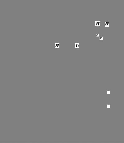
Multiphasic models of cell mechanics |
95 |
(a)CELL AND PERICELLULAR MATRIX MODEL
(b)
R
R
Fig. 5-6. (a) Biphasic finite element mesh of the micropipette aspiration experiment. The cell and pericellular matrix were modeled using an axisymmetric mesh with bilinear quadrilateral elements (342 nodes, 314 elements). (b) Transient response of normal and osteoarthritic chondrons (cell with the pericellular matrix), and the associated biphasic prediction of their mechanical behavior. The transient mechanical behavior of the PCM was well-described by a biphasic model, suggesting that the viscoelastic response of the pericellular matrix is attributable to flow-dependent effects, similar to that of the extracellular matrix. From Alexopoulos et al., 2005 with permission.
is recorded. These techniques have generally used elastic or viscoelastic models to calculate the equilibrium or dynamic moduli of cells over a range of frequencies. In one set of cell indentation experiments, MG63 osteoblast-like cells were modeled with either a linear elasticity solution of half-space indentation or the linear biphasic theory under the assumption that the viscoelastic behavior of each cell was due to the interaction between the solid cytoskeletal matrix and the cytoplasmic fluid (Shin and Athanasiou, 1999). The intrinsic biphasic material properties (aggregate modulus, Poisson’s ratio, and permeability) were determined by curve-fitting the experimental surface reaction force and deformation with a linear biphasic finite element code in conjunction with optimization routines. These cells exhibited a compressive aggregate modulus