

96F. Guilak et al.
of 2.05 ± 0.89 kPa with a Poisson ratio of 0.37 ± 0.03. These properties are on the
same order of magnitude as the elastic properties determined using other techniques (Trickey et al., in press), although the permeability of 1.18 ± 0.65 × 10⁻¹⁰ m4/N·s
is several orders of magnitude higher than that estimated for chondrocytes using micropipette aspiration (Trickey et al., 2000).
Analysis of cell–matrix interactions using multiphasic models
Previous studies suggest that cells have the ability to respond to the local stress-strain state within the extracellular matrix, thus suggesting that cellular response reflects the history of the time-dependent and spatially varying changes in the mechanical environment of the cells. The use of multiphasic models for cells has been of particular value in theoretical models of cell–matrix interactions that seek to model the stressstrain and fluid-flow environment of single cells within a tissue matrix. However, the relationship between the stress-strain and fluid-flow fields at the macroscopic “tissue” level and at the microscopic “cellular” level are not fully understood. To directly test such hypotheses, it would be important to have accurate knowledge of the local stress and deformation environment of the cell. In this respect, theoretical models of cells and tissues are particularly valuable in that they may be used to provide information on biophysical parameters that cannot be measured experimentally in situ at the cellular level, for example, the stress-strain, physicochemical, and electrical states in the immediate vicinity of the cell.
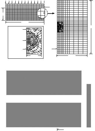
Based on existing experimental data on the deformation behavior and biomechanical properties of articular cartilage and chondrocytes, a multiscale biphasic finite element model was developed of the chondrocyte as a spheroidal inclusion embedded within the extracellular matrix of a cartilage explant (Fig. 5-7). In these studies, the cell membrane was neglected, and it was assumed that the cell was freely permeable to water to allow for changes in volume via transport of interstitial water in an out of the cell. Finite element analysis of the stress, strain, fluid flow, and hydraulic fluid pressure were made of a configuration simulating a cylindrical cartilage specimen (5 mm × 1 mm) subjected to a step load in an unconfined compression experiment. A parametric analysis was performed by varying the mechanical properties of the cell over 5–7 orders of magnitude relative to the properties of the ECM. Using a range of chondrocyte biphasic properties reported in the literature (E 0.5 − 1 kPa,
k 10−10 − 10−15 m4/N·s, ν 0.1 − 0.4) (Shin and Athanasiou, 1999; Trickey et al., 2000; Trickey et al., 2006), the distribution of stress at the cellular level was found to be time varying and inhomogeneous, and it differed significantly from that in the bulk extracellular matrix. At early time points (<100 s) following application of the load, the chondrocytes were exposed primarily to shear stress and strain and hydraulic fluid pressure, with little volume change. At longer time periods, changes in cell shape and volume were predicted coincident with exudation of the interstitial fluid (Fig. 5-7). The large difference ( 3 orders of magnitude) in the elastic properties of the chondrocyte and of the extracellular matrix results in the presence of stress concentrations at the cell–matrix border and a nearly two-fold increase in strain and dilatation (volume change) at the cellular level, as compared to that at the macro-level. The presence of a narrow “pericellular matrix” with different properties than that of the chondrocyte or extracellular matrix significantly altered the principal stress and strain magnitudes
Multiphasic models of cell mechanics |
97 |
(a) |
Macro-scale model |
Micro-scale model |
|
0.5 mm
2.5 mm
Extracellular |
Matrix |
Chondrocyte |
Pericellular |
Matrix |
10 µm |
 Macro-scale
Element
100 µm
Micro-scale
Element
50 µm
(b) |
t *=0 |
t *=0.1 |
t*=0.2 |
|
|
Normalized |
|||
|
|
|
|
|
|
|
|
|
Principal |
|
|
|
|
Stress |
|
|
|
|
1.8 |
|
|
|
|
1.5 |
|
t*=0.5 |
t*=1 |
t *=2 |
1.2 |
|
|
|||
|
|
|
|
0.9 |
|
|
|
|
0.6 |
0.3
0.0
1 0 µm 

Fig. 5-7. (a) A biphasic multiscale finite element method was used to model the mechanical environment of a single cell within the cartilage extracellular matrix. The “macro-scale” response of a cartilage explant in a state of unconfined compression was the first model. From this solution, a linear interpolation of the time-history of the kinematic boundary conditions within a 50 × 100 µm region were then applied to a “micro-scale” finite element mesh that incorporated a chondrocyte (10 µm diameter) and its pericellular matrix (2.5 µm thick) embedded within and attached to the extracellular matrix. Using this technique, it is assumed that due to their low volume fraction (<10%), the cells do not contribute mechanically to the macroscopic properties and behavior of the extracellular matrix. (b) Predictions of the compressive stress in the cell and extracellular matrix versus time. A gray-scale image of one-quarter of the cell is shown within the matrix. Stress is normalized to the far-field extracellular matrix stress at equilibrium, and time is normalized to the biphasic gel time (t = t /τgel). At early times following loading, low magnitudes of solid stress were observed, as the total stress in the tissue was borne primarily by pressurization of the interstitial fluid. With time, stresses were transferred to the solid phase and increased stress concentrations are observed at the cell–matrix boundary. From Guilak and Mow, 2000.
within the chondrocyte, suggesting a functional biomechanical role for this tissue region. These findings suggest that even under simple compressive loading conditions, chondrocytes are subjected to a complex local mechanical environment consisting of tension, compression, shear, and hydraulic pressure. Knowledge of the magnitudes
98F. Guilak et al.
and distribution of local stress/strain and fluid-flow fields in the extracellular matrix around the chondrocytes is an important step in the interpretation of studies of mechanical signal transduction in cartilage explant culture models.
In other tissues, anisotropic behavior may play an important role in defining the micromechanical environment of the cell. For example, cellular response to mechanical loading varies between the anatomic zones of the intervertebral disc, and this difference may be related to differences in the structure and mechanics of both cells and extracellular matrix, which are expected to cause differences in the physical stimuli (such as pressure, stress, and strain) in the cellular micromechanical environment (Guilak et al., 1999; Baer et al., 2003). In other studies, finite element analyses have been used to model flow-dependent viscoelasticity using the biphasic theory for soft tissues; finite deformation effects using a hyperelastic constitutive law for the solid phase; and material anisotropy by including a fiber-reinforced continuum law in the hyperelastic strain energy function. The model predicted that the cellular micromechanical environment varies dramatically depending on the local tissue stiffness and anisotropy. Furthermore, the model predicted that stress-strain and fluid-flow environment is strongly influenced by cell shape, suggesting that the geometry of cells in situ may be an adaptation to reduce cellular strains during tissue loading.
With similar multiscaling techniques, other studies have used triphasic constitutive models to predict the physicochemical environment of cells within charged, hydrated tissues (Likhitpanichkul et al., 2003). These studies also show that in addition to nonhomogeneous stress-strain and fluid-flow fields within the extracellular matrix, cells may also be exposed to timeand spatially varying osmotic pressure and electric fields due to the coupling between electrical, chemical, and mechanical events in the cell and in the surrounding tissues. Such methods may provide new insight into the physical regulatory mechanisms that influence cell behavior in situ.
Summary
Multiphasic approaches have important advantages and disadvantages relative to more classic single-phase models. The disadvantages are based primarily on the added complexity required for computational models. In most cases, analytical solutions are intractable and numerical methods such as finite element modeling are required. Furthermore, additional experimental tests are necessary to determine the intrinsic mechanical contributions of the different phases to the overall behavior of the cell. However, multiphasic models may provide a more realistic representation of the physical events that govern cell mechanical behavior. Furthermore, as most current multiphasic models are based on a continuum approach, the constitutive models describing each phase can be selected independently to best describe the empirically observed behavior of the cell. A multiphasic approach may also be combined with other structurally based models (such as, tensegrity models), and thus may provide a versatile modeling approach for examining the interactions of the different constitutive phases governing cell mechanical behavior.
Multiphasic models of cell mechanics |
99 |
References
Alexopoulos, L.G., Haider, M.A., Vail, T.P., Guilak, F. (2003). Alterations in the mechanical properties of the human chondrocyte pericellular matrix with osteoarthritis. J. Biomech. Eng., 125, 323–333.
Alexopoulos, L.G., Williams, G.M., Upton, M.L., Setton, L.A., Guilak, F. (2005). Osteoarthritic changes in the biphasic mechanical properties of the chondrocyte pericellular matrix in articular cartilage. J. Biomech., 38, 509–517.
Ateshian, G.A., Likhitpanichkul, M., Hung, C.T. (2006). A mixture theory analysis for passive transport in osmotic loading of cells. J. Biomech., 39, 464–475.
Baaijens, F.P. (1998). Mixed finite element methods for viscoelastic flow analysis: A review. J. of Non-Newtonian Fluid Mechanics 79, 361–385.
Baaijens, F.P.T., Trickey, W.R., Laursen, T.A., Guilak, F. (2005). Large deformation finite element analysis of micropipette aspiration to determine the mechanical properties of the chondrocyte. An. Biomed. Eng., 33, 494–501.
Bachrach, N.M., Valhmu, W.B., Stazzone, E., Ratcliffe, A., Lai, W.M., Mow, V.C. (1995). Changes in proteoglycan synthesis of chondrocytes in articular cartilage are associated with the timedependent changes in their mechanical environment. J. Biomech., 28, 1561–1570.
Baer, A.E., Laursen, T.A., Guilak, F., Setton, L.A. (2003). The micromechanical environment of intervertebral disc cells determined by a finite deformation, anisotropic, and biphasic finite element model. J. Biomech. Eng., 125, 1–11.
Bowen, R.M. (1980). Incompressible porous media models by use of the theory of mixtures. Internat. J. Eng. Sci., 18, 1129–1148.
Buschmann, M.D., Hunziker, E.B., Kim, Y.J., Grodzinsky, A.J. (1996). Altered aggrecan synthesis correlates with cell and nucleus structure in statically compressed cartilage. J. Cell Sci., 109, 499–508.
Cantiello, H.F., Patenaude, C., Zaner, K. (1991). Osmotically induced electrical signals from actin filaments. Biophys. J., 59, 1284–1289.
Cohen, B., Lai, W.M., Mow, V.C. (1998). A transversely isotropic biphasic model for unconfined compression of growth plate and chondroepiphysis. J. Biomech. Eng., 120, 491–496.
Daily, B., Elson, E.L., Zahalak, G.I. (1984). Cell poking. Determination of the elastic area compressibility modulus of the erythrocyte membrane. Biophys. J., 45, 671–682.
DiSilvestro, M.R., Suh, J.K. (2002). Biphasic poroviscoelastic characteristics of proteoglycandepleted articular cartilage: simulation of degeneration. An. Biomed. Eng., 30, 792–800.
Dong, C., Skalak, R., Sung, K.L. (1991). Cytoplasmic rheology of passive neutrophils. Biorheology, 28, 557–567.
Duszyk, M., Schwab, B.D., Zahalak, G.I., Qian, H., Elson, E.L. (1989). Cell poking: quantitative analysis of indentation of thick viscoelastic layers. Biophys. J., 55, 683–690.
Evans, E., Yeung, A. (1989). Apparent viscosity and cortical tension of blood granulocytes determined by micropipet aspiration. Biophys. J., 56, 151–160.
Evans, E.A., 1989. Structure and deformation properties of red blood cells: Concepts and quantitative methods. Methods Enzymol., 173, 3–35.
Greco, F., Specchia, N., Falciglia, F., Toesca, A., Nori, S. (1992). Ultrastructural analysis of the adaptation of articular cartilage to mechanical stimulation. Italian J. of Orthopaedics and Traumatology, 18, 311–321.
Gu, W.Y., Lai, W.M., Hung, C.T., Liu, Z.P., Mow, V.C. (1997). Analysis of transient swelling and electrical responses of an isolated cell to sudden osmotic loading. Adv. in Bioengin., BED36, 189–190.
Gu, W.Y., Lai, W.M., Mow, V.C. (1998). A mixture theory for charged-hydrated soft tissues containing multi-electrolytes: Passive transport and swelling behaviors. J. Biomech. Eng., 120, 169–180.
Guilak, F. (1995). Compression-induced changes in the shape and volume of the chondrocyte nucleus. J. Biomech., 28, 1529–1542.
100F. Guilak et al.
Guilak, F., Erickson, G.R., Ping Ting-Beall, H. (2002). The effects of osmotic stress on the viscoelastic and physical properties of articular chondrocytes. Biophys. J., 82, 720–727.
Guilak, F., Mow, V.C. (2000). The mechanical environment of the chondrocyte: A biphasic finite element model of cell–matrix interactions in articular cartilage. J. Biomech., 33, 1663–1673.
Guilak, F., Ratcliffe, A., Mow, V.C. (1995). Chondrocyte deformation and local tissue strain in articular cartilage: A confocal microscopy study. J. Orthop. Res., 13, 410–421.
Guilak, F., Ting-Beall, H.P., Baer, A.E., Trickey, W.R., Erickson, G.R., Setton, L.A. (1999). Viscoelastic properties of intervertebral disc cells – Identification of two biomechanically distinct cell populations. Spine, 24, 2475–2483.
Haider, M.A. (2004). A radial biphasic model for local cell–matrix mechanics in articular cartilage.
SIAM J. Appl. Math., 64, 1588–1608.
Haider, M.A., Guilak, F. (2000). An axisymmetric boundary integral model for incompressible linear viscoelasticity: Application to the micropipette aspiration contact problem. J. Biomech. Eng., 122, 236–244.
Haider, M.A., Guilak, F. (2002). An axisymmetric boundary integral model for assessing elastic cell properties in the micropipette aspiration contact problem. J. Biomech. Eng., 124, 586–595.
Hochmuth, R.M. (2000). Micropipette aspiration of living cells. J. Biomech., 33, 15–22.
Holmes, M.H., Lai, W.M., Mow, V.C. (1985). Singular perturbation analysis of the nonlinear, flow-dependent compressive stress relaxation behavior of articular cartilage. J Biomech. Eng., 107, 206–218.
Huyghe, J.M., Houben, G.B., Drost, M.R., van Donkelaar, C.C. (2003). An ionised/non-ionised dual porosity model of intervertebral disc tissue. Biomech. and Model. in Mechanobiol., 2, 3–19.
Iatridis, J.C., Setton, L.A., Foster, R.J., Rawlins, B.A., Weidenbaum, M., Mow, V.C. (1998). Degeneration affects the anisotropic and nonlinear behaviors of human anulus fibrosus in compression. J. Biomech., 31, 535–544.
Ingber, D.E. (2003). Tensegrity I. Cell structure and hierarchical systems biology. J. Cell Sci., 116, 1157–1173.
Jones, W.R., Ting-Beall, H.P., Lee, G.M., Kelley, S.S., Hochmuth, R.M., Guilak, F. (1999). Alterations in the Young’s modulus and volumetric properties of chondrocytes isolated from normal and osteoarthritic human cartilage. J. Biomech., 32, 119–127.
Karcher, H., Lammerding, J., Huang, H., Lee, R.T., Kamm, R.D., Kaazempur-Mofrad, M.R. (2003). A three-dimensional viscoelastic model for cell deformation with experimental verification. Biophys. J., 85, 3336–3349.
Kedem, O., Katchalsky, A. (1958). Thermodynamic analysis of the permeability of biological membranes to non-electrolytes. Biochim. Biophys. Acta 27, 229–246.
Koay, E.J., Shieh, A.C., Athanasiou, K.A. (2003). Creep indentation of single cells. J. Biomech. Eng., 125, 334–341.
Lai, W.M., Hou, J.S., Mow, V.C. (1991). A triphasic theory for the swelling and deformation behaviors of articular cartilage. J. Biomech. Eng., 113, 245–258.
Lai, W.M., Mow, V.C., Roth, V. (1981). Effects of nonlinear strain-dependent permeability and rate of compression on the stress behavior of articular cartilage. J. Biomech. Eng., 103, 61–66.
Likhitpanichkul, M., Sun, D.D., Guo, X.E., Lai, W.M., Mow, V.C. (2003). The mechanoelectrochemical environment of chondrocytes in articular cartilage explants under unconfined compression: Emphasis on the cell matrix interactions. Proceedings of the 2003 Summer Bioengineering Conference, Key Biscayne, FL.
Mak, A.F. (1986a). The apparent viscoelastic behavior of articular cartilage – The contributions from the intrinsic matrix viscoelasticity and interstitial fluid flows. J. Biomech. Eng., 108, 123–130.
Mak, A.F. (1986b). Unconfined compression of hydrated viscoelastic tissues: A biphasic poroviscoelastic analysis. Biorheology, 23, 371–383.
Mak, A.F.T., Huang, D.T., Zhang, J.D., Tong, P. (1997). Deformation-induced hierarchial flows and drag forces in bone canaliculi and matrix microporosity. J. Biomech., 20, 11–18.
Multiphasic models of cell mechanics |
101 |
Maughan, D.W., Godt, R.E. (1989). Equilibrium distribution of ions in a muscle fiber. Biophys. J., 56, 717–722.
Mow, V.C., Holmes, M.H., Lai, W.M. (1984). Fluid transport and mechanical properties of articular cartilage: A review. J. Biomech., 17, 377–394.
Mow, V.C., Kuei, S.C., Lai, W.M., Armstrong, C.G. (1980). Biphasic creep and stress relaxation of articular cartilage in compression: Theory and experiments. J. Biomech. Eng., 102, 73–84.
Needham, D., Hochmuth, R.M. (1992). A sensitive measure of surface stress in the resting neutrophil. Biophys. J., 61, 1664–1670.
Oster, G. (1984). The mechanochemistry of cytogels. Physica, 12D, 333–350.
Oster, G. (1989). Cell motility and tissue morphogenesis. In: Cell Shape: Determinants, Regulation, and Regulatory Role. Stein, W.D. and Bronner, F. (Eds.) San Diego, CA, Academic Press, pp. 33–61.
Pollack, G.H. (2001). Cells, gels and the engines of life: A new, unifying approach to cell function. Seattle, WA, Ebner & Sons.
Poole, C.A. (1992). Chondrons: the chondrocyte and its pericellular microenvironment. In: Articular Cartilage and Osteoarthritis. Kuettner, K.E., Schleyerbach, R., Peyron, J.G. and Hascall, V.C. (Eds.) New York, London: Academic Press, pp. 201–220.
Poole, C.A. (1997). Articular cartilage chondrons: Form, function and failure. J. Anat., 191 (Pt 1), 1–13.
Poole, C.A., Flint, M.H., Beaumont, B.W. (1987). Chondrons in cartilage: Ultrastructural analysis of the pericellular microenvironment in adult human articular cartilages. J. Orthop. Res., 5, 509–522.
Radmacher, M., Tillmann, R.W., Fritz, M., Gaub, H.E. (1992). From molecules to cells: Imaging soft samples with the atomic force microscope. Science, 257, 1900–1905.
Sato, M., Theret, D.P., Wheeler, L.T., Ohshima, N., Nerem, R.M. (1990). Application of the micropipette technique to the measurement of cultured porcine aortic endothelial cell viscoelastic properties. J. Biomech. Eng., 112, 263–268.
Sengers, B.G., Oomens, C.W., Baaijens, F.P. (2004). An integrated finite-element approach to mechanics, transport and biosynthesis in tissue engineering. J. Biomech. Eng., 126, 82–91.
Setton, L.A., Zhu, W., Mow, V.C. (1993). The biphasic poroviscoelastic behavior of articular cartilage: Role of the surface zone in governing the compressive behavior. J. Biomech., 26, 581–592.
Shin, D., Athanasiou, K. (1999). Cytoindentation for obtaining cell biomechanical properties. J. Orthop. Res., 17, 880–890.
Shroff, S.G., Saner, D.R., Lal, R. (1995). Dynamic micromechanical properties of cultured rat atrial myocytes measured by atomic force microscopy. Am. J. Physiol., 269, C286– 292.
Szirmai, J.A., 1974. The concept of the chondron as a biomechanical unit. In: Biopolymer und Biomechanik von Bindegewebssystemen. Hartmann, F. (Ed.) Berlin, Academic Press, pp. 87.
Spilker, R.L., Donzelli, P.S., Mow, V.C. (1992). A transversely isotropic biphasic finite element model of the meniscus. J. Biomech., 112, 1027–1045.
Sung, K.L., Schmid-Schonbein, G.W., Skalak, R., Schuessler, G.B., Usami, S., Chien, S. (1982). Influence of physicochemical factors on rheology of human neutrophils. Biophys. J., 39, 101–106.
Theret, D.P., Levesque, M.J., Sato, M., Nerem, R.M., Wheeler, L.T. (1988). The application of a homogeneous half-space model in the analysis of endothelial cell micropipette measurements. J. Biomech. Eng., 110, 190–199.
Thoumine, O., Ott, A. (1997). Comparison of the mechanical properties of normal and transformed fibroblasts. Biorheology, 34, 309–326.
Ting-Beall, H.P., Needham, D., Hochmuth, R.M. (1993). Volume and osmotic properties of human neutrophils. Blood, 81, 2774–2780.
Trickey, W.R., Baaijens, F.P.T., Laursen, T.A., Alexopoulos, L.G., Guilak, F. (2006). Determination of the Poisson’s ratio of the cell: Recovery properties of chondrocytes after release from complete micropipette aspiration. J. Biomech., (in press) 39, 78–87.
Trickey, W.R., Lee, M., Guilak, F. (2000). Viscoelastic properties of chondrocytes from normal and osteoarthritic human cartilage. J. Orthop. Res., 18, 891–898.
102F. Guilak et al.
Truesdell, C., Toupin, R., 1960. The classical field theories. In: Handbuch der Physik. Flugge, S. (Ed.) Berlin, Springer-Verlag, pp. 226–793.
Yeung, A., Evans, E. (1989). Cortical shell-liquid core model for passive flow of liquid-like spherical cells into micropipets. Biophys. J., 56, 139–149.
Zahalak, G.I., McConnaughey, W.B., Elson, E.L. (1990). Determination of cellular mechanical properties by cell poking, with an application to leukocytes. J. Biomech. Eng., 112, 283–294.