

68 J. Fredberg and B. Fabry
η E L L
δF
}quench (a)
LSE
}quench (b)
1.2
1.1 |
x |
(c) |
1.0
cold
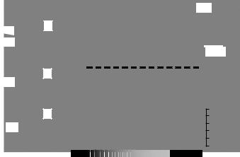
Fig. 3-10. Evolution of mechanical properties of bovine tracheal smooth muscle during contraction against a constant mean force on which force fluctuations (0.2 Hz) of graded amplitude δ F were superimposed. (a) Mean muscle length L (relative to optimal length L0). (b) Loop stiffness (percentage of maximum isometric value). (c) hysteresivity η and noise temperature x. LSE is the statically equilibrated length of the muscle after 120 min. of unperturbed contraction against a constant load of 32% of maximum force (F0). Adapted from Fredberg, 2000.
tidal stretches are terminated (Figs. 3-9 and 3-10), however, the noise temperature is suddenly lowered, and all plastic changes might become trapped, or quenched, so that the muscle is unable to return to maximum force and stiffness (Fig. 3.10), or maximum shortening (Fig. 3-10) (Fabry and Fredberg, 2003).
The glass hypothesis predicts, therefore, that the cell ought to be able to adapt faster to step-length changes imposed while the cell is transiently ‘hot’ (that is, early in activation), and far less so after it has cooled in the process of sustained activation (Fabry and Fredberg, 2003; Gunst and Fredberg, 2003). Indeed, Gunst and colleagues showed that a step change of muscle length alters the level of the subsequent force plateau to a degree that depends mostly on the timing of the length change with respect to stimulus onset (Gunst, Meiss et al., 1995; Gunst and Fredberg, 2003).
Conclusion
The behavior of soft glasses, and the underlying notion of the noise temperature, might provide a unifying explanation of the ability of the cytoskeletal lattice to deform, to flow, and to remodel. Such a view does not point to specific molecular processes that occur, but instead derives the mechanical properties from generic features: structural elements that are discrete, numerous, aggregated with one another via weak interactions, and arrayed in a geometry that is structurally disordered and metastable. We have proposed here that these features may comprise the basis of CSK rheology and remodeling.
References
Alcaraz, J., L. Buscemi, et al. (2003). Microrheology of human lung epithelial cells measured by atomic force microscopy. Biophys. J., 84, 2071–9.
The cytoskeleton as a soft glassy material |
69 |
An, S. S., B. Fabry, et al. (2004). Role of heat shock protein 27 in cytoskeletal remodeling of the airway smooth muscle cell. J. Appl. Physiol., 96, 1701–13.
Bouchaud, J. (1992). Weak ergodicity breaking and aging in disordered systems. J. Phys. I., 2, 1705– 13.
Butler, J. P., I. M. Tolic-Norrelykke, et al. (2002). Traction fields, moments, and strain energy that cells exert on their surroundings. Am. J. Physiol. Cell Physiol., 282, C595–605.
Crandall, S. H. (1970). The role of damping in vibration theory. J. Sound Vibr., 11, 3–18.
Crick, F. H. C. and A. F. W. Hughes (1950). The physical properties of cytoplasm. Exp. Cell Res., 1, 37–80.
Fabry, B. and J. J. Fredberg (2003). Remodeling of the airway smooth muscle cell: are we built of glass? Respir. Physiol. Neurobiol., 137, 109–24.
Fabry, B., G. N. Maksym, et al. (2001). Scaling the microrheology of living cells. Phys. Rev. Lett., 87, 148102.
Fabry, B., G. N. Maksym, et al. (2003). Time scale and other invariants of integrative mechanical behavior in living cells. Phys. Rev. E., 68, 041914.
Fabry, B., G. N. Maksym, et al. (2001). Time course and heterogeneity of contractile responses in cultured human airway smooth muscle cells. J. Appl. Physiol., 91, 986–94.
Fredberg, J. J. (2000). Airway smooth muscle in asthma. Perturbed equilibria of myosin binding. Am. J. Respir. Crit. Care Med., 161, S158–60.
Fredberg, J. J., D. Bunk, et al. (1993). Tissue resistance and the contractile state of lung parenchyma.
J. Appl. Physiol., 74, 1387–97.
Fredberg, J. J., D. Inouye, et al. (1997). Airway smooth muscle, tidal stretches, and dynamically determined contractile states. Am. J. Respir. Crit. Care Med., 156, 1752–9.
Fredberg, J. J., K. A. Jones, et al. (1996). Friction in airway smooth muscle: mechanism, latch, and implications in asthma. J. Appl. Physiol., 81, 2703–12.
Fredberg, J. J. and D. Stamenovic (1989). On the imperfect elasticity of lung tissue. J. Appl. Physiol., 67, 2408–19.
Fung, Y. C. (1967). Elasticity of soft tissues in simple elongation. Am. J. Physiol., 213, 1532–44. Goldmann, W. H. and R. M. Ezzell (1996). Viscoelasticity in wild-type and vinculin-deficient (5.51)
mouse F9 embryonic carcinoma cells examined by atomic force microscopy and rheology. Exp. Cell Res., 226, 234–7.
Gunst, S. J. and J. J. Fredberg (2003). The first three minutes: smooth muscle contraction, cytoskeletal events, and soft glasses. J. Appl. Physiol., 95, 413–25.
Gunst, S. J., R. A. Meiss, et al. (1995). Mechanisms for the mechanical plasticity of tracheal smooth muscle. Am. J. Physiol., 268, C1267–76.
Hai, C. M. and R. A. Murphy (1989). Cross-bridge dephosphorylation and relaxation of vascular smooth muscle. Am. J. Physiol., 256, C282–7.
Hantos, Z., B. Daroczy, et al. (1990). Modeling of low-frequency pulmonary impedance in dogs.
J. Appl. Physiol., 68, 849–60.
Hildebrandt, J. (1969). Comparison of mathematical models for cat lung and viscoelastic balloon derived by Laplace transform methods from pressure-volume data. Bull. Math. Biophys., 31, 651–67.
Hill, A. V. (1965). Trails and Trials in Physiology (pp. 14–15). London, E. Arnold.
Hubmayr, R. D., S. A. Shore, et al. (1996). Pharmacological activation changes stiffness of cultured human airway smooth muscle cells. Am. J. Physiol., 271, C1660–8.
Kawai, M. and P. W. Brandt (1980). Sinusoidal analysis: a high resolution method for correlating biochemical reactions with physiological processes in activated skeletal muscles of rabbit, frog and crayfish. . J Muscle Res. Cell Motil., 1, 279–303.
Kimball, A. L. and D. E. Lovell (1927). Internal friction in solids. Phys. Rev., 30, 948–959. Kohlrausch, F. (1866). Beitr¨age zur Kenntniss der elastischen Nachwirkung. Ann. Phys. Chem.,
128, 1–20, 207–227, 399–419.
Kohlrausch, R. (1847). Nachtrag ueber die elastische Nachwirkung beim Coconund Glasfaden, und die hygroskopische Eigenschaft des ersteren. Ann. Phys. Chem., 72, 393–8.
Lau, A. W., B. D. Hoffman, et al. (2003). Microrheology, stress fluctuations, and active behavior of living cells. Phys. Rev. Lett., 91, 198101.
70J. Fredberg and B. Fabry
Laudadio, R. E., E. J. Millet, et al. (2005). Rheology of the rat airway smooth muscle cell: scaling of responses to actin modulation. Am. J. Physiol., in review.
Lenormand, G., E. Millet, et al. (2004). Linearity and time-scale invariance of the creep function in living cells. J. Royal Soc. Interface, 1, 91–7.
Mahaffy, R. E., C. K. Shih, et al. (2000). Scanning probe-based frequency-dependent microrheology of polymer gels and biological cells. Phys. Rev. Lett., 85, 880–3.
Maksym, G. N., B. Fabry, et al. (2000). Mechanical properties of cultured human airway smooth muscle cells from 0.05 to 0.4 Hz. J. Appl. Physiol., 89, 1619–32.
Mijailovich, S. M., J. P. Butler, et al. (2000). Perturbed equilibria of myosin binding in airway smooth muscle: bond-length distributions, mechanics, and ATP metabolism. Biophys. J., 79, 2667–81.
Mijailovich, S. M., M. Kojic, et al. (2002). A finite element model of cell deformation during magnetic bead twisting. J. Appl. Physiol., 93, 1429–36.
Murphy, R. A. (1988). Muscle cells of hollow organs. News Physiol. Sci., 3, 124–8.
Navajas, D., S. Mijailovich, et al. (1992). Dynamic response of the isolated passive rat diaphragm strip. J. Appl. Physiol., 73, 2681–92.
Puig-de-Morales, M., E. Millet, et al. (2004). Cytoskeletal mechanics in adherent human airway smooth muscle cells: probe specificity and scaling of protein-protein dynamics. Am. J. Physiol. Cell Physiol., 287, C643–54.
Satcher, R. L., Jr. and C. F. Dewey, Jr. (1996). Theoretical estimates of mechanical properties of the endothelial cell cytoskeleton. Biophys. J., 71, 109–18.
Shroff, S. G., D. R. Saner, et al. (1995). Dynamic micromechanical properties of cultured rat atrial myocytes measured by atomic force microscopy. Am. J. Physiol., 269, C286–92.
Sollich, P. (1998). Rheological constitutive equation for a model of soft glassy materials. Phys. Rev. E., 58, 738–59.
Sollich, P., F. Lequeux, et al. (1997). Rheology of soft glassy materials. Phys. Rev. Lett., 78, 2020–2023.
Suki, B., R. Peslin, et al. (1989). Lung impedance in healthy humans measured by forced oscillations from 0.01 to 0.1 Hz. J. Appl. Physiol., 67, 1623–9.
Wang, L., P. D. Pare, et al. (2001). Effect of chronic passive length change on airway smooth muscle length-tension relationship. J. Appl. Physiol., 90, 734–40.
Wang, N., I. M. Tolic-Norrelykke, et al. (2002). Cell prestress. I. Stiffness and prestress are closely associated in adherent contractile cells. Am. J. Physiol. Cell Physiol., 282, C606–16.
Warshaw, D. M., D. D. Rees, et al. (1988). Characterization of cross-bridge elasticity and kinetics of cross-bridge cycling during force development in single smooth muscle cells. J. Gen. Physiol., 91, 761–79.
Weber, W. (1835). Ueber die Elasticitaet der Seidenfaeden. Annalen der Physik und Chemie, 34, 247–57.
Weber, W. (1841). Ueber die Elasticitaet fester Koerper. Annalen der Physik und Chemie, 54, 1–18. Yamada, S., D. Wirtz, et al. (2000). Mechanics of living cells measured by laser tracking
microrheology. Biophys. J., 78, 1736–47.