

MusculoSkeletal Exam
.pdfChapter 3 Overview of the Spine and Pelvis
Skull
C-1 (Atlas)
C-2 (Axis)
Odontoid |
|
process |
C-1 (Atlas) |
Spinal cord |
|
C-2 (Axis)
Figure 3.3 The skull rests on the atlas (C1); the head rotates about the odontoid process as if it were a gatepost.
Below C2, the point of maximum flexion–extension movement is at the C4–C5 and C5–C6 levels. Hence, it is at these sites that osteoarthritic degeneration is most commonly seen. The consequence of this frequency of osteoarthritic degeneration is that radicular symptomatology secondary to cervical osteoarthritis most commonly affects the C4, C5, and C6 nerve roots. This is due to foraminal and disc space narrowing (stenosis) caused by degenerative changes and osteophyte formation at these levels. One additional anatomical curiosity of the cervical spine involves the vertebral artery becoming entombed within the vertebral processes of C2, C3, C4, and C5 as it travels proximal toward the skull. This tethering of the vertebral artery within the bony vertebrae can create a stress point to the vessel with extreme movement of the cervical spine.
The 12 thoracic vertebrae are stabilized by the rib cage into a relatively immobile segment. There are four localized points of significant stress created at the proximal and distal ends of the thoracic spine, at the cervicothoracic and thoracolumbar junctions. This is due to the abrupt change in stiffness at these points.
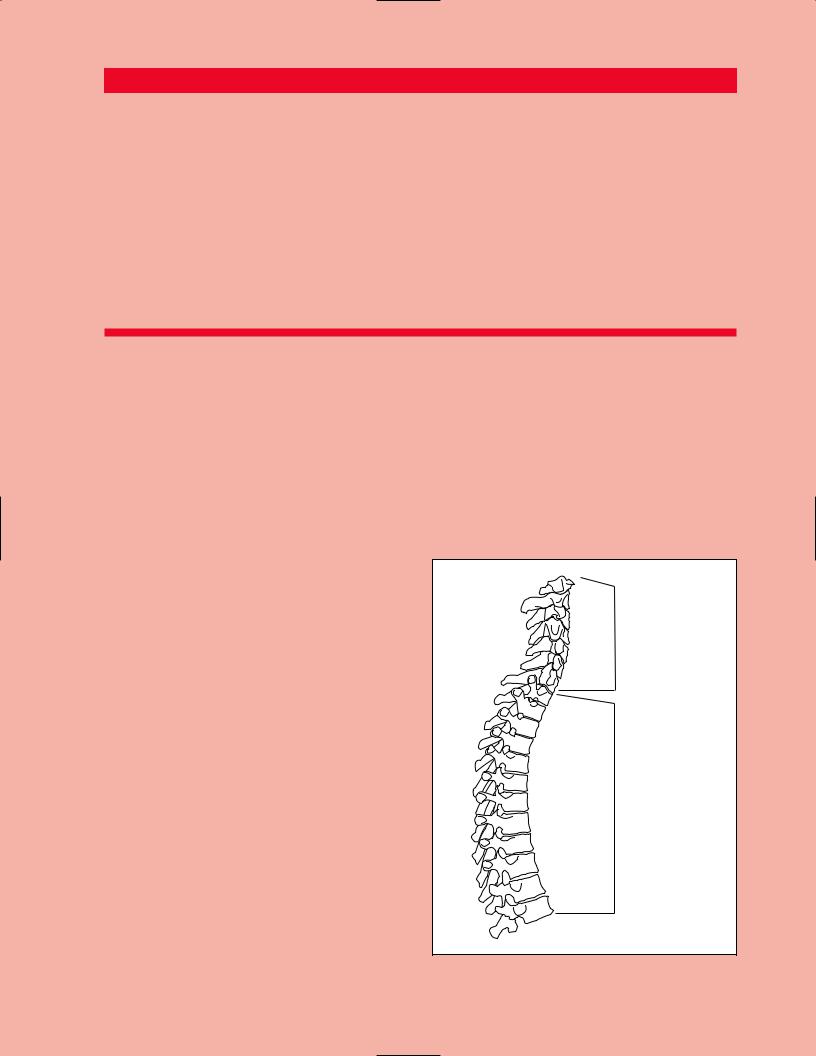
The five segments of the lumbar spine are very large versions of those found in the cervical spine. This is consistent with the increased load to which they are subjected and the fact that their purpose is to permit motion in all three planes between the rigid pelvis below and the semirigid thorax above. Like the cervical spine, the lumbar spine is a common site of degenerative change. It has been said that the human lumbar spine has not yet sufficiently evolved to accommodate the erect
bipedal stance. This is certainly borne out by the fact that back pain is almost a universal ailment among humans at some point during their lives. Of particular importance are the L4–L5 and L5–S1 articulations. A forwardfacing convexity called lordosis is quite pronounced at these levels. This lordosis accounts for the slight “hollow” one normally perceives at the region of the low back when lying on the floor with the lower extremities fully extended. This lordosis creates a tremendous forward pressure on the vertically oriented facet joints, which serve to stabilize the lower lumbar segments against forward translation. This constant forward pressure may explain the high frequency of degenerative change seen within these particular facet articulations.
The lumbar, thoracic, and cervical spinal segments rest on a large triangular structure called the sacrum. The sacrum is formed by the fusion of five vertebral segments into one large triangular bone. Similar to the keystone at the top of the arch, the sacrum is keystoned into the pelvic ring between the ilia (innominates). It is held in place by a combination of extremely strong ligaments and a synchondrosis with each iliac wing.
Beneath the sacrum are the vertebral segments of the coccyx. Seen on lateral x-ray films, the coccyx has the appearance of a short tail. It actually represents the vestigial remnants of the tails that existed on our ancestors. Occasionally, an infant will be born with accessory coccygeal segments or an actual tail, which will require surgical removal. The coccyx serves to protect the structures of the lower pelvis and acts as an attachment for some of the lower pelvic musculature and ligaments.
35
Chapter 4
The Cervical Spine
and Thoracic Spine
Cervical spine
Thoracic spine
Please refer to Chapter 2 for an overview of the sequence of a physical examination. For purposes of length and to avoid having to repeat anatomy more than once, the palpation section appears directly after the section on subjective examination and before any section on testing, rather than at the end of each chapter. The order in which the examination is performed should be based on your experience and personal preference as well as the presentation of the patient.
Observation
The cervical spine is a flexible column that supports the weight of the head and provides a protected pathway for the spinal cord as it descends. It protects the vertebral arteries, the internal jugular veins, and the sympathetic chain of the autonomic nervous system (ANS). It is imperative that special care be taken to monitor these structures during the examination. The distinctive arrangement of the articulations of the upper cervical spine continuing with the facet joints of the
Inion |
Mastoid process |
|
Mandible |
||
|
Superior nuchal line
|
C1 transverse |
|
C1 |
process |
|
Facet joint |
||
C2 |
||
|
Hyoid bone |
|
C6 |
Thyroid cartilage |
|
First cricoid ring |
||
C7 |
||
|
||
T2 |
Carotid tubercle |
|
|
Trachea |
|
T4 |
Sternum |
Spinous processes
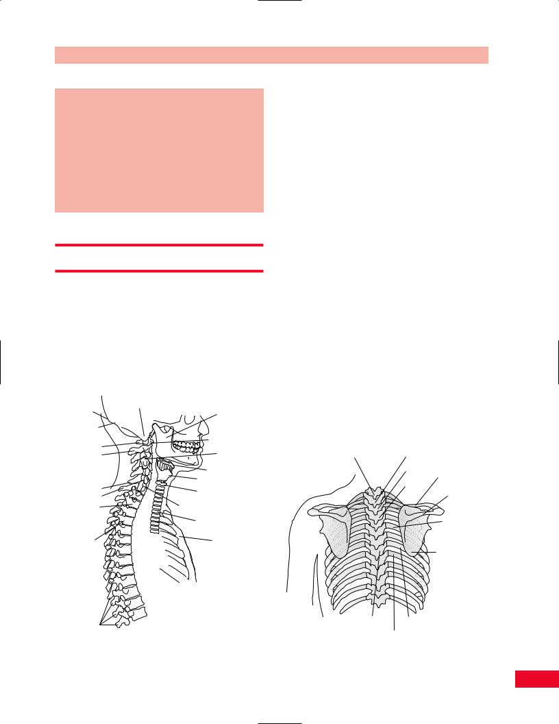
Figure 4.1 Overview of the neck with anterior–posterior relationships.
Chapter 4 The Cervical Spine and Thoracic Spine
lower cervical spine allows for the head to move through space. The muscles and ligaments create a great deal of stability as they counteract the inertia of the head. There is also a unique interaction with the shoulder girdle because of the many mutual muscle attachments. In contrast, the thoracic spine is quite rigid because of its attachment to the rib cage. Active motion is therefore much more restricted. See Figures 4.1–4.4.
Note the manner in which the patient is sitting in the waiting room. Notice how the patient is posturing the head, neck, and upper extremity. Is the patient’s head forward or laterally bent? Is the patient supporting their head with their hands or is he or she wearing a cervical collar? Is the arm relaxed at the side or is it being cradled for protection? How willing is the patient to turn the head or look up at you as you approach? Will the patient use the upper extremity? Will he or she extend the arm to you to shake your hand? Pain may be altered by changes in position, so watch the patient’s facial expression for indications as to their pain level.
Observe the patient as he or she assumes the standing position and note their posture. Pay particular attention to the position of the head, cervical spine, and thoracic kyphosis. Note the height of the shoulders and their relative positions. Once the patient starts to ambulate, observe whether he or she is willing to swing their arms. Arm swing can be limited by pain or loss of motion. Once the patient is in the examination room, ask him or her to disrobe. Observe his or her willingness to bend the head to allow for removal of
Vertebra |
Spinous |
|
prominens (C7) |
process of T1 |
|
|
T2 |
Clavicle |
|
T3 |
Spine of |
|
|
scapula |
|
|
Rib angle |
|
|
Inferior angle |
|
|
of scapula |
|
|
(T7 vertebra) |
T7 |
7th rib |
8th rib
Figure 4.2 Overview of the posterior thorax.
37

The Cervical Spine and Thoracic Spine Chapter 4
|
Vertex of head |
Temporal |
Temporal |
|
|
|
artery |
|
bone |
|
|
|
Frontal |
|
Parietal |
|
bone |
|
|
|
bone |
|
Orbit |
Nasal bone
|
Zygoma |
Sternocleidomastoid |
Scalene |
|
muscle |
muscles |
|
Occipital |
Maxilla |
|
|
bone |
|
|
|
|
|
|
|
Mastoid |
Parotid |
|
|
process |
duct |
|
|
|
Mandible |
|
|
|
Parotid |
|
|
|
gland |
|
|
|
Figure 4.3 Overview of the skull. |
|
|
the shirt. Note the ease with which upper extremities are used and the rhythm of the movements. Observe the posture of the head, neck, and upper back. Observe for symmetry of bony structures. Observe the clavicles and the sternum. An uneven contour may be present secondary to a healed fracture. Observe the scapulae and determine whether they are equidistant from the spine and are lying flat on the rib cage. Is a subluxation present at the glenohumeral joint and if so, to what degree? Notice the size and contour of the deltoid muscle and compare to the opposite deltoid. Observe for any areas of atrophy in the upper extremities. Pay attention to the rib cage. Does the patient have a barrel chest? Observe the patient’s breathing pattern. Is he or she a mouth breather? Note the degree and symmetry of expansion bilaterally.
Subjective Examination
Since the cervical spine is quite flexible, it is an area very commonly affected by osteoarthritis, inflammation, and trauma. You should inquire about the nature and location of the patient’s complaints and their duration and intensity. Note if the pain travels up to the patient’s head or distally to below the elbow. The
Figure 4.4 The sternocleidomastoid muscle acts both as a cervical flexor and lateral rotator of the cervical spine. The scaleni muscles act to bend the cervical spine laterally and also assist in flexion.
behavior of the pain during the day and night should also be addressed. Is the patient able to sleep or is he or she awakened during the night? What position does the patient sleep in? How many pillows do they use? What type of pillow is used?
You should determine the patient’s functional limitations. Can the patient independently support the head upright? Is he or she able to read, drive, or lift heavy objects? If the patient complains of radicular pain, ask questions regarding use of the upper extremity. Is the patient able to comb their hair, fasten a bra, bring their hand to their mouth to eat, or remove their jacket? Is the radicular pain associated with numbness or tingling in the arm or hand? Does the patient regularly participate in any vigorous sports or work related activity that would stress the neck and upper back? What is the patient’s occupation? Working at a computer or constant use of the telephone can influence the patient’s symptoms.
If the patient reports a history of trauma, it is important to note the mechanism of injury. The direction of force, the position of the head and neck during impact, and the activity the patient was participating in at the time of the injury all contribute to an
38

understanding of the resulting problem and help to better direct the examination. If the patient was involved in a motor vehicle accident, it is important to determine whether he or she was the driver or the passenger. Did the patient strike their head during the accident? Did the patient suffer a loss of consciousness and if so, for how long? Was the patient wearing a seatbelt and if so, what type? The degree of pain, swelling, and disability at the time of the trauma and within the next 24 hours should be noted. Does the patient have a previous history of the same injury?
Is the pain constant or intermittent? The answer to this question will give you information as to whether the pain is chemical or mechanical in nature. Can the pain be altered by position? If the pain is altered by position, then one can assume that there is a mechanical basis. Consider the factors that make the patient’s complaints increase or ease. Does the pain increase when the patient takes a deep breath? This may be secondary to a musculoskeletal problem or a spaceoccupying lesion. Does coughing, sneezing, or bearing down increase the symptoms? Increased pain with greater intra-abdominal pressure may be secondary to a space-occupying lesion. Does the patient complain of gastrointestinal problems? Pain may be referred from the viscera to the thoracic spine. If the patient has a central nervous system disorder including a compression of the spinal cord, he or she may present with the following complaints: headaches, dizziness, seizures, nausea, blurred vision, or nystagmus. The patient may notice difficulty swallowing secondary to an anterior disc bulge or a change in the quality of his or her voice. The patient may experience difficulty with the lower extremities and gait disorders. How easily is the patient’s condition irritated and how quickly can the symptoms be relieved? The examination may need to be modified if the patient reacts adversely with very little activity and requires a long time for relief.
The patient’s disorder may be related to age, gender, ethnic background, body type, static and dynamic posture, occupation, leisure activities, hobbies, and general activity level. It is important to inquire about any change in daily routine and any unusual activities that the patient has participated in.
You should inquire about the nature, location, duration, and intensity of the complaints. The location of the symptoms may provide some insight into the etiology of the complaints. For example, pain that is located over the lateral aspect of the shoulder may actually be referred from C5.
(Please refer to Box 2.1, p. 18 for typical questions for the subjective examination.)
Chapter 4 The Cervical Spine and Thoracic Spine
Paradigm for a herniated cervical disc
A 45-year-old male presents 2 days after the car he was driving was struck from behind. At the time of the accident, he had immediate pain in the posterior aspect of his neck which radiated down the entire right upper extremity into the small finger of his right hand. He noted weakness in his grip and loss of dexterity in fine motor movements of the digits of his right hand. There was also a sensation of “pins and needles” in the ring and small fingers. He gave no history of complaints relative to his head or neck existing prior to the accident.
On physical examination, the patient is able to ambulate independently without support. He holds his neck in a rigid posture and resists neck rotation in any direction. He has full active movement of his upper extremities, but has weakness in the grip of his right hand. Biceps and triceps reflexes appear to be equal bilaterally. However, there is diminished light touch on the ulnar aspect of the hand. Pain is produced on vertical compression of the cervical spine and with passive cervical spine extension. The patient can actively forward flex his neck 20 degrees without causing himself distress. His lower extremity exam is unremarkable. X-rays demonstrate loss of cervical lordosis and narrowing of the C6–C7 disc space without fracture or displacement of the bony structures. There are signs of mild early osteoarthritis of the facet joints at the mid cervical levels.
This is a paradigm for an acute herniated cervical disc because of: A history of acute trauma
No prior history of symptoms
Immediate onset of pain and neurological symptoms at the time of injury
Inability to extend the cervical spine
Limited painless active flexion of the cervical spine Pain with vertical compression
Motor and sensory deficits in a specific distribution
Gentle Palpation
The palpatory examination is started with the patient in the standing position. This allows you to see the influence of the lower extremities on the trunk and the lumbar spine in the weight-bearing position. If the patient has difficulty standing, he or she may sit on a stool with the back toward you. The patient must be sufficiently disrobed so that the thoracic spine and neck are exposed. You should first search for areas of localized effusion, discoloration, birthmarks, open sinuses or drainage, incisional areas, bony contours and alignment, muscle girth, symmetry, and skinfolds. A café au lait spot or a “faun’s” beard most commonly found in the lumbar spine might be indicative of a spina bifida occulta. Remember to use the dominant eye (see p. 18) (Bourdillon et al., 1992) when
39
The Cervical Spine and Thoracic Spine Chapter 4
Inion
Inion
Figure 4.5 Palpation of the inion.
Superior nuchal line
Superior nuchal line
Figure 4.6 Palpation of the superior nuchal line.
40
Chapter 4 The Cervical Spine and Thoracic Spine
checking for alignment or symmetry. Failure to do this can alter the findings. You should not have to use deep pressure to determine areas of tenderness or malalignment. It is important to use firm but gentle pressure, which will enhance your palpatory skills. By having a sound basis of cross-sectional anatomy, you should not have to physically penetrate through several layers of tissue to have a good sense of the underlying structures. Remember, if the patient’s pain is increased at this point in the examination, the patient will be very reluctant to allow you to continue, or may become more limited in his or her ability to move.
Palpation is most easily performed with the patient in a relaxed position. Although the initial palpation may be performed with the patient standing or sitting, the supine, sidelying, or prone positions allow for easier access to the bony and soft-tissue structures.
Posterior Aspect
The easiest position for palpation of the posterior structures is with the patient supine and the examiner sitting behind the patient’s head. You can rest your forearms on the table, which enables you to relax your hands during palpation.
Bony Structures
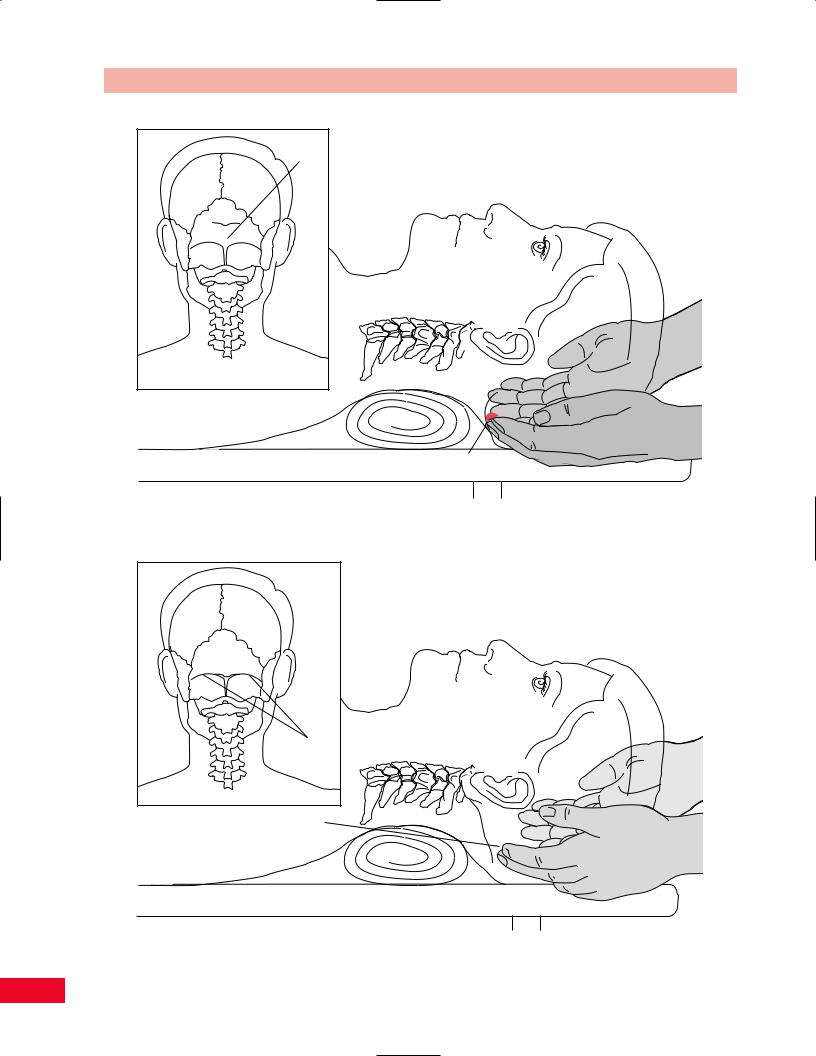
Inion (External Occipital Protuberance)
Place your fingers on the middle of the base of the skull and move slightly superiorly into the hairline and you will feel a rounded prominence, which is the inion (Figure 4.5). This is often referred to as the “bump of knowledge.”
Superior Nuchal Line
Place your fingers on the inion and move laterally and inferiorly diagonally toward the mastoid process. You will feel the ridge of the superior nuchal line under your fingers (Figure 4.6).
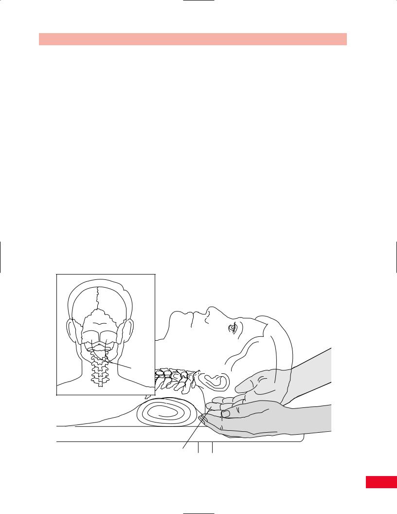
Occiput
Place your hands under the base of the patient’s head and allow your fingertips to rest on the most inferior aspect. This area is the occiput (Figure 4.7).
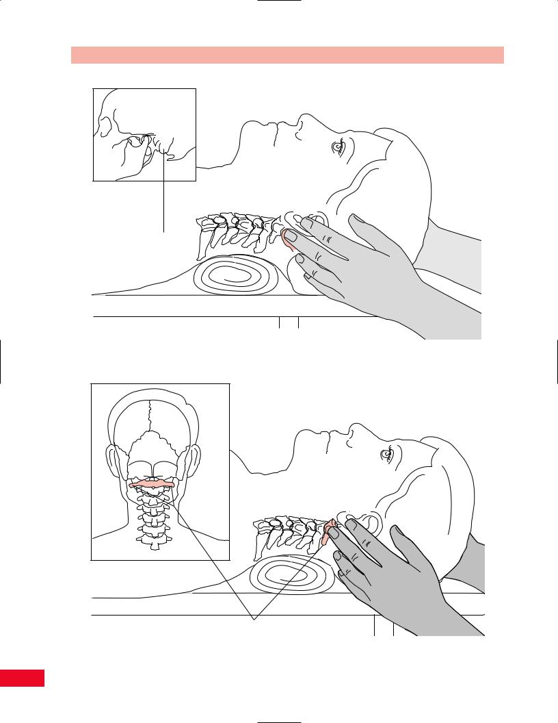
Mastoid Processes
Place your fingers directly under the patient’s earlobes and you will feel a rounded prominence on each side under your fingers. These are the mastoid processes (Figure 4.8).
Occiput
Occiput
Figure 4.7 Palpation of the occiput.
41
The Cervical Spine and Thoracic Spine Chapter 4
Mastoid
process
Figure 4.8 Palpation of the mastoid process.
Transverse process of C1
Figure 4.9 Palpation of the transverse process of C1.
42
Chapter 4 The Cervical Spine and Thoracic Spine
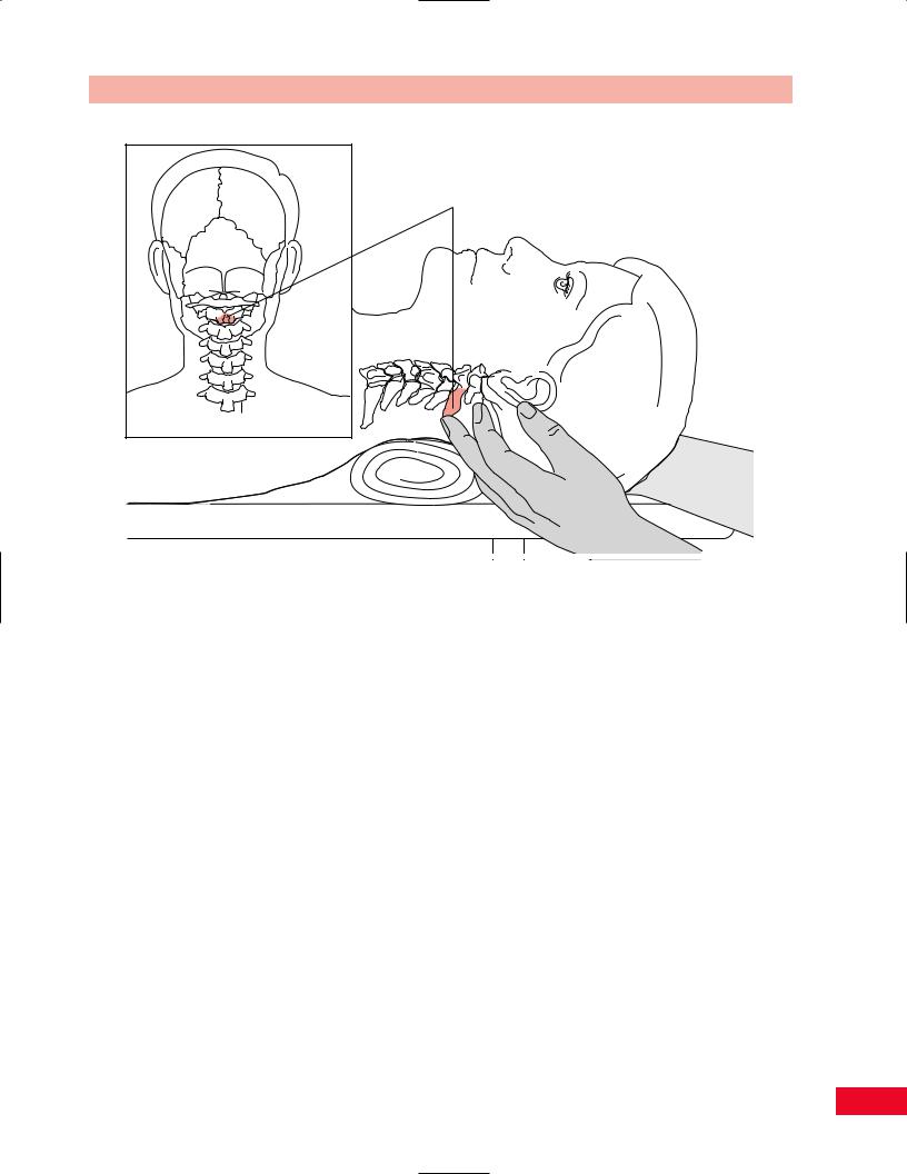
Spinous process of C2
Figure 4.10 Palpation of the spinous process of C2.
Transverse Processes of C1
Place your fingers anterior to the mastoid processes and in the space between the mastoid processes and the angle of the mandible, you will find the projection of the transverse processes of C1 (Figure 4.9). Although they can be deep, be careful not to press too firmly since they are often tender to palpation even in the normal patient.
Spinous Process of C2
Place your finger on the inion and move inferiorly into an indentation (posterior arch of C1). As you continue to move inferiorly, the rounded prominence that you feel is the spinous process of C2 (Figure 4.10).
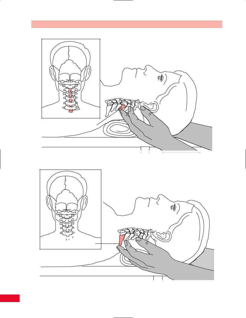
Spinous Processes
Place your middle fingers in the upper portion of the midline of the posterior aspect of the neck. You will feel blunt prominences under your fingers. These are the spinous processes (Figure 4.11). The spinous processes are often bifurcated, which you may be able to sense as you palpate them. You can start counting the spinous processes from C2 (location described above) caudally. You will notice the cervical lordosis as you palpate. Notice that the spinous processes of C3, C4,
and C5 are deeper and closer together, making them more difficult to differentiate individually.
Spinous Process of C7
The spinous process of C7 is normally the longest of all the cervical spinous processes (Figure 4.12). It is referred to as the prominens. However, it may be the same length as the spinous process of T1. To determine whether you are palpating C7 or T1, locate the spinous process you assume is C7. Place one finger on the spinous process that you presume is C7, and one over C6 and T1, and then have the patient extend the head slightly. The C6 vertebra will drop off slightly at the beginning of the movement, followed by C7 with a slight increase in extension, and T1 will not drop off at all. The T1 spinous process is immobilized by the first ribs and therefore does not move.
Articular Pillar (Facet Joints)
Move your fingers laterally approximately 1 in. from the spinous processes, over the erector spinae until you find a depression. You will be on the articular pillar. As you palpate in a caudal direction, you will be able to differentiate the joint lines of the facet joints: they
43
The Cervical Spine and Thoracic Spine Chapter 4
Spinous processes of C3 to C7
Spinous process of C4
Figure 4.11 Palpation of the spinous processes.
 Spinous
process of C7
Figure 4.12 Palpation of the spinous process of C7.
44