

MusculoSkeletal Exam
.pdfChapter 6 The Lumbosacral Spine
Knee extension (quadriceps)
Motor
L1
L2
 L3
L3 
L4
L5
Sensation |
Reflex |
Quadriceps reflex
L2 L2
L3 L3
L4
L4 L5
L3 dermatome
Key ( ) sensory areas
S1 |
S1 |
S1
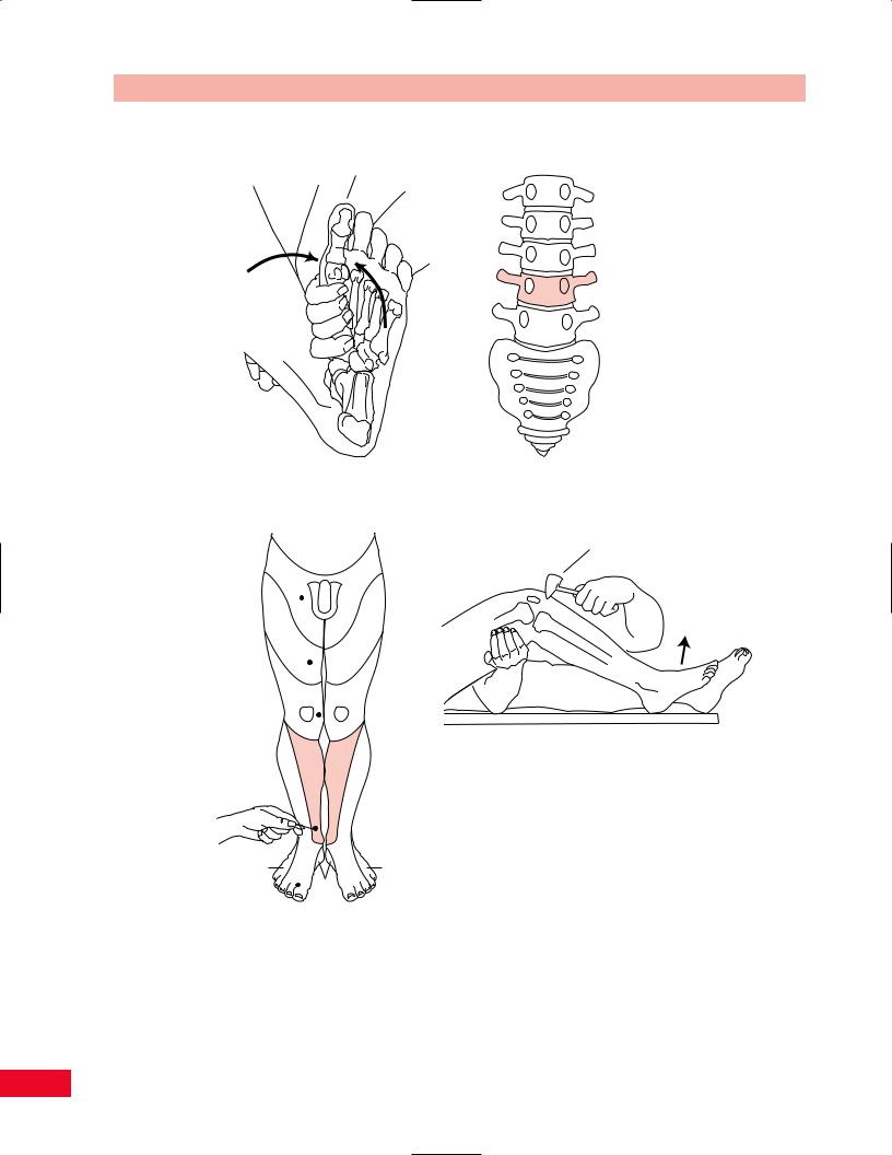
Figure 6.49 The L3 root level.
The L3 Level
Muscle Testing
The L3 root level (Figure 6.49) is best tested by examining the quadriceps muscle, which extends the knee. This is performed by having the patient sit on the edge of the examining table with the knees bent to 90 degrees. Ask the patient to extend the knee as you
apply resistance to the anterior aspect of the lower leg (see p. 364 for further information).
Sensation Testing
The L3 dermatome is located on the anteromedial aspect of the thigh. It extends just below the medial aspect of the knee. The key sensory area for L3 is located just medial to the patella.
125
The Lumbosacral Spine Chapter 6
Motor
Dorsiflexion (tibialis anterior)
L1
L2
L3
L4 
L5
Sensation |
Reflex |
Quadriceps reflex
L1 |
|
L1 |
|
|
S |
|
|
3 |
L2 |
|
L2 |
L3 |
|
L3 |
L5 |
L4 |
L4 |
|
||
|
|
L5 |
L4 dermatome
S1 |
S1 |
S1
( ) Key sensory areas
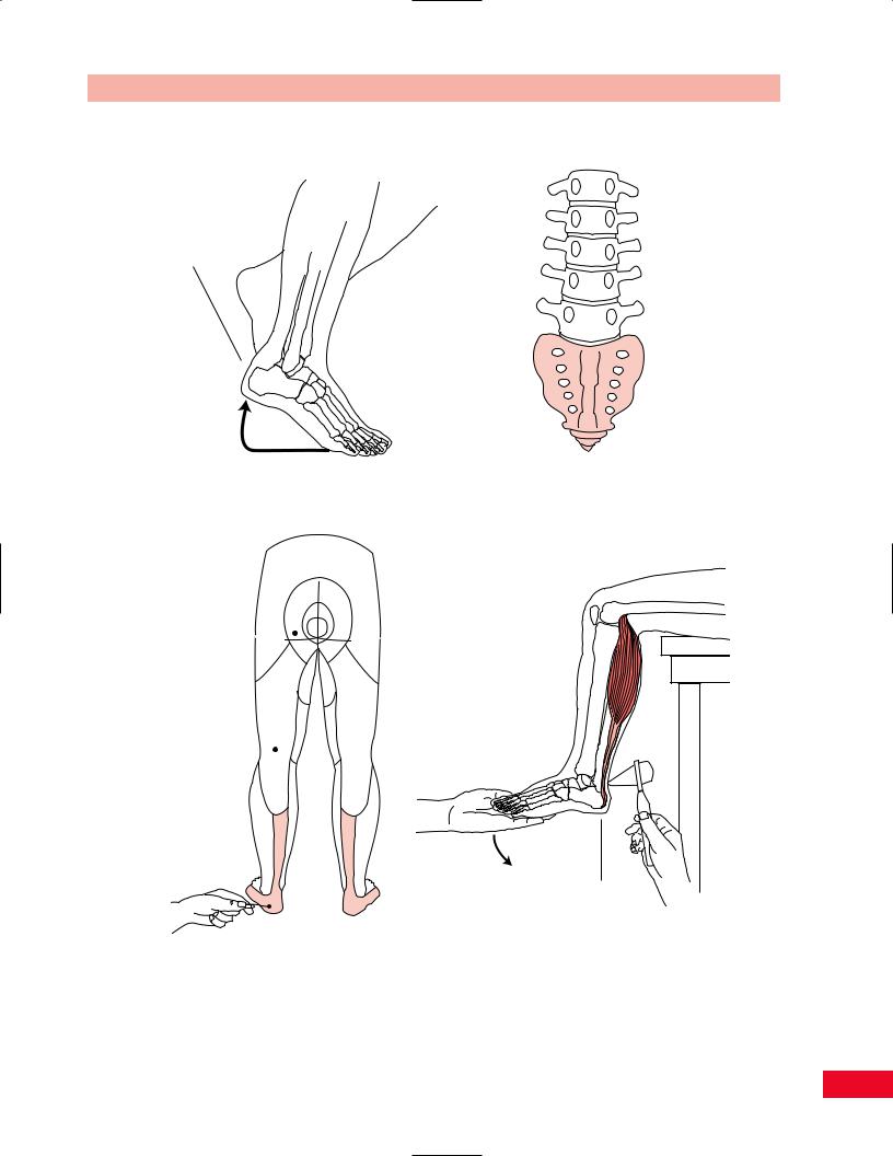
Figure 6.50 The L4 root level.
126

Chapter 6 The Lumbosacral Spine
Reflex Testing
There is no specific reflex for the L3 level. The L3 nerve root does contribute to the quadriceps reflex at the knee (see below).
The L4 Level
Muscle Testing
The L4 nerve root (Figure 6.50) is best examined by testing dorsiflexion, which is performed by the tibialis anterior muscle. The patient is in a sitting position or supine. Ask the patient to bring the foot upward and inward, bending at the ankle, while you apply resistance to the dorsum of the foot.
Sensation Testing
The L4 dermatome is located over the medial aspect of the leg and extends beyond the medial malleolus. The key sensory area of L4 is located just proximal to the medial malleolus.
Reflex Testing
L4 is tested by examining the quadriceps reflex. The patient is sitting with the legs over the edge of the table. Tap the patellar tendon with a reflex hammer and observe for quadriceps contraction and extension of the knee.
The L5 Level
Muscle Testing
The L5 nerve root (Figure 6.51) is best tested by examining the extensor hallucis longus muscle, which extends the great toe’s distal phalanx. The patient is sitting or supine. Ask the patient to raise the great toe as you apply resistance to the distal phalanx.
Sensation Testing
The L5 dermatome is located on the anterolateral region of the leg and extends onto the dorsal aspect of the foot. The L5 key sensory area is located just proximal to the second web space on the dorsal aspect of the foot.
Reflex Testing
The medial hamstring jerk can be used to test the L5 nerve root. This is performed with the patient supine. Support the lower leg with your forearm and place your thumb over the distal medial hamstring tendon in the popliteal fossa. Tap your thumb with the reflex hammer and observe for knee flexion.
The S1 Level
Muscle Testing
The S1 nerve root (Figure 6.52) is best tested by examining plantar flexion of the foot by the gastrocnemius and soleus muscles. This is performed by asking the patient to stand up on the toes.
Sensation Testing
The S1 dermatome is located on the posterior aspect of the calf and extends distally to the heel and then laterally along the dorsum of the foot. The key sensory area for S1 is located lateral to the insertion of the Achilles tendon on the foot.
Reflex Testing
The S1 nerve root is tested by examining the ankle jerk. The patient is sitting with the legs hanging over the edge of the table. Gently apply light pressure to the plantar aspect of the foot and ask the patient to relax as you tap the Achilles tendon with the reflex hammer. Observe the patient for plantar flexion of the foot and contraction of the calf muscles.
The S2, S3, and S4 Levels
Muscle Testing
The S2 through S4 nerve roots supply the urinary bladder and the intrinsic muscles of the foot.
Sensation Testing
The S2 dermatome is located on the posterior aspect of the thigh and extends distally to the midcalf. The key sensory area is located in the center of the popliteal fossa. The S3, S4, and S4 dermatomes are located concentrically around the anus, with the S3 dermatome forming the outermost ring.
The Superficial Reflexes
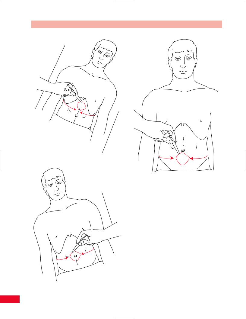
The upper, middle, and lower abdominal skin reflexes, the cremasteric reflex, and Babinski’s reflex are tested to examine the upper motor neurons of the pyramidal tract. These reflexes are exaggerated in upper motor neuron diseases, such as strokes and proximal spinal cord injuries.
Upper Abdominal Skin Reflex (T5–T8) (Figure 6.53) The patient is in a supine position and relaxed with the arms at the sides and the knees gently flexed. The skin over the lower part of the rib cage is stroked with
127

The Lumbosacral Spine Chapter 6
Motor
Big toe extension (extensor hallucis longus)
Sensation
L2 L2
L3 L3
L5 |
L4 |
L4 |
L5 |
L1
L2
L3
L4
L5
Reflex
Medial hamstring jerk
L5 dermatome
S1 S1
Key ( )
sensory areas |
S1 |
|
Figure 6.51 The L5 root level.
128
Chapter 6 The Lumbosacral Spine
|
Motor |
|
|
L1 |
|
|
L2 |
|
Plantar flexion |
L3 |
|
(gastrocnemius, soleus) |
||
|
||
|
L4 |
|
|
L5 |
|
|
S1 |
Sensation |
Reflex |
S 3
S 4
S 5
L2 L2
L |
L |
3 |
3 |
|
L |
L |
|
|
4 |
4 |
|
S1 dermatome |
S1 |
S1 |
|
L5 |
L5 |
||
|
Key ( ) sensory areas
Ankle jerk
Figure 6.52 The S1 root level.
129
The Lumbosacral Spine Chapter 6
Figure 6.53 The upper abdominal skin reflex (T5–T8).
Figure 6.55 The lower abdominal skin reflex (T11, T12).
Figure 6.54 The midabdominal skin reflex (T9–T11).
a fingernail or key from laterally to medially. Observe the patient for contraction of the upper abdominal muscles on the same side. You may also note movement of the umbilicus to the same side as the scratch.
Midabdominal Skin Reflex (T9–T11) (Figure 6.54) Perform the test above, but this time at about the level of the umbilicus. The response is similar to that for the upper abdominal skin reflex.
Lower Abdominal Skin Reflex (T11–T12) (Figure 6.55) Perform the test as above, but this time over the level of the iliac crest to the hypogastric region. Again observe for contraction of the lower abdominal muscles on the same side and movement of the umbilicus in the direction of the scratch.
Cremasteric Reflex (L1–L2) (Figure 6.56)
This test is performed in men only. The inner aspect of the thigh is scratched with the handle of the reflex
130
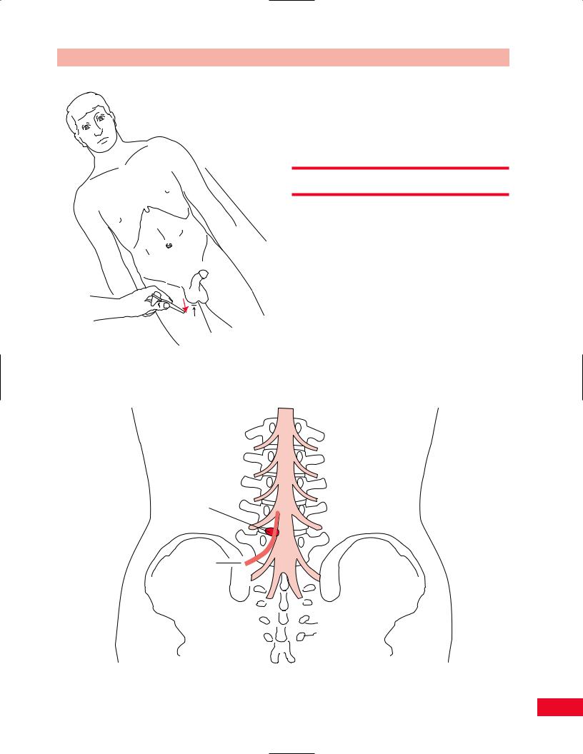
Figure 6.56 The cremasteric reflex. Note that immediate movement of the scrotum upward is a positive test result.
L1
L2
L3
L4-L5 L4 disc
L5
L5 nerve root
Chapter 6 The Lumbosacral Spine
hammer from the pubis downward. You will note an immediate contraction of the scrotum upward on the same side. An irregular or slow rise of the testis on the same side is not a positive response.
Special Tests
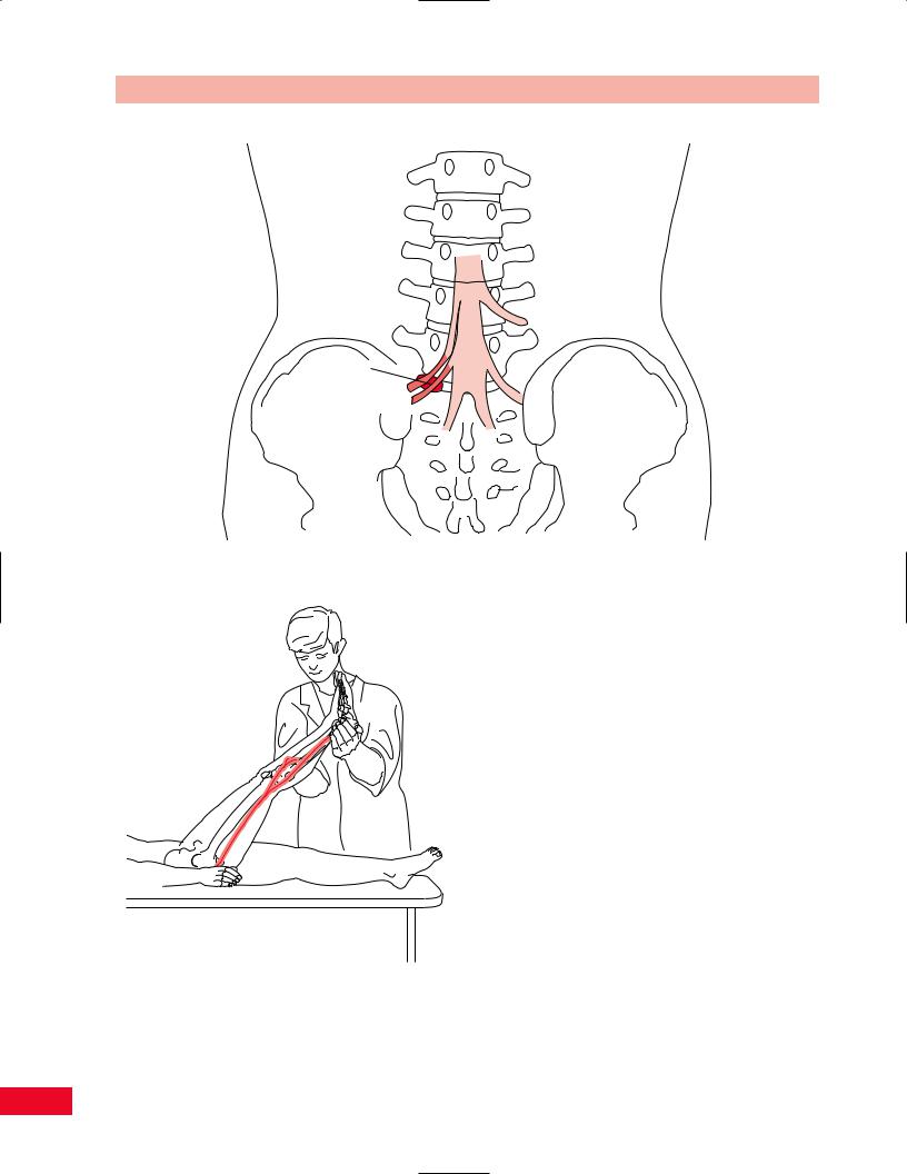
Straight-Leg Raise Test
This test is performed to stretch the sciatic nerve and its dural covering proximally. In patients who have a herniated disc at L4–L5 or L5–S1 (Figures 6.57 and 6.58) that is causing pressure on the L5 or S1 nerve roots, stretching the sciatic nerve will frequently cause worsening of the lower-extremity pain or parasthesias or both. The test is performed by asking the patient to lie supine (Figure 6.59). With the patient’s knee extended, take the patient’s foot by the heel and elevate the entire leg from the examining table. As the leg is raised beyond approximately 75 degrees, the sciatic nerve is being stretched. The patient will complain of increased lower-extremity pain or parasthesias on the
Figure 6.57 A posterolateral herniation of the L4–L5 disc can cause pressure and injury to the L5 nerve root.
131
The Lumbosacral Spine Chapter 6
L1
L2
L3
L4
L5
L5,S1 disc
L5 nerve root
S1 nerve root
Figure 6.58 A posterolateral herniation of the L5–S1 disc can cause pressure and injury to the L5 and S1 nerve roots.
Figure 6.59 Straight-leg raising test. Between 35 and 70 degrees, the L5 and S1 nerve roots may be stretched against an intervertebral disc. Flexing the hip more than 70 degrees causes stress on the lumbar spine.
side that is being examined. This is a positive response on the straight-leg raising test. If the patient complains of pain down the opposite leg, this is called a positive crossed response on the straight-leg raising test and is very significant for a herniated disc. The patient may also complain of pain in the posterior part of the thigh, which is due to tightness of the hamstrings.
You can determine whether the pain is caused by tight hamstrings or is of a neurogenic origin by raising the leg up to the point where the patient complains of leg pain, and then lowering the leg slightly (Figure 6.60). This should reduce the pain in the leg. Now passively dorsiflex the patient’s foot to increase the stretch on the sciatic nerve. If this maneuver causes pain, then the pain is neurogenic in origin. If this movement is painless, then the patient’s discomfort is caused by hamstring tightness.
Variations on the Straight-Leg Raise Test
The tibial nerve can be stretched by first dorsiflexing the ankle and everting the foot, and then performing a straight-leg raise test. The test is abnormal if the patient complains of pain or numbness in the plantar aspect of the foot that is relieved by returning the foot to the neutral position.
132
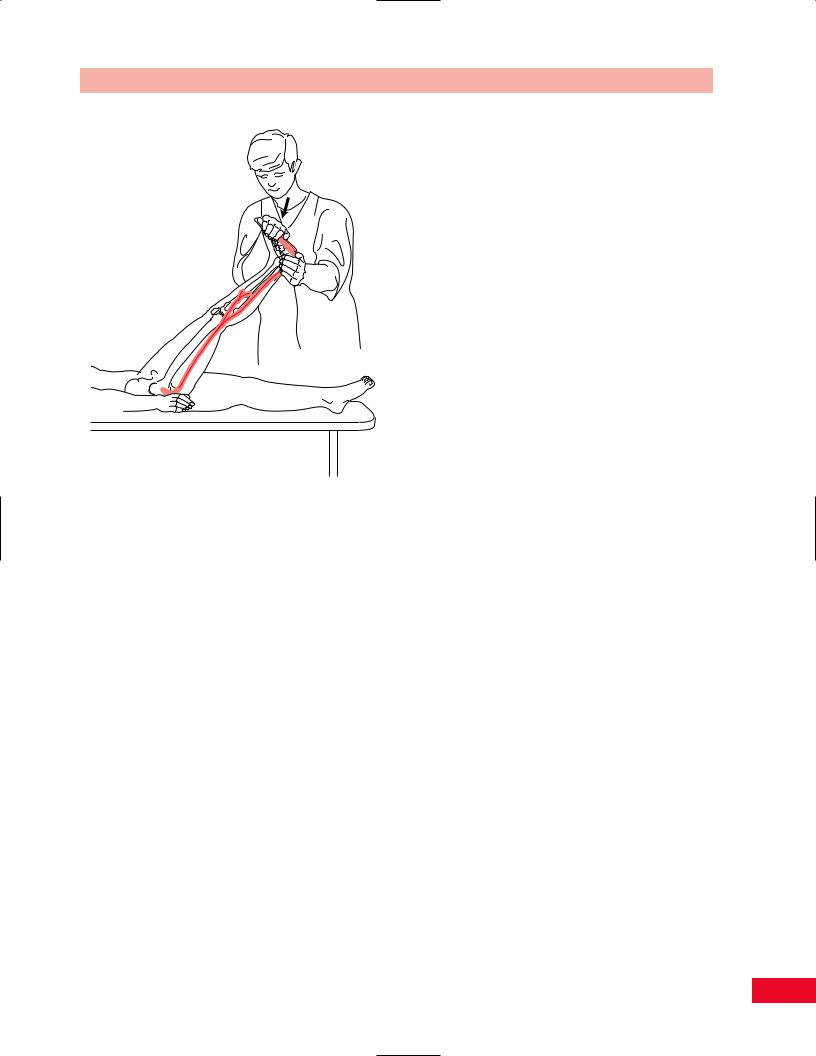
Figure 6.60 By lowering the leg slightly to the point where the patient stops feeling pain or parasthesias in the leg, and then dorsiflexing the ankle, you can determine whether the pain in the leg is due to tight hamstrings or has a neurogenic origin.
If the pain is reproduced on dorsiflexion of the ankle after the hamstrings have been relaxed by lowering the leg slightly, then the pain has a neurogenic origin.
The peroneal nerve can be stretched by first plantarflexing the ankle and inverting the foot, and then performing a straight-leg raise test. The test is abnormal if the patient complains of pain or numbness on the dorsum of the foot that is relieved by returning the foot to the neutral position.
The test can be conducted in two ways: Either the ankle or the leg can be positioned first. You choose what order to perform the test by first positioning the body part closest to the symptoms. For example, if the pain is in the buttock, use the straight-leg raise test first and position the ankle afterwards. If the pain is in the foot, position the ankle first (Butler, 1991).
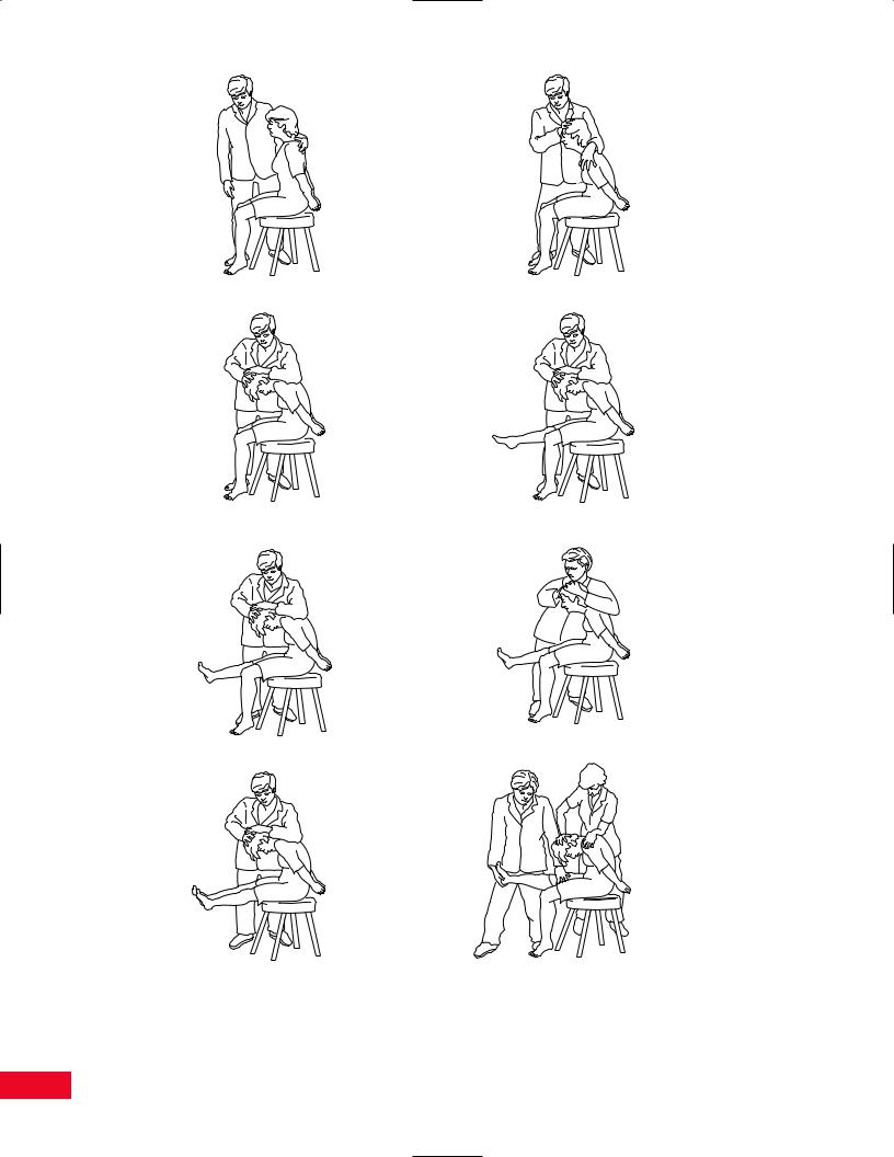
The Slump Test
The slump test (Figure 6.61) is a neural tension test which is indicated when the patient complains of spinal symptoms. The test is conducted as follows:
Chapter 6 The Lumbosacral Spine
Patient Position: The patient is sitting with both lower extremities supported with the upper extremities behind the back and the hands clasped.
Instruct the patient to “sag.” Overpressure can be added to increase the degree of flexion. Maintain flexion and then ask the patient to bend the neck towards the chest. Overpressure can be added and the symptoms are reassessed. While maintaining the position, instruct the patient to extend one knee and reassess. Then ask the patient to dorsiflex the ankle and reassess. Release neck flexion and reassess. Ask the patient to flex the neck again and repeat the process on the other leg. Finally both legs can be extended simultaneously.
Normal responses can include pain at T8–9 in approximately 50% of patients, pain on the posterior aspect of the extended knee, decreased range of motion (ROM) in dorsiflexion, and a release of symptoms and an increase in range when neck flexion is released (Butler, 1991). Worsening of neurological symptoms can be indicative of pathology secondary to tension in the nervous system.
Femoral Stretch Test
This test (Figure 6.62) is useful in determining whether the patient has a herniated disc in the L2–L4 region. The purpose of the test is to stretch the femoral nerve and the L2–L4 nerve roots. The patient is lying on the side, with the test side up. The test can also be performed with the patient lying prone. Support the patient’s lower extremity with your arm, cradling the knee and leg. The test leg is extended at the hip and flexed at the knee. If this maneuver causes increased pain or parasthesias in the anterior medial part of the thigh or medial part of the leg, it is likely that the patient has a compressive lesion of the L2, L3, or L4 nerve roots, such as an L2–L3, L3–L4, or L4–L5 herniated disc.
Hoover Test
This test is useful in identifying a malingering patient who is unable to raise the lower extremity from the examining table while lying supine. The test is performed by taking the patient’s heels in your hands while the legs are flat on the table. Ask the patient to raise one of the legs off the table while maintaining the knee in an extended position. Normally, the opposite leg will press downward into your hand. If the patient states that he or she is trying to raise the leg and there is no downward pressure in your opposite hand, it is likely that the patient is malingering.
133
Slump stage 1 |
Slump stage 2 |
Slump stage 3 |
Slump stage 4 |
Slump stage 5 |
Slump stage 6 |
Testing bilateral knee |
The Slump Test with |
extension in Slump |
an assistant |
Figure 6.61 The slump test. Adapted from Butler D. Mobilization of the Nervous System. New York, Churchill Livingstone, 1991.
134