MusculoSkeletal Exam
.pdfinvolved in clinical syndromes that they bear specific mention. They are the carpal tunnel, which contains the median nerve together with the flexor tendons of the digits, and the tunnel of Guyon, which contains the ulnar nerve. Injury to the median nerve presents as parasthesias, atrophy, and loss of sensation to the thenar eminence of the thumb, the index and long fingers, and the radial half of the ring finger. Compression injury of the ulnar nerve will affect the medial aspect of the hand (hypothenar eminence, small finger, and ulnar half of the ring finger), together with the ulnar intrinsic muscles of the hand. This muscular compromise will lead to classic posturing of the digits called the benediction hand, referring to the appearance of a priest’s hand when giving a blessing (see p. 283).
Observation
The examination should begin in the waiting room before the patient is aware of the examiner’s observation. Information regarding the degree of the patient’s disability, level of functioning, posture, and gait can be observed. The clinician should pay careful attention to the patient’s facial expressions with regard to the degree of discomfort the patient is experiencing. The information gathered in this short period can be very useful in creating a total picture of the patient’s condition.
Note the manner in which the patient is sitting and how he or she is posturing the upper extremity. Is the arm relaxed at the side or is the patient cradling it for protection? What is the relaxed posture of the hand itself? Note whether the wrist or hand is edematous. Note the shape of the hand and if there are any changes in contour. The patient may have a protuberance secondary to a ganglion, nodule, or bony dislocation. Note any bony deformities. The patient may have a swan neck, boutonnière deformity, or claw fingers. Compare one hand to the other, remembering that the dominant hand may be larger in the normal individual. What is the cosmetic appearance of the hand? Many patients are extremely self-conscious of how their hands look.
How willing is the patient to use the upper extremity? Will he or she allow you to shake their hand? Is the patient able to move the hand in an effortless and coordinated fashion or is he or she stiff and uncoordinated? Is the patient willing to bear weight on an extended wrist? Watch as the patient gets up from the chair to see whether he or she pushes down with the wrist. Pain may be altered by changes in position so watch
Chapter 10 The Wrist and Hand
the patient’s facial expression to give you insight into the pain level.
Observe the patient as he or she assumes the standing position. Observe the patient’s posture. Pay particular attention to the position of the head, cervical spine, and the thoracic kyphosis. Note the height of the shoulders and their relative positions. Once the patient starts to ambulate, observe whether he or she is willing to swing the arms. Arm swing can be limited by either loss of motion, pain, or neurological damage.
Once the patient is in the examination room, ask him or her to disrobe. Observe the ease with which the patient uses the upper extremities and the rhythm of the movements. Observe for symmetry of bony structures. Note the carrying angle with the upper extremity postured in the anatomical position. Observe the hand on all surfaces. Observe for areas of muscle wasting that may be secondary to peripheral nerve lesions. Inspect for scars, open lesions, abrasions, calluses, color, hair growth patterns, nails, and the presence of any trophic changes. Abnormalities may be secondary to reflex sympathetic dystrophy, shoulder hand syndrome, Raynaud’s disease, or peripheral vascular or metabolic disease. Observe the skin. Is the skin smooth with a loss of the creases? Is there an increase in moisture or a decrease in sensibility? Spindlelike fingers can be secondary to systemic lupus erythematosus, long-standing neuropathy, or rheumatoid arthritis. Clubbing and cyanosis of the nails may be secondary to pulmonary disease.
Subjective Examination
The wrist and hand are extremely active structures that are both complicated and delicate. They are very vulnerable to injury. Since they are non-weight-bearing, problems are most commonly related to overuse syndromes, inflammation, and trauma. You should inquire about the nature and location of the patient’s complaints as well as their duration and intensity. Note whether the pain travels up to the elbow. The behavior of the pain during the day and night should also be addressed.
You should determine the patient’s functional limitations. How much was the patient able to do before the onset of symptoms? Which is the dominant hand? How incapacitated does the patient consider himself or herself to be? Question the patient regarding actual use of the upper extremity. Is the patient able to comb his hair, fasten her bra, fasten buttons, handle small objects, or feed himself? Is this injury traumatic in
235

The Wrist and Hand Chapter 10
nature? What was the mechanism of the injury? Does the patient regularly participate in any vigorous sport activity that would stress the wrist or hand? What is the patient’s occupation? Does he or she use tools or spend a great deal of time at a computer terminal stressing the wrist and hand repetitively?
If the patient reports a history of trauma, it is important to note the mechanism of injury. The direction of the force, the position of the upper extremity, and the activity the patient was participating in at the time of the injury contribute to your understanding of the resulting problem and help you to better direct your examination. The degree of pain, edema, and disability at the time of the trauma and within the initial 24 hours should be noted. Does the patient have a previous history of the same injury? Does the patient report any clicking, grating, or snapping? This may be due to a stenosing tenosynovitis or to a loose body. Is any grating present? This may be due to osteoarthritis.
The patient’s disorder may be related to age, gender, ethnic background, body type, static and dynamic posture, occupation, leisure activities, hobbies, and general activity level. It is important to inquire about any change in daily routine and any unusual activities that the patient has participated in. The location of the symptoms may give you some insight into the etiology of the complaints. The cervical spine and the shoulder can refer pain to the wrist and hand and should be included as part of the examination. The most common nerve roots that refer pain are C6, C7, C8, and T1. (Please refer to Box 2.1, p. 18 for typical questions for the subjective examination.)
Gentle Palpation
The palpatory examination is started with the patient in the sitting position. You should first examine for areas of localized effusion, discoloration, birthmarks, calluses, open sinuses or drainage, incisional areas, abrasions, bony contours, muscle girth and symmetry, and skin creases. You should not have to use deep pressure to determine areas of tenderness or malalignment. It is important to use firm but gentle pressure, which will enhance your palpatory skills. By having a sound basis of cross-sectional anatomy, you will not need to physically penetrate through several layers of tissue to have a good sense of the underlying structures. Remember that if you increase the patient’s pain at this point in the examination, the patient will be very reluctant to
Paradigm for carpal tunnel syndrome
A 45-year-old female office worker presents with complaints of “pins and needles” in her dominant right hand. She states that she has been having pain in her neck, upper arm and thumb. Her symptoms seem to be aggravated by typing for long periods of time at her word processor. She is often awakened during the night by pain in her hand. She can find relief by shaking her hand or holding it under warm water. She recalls no injuries to either her hand or neck; and has no difficulty rotating her head when driving. She has recently been told that she has an “underactive thyroid” and has had a twenty pound weight gain. The remainder of her medical history is unremarkable.
On physical examination, the patient is a slightly obese woman in no apparent distress. She has full range of motion of her cervical spine and upper extremity joints without pain. There are no symptoms produced with vertical compression applied to the head and neck. She has diminished light touch in the distal palmar aspects of the thumb, index and long fingers. She has positive Tinel’s and Phalen’s tests. X-rays of the cervical spine and hand are reported as showing no pathology. Electrodiagnostic studies (electromyography (EMG) and nerve conduction) demonstrate no motor deficits in the upper extremity; but confirm increased latency of the signal across the wrist.
This paradigm is consistent with distal rather than proximal nerve injury because of:
A history of repetitive hand/wrist movements No history of trauma to the neck
History of a possible contributory collateral medical condition Symptoms suggestive of compromised circulation to the nerve (see Table 10.2)
allow you to continue and may become more limited in his or her ability to move.
Palpation is most easily performed with the patient in a relaxed position. The sitting position with the extremity supported on a table is preferred for ease of examination of the wrist and hand. Remember that for palpation of all the structures described, the hand is in the anatomical position. While locating the bony landmarks, it is also useful to pay attention to areas of increased or decreased temperature and moisture.
This will help you identify areas of acute and chronic inflammation.
Anterior (Palmar) Aspect
Bony Structures
Since thick skin and fascia cover the palm, the bony structures are more difficult to palpate on the anterior surface. The carpal bones are more accessible and easier to identify from the dorsal aspect. Descriptions of their locations are found later in this chapter.
236
Soft-Tissue Structures
Start your palpation by observing the superficial palmar surface. The skin is thicker than on the dorsal aspect. The skin contains many sweat glands but is free of hair. Observe the creases in both the longitudinal and transverse directions. You will notice that the longitudinal creases are more distinct when the patient opposes the thumb. The transverse creases are more distinct when the patient flexes the metacarpophalangeal joints. Note the absence of the fibro-fatty tissues and how the skin is securely attached to the deep fascia in the area of the skeletal joints forming the creases. They are useful to identify the underlying anatomical structures. At the level of the wrist you will notice the proximal and then the distal wrist creases. On the lateral side, notice the thenar (radial longitudinal) crease that surrounds the thenar eminence.
Continuing distally, note the proximal palmar (flexion) crease, which begins simultaneously with the thenar crease, just proximal to the head of the second metacarpal. It travels medially across the palm along the middle of the shafts of the third through fifth metacarpals.
The distal palmar (transverse) crease is located on the palmar surface of the heads of the second or third through fifth metacarpals. It becomes more distinct as the patient flexes the metacarpophalangeal joints.
As you continue distally you will notice the proximal digital creases located at the level of the finger webs. There are no joints under these creases. The metacarpophalangeal joints are about 2 cm proximal to the proximal digital creases.
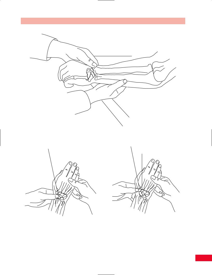
The proximal and distal interphalangeal creases lie superficial to the proximal and distal interphalangeal joints and deepen as the joints are flexed (Figure 10.2).
To enable you to more easily organize the palpation of the deeper soft tissues, the anterior surface of the hand can be divided into three areas: the medial, middle, and lateral compartments. Each compartment is described, from proximal to distal.
Medial (Ulnar) Compartment
Flexor Carpi Ulnaris. Move your fingers to the medial palmar surface and locate the pisiform bone (see description on p. 242). The tendon of the flexor carpi ulnaris is palpable proximal to its attachment on the pisiform (Figure 10.3). The tendon becomes more distinct when you resist wrist flexion and ulnar deviation.
Chapter 10 The Wrist and Hand
Distal interphalangeal crease
 Proximal interphalangeal crease
 Proximal digital creases
 Proximal palmar crease Distal palmar crease
Thenar crease
Distal wrist crease Proximal wrist crease
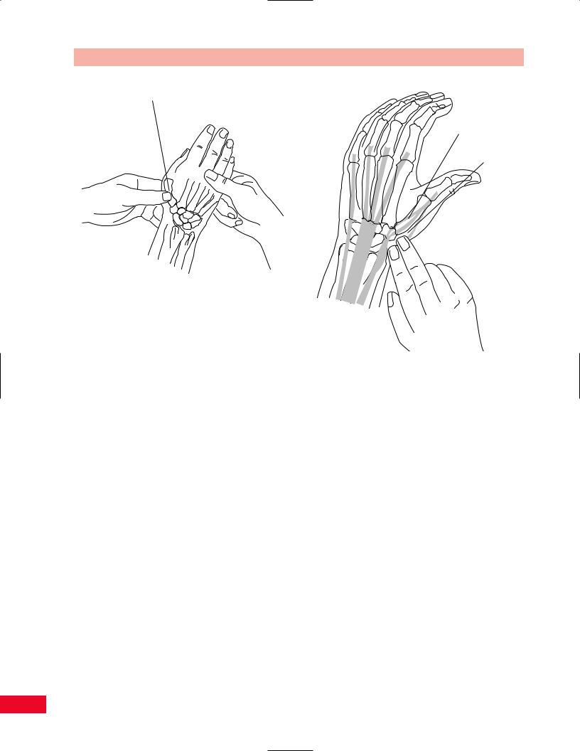
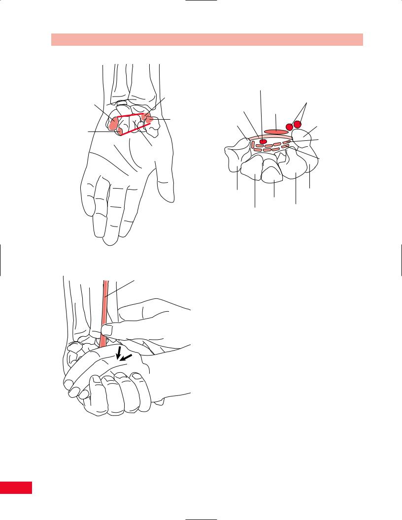
Figure 10.2 Palpation of the palmar surface of the hand.
Flexor carpi ulnaris tendon
Figure 10.3 Palpation of the flexor carpi ulnaris.
237

The Wrist and Hand Chapter 10
Ulnar
Artery
Figure 10.4 Palpation of the ulnar artery. |
Figure 10.5 Palpation of the hypothenar eminence. |
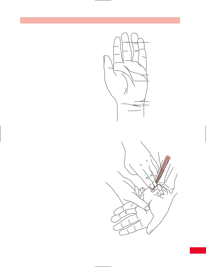
Ulnar Artery. The ulnar pulse can be palpated on the medial volar surface of the wrist (Figure 10.4). Press against the distal aspect of the ulna, just proximal to the pisiform, to facilitate palpating the pulse. Remember not to press too hard since the pulse will be obliterated.
Ulnar Nerve. The ulnar nerve passes into the hand lateral to the pisiform, medial and posterior to the ulnar artery, and then under the hook of the hamate. It is not readily palpable in the wrist, but it is at the medial elbow (see p. 201).
Hypothenar Eminence. Place your fingers on the pisiform and move distally until you reach the distal palmar crease. You will feel the longitudinal bulk of the muscle bellies of the hypothenar eminence (Figure 10.5). The eminence is composed of the palmaris brevis, abductor digit minimi, flexor digiti minimi brevis, and the opponens digiti minimi. It is not possible to differentiate between these muscles on palpation. Examine the hypothenar eminence and compare it to the one in the opposite hand for size and symmetry. Atrophy and diminished sensation can be indicative of compression of the ulnar nerve in the elbow in the cubital tunnel or in the canal of Guyon secondary to trauma or from
compression from a ganglion. Power grip will be significantly impaired.
Middle Compartment
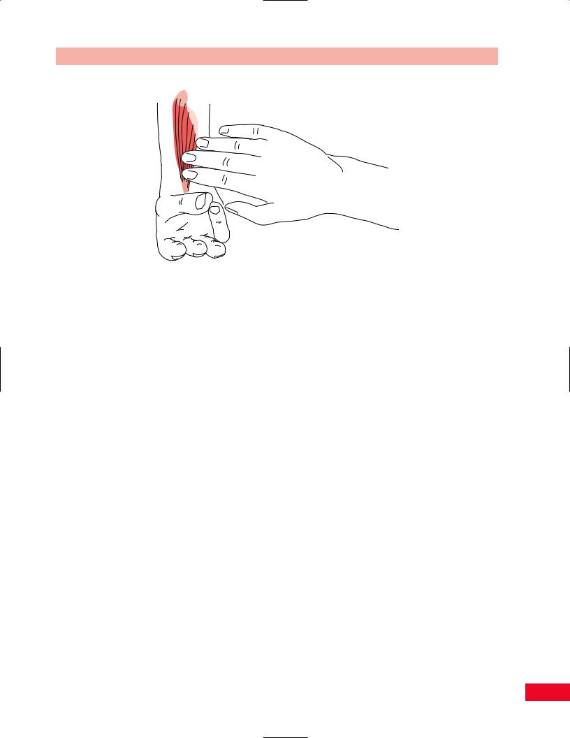
Palmaris Longus. Continue to move laterally along the anterior surface of the wrist. The long thin tendon in the middle is the palmaris longus. It can be palpated just proximal to its attachment to the anterior distal surface of the flexor retinaculum and the palmar aponeurosis (Figure 10.6). The tendon becomes more distinct when the patient flexes the wrist and approximates the thenar and hypothenar eminences, causing a tensing of the palmar fascia. The palmaris longus is absent in either one or both wrists in approximately 13% of the population. However, its absence does not alter the patient’s function (Moore and Dalley, 1999). When the tendon is present, it may be useful as a donor site for twostage tendon reconstructive surgery. The tendon can be used to help locate the median nerve, which runs just lateral to it at the wrist.
Flexor Digitorum Profundus and Superficialis. The tendons of the flexor digitorum profundus and superficialis travel in a common sheath passing under the
238
Chapter 10 The Wrist and Hand
Figure 10.6 Palpation of the palmaris longus.
flexor retinaculum and deep to the palmar aponeurosis. They then divide, are covered by synovial membrane, and enter into individual osseofibrous digital tunnels. In some individuals it is possible to palpate the individual tendons as they travel along the palm toward the digits. Ask the patient to flex and extend the fingers and you will feel the tendon become more prominent as the fingers contract into flexion and taut as they move toward extension.
If snapping, “clunking,” or grating in the tendon is noted with either flexion or extension, then a trigger finger may be present. This is caused by swelling of the tendon, which creates difficulty in gliding under the pulley at the metacarpal head during movement.
Carpal Tunnel. The carpal tunnel is created by the flexor retinaculum covering the anterior concavity of the carpal bones. Its floor is formed medially by the pisiform and the hook of the hamate and laterally by the tubercle of the scaphoid and the tubercle of the trapezium (Figure 10.7). The tunnel allows the flexor tendons of the digits and the median nerve to travel through to the hand. It is extremely significant clinically because of the frequency of compression of the median nerve secondary to edema, fracture, arthritis, cumulative trauma or repetitive motion injuries. This is referred to as carpal tunnel syndrome. You can confirm the diagnosis with electrodiagnostic testing.
Palmar Aponeurosis. The palmar aponeurosis is a triangular shaped fascia found in the palm of the hand covering the long finger flexors. It divides into four bands, which join with the fibrous finger sheaths. It is
palpable as a resistance to your pressure in the center of the palm while the fingers are in extension. Marked finger flexion at the metacarpophalangeal joints with increased fibrosis of the palmar fascia is indicative of a Dupuytren-like contracture.
Lateral (Radial) Compartment
Flexor Carpi Radialis. If you continue to move laterally from the palmaris longus, the next tendon you will palpate is the flexor carpi radialis (Figure 10.8). The tendon is palpable at the level of the wrist as it passes into the hand and attaches at the base of the second metacarpal. In some individuals the muscle may be absent. The tendon is made more distinct by providing resistance during wrist flexion and radial deviation.
Radial Artery. The radial pulse can be palpated just lateral to the tendon of the flexor carpi radialis. Press against the radius to facilitate palpating the pulse. Remember not to press too hard or the pulse will be obliterated.
Thenar Eminence. The thenar eminence is comprised of the three short muscles of the thumb: abductor pollicis brevis, flexor pollicis brevis, and opponens pollicis. It is located at the base of the thumb and is a thick, fleshy prominence that is freely movable on palpation. It is demarcated by the thenar crease.
Compare both hands for symmetry, especially paying attention to the size, shape, and feel of the thenar eminence. Note that the dominant side may be noticeably larger, particularly when the individual engages
239
The Wrist and Hand Chapter 10
|
|
|
Median nerve |
|
|
Scaphoid |
|
Ulnar A & N |
|
Pisiform |
tuberosity |
Transverse carpal |
||
|
Palmaris |
|||
|
|
ligament |
longus |
|
|
|
Tubercle of |
||
|
Tubercle of |
|
||
Hook of |
trapezium |
Pisiform |
||
trapezium |
||||
hamate |
|
|
||
|
|
|
||
|
|
|
Flexor |
|
|
Carpal tunnel |
|
digitorum |
|
|
|
superficialis |
||
|
|
|
||
|
|
Flexor |
Flexor |
|
|
|
digitorum |
||
|
|
pollicis |
||
|
|
profundus |
||
|
|
longus |
||
|
|
Trapezium |
Triquetrum |
|
|
|
|
Capitate |
|
|
|
|
Lunate |
|
|
|
Trapezoid |
||
Figure 10.7 The carpal tunnel.
Flexor carpi radialis
Figure 10.8 Palpation of the flexor carpi radialis.
in racquet sports or is a manual laborer. Notice any atrophy. The muscles are innervated by the recurrent branch of the median nerve and may be affected when the individual has carpal tunnel syndrome. The thenar
eminence may actually become hollow in advanced stages of the pathology.
Fingers. Observe the bony alignment of the phalanges. They should be symmetrical and straight from both the anterior–posterior and medial–lateral views. The patient may present with a swan neck deformity secondary to a contracture of the intrinsic muscles. This is often seen in patients with rheumatoid arthritis. A boutonnière deformity may be present when the patient ruptures or lacerates the central tendinous slip of the extensor digitorum communis tendon, which allows the lateral bands to migrate volarly. This can occur secondary to trauma. In rheumatoid arthritis the balance between the flexors and extensors is disturbed, not allowing the central slip to pull properly and letting the lateral bands migrate volarly. Claw fingers can also be present secondary to loss of the intrinsic muscles and a subsequent overactivity of the extrinsic muscles.
Note the presence of any nodules. Heberden’s nodes can be found on the dorsal aspect of the distal interphalangeal joint and are associated with osteoarthritis. Bouchard’s nodes can be found on the dorsal aspect of the proximal interphalangeal joint and are associated with rheumatoid arthritis.
240
Examine the finger pads. They are highly innervated and vascularized. The pads are especially susceptible to infection because of their location and use. Note any area of edema, erythema, and warmth. Osler’s nodes may be present secondary to subacute bacterial endocarditis.
Medial (Ulnar) Aspect
Bony Structures
Ulna Styloid Process
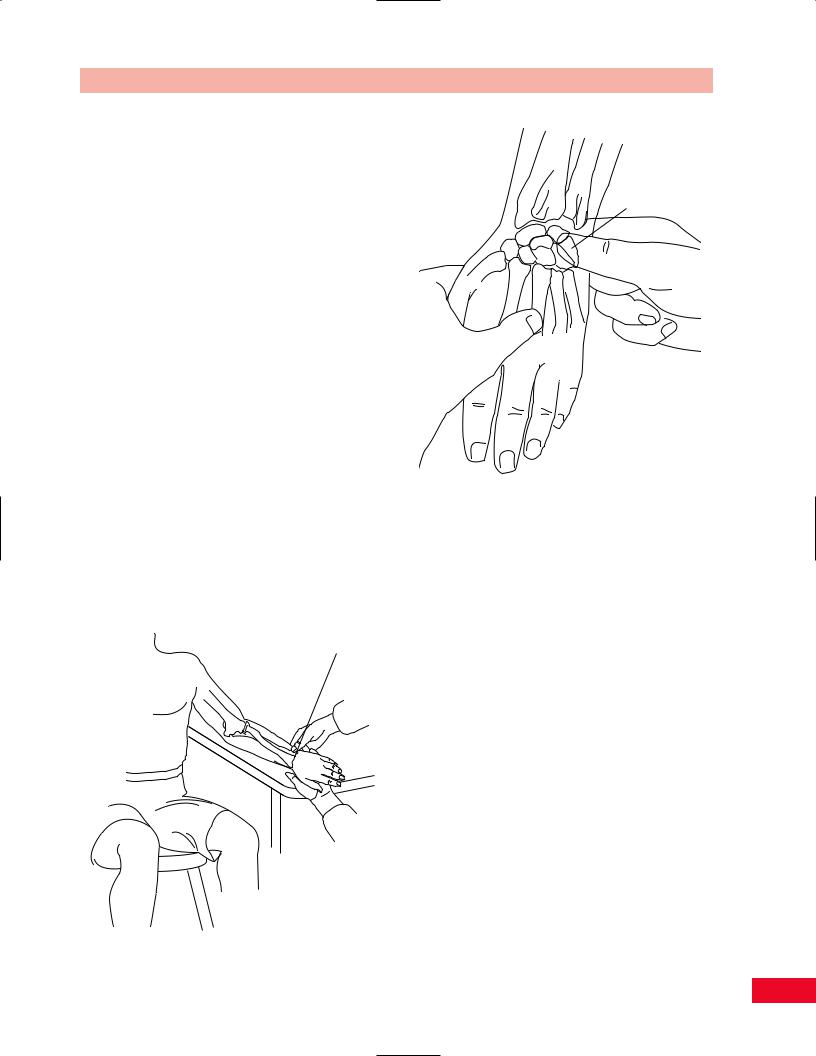
Place your fingers along the shaft of the ulna (see description on p. 206) and follow it distally until you come to the rounded prominence of the ulna styloid process. It is more defined than the radial styloid process. The ulna styloid process is located more proximal than its radial counterpart and slightly more posterior (Figure 10.9). The ulna styloid process does not have a direct articulation with the carpal bones.
Triquetrum
Palpate the ulna styloid process and continue to move distally on the medial aspect of the wrist. You will first find the space for the articular meniscus, and then feel the rounded surface of the triquetrum. Move the patient’s hand into radial deviation and the triquetrum will move medially into your finger (Figure 10.10). The dorsal aspect can also be palpated
Ulnar styloid
Figure 10.9 Palpation of the ulna styloid process.