

MusculoSkeletal Exam
.pdfSupraspinatus muscle-tendon
Coracoacromial ligament
Subacromial space
Subacromial bursa
Long head of biceps tendon
Figure 8.5 The subacromial space is defined superiorly by the acromioclavicular bony arch and the coracoacromial ligament. Inferiorly, it is defined by the humeral head. Within this space lies the subacromial bursa and the tendons of the supraspinatus and long head of the biceps muscles.
from this condition can lead not only to a chronically disabling condition, with erosion of the rotator cuff tissues, but also to an attempt to compensate for the loss of glenohumeral motion with scapulothoracic movement. Excessive stress can be created on the cervical spine due to the muscular effort of the proximal back and shoulder muscles to compensate for the lack of glenohumeral movement.
When the biceps tendon has become chronically inflamed by either frictional wear beneath the acromial arch or chronic tendinitis, it is at risk of rupture. If this occurs, the humeral head depression is compromised. The humerus will then ride superiorly within the glenoid, increasing pressure between the humeral head and the acromial arch. This will also prevent clearance of the greater tuberosity of the humerus beneath the acromion during abduction and will result in limited shoulder motion. The resultant cycle of pain and guarded range of motion produces an increasing pattern of upper-extremity dysfunction.
Shoulder movement therefore represents a complex interplay of multiple articulations and soft tissues, which must be recognized and appreciated for their delicate interrelationship.
Observation
Note the manner in which the patient is sitting in the waiting room. Notice how the patient postures the upper extremity. Is the arm relaxed at the side or is
Chapter 8 The Shoulder
the patient cradling it for protection? How willing is the patient to use the upper extremity? Will the patient extend the arm to you to shake your hand? Pain may be altered by changes in position. Therefore, it is important to watch the patient’s facial expression, which may give you insight into the patient’s pain level.
Observe the patient as he or she assumes the standing position and note their posture. Pay particular attention to the position of the head, cervical and thoracic spine. Note the height of both shoulders and their relative positions. Once the patient starts to ambulate, observe whether he or she is willing to swing the arms, as pain or loss of motion can limit arm swing. Once the patient is in the examination room, ask him or her to disrobe. Observe the ease with which the patient uses the upper extremities and the rhythm of the movements. Observe for symmetry of bony structures. From the front, observe the clavicles. An uneven contour may be present secondary to a healed fracture. Follow the clavicle and determine whether the acromioclavicular and sternoclavicular joints are at equal heights. From the back, observe the scapulae and determine whether they are equidistant from the spine and laying flat on the rib cage. Is one scapula structurally higher, as in Sprengel’s deformity? Is a visible subluxation present at the glenohumeral joint? Notice the size and contour of the deltoid and compare both sides for atrophy or hypertrophy.
Subjective Examination
The glenohumeral joint is a flexible joint held by muscles that allow a wide range of movement. The shoulder is non-weight-bearing; therefore, problems are most commonly related to overuse syndromes, inflammation, and trauma. You should inquire about the nature and location of the patient’s complaints as well as their duration and intensity. Note if the pain travels below the elbow. This may be an indication that the pain is originating from the cervical spine. The behavior of the pain during the day and night should also be addressed. Is the patient able to sleep on the involved shoulder or is he or she awakened during the night? Is the patient able to lie down to sleep or forced to sleep in a reclining chair? This will give you information regarding the patient’s reaction to changes in position, activity, and swelling.
You want to determine the patient’s functional limitations. Question the patient regarding the use of the
145
The Shoulder Chapter 8
Paradigm for a chronic impingement syndrome of the shoulder
A 45-year-old man presents with a complaint of right shoulder pain. The pain has been episodic for at least 10 years, but has become more severe, constant and limiting in activities of daily living (ADL) over the past 3 months. There has been no recent trauma to the upper extremity, but the patient had fallen onto the right shoulder skiing 25 years ago. At that time he had limited use of his right dominant arm for 4 weeks. Eventually he recovered “full” use of that limb and has participated in regular athletic activities. Three months ago, the patient had been traveling extensively on business. He developed pain in the superior shoulder and lateral aspect of the arm. It is not aggravated by movement of the head and neck, and is not associated with “pins and needles” or “electric shock” sensations in any part of the upper extremity. He has noticed that there is often a sensation or sound of “rubbing” and “popping” in the area of the shoulder when reaching overhead.
On physical exam the patient lacks the terminal 20 degrees of shoulder external rotation due to pain. He shows full strength and no evidence of shoulder instability. His right acromioclavicular (A/C) joint is larger and more tender as compared with that on the opposite side. There are no neurological deficits found and he has a negative cervical spine exam. X-rays show normal glenohumeral alignment; there is hypertrophy of the A/C joint with elevation of the clavicle. There is slight sclerosis on the superior margin of the greater tuberosity and minimal narrowing of the subacromial space.
This paradigm is most consistent with chronic subacromial impingement because of:
A history of prior injury with apparent full recovery Delayed onset of symptoms
A history of recent aggravating event(s) Crepitus on range of motion without instability
upper extremity. Is the patient able to comb his hair, fasten her bra, bring his hand to his mouth to eat, or remove her jacket? Can the patient reach for objects that are above shoulder height? Can the patient lift or carry? Does the patient regularly participate in any vigorous sports activity that would stress the shoulder?
What is the patient’s occupation? Are there job-related tasks that involve excessive or improper shoulder use?
If the patient reports a history of trauma, it is important to note the mechanism of injury. The direction of the force and the activity being participated in at the time of the injury contribute to your understanding of the resulting problem and help you to better direct your examination. The degree of pain, swelling, and disability noted at the time of the trauma and within the first 24 hours should be noted. Does the patient have a previous history of the same type of injury or other injury to the same location?
The patient’s disorder may be related to age, gender, ethnic background, body type, static and dynamic posture, occupation, leisure activities, hobbies, and general activity level. Therefore, it is important to inquire about any change in daily routine and any unusual activities in which the patient has participated.
The location of the symptoms may give you some insight as to the etiology of the complaints. Pain located over the lateral part of the shoulder may be referred from C5. The temporomandibular joint and elbow can also refer pain to the shoulder. In addition, particular attention should be paid to the possibility of referred pain from the viscera, especially the heart, gallbladder, and pancreas. (Please refer to Box 2.1, p. 18 for typical questions for the subjective examination.)
Gentle Palpation
The palpatory examination is started with the patient in the supine position. You should first examine for areas of localized effusion, discoloration, birthmarks, open sinuses or drainage, incisions, bony contours, muscle girth and symmetry, and skinfolds. You should not have to use deep pressure to determine areas of tenderness or malalignment. It is important to use a firm but gentle pressure, this will enhance your palpatory skills. If you have a sound basis of cross-sectional anatomy, you will not need to physically penetrate through several layers of tissue to have a good sense of the underlying structures. Remember that if you increase the patient’s pain at this point in the examination, the patient will be very reluctant to allow you to continue, and his or her ability to move may become more limited.
Palpation is best performed with the patient in a relaxed position. Although palpation may be performed with the patient standing, the sitting position is preferred for ease of shoulder examination. While locating the bony landmarks, you should pay attention to areas of increased or decreased temperature and moisture to identify areas of acute or chronic inflammation.
Anterior View
Bony Structures
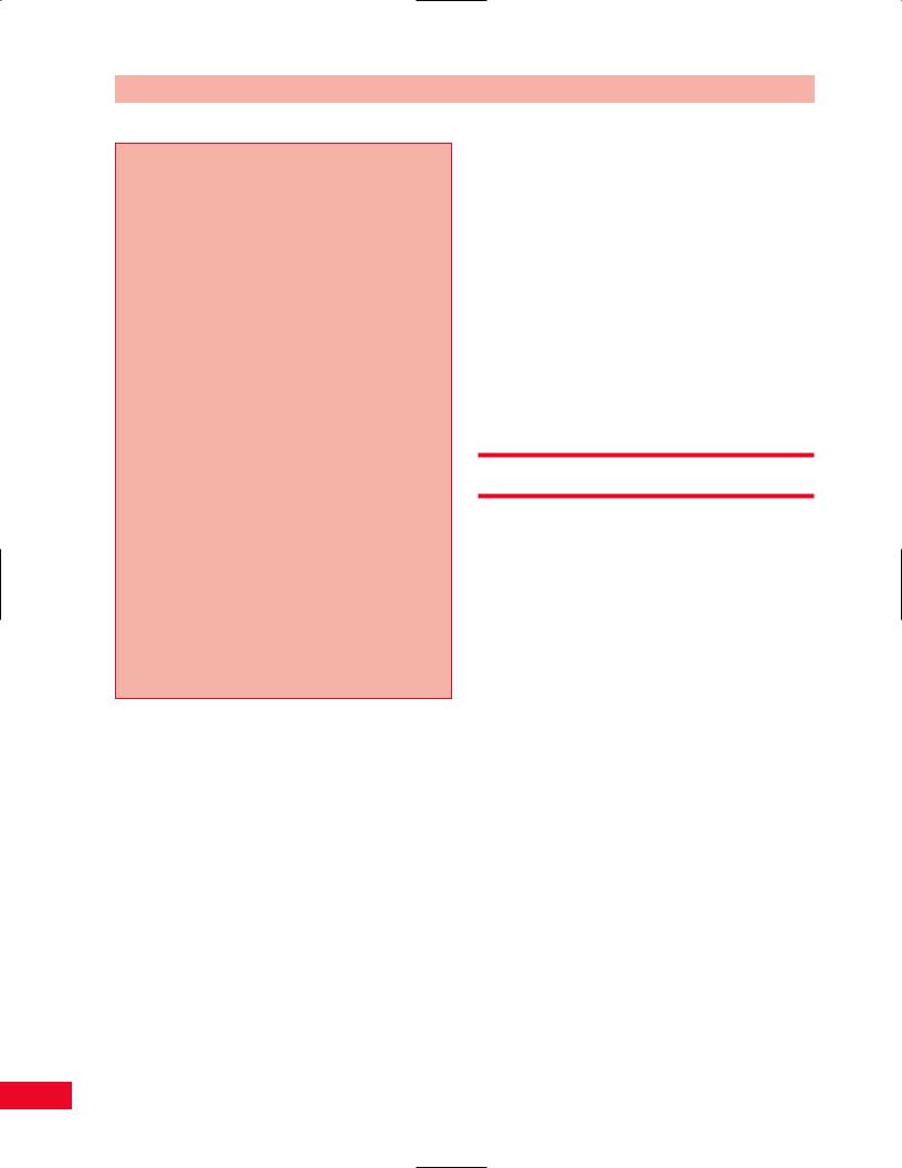
Suprasternal Notch
Stand facing the seated patient and use your middle or index finger to locate the triangular notch between the two clavicles. This is the suprasternal notch (Figure 8.6).
146
Suprasternal notch
Figure 8.6 Palpation of the suprasternal notch.
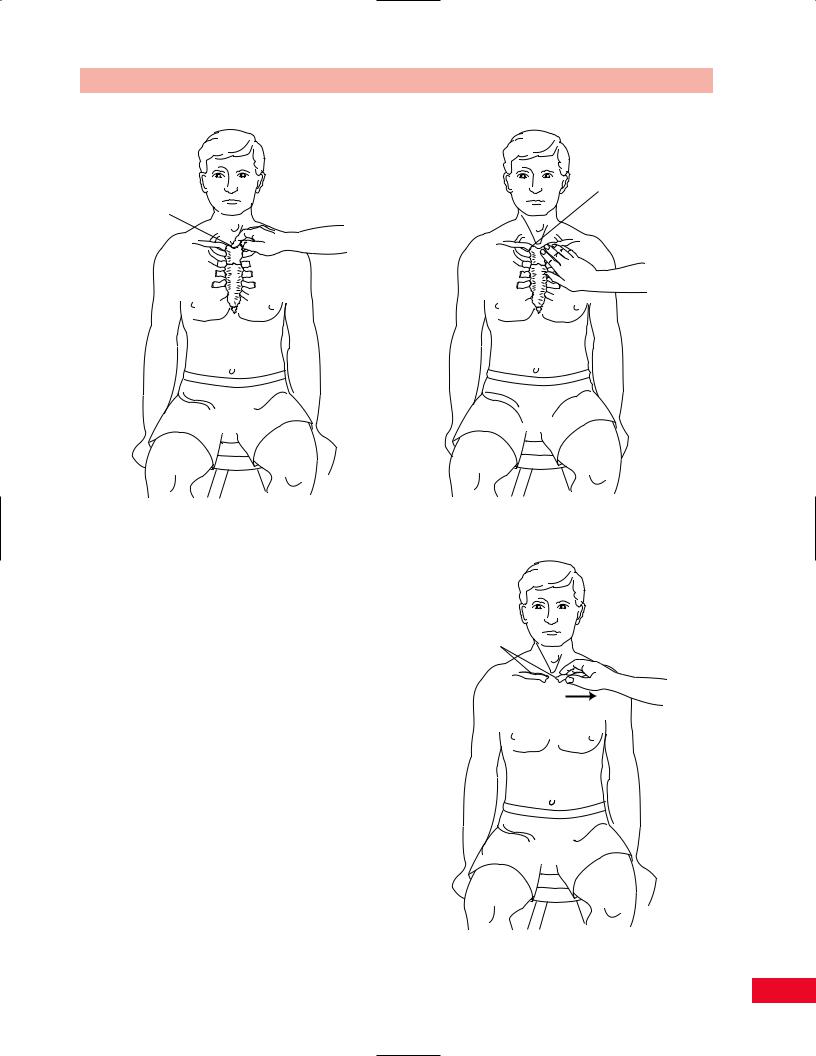
Sternoclavicular Joint
Move your fingers slightly superiorly and laterally from the center of the suprasternal notch until you feel the joint line between the sternum and the clavicle (Figure 8.7). The joints should be examined simultaneously to allow for comparison of heights and location. A superior and medial displacement of the clavicle may be indicative of dislocation of the sternoclavicular joint. You can get a better sense of the exact location and stability of the sternoclavicular joint by having the patient shrug the shoulders while you palpate the joint and the upward motion of the clavicles.
Clavicle
Continue to move laterally from the sternoclavicular joint along the superiorly and anteriorly curved bony surface of the clavicle. The bony surface should be smooth and continuous. Any area of increased prominence, sense of motion, crepitus, or pain along the shaft may be indicative of a fracture. In addition, the platysma muscle passes over the clavicle as it courses up the neck and can be palpated by having the patient strongly pull the corners of the mouth in a downward direction (Figure 8.8). The supraclavicular lymph
Chapter 8 The Shoulder
Sternoclavicular
joint
Figure 8.7 Palpation of the sternoclavicular joint.
Clavicle
Figure 8.8 Palpation of the clavicle.
147
The Shoulder Chapter 8
Acromioclavicular |
Acromion |
|
joint |
||
|
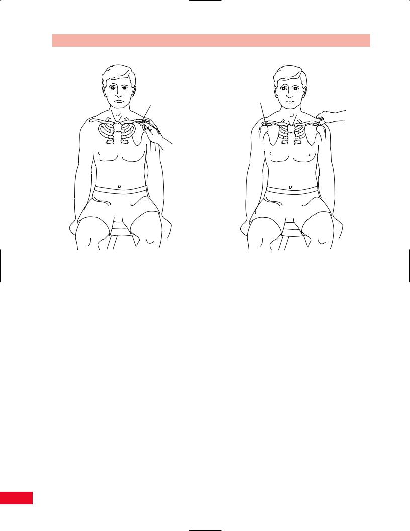
Figure 8.9 Palpation of the acromioclavicular joint. |
Figure 8.10 Palpation of the acromion process. |
nodes are found on the superior surface of the clavicle, lateral to the sternocleidomastoid. If you notice any enlargement or tenderness, an infection or malignancy should be suspected.
Acromioclavicular Joint
Continue to palpate laterally along the clavicle from the convexity to where it becomes concave to the most lateral aspect of the clavicle, just medial to the acromion. You will be able to palpate the acromioclavicular joint line where the clavicle is slightly superior to the acromion (Figure 8.9). You can get a better sense of its location by asking the patient to extend the shoulder while you palpate the movement at the acromioclavicular joint. The acromioclavicular joint is susceptible to osteoarthritis, crepitus, and tenderness, which can be noted with palpation. Pain with movement and swelling in the joint may be indicative of acromioclavicular joint subluxation. If the joint is severely traumatized, usually by a fall directly on the shoulder, a dislocation may occur and the clavicle may be displaced superiorly and posteriorly. The acromioclavicular joint is one area in which the pain is felt locally and is not referred.
Acromion Process
Palpate past the lateral aspect of the acromioclavicular joint and palpate the broad, flattened surface of the acromion between your index finger and thumb (Figure 8.10).
Greater Tuberosity of the Humerus
Allow your fingers to follow to the most lateral aspect of the acromion and they will drop off inferiorly onto the greater tuberosity of the humerus (Figure 8.11).
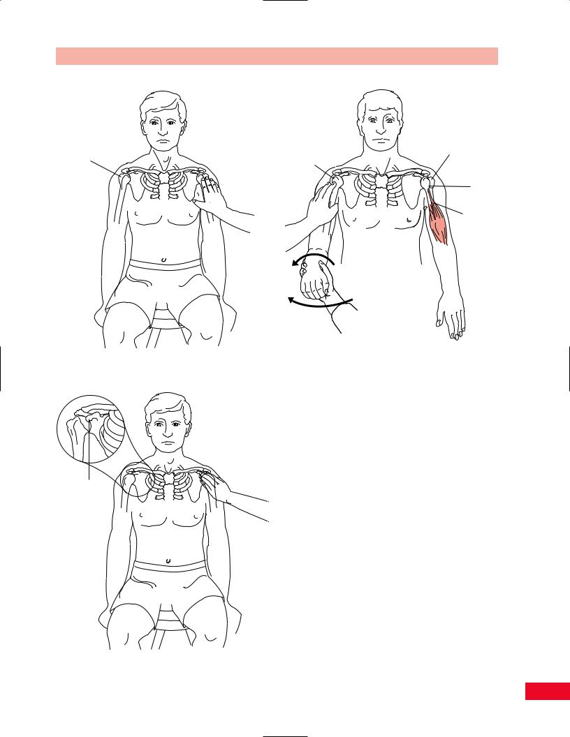
Coracoid Process
Move your fingers on a diagonal inferiorly and medially from the acromioclavicular joint. Gently place your middle finger deep into the deltopectoral triangle until you locate the bony prominence of the coracoid process, which is normally tender to palpation (Figure 8.12). The coracoid process is the attachment of the pectoralis minor, the coracobrachialis, and the short head of the biceps.
Bicipital Groove
Have the patient position the upper extremity at the side so that the arm is in midposition between internal
148
Chapter 8 The Shoulder
Greater tuberosity
Figure 8.11 Palpation of the greater tuberosity of the humerus.
Greater Lesser tuberosity tuberosity
Bicipital groove
Biceps tendon
Figure 8.13 Palpation of the bicipital groove.
Coracoid process
Figure 8.12 Palpation of the coracoid process.
and external rotation, and the forearm is in midposition between pronation and supination. Move your fingers laterally from the coracoid process, onto the lesser tuberosity of the humerus, and finally into the bicipital groove. The groove can be difficult to palpate if the patient has a hypertrophied deltoid. It may be helpful to locate the medial and lateral epicondyles of the humerus, making sure that they are in the frontal plane. Find the midpoint of the humerus and trace proximally to find the bicipital groove. The groove contains the tendon of the long head of the biceps and its synovial sheath and can therefore be tender to palpation. Ask the patient to internally rotate the arm and you will then feel your finger roll out of the groove and onto the greater tuberosity of the humerus (Figure 8.13).
Soft-Tissue Structures
Sternocleidomastoid
To facilitate palpating the sternocleidomastoid muscle, have the patient bend the neck toward the side you are palpating and simultaneously rotate away. This movement allows the muscle to be more prominent and therefore easier to locate. Palpate the distal attachments on the manubrium of the sternum and the medial
149
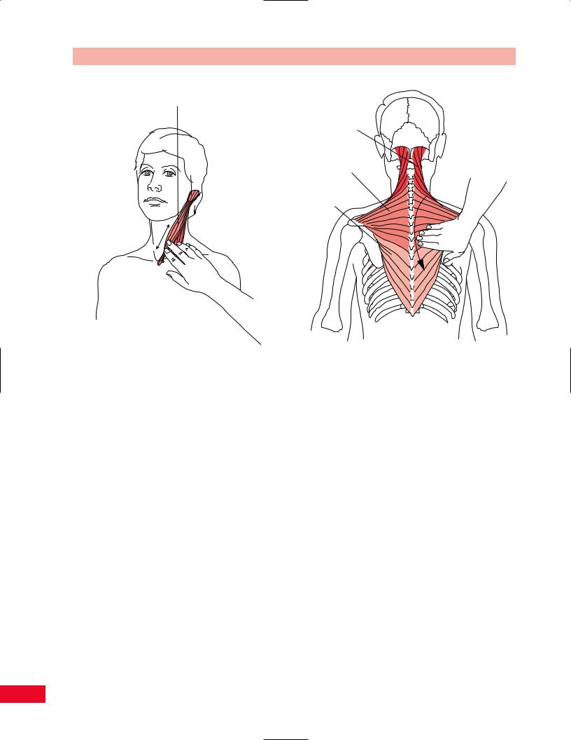
The Shoulder Chapter 8
Sternocleidomastoid muscle
Figure 8.14 Palpation of the sternocleidomastoid muscle.
Upper trapezius
Middle trapezius
Lower trapezius
Figure 8.15 Palpation of the trapezius muscle.
aspect of the clavicle and follow the muscle superiorly and laterally to its attachment on the mastoid process. The sternocleidomastoid is the anterior border of the anterior triangle of the neck and is a useful landmark for palpating enlarged lymph nodes (Figure 8.14).
Trapezius
Stand behind the seated patient. Differences in contour and expanse can be easily noted as you observe the patient prior to palpation. To enable you to palpate the fibers of the upper trapezius, allow your fingers to travel laterally and inferiorly from the external occipital protuberance to the lateral third of the clavicle. The muscle is a flat sheet but feels like a cordlike structure because of the rotation of the fibers. It is frequently tender to palpation and often very tight, secondary to tension or trauma. You can palpate the muscle using your thumb on the posterior aspect and your index and middle fingers anteriorly. The fibers of the lower trapezius can be traced as they attach from the medial aspect of the spine of the scapula, running medially and inferiorly to the spinous processes of the lower thoracic vertebrae. The fibers can be made more prominent by asking the patient to depress the scapula. The fibers of the middle trapezius can be palpated from
the acromion to the spinous processes of the seventh cervical and upper thoracic vertebrae. The muscle is made more prominent by asking the patient to adduct the scapulae (Figure 8.15).
Pectoralis Major
The pectoralis major is located on the anterior surface of the shoulder girdle. It is palpated from its attachment on the sternal aspect of the clavicle and along the sternum to the sixth or seventh rib, to its lateral attachment to the crest of the greater tubercle of the humerus. It creates the inferior aspect of the deltopectoral groove where it lies next to the deltoid muscle. The muscle forms the anterior wall of the axilla (Figure 8.16).
Deltoid
The deltoid has proximal attachments to the lateral clavicle, acromion, and spine of the scapula. The fibers then converge and insert onto the deltoid tuberosity of the humerus. The deltoid has a large rounded mass, creating the full contour of the shoulder (Figure 8.17). Atrophy can be caused by injury to the upper trunk of the brachial plexus or to the axillary nerve, following fracture or dislocation of the humerus. Start
150
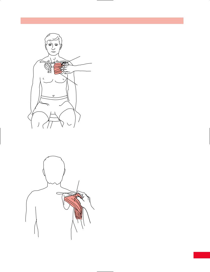
Deltopectoral
groove
Pectoralis
major
Figure 8.16 Palpation of the pectoralis major muscle.
Deltoid
Figure 8.17 Palpation of the deltoid muscle.
Chapter 8 The Shoulder
your examination by standing in front of the patient. Allow your hand to travel from the clavicle inferiorly and laterally as you feel the fullness of the muscle. Take note that the anterior fibers are superficial to the bicipital groove, making it difficult to distinguish whether tenderness in this area is from the muscle itself or the underlying structures. Continue by following the middle fibers from the acromion to the deltoid tuberosity. Note that the middle fibers overlie the subdeltoid bursa. If the patient has bursitis, careful examination of the area will help differentiate the tender structures. A neoplasm affecting the diaphragm or cardiac ischemia can refer pain to the deltoid.
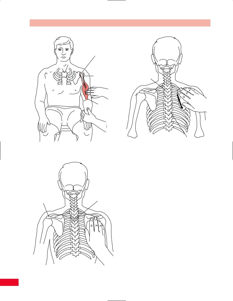
Biceps
Stand in front of the seated patient. Palpate the bicipital groove as described previously. Trace the long head of the biceps tendon inferiorly through the groove as it attaches to the muscle belly. Tenderness of the biceps tendon on palpation may be indicative of tenosynovitis. This is also a site for subluxation or dislocation of the biceps tendon. The tendon of the short head can be palpated on the coracoid process as previously described. The muscle belly is more prominent when the patient is asked to flex the elbow. The distal aspect of the belly and the biceps tendon can be palpated at its insertion on the bicipital tuberosity of the radius. Palpate for continuity of the belly and tendon (Figure 8.18). If a large muscle bulge is noted on the distal anterior aspect of the humerus with a concavity above it, you should suspect a rupture of the long head of the biceps. A subluxation of the biceps tendon secondary to a rupture of the transverse humeral ligament is known as a snapping shoulder.
Posterior Aspect
Bony Structures
Spine of the Scapula
Palpate the posterior aspect of the acromion medially along the ridge of the spine of the scapula as it tapers at the medial border. The spine of the scapula is located at the level of the spinous process of the third thoracic vertebra (Figure 8.19). The spine of the scapula separates the supraspinous and infraspinous fossae and serves as the attachment of the supraspinatus and infraspinatus muscles.
Medial (Vertebral) Border of the Scapula
Move superiorly from the medial aspect of the spine of the scapula until you palpate the superior angle,
151
The Shoulder Chapter 8
Tendon of short head of the biceps

 Tendon of long head of the biceps
Bicipital aponeurosis
Figure 8.18 Palpation of the biceps muscle.
Spine of scapula |
T3 |
Figure 8.19 Palpation of the spine of the scapula.
T2
T7
Figure 8.20 Palpation of the medial border of the scapula.
which is located at the level of the second thoracic vertebra. This area serves as the attachment of the levator scapulae and is often tender to palpation. In addition, it is frequently an area of referred pain from the cervical spine. Continue inferiorly along the medial border and note whether it lies flat on the rib cage. If the border wings away from the cage, it may be indicative of a long thoracic nerve injury. Notice the attachment of the rhomboideus major along the length of the medial border from the spine to the inferior angle. The inferior angle of the scapula is located at the level of the seventh thoracic vertebra and serves as the attachment of the latissimus dorsi and serratus anterior (Figure 8.20).
Lateral Border of the Scapula
Continue superiorly and laterally from the inferior angle along the lateral border of the scapula. The lateral border is less defined than the medial border because of the muscle attachments of the subscapularis anteriorly and the teres major and minor posteriorly. The attachment of the long head of the triceps can also be palpated on the infraglenoid tubercle, which is at the superior aspect of the lateral border (Figure 8.21).
152

Chapter 8 The Shoulder
Rhomboids
Inferior angle of scapula
Lateral border of scapula
Figure 8.21 Palpation of the lateral border of the scapula.
Figure 8.22 Palpation of the rhomboideus major and minor muscles.
Soft-Tissue Structures
Rhomboideus Major and Minor
The rhomboideus major originates from the spinous processes of T2–T5 and inserts on the medial border of the scapula between the spine and the inferior angle. The rhomboideus minor attaches from the ligamentum nuchae and the spinous processes of C7 and T1 to the medial border at the root of the spine of the scapula. Stand behind the seated patient. The muscles can be located at the vertebral border of the scapula. You can more easily distinguish the muscle by having the patient place the hand behind the waist and adduct the scapula (Figure 8.22).
Latissimus Dorsi
The latissimus dorsi attaches distally to the spinous processes of T6–T12, inferior three or four ribs, the inferior angle of the scapula, thoracolumbar fascia, iliac crest, and converges proximally to the intertubercular groove of the humerus. Palpation of the superior portion is discussed in the section on the posterior wall of the axilla. Continue to slide your hand along the muscle belly in an inferior and medial direction until
you reach the iliac crest. The fibers are much harder to differentiate as you move inferiorly (Figure 8.23).
Medial Aspect
Soft-Tissue Structures
Axilla
The axilla has been described as a pentagon (Moore and Dalley, 1999) created by the pectoralis major and minor anteriorly; the subscapularis, latissimus dorsi, and teres major posteriorly; the first four ribs with their intercostal muscles covered by the serratus anterior medially; and the proximal part of the humerus laterally. The interval between the outer border of the first rib, the superior border of the scapula, and the posterior aspect of the clavicle forms the apex. The axillary fascia and skin make up the base. To examine the axilla, face the seated patient. Support the patient’s abducted upper extremity by supporting the forearm with the elbow flexed. Allow your opposite hand to gently but firmly palpate. Remember that this area is especially ticklish to palpation. The axilla is clinically significant because it allows for passage of the
153

The Shoulder Chapter 8
Axillary lymph nodes
Brachial artery
Figure 8.24 Palpation of the axilla.
Figure 8.23 Palpation of the latissimus dorsi muscle.
brachial plexus and the axillary artery and vein to the upper extremity.
Allow your fingers to palpate the anterior wall and grasp the pectoralis major between your thumb, index and middle fingers. Move to the medial aspect of the axilla and palpate along the ribs and serratus anterior. Move superiorly into the axilla and gently pull the tissue inferiorly, rolling it along the rib cage, to palpate the lymph nodes. Normal lymph nodes should not be palpable in an adult. If palpable nodes are found, they should be noted since they are indicative of either an inflammation or a malignancy. Continue to palpate laterally and you will note the brachial pulse as you press against the proximal aspect of the humerus, located between the biceps and triceps muscles. Continue to palpate the posterior wall and grasp the latissimus dorsi between your thumb, index, and middle fingers. While palpating the muscles, pay attention to their tone and size. Note whether they are symmetrical bilaterally (Figure 8.24).
Serratus Anterior
The description of this palpation is found in the previous section on the axilla. The serratus anterior is important since it secures the medial border of the scapula
to the rib cage (Figure 8.25). Weakness or denervation will be observed as a winged scapula.
Lateral Aspect
Soft-Tissue Structures
Rotator Cuff
When the patient rests the arm at the side, the rotator cuff tendons are located under the acromial arch at the point of their attachment to the greater tubercle of the humerus. These tendons are referred to as the SIT muscles by virtue of the order of their attachment from anterior to posterior: supraspinatus, infraspinatus, and teres minor. The remaining muscle of the rotator cuff is the subscapularis and is not palpable in this position. To gain easier access to the tendons, ask the patient to bring the arm behind the waist in internal rotation and extension (Figure 8.26). You will be able to distinguish the tendons as a unit over the anterior aspect of the greater tubercle. If an inflammation is present, palpation of the tendons will cause pain.
Cyriax (1984) described a more specific method of palpation of the individual tendons. To locate the supraspinatus tendon, have the patient bend the elbow
154