

MusculoSkeletal Exam
.pdfUpper Lateral head pterygoid
muscle Lower head
Medial pterygoid muscle
Figure 5.18 Lateral and medial pterygoid muscles.
Masseter muscle (superficial)
Figure 5.19 Masseter.
Temporalis muscle
Coronoid process
Figure 5.20 Temporalis.
Chapter 5 The Temporomandibular Joint
mouth from the closed position as you resist their effort. Normally, the patient will be able to overcome maximal resistance.
Jaw Closing
The masseter (Figure 5.19) and temporalis (Figure 5.20) are the primary muscles that close the mouth. The medial pterygoid (Figure 5.18) assists them.
•Position of patient: Sitting, facing you.
•Resisted test: Ask the patient to close their mouth tightly and then attempt to open their jaw by pulling down on the mandible.
Reflex Testing
Jaw Jerk
The trigeminal (fifth cranial) nerve mediates the jaw reflex. This reflex results from contraction of the masseter and temporalis muscles following a tap on the chin (mandible). To perform the reflex, the patient should relax the jaw in the resting position, with the mouth slightly open. Place your index and long fingers under the lip, on the chin, and tap your fingers with the reflex hammer (Figure 5.21). A normal response is closure of the mouth. An exaggerated response indicates an upper motor neuron lesion. A reduced response indicates a trigeminal nerve disorder.
Figure 5.21 Jaw jerk: Make sure the patient is relaxed.
95

Chapter 6
The Lumbosacral
Spine
Lumbar vetebrae
Sacrum
Coccyx

Chapter 6 The Lumbosacral Spine
Please refer to Chapter 2 for an overview of the sequence of a physical examination. For purposes of length and to avoid having to repeat anatomy more than once, the palpation section appears directly after the section on subjective examination and before any section on testing, rather than at the end of each chapter. The order in which the examination is performed should be based on your experience and personal preference as well as the presentation of the patient.
Observation
The lumbar spine and sacroiliac joints are intimately related. They function together to support the upper body and transmit weight through the pelvis to the lower extremities. In addition they receive ground reaction forces through the lower extremities at the time of heel strike and through the stance phase of gait. A disruption of the balance of these forces can inflict an injury to either or both.
Observe the patient in your waiting room. Is the patient able to sit or is he or she pacing because sitting is too uncomfortable? If the patient is sitting, is he or she symmetrical or leaning to one side? This may be due to pain in the ischial tuberosity secondary to bursitis, sacroiliac dysfunction, or radiating pain from the low back. Pain may be altered by changes in position. Watch the patient’s facial expression to give you insight into their pain level.
Observe the patient as he or she assumes the standing position. How difficult is it for the patient to go from flexion to extension? Can the patient evenly distribute weight between both lower extremities? Observe the patient’s posture. Note any structural deformities such as kyphosis or scoliosis. Are the spinal curves normal, diminished, or exaggerated? Observe the patient’s total spinal posture from the head to the sacral base. It is also important to recognize the influence of the lower extremities. Observe any structural deviations in the hips, knees, and feet. Once the patient starts to ambulate, a brief gait analysis should be initiated. Note any gait deviations and whether the patient requires or is using an assistive device. Details and implications of deviations are discussed in the chapter on gait.
Subjective Examination
Inquire about the etiology of the patient’s symptoms. Was there a traumatic incident or did the pain develop insidiously? Is this the first episode or does the patient have a prior history of low back pain? Is the patient pregnant or has she recently delivered a baby? Is the patient’s symptomatology related to her menstrual cycle? Pregnancy and menstruation influence the degree of ligamentous laxity, making the patient more susceptible to injury. Is the pain constant or intermittent? Can the pain be altered by position? What exaggerates or alleviates the patient’s complaints? Does coughing, sneezing, or bearing down increase the symptoms? Increased pain with increased intra-abdominal pressure may be secondary to a spaceoccupying lesion such as a tumor or a herniated disc. How easily is the patient’s condition irritated and how quickly can the symptoms be relieved? Your examination may need to be modified if the patient reacts adversely with very little activity and requires a long time for relief.
The patient’s disorder may be related to age, gender, ethnic background, body type, static and dynamic posture, occupation, leisure activities, hobbies, and general activity level. It is important to inquire about any change in daily routine and any unusual activities in which the patient may have participated. If an incident occurred, the details of the mechanism of injury are important to help direct your examination.
You should inquire about the nature and location of the complaints as well as the duration and intensity of the symptoms. The course of the pain during the day and night should be addressed. The location of the symptoms may give you some insight into the etiology of the complaints. Pain, numbness, or tingling that is located over the anterior and lateral part of the thigh may be referred from L3 or L4. Pain into the knee may be referred from L4 or L5 or from the hip joint. The patient may complain about pain over the lateral or posterior aspect of the greater trochanter, which may be indicative of trochanteric bursitis or piriformis syndrome. Note any pain or numbness in the saddle (perineal) area. This may be indicative of radiation from S2 and S3. Inquire about any changes in bowel, bladder, and sexual function. Alteration of these functions may be indicative of sacral plexus problems. (Please refer to Box 2.1, p. 18 for typical questions for the subjective examination.)
97
The Lumbosacral Spine Chapter 6
Paradigm for a neoplasm of the lumbar spine
A 65-year-old bank executive presents with acute pain in the mid low back region. There has been a slow insidious increasing discomfort for the past 3 months which has become severe during the past week. There has been no history of trauma. He describes his pain as being worse at night and relieved with standing. His pain is not made worse by coughing, sneezing, or straining during a bowel movement.
On a physical examination, the patient appears to be in mild discomfort while seated. He is independent in transfer to and from the exam table and dressing. He has no tension signs with straight leg raise, reflexes are equal bilaterally, as is strength. There is pain on percussion over the mid lumbar spinous processes. X-rays suggest an absence of the right pedicle of the third lumbar vertebrae.
This paradigm is characteristic of a spinal neoplasm because of: No history of a trauma
Pain at rest, relieved on standing No evidence of nerve involvement
Gentle Palpation
The palpatory examination starts with the patient standing. This allows you to see the influence of the lower extremities on the trunk in the weight-bearing position. If the patient has difficulty standing, he or she may sit on a stool with their back toward you. The patient must be sufficiently disrobed so that the entire back is exposed. You should first search for areas of localized effusion, discoloration, birthmarks, open sinuses or drainage, and incisional areas. Note the bony contours and alignment, muscle girth and symmetry, and skinfolds. A café au lait spot or a “faun’s” beard may be indicative of spina bifida occulta or neurofibromatosis. Remember to use your dominant eye when checking for alignment or symmetry. Failure to do this can alter your findings. You should not have to use deep pressure to determine areas of tenderness or malalignment. It is important to use a firm but gentle pressure, which will enhance your palpatory skills. If you have a sound basis of cross-sectional anatomy, it should not be necessary to physically penetrate through several layers of tissue to have a good sense of the underlying structures. Remember that if you increase the patient’s pain at this point in the examination, the patient will be very reluctant to allow you to continue, or may become more limited in his or her ability to move.
Palpation is most easily performed with the patient in a relaxed position. Although the initial palpation
can be performed with the patient standing or sitting, the supine, side-lying, and prone positions may also be used to allow for easier access to the bony and softtissue structures.
Posterior Aspect
Bony Structures
Iliac Crest
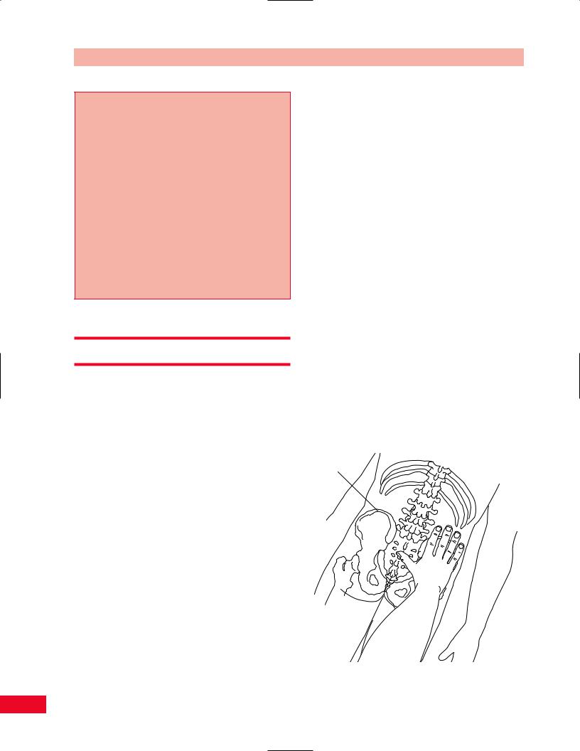
The iliac crest is very prominent since it is so superficial and is therefore easy to palpate. Place your extended hands so that the index fingers are at the waist. Allow your hands to press medially and rest on the superior aspect of the ridge of the crests. Then place your thumbs on the lumbar spine in line with the fingers on the crests. The L4–L5 vertebral interspace is located at this level. This is a useful starting landmark when palpating the lumbar spinous processes (Figure 6.1).
Iliac crests that are uneven in height may occur secondary to a leg-length difference, a pelvic obliquity, or a sacroiliac dysfunction.
Spinous Processes
The spinous processes of the lumbar spine are quadrangular in shape and are positioned in a horizontal fashion just posterior to the vertebral body. Locate the posterior superior iliac spine and allow your finger to drop off in the medial and superior direction at a
Iliac crest
Figure 6.1 Palpation of the iliac crest.
98
Chapter 6 The Lumbosacral Spine
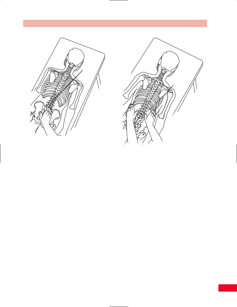
Spinous processes
Transverse
processes
Figure 6.2 Palpation of the spinous processes.
Figure 6.3 Palpation of the transverse processes.
30-degree angle. You will locate the spinous process of L5. Another consistent method of locating the vertebra is by placing your hands on the iliac crest and moving medially where you will find the vertebral interspace of L4–5. You can count up the spinous processes from either of these starting points. You can locate the spinous process of L1 by locating the twelfth rib and moving your hand medially and down one level. If you choose this method, then locate the remaining spinous processes by counting down to L5 (Figure 6.2).
Tenderness or a palpable depression from one level to another may indicate the absence of the spinous process or a spondylolisthesis.
Transverse Processes
The transverse processes of the lumbar spine are long and thin and are positioned in a horizontal fashion. They vary in length, with L3 being the longest and L1 and L5 the shortest. The L5 transverse process is most easily located by palpating the posterior superior iliac spine and moving medially and superiorly at a 30to 45-degree angle. The transverse processes are more difficult to palpate in the lumbar spine because of the thickness of the overlying tissue. They are most easily identified in the trough located between the spinalis and the longissimus muscles (Figure 6.3).
Posterior Superior Iliac Spines
The posterior superior iliac spines (PSIS) can be found by placing your extended hands over the superior aspect of the iliac crests and allowing your thumbs to reach diagonally in an inferior medial direction until they contact the bony prominence. Have your thumbs roll so that they are under the PSISs and are directed cranially to more accurately determine their position. Many individuals have dimpling that makes the location more obvious. However, you should be careful because dimpling is not present in all individuals, and if it is present, it may not coincide with the posterior superior iliac spines. If you move your thumbs in a medial and superior angle of approximately 30 degrees, you will come in contact with the posterior arch of L5. If you move your thumbs at a caudad and inferior angle of approximately 30 degrees, you will come in contact with the base of the sacrum. If you are having difficulty, you may also locate the posterior superior iliac spines by following the iliac crests posteriorly and then inferiorly until you arrive at the spines (Figure 6.4).
Sacroiliac Joint
The actual joint line of the sacroiliac joint is not palpable because it is covered by the posterior aspect of the
99

The Lumbosacral Spine Chapter 6
Sacroiliac |
|
Sacral base |
|
joint |
L4 |
||
|
|||
|
|
||
|
L5 |
Sacrum |
|
|
S2 |
|
Figure 6.4 Palpation of the posterior superior iliac spine. |
Figure 6.6 Palpation of the sacral base. |
L5
PSIS |
S2 Spinous |
|
process |
Figure 6.5 Palpation of the sacroiliac joint.
innominate bone. You can get a sense of its location by allowing your thumb to drop off medially from the posterior superior iliac spine. The sacroiliac joint is located deep to this overhang at approximately the second sacral level (Figure 6.5).
Sacral Base
Locate the posterior superior iliac spines on both sides (described above). Allow your thumbs to drop off medially and then move anteriorly until you contact the sacral base. This area is also referred to as the sacral sulcus (Figure 6.6). Palpation of the sacral base is useful in determining the position of the sacrum.
Inferior Lateral Angle
Place your fingers on the inferior midline of the posterior aspect of the sacrum and locate a small vertical depression, this is the sacral hiatus. Move your fingers laterally approximately 3/4 in. and you will be on the inferior lateral angle (Figure 6.7).
Ischial Tuberosity
You can place you thumbs under the middle portion of the gluteal folds at approximately the level of the greater trochanters. Allow your thumb to face
100

Sacral hiatus
Inferior lateral angle
Figure 6.7 Palpation of the inferior lateral angle.
Ischial tuberosity
Figure 6.8 Palpation of the ischial tuberosity.
Chapter 6 The Lumbosacral Spine
Coccyx
Figure 6.9 Palpation of the coccyx.
superiorly and gently probe through the gluteus maximus until the thumb is resting on the ischial tuberosity. Some people find it easier to perform this palpation with the patient lying on the side with the hip flexed, allowing the ischial tuberosity to be more accessible since the gluteus maximus is pulled up, reducing the muscular cover (Figure 6.8). If this area is tender to palpation, it may be indicative of an inflammation of the ischial bursa or an ischiorectal abscess.
Coccyx
The tip of the coccyx can be found in the gluteal cleft. To palpate the anterior aspect, which is essential to determine the position, a rectal examination must be performed (Figure 6.9). Pain in the coccyx is referred to as coccydynia and is usually secondary to direct trauma to the area.
Soft-Tissue Structures
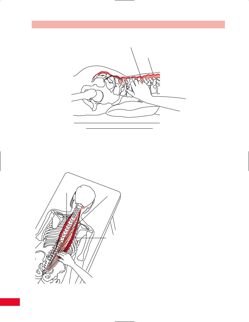
Supraspinous Ligament
The supraspinous ligament joins the tips of the spinous processes from C7 to the sacrum. This powerful fibrous cord that is blended with the fascia is denser and wider in the lumbar than in the cervical and thoracic spines.
101
The Lumbosacral Spine Chapter 6
Interspinous ligament
Supraspinous ligament
Figure 6.10 Palpation of the supraspinous ligament.
The ligament can be palpated by placing your fingertip between the spinous processes. The tension of the ligament is more easily noted if the patient is in a slight degree of flexion (Figure 6.10).
Spinalis
Longissimus
Iliocostalis
Figure 6.11 Palpation of the erector spinae muscles.
Erector Spinae (Sacrospinalis) Muscle
The erector spinae muscle forms a thick fleshy mass in the lumbar spine. The intermediate muscles of the group are the spinalis (most medial), longissimus, and iliocostalis (most lateral) muscles. They are easily palpated just lateral to the spinous processes. Their lateral border appears to be a groove (Figure 6.11). This muscle is often tender and in spasm in patients with an acute low-back pain.
Quadratus Lumborum Muscle
Place your hands over the posterior aspect of the iliac crest. Press medially in the space below the rib cage and you will feel the tension of the quadratus lumborum as it attaches to the iliolumbar ligament and the iliac crest (Figure 6.12). The muscle can be made more distinct by asking the patient to lift the pelvis towards the thorax. The quadratus lumborum is important in the evaluation of the lumbar spine. It can adversely affect alignment and muscle balance because of its attachment to the iliolumbar ligament. It can also play a role in changing pelvic alignment because of its intimate relationship to the iliac crest.
Sacrotuberous Ligament
Place the patient in the prone position and locate the ischial tuberosities as described above. Allow your thumbs to slide off in a medial and superior direction. You will feel a resistance against your thumbs, which is the sacrotuberous ligament (Figure 6.13).
102

Chapter 6 The Lumbosacral Spine
Quadratus lumborum
Figure 6.12 Palpation of the quadratus lumborum muscle.
Sacrospinus ligament
Sacrotuberous ligament
Figure 6.13 Palpation of the sacrotuberous ligament.
103
The Lumbosacral Spine Chapter 6
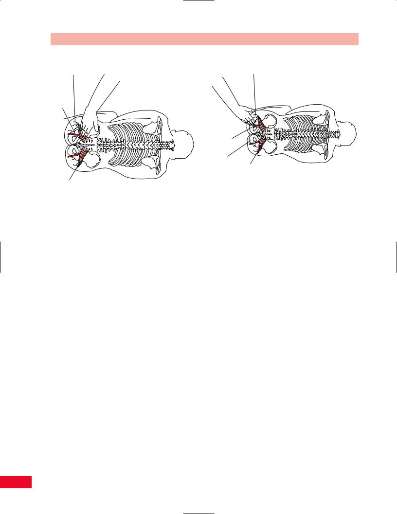
Greater trochanter
Sciatic nerve
Piriformis muscle
Figure 6.14 Palpation of the piriformis muscle.
Greater trochanter
Sciatic nerve
Ischial tuberosity
Piriformis muscle
Figure 6.15 Palpation of the sciatic nerve.
Side-Lying Position
Soft-Tissue Structures
Piriformis Muscle
The piriformis muscle is located between the anterior inferior aspect of the sacrum and the greater trochanter. This muscle is very deep and is normally not palpable. However, if the muscle is in spasm, a cordlike structure can be detected under your fingers as you palpate the length of the muscle (Figure 6.14). Because of its attachment to the sacrum, the piriformis is able to influence the alignment of the sacrum by pulling it anteriorly. The sciatic nerve runs either under, over, or through the muscle belly. Compression or irritation of the nerve can occur when the muscle is in spasm.
Sciatic Nerve
The sciatic nerve is most easily accessed while the patient is lying on the side, which allows the nerve to have less muscle cover since the gluteus maximus is flattened. Locate the midposition between the ischial tuberosity and greater trochanter. The nerve usually travels under the piriformis muscle, but in some patients it pierces the muscle. You may be able to roll the nerve under your fingers if you take up the softtissue slack. Tenderness in this area can be due to an irritation of the sciatic nerve secondary to lumbar disc disease or a piriformis spasm (Figure 6.15).
Anterior Aspect
Bony Structures
Anterior Superior Iliac Spine
Place your hands on the iliac crests and allow your thumbs to reach anteriorly and inferiorly, on a diagonal, toward the pubic ramus. The most prominent protuberance is the anterior superior iliac spine (ASIS). Roll the pads of your thumbs in a cranial direction so that they can rest under the anterior superior iliac spines, to determine their position most accurately. This area is normally superficial but can be obscured in an obese patient. Differences in height from one side to the other may be due to an iliac rotation or shear (Figure 6.16).
Pubic Tubercles
The patient should be in the supine position. Stand so that you face the patient and start the palpation superior to the pubic ramus. Place your hands so that your middle fingers are on the umbilicus and allow your palms to rest over the abdomen. The heel of your hand will be in contact with the superior aspect of the pubic tubercles. Then move your finger pads directly over the tubercles to determine their relative position. They are located medial to the greater trochanters and the inguinal crease. Make sure that your dominant eye is in the midline. The tubercles are normally tender to palpation. If they are asymmetrical either in height or in an anterior–posterior dimension, there may be a subluxation or dislocation, or a sacroiliac dysfunction (Figure 6.17).
104