

7 Mediastinum
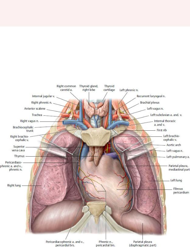
The mediastinum is the region within the thorax between the right and left pulmonary cavities (Fig 7.1; see Table 5.1). The ster-num and the costal cartilages of ribs 1 through 7 form its anterior boundary, and the thoracic vertebrae form its posterior boundary. The mediastinum contains the heart, great vessels, and pericar-dium, as well as the esophagus, trachea, thymus, and associated neurovasculature (Fig. 7.2).
Fig. 7.1 Thoracic cavity
Opened thoracic cavity. Removed: Thoracic wall; connective tissue of anterior mediastinum. (From Schuenke M, Schulte E, Schumacher U. THIEME Atlas of Anatomy, Vol 2. Illustrations by Voll M and Wesker K. 3rd ed. New York: Thieme Publishers; 2020.)
7.1 Regions of the Mediastinum (Table 7.1)
—A horizontal plane passing through the sternal angle (at the T4–T5 intervertebral disk) divides the region into a superior mediastinum, bounded above by the superior thoracic aperture, and an inferior mediastinum limited inferiorly by the thoracic diaphragm.
—The inferior mediastinum is further divided into
•the anterior mediastinum, a narrow space posterior to the sternum and anterior to the pericardium;
•the middle mediastinum, the largest section of the inferior mediastinum, which contains the pericardium, heart, and major vessels; and
•the posterior mediastinum, a small area posterior to the pericardium and anterior to the 5th through 12th thoracic vertebrae.
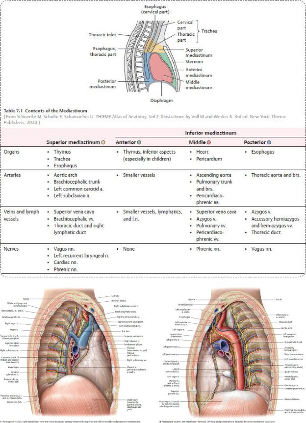
Fig. 7.2 Mediastinum
Divisions of the mediastinum. (From Schuenke M, Schulte E, Schumacher U. THIEME Atlas of Anatomy, Vol 2. Illustrations by Voll M and Wesker K. 3rd ed. New York: Thieme Publishers; 2020.) (From Gilroy AM, MacPherson BR, Wikenheiser JC. Atlas of Anatomy. Illustrations by Voll M and Wesker K. 4th ed. New York: Thieme Publishers; 2020.)
7.2 Anterior Mediastinum
Thymus
The thymus is a gland of the immune system, responsible for mat-uration of T- lymphocytes (Fig. 7.3).
—In childhood, the thymus presents as a large bilobed organ that overlies the heart and great vessels in the superior and anterior mediastinum.
—At puberty, high levels of circulating sex hormones cause the gland to atrophy.
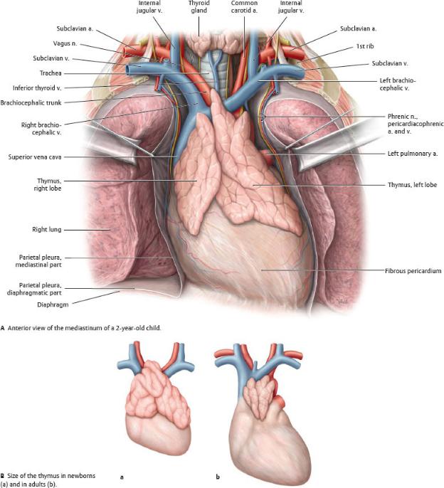
Fig. 7.3 The thymus in newborns and adults
The thymus of newborns is much larger than those of adults and extends inferiorly into the anterior part of the inferior mediastinum. Because it atrophies during puberty, the thymus in adults is small and extends only into the superior mediastinum. (From Schuenke M, Schulte E, Schumacher U. THIEME Atlas of Anatomy, Vol 2. Illustrations by Voll M and Wesker K. 3rd ed. New York: Thieme Publishers; 2020.)
7.3 Middle Mediastinum: Pericardium and
Pericardial Cavity
Pericardium
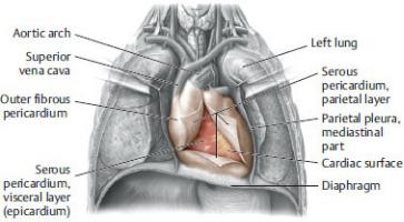
The pericardium, a double-layered fibroserous membrane, forms the pericardial sac that surrounds the heart and the origins of the great vessels
(Figs. 7.4 and 7.5).
—The pericardium is composed of two layers: an outer fibrous layer and an inner serous layer.
1.The outer fibrous pericardium is composed of tough inelastic connective tissue. It is attached inferiorly to the diaphragm and is continuous superiorly with the tunica adventitia (outer layer) of the great vessels.
2.The thin serous pericardium consists of parietal and visceral layers
◦The parietal layer of serous pericardium lines the inner surface of the fibrous pericardium.
◦The visceral layer of serous pericardium firmly adheres to the outer surface of the heart as the epicardium. This layer is continuous with the parietal layer of the serous pericardium at the root of the great vessels.
—Pericardiacophrenic arteries, branches of the internal thoracic arteries, provide the main blood supply to the pericardium. Veins that accompany the arteries drain into the superior vena cava.
—The vagus (cranial nerve X) and phrenic nerves (C3–C5) and branches from the sympathetic trunks innervate the pericardium.
—Pericardial pain is often referred via the phrenic nerve to the skin of the ipsilateral supraclavicular region (dermatomes C3–C5).
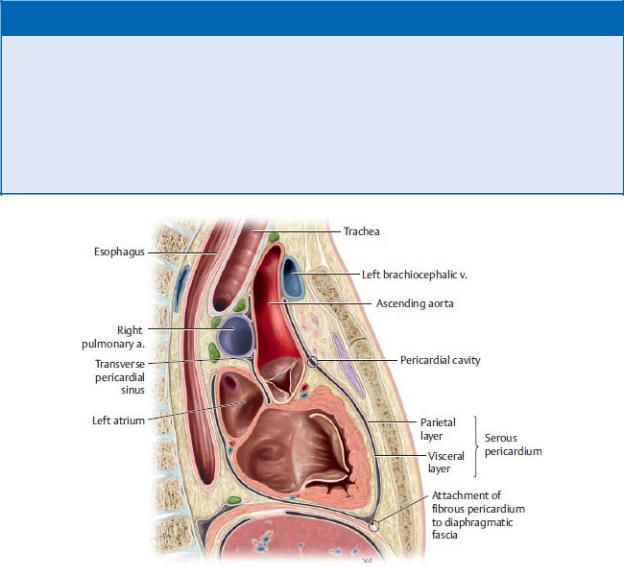
Fig. 7.4 Pericardium
Anterior view of opened thorax. Removed: Thymus. Reflected: Flaps of the fibrous pericardium. (From Gilroy AM, MacPherson BR, Wikenheiser JC. Atlas of Anatomy. Illustrations by Voll M and Wesker K. 4th ed. New York: Thieme Publishers; 2020.)
BOX 7.1: CLINICAL CORRELATION
PERICARDITIS
Pericarditis is an inflammation of the pericardium, which causes sharp, retrosternal or epigastric pain and a characteristic pericardial friction rub (a sound like the rustle of silk) that is heard on auscultation. This is caused by friction from the roughened layers of the inflamed pericardium rubbing together. Pericarditis can lead to pericardial effusion (fluid in the pericardial cavity) or cardiac tamponade (abnormal accumulation of fluid in the pericardial cavity that prevents venous return to the heart) and may be accompanied by dyspnea (shortness of breath) and peripheral edema (swelling).
Fig. 7.5 Serous pericardial reflections
Sagittal section through the mediastinum. Note the continuity of the parietal serous and visceral serous pericardia. (From Schuenke M, Schulte E, Schumacher U. THIEME Atlas of Anatomy, Vol 2. Illustrations by Voll M and Wesker K. 3rd ed. New York: Thieme Publishers; 2020.)
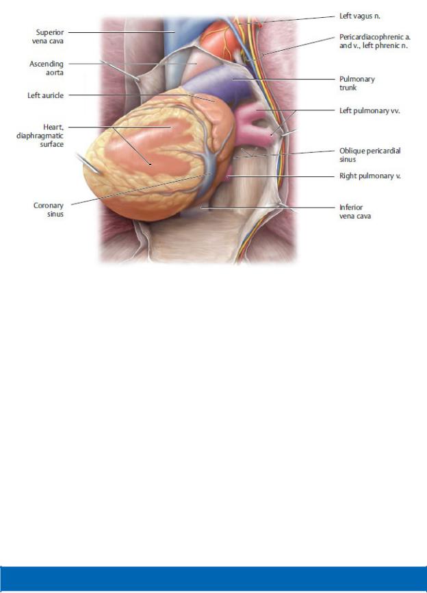
Fig. 7.6 Posterior pericardial cavity
Anterior view. The heart has been elevated to partially visualize the posterior pericardial cavity and the oblique pericardial sinus. (From Gilroy AM, MacPherson BR, Wikenheiser JC. Atlas of Anatomy. Illustrations by Voll M and Wesker K. 4th ed. New York: Thieme Publishers; 2020.)
The Pericardial Cavity
The pericardial cavity is the space within the pericardial sacbetween the parietal and visceral layers of the serous pericardium (Figs. 7.6 and 7.7).
—The pericardial cavity is filled with a thin layer of serous fluid that allows for frictionless movement of the heart.
—Two pericardial recesses form where the serous pericardium reflects around the roots of the great vessels:
1.The transverse pericardial sinus is a passage between the inflow channels (superior vena cava and pulmonary veins) and the outflow channels (aorta and pulmonary trunk) of the heart.
2.The oblique pericardial sinus is a recess of the pericardial cavity posterior to the heart between the right and left pulmonary veins.
BOX 7.2: CLINICAL CORRELATION
CARDIAC TAMPONADE
Cardiac tamponade is a life-threatening condition in which fluid collects in the pericardial space. The increased intrapericardial pressure restricts the filling of the heart during diastole and reduces cardiac output but increases the heart rate. Inhibition of venous return to the heart causes peripheral edema, hepatomegaly (enlarged liver), and increased venous pressure (often noted by distension of the internal jugular vein). Pericardiocentesis, aspiration of the fluid from the pericardial space, can relieve the tamponade.
Fig. 7.7 Pericardial recesses Posterior pericardium, anterior view. Removed: Anterior pericardium and heart. Revealed: The posterior pericardium and the double-headed arrow indicates the course of the transverse pericardial sinus, the passage between the reflections of the serous layer of the pericardium around the arterial and venous great vessels of the heart. (From Schuenke M, Schulte E, Schumacher U. THIEME Atlas of Anatomy, Vol 2. Illustrations by Voll M and Wesker K. 3rd ed. New York: Thieme Publishers; 2020.)
BOX 7.3: CLINICAL CORRELATION
SURGICAL SIGNIFICANCE OF THE TRANSVERSE PERICARDIAL SINUS
During cardiac surgery, the surgeon is able to isolate (and clamp) the heart’s outflow tracts, the aorta and pulmonary trunk, from its inflow tracts, the superior vena cava and pulmonary veins, by passing the clamps through the transverse pericardial sinus.
7.4 Middle Mediastinum: The Heart
General Features
— The heart is a hollow muscular organ located in the middle mediastinum

within the pericardial sac. It rests on the central tendon of the diaphragm and is flanked on either side by the right and left pulmonary cavities (Fig. 7.8).
—The heart has a conical shape. The base, anchored by the great vessels, is on its superior and posterior surfaces. The apex, located approximately at the 5th intercostal space, projects anteriorly, inferiorly, and to the left and moves freely within the pericardial sac.
—Internally, the heart is divided into four chambers: the right and left atria and the right and left ventricles.
Fig. 7.8 Heart in situ
Anterior view of the opened thorax. Removed: Thymus and anterior pericardium. Revealed: Heart. (From Schuenke M, Schulte E, Schumacher U. THIEME Atlas of Anatomy, Vol 2. Illustrations by Voll M and Wesker K. 3rd ed. New York: Thieme Publishers; 2020.)
•The right and left atria, separated by an interatrial septum, are the inflow chambers of the heart, receiving blood from the systemic circulation on the right and pulmonary circulation on the left.
•The right and left ventricles, separated by an interventric-ular septum, are the outflow chambers of the heart. Blood flows from the right ventricle into the pulmonary circula-tion and from the left ventricle into the systemic circulation.
—Two small appendages, the right and left auricles, are extensions of the atria and are visible externally.
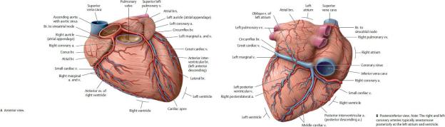
—The surfaces of the heart (Fig. 7.9) are
•the sternocostal surface on the anterior side of the heart, formed mostly by the right ventricle with portions of the right atrium and left ventricle;
•the base on the posterior and superior sides of the heart, formed by the left atrium and a portion of the right atrium; and
•the diaphragmatic surface on the inferior side of the heart, formed by the left and right ventricles.
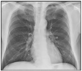
—The borders of the heart define the cardiac shadow that is seen on radiographic images (Table 7.2).
—Three grooves on the external surface of the heart can be used to determine the position of the chambers:
1. The coronary sulcus encircles the heart between the atria and ventricles. Because the heart has an oblique orienta-tion, the sulcus is nearly vertical.
2. The anterior interventricular sulcus is a longitudinal groove on the anterior surface of the heart that marks the position of the interventricular septum.
3. The posterior interventricular sulcus is a longitudinal groove on the diaphragmatic surface of the heart that marks the position of the interventricular septum.
—The crux of the heart is a point on the posterior surface of the heart where the coronary (atrioventricular) and interventric-ular sulci meet. It marks the junction of the four chambers of the heart.
—The wall of the heart consists of three layers:
1. The epicardium, the thin outermost layer, formed by the visceral layer of the serous pericardium
2. The myocardium, the thick layer of cardiac muscle, thickest in the walls of the ventricles
3. The endocardium, the thin internal layer, which lines the chambers and valves of the heart
—A cardiac skeleton of dense fibrous connective tissue forms four fibrous anuli (rings) and intervening trigones that separate the chambers of the heart, provide anchoring points for cardiac muscle fibers and cardiac valves, and insulate electrical impulses of the heart’s conduction system (Fig. 7.10).
Table 7.2 Borders of the Heart |
|
|
|
|
|
Border |
Defining Structures |
|
Right cardiac border |
• |
Right atrium |
|
• |
Superior vena cava |
Apex |
• |
Left ventricle |
|
|
|
Left cardiac border |
• |
Aortic arch (“aortic knob”) |
|
• |
Pulmonary trunk |
|
• |
Left atrium |
|
• |
Left ventricle |
Inferior cardiac border |
• |
Left ventricle |
|
• |
Right ventricle |
Radiographic appearance of the heart
(From Gunderman R. Essential Radiology, 3rd ed. New York: Thieme Publishers; 2014.)
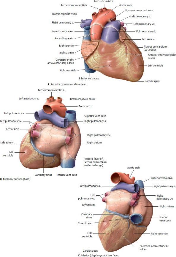

Fig. 7.9 Surfaces of the heart
The heart has three surfaces: anterior (sternocostal), posterior (base), and inferior (diaphragmatic). (From Schuenke M, Schulte E, Schumacher U. THIEME Atlas of Anatomy, Vol 2. Illustrations by Voll M and Wesker K. 3rd ed. New York: Thieme Publishers; 2020.)
Fig. 7.10 Cardiac skeleton
Superior view. Red dotted circles indicate attachment sites of papillary muscles on valves. (From Schuenke M, Schulte E, Schumacher U. THIEME Atlas of Anatomy, Vol 2. Illustrations by Voll M and Wesker K. 3rd ed. New York: Thieme Publishers; 2020.)
The Atria (Figs. 7.11 and 7.12)
The atria are the thin-walled inflow chambers of the heart.
—The right atrium receives the superior and inferior venae cavae from the systemic circulation and the cardiac veins from the heart. The left atrium receives the pulmonary veins from the lungs.
—Each atrium is associated with an auricle, a small pouch that expands the capacity of the atrium and whose roughened walls contain pectinate muscles.
—A depression on the right ride of the interatrial septum, the oval fossa (fossa ovalis), is a remnant of the oval foramen (foramen ovale), an opening through which blood was shunted from the right to left atria in the prenatal circulation.
—The right atrium is divided into two parts by a muscular ridge, the terminal crest (crista terminalis). The ridge is evident on the outside of the heart as the terminal sulcus. The two parts of the right atrium are
1. the venous sinus (sinus venarum), a smooth-walled region on the
posterior wall that contains the openings of the superior vena cava, inferior vena cava, coronary sinus, and anterior cardiac veins; and
2.the atrium proper, the anterior muscular portion that, like the right auricle, contains pectinate muscles.
—The left atrium is smaller but thicker walled than the right atrium and receives the four to five pulmonary veins from the lungs. The atrial walls are smooth, with the pectinate muscles confined to the left auricle.
The Ventricles (Figs. 7.11 and 7.12)
The ventricles are thick-walled chambers that connect to the out-flow channels of the heart: the right ventricle to the pulmonary artery and the left ventricle to the aorta.
—The walls of the ventricles are marked with a meshwork of thick muscular ridges known as trabeculae carneae.
—Most of the interventricular septum is muscular, but there is a small membranous part at the superior end that is a common site of septal defects.
—The right ventricle is the smaller and thinner walled of the two ventricles. A muscular ridge, the supraventricular crest, separates it into two parts:
1.The right ventricle proper, the inflow portion of the ventricle that receives blood from the right atrium
◦An anterior and a posterior papillary muscle arise from its floor, and a septal papillary muscle arises from the interventricular septum.
◦A muscular septomarginal trabecula (moderator band) extends from the septum to the base of the anterior papillary muscle and carries a part of the electrical conduction system (the right branch of the atrioventricular bundle) that facilitates the coordinated contraction of the papillary muscle.
2.The conus arteriosus (infundibulum), the smooth-walled outflow channel through which blood flows into the pulmonary trunk
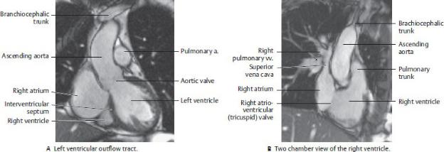
Fig. 7.11 MRI of the Heart
(From Moeller TB, Reif E. Pocket Atlas of Sectional Anatomy, Vol 2, 3rd ed. New York: Thieme; 2007.)
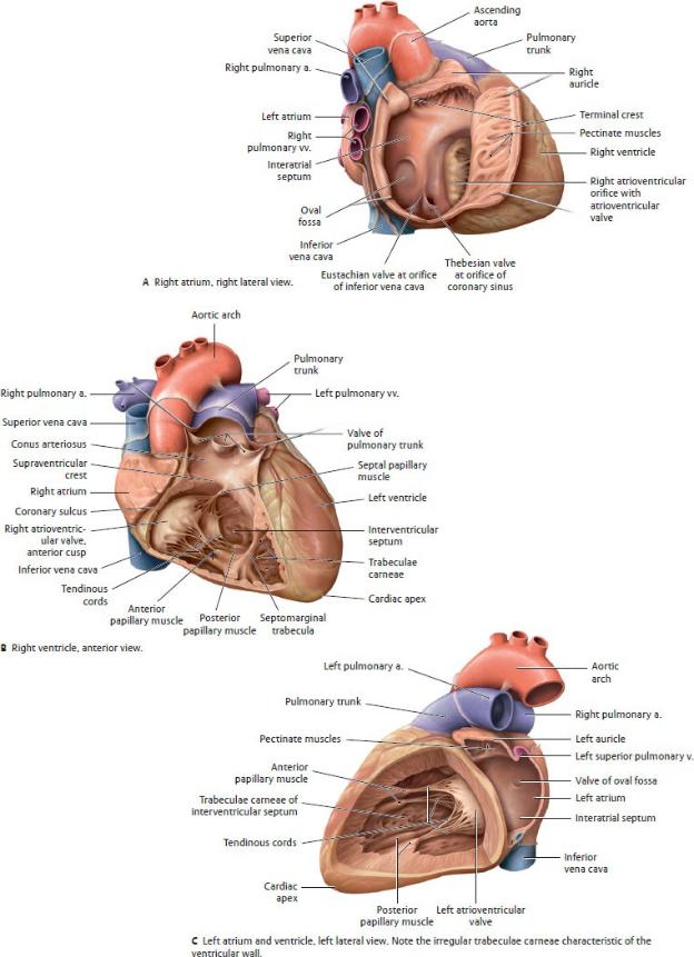

Fig. 7.12 Chambers of the heart
(From Schuenke M, Schulte E, Schumacher U. THIEME Atlas of Anatomy, Vol 2. Illustrations by Voll M and Wesker K. 3rd ed. New York: Thieme Publishers; 2020.)
—The left ventricle, which includes the apex of the heart, is the thickestwalled chamber of the heart. Similar to the right ventricle, the left is divided into inflow and outflow portions:
1.The left ventricle proper, which receives blood from the left atrium. A large anterior and small posterior papillary muscle arise from its floor
2.The aortic vestibule, the smooth-walled outflow channel through which blood flows into the aorta
BOX 7.4: DEVELOPMENTAL CORRELATION
TETRALOGY OF FALLOT
Tetralogy of Fallot is a rare combination of four congenital cardiac defects: pulmonary stenosis, overriding aorta, ventricular septal defect (VSD), and right ventricular hypertrophy. Symptoms include cyanosis, dyspnea (shortness of breath), fainting, finger clubbing, fatigue, and prolonged crying in infants.
Valves of the Heart
There are two types of cardiac valves: atrioventricular and semi lunar (Fig. 7.13).
1.Atrioventricular valves separate the atria from the ventricles and prevent regurgitation of blood into the atria during con-traction of the ventricles.
•The atrioventricular valves are made up of cusps, thin leaflets with free inner margins and outer margins that are attached to the fibrous rings of the cardiac skeleton.
•Slender threads called tendinous cords (chordae tend-ineae) attach the free edges of the valve leaflets to the papillary muscles in the ventricles. These cords maintain closure of the valves and prevent regurgitation of blood during ventricular contraction. Each cusp attaches to tendinous cords from more than one papillary muscle.
Fig. 7.13 Cardiac valves
(From Gilroy AM, MacPherson BR, Wikenheiser JC. Atlas of Anatomy. Illustrations by Voll M and Wesker K. 4th ed. New York: Thieme Publishers; 2020.)
•The atrioventricular valves include
◦the tricuspid valve, which separates the right atrium from the right ventricle and is composed of anterior, posterior, and septal cusps; and
◦the bicuspid (or mitral) valve, which separates the left atrium from the left ventricle and is composed of anterior and posterior cusps. The anterior cusp is immediately adjacent to, and continuous with, the wall of the aorta.

BOX 7.5: CLINICAL CORRELATION
MITRAL VALVE PROLAPSE
Mitral valve prolapse is a condition in which one or both leaflets of the mitral valve prolapse (evert) into the left atrium, allowing regurgitation of blood through the mitral valve during systole. This condition is usually asymptomatic but is noted by a midsys-tolic click (valve prolapsing) and murmur (regurgitation).
2.Semilunar valves prevent outflow from the ventricles as the chambers fill and backflow of blood into the ventricles after it has been expelled.
•Each valve is composed of three semilunar cusps with free inner margins and attached outer margins. A sinus, or pocket, is created between each cusp and the vessel wall. The thickened free margin of the cusp, the lunule, is the point of contact of the cusps. A nodule marks the center of the lunule.
•The semilunar valves include the following:
◦The pulmonary semilunar valve (pulmonary valve), is located in the pulmonary trunk at the top of the conus arteriosus, where it moderates blood flow through the right ventricular outflow channel. Its cusps are in the anterior, right, and left positions.
◦The aortic semilunar valve (aortic valve) is located within the aorta immediately adjacent to the mitral valve, where it moderates blood flow through the left ventricular outflow channel. Its cusps are in the posterior, right, and left positions. The coronary arteries arise from the sinuses above the right and left cusps.
BOX 7.6: CLINICAL CORRELATION
AORTIC VALVE STENOSIS
Stenosis of the aortic valve is the most common valve abnormal-ity. Calcifications of the valve leaflets narrow the outflow tract and lead to overload of the left ventricle, resulting in left ventricular hypertrophy.
Heart Sounds and Auscultation Sites
When the heart valves close, they produce characteristic sounds described as “lub dub.”
—Closure of the tricuspid and mitral valves during contraction of the ventricles produces the first sound (“lub”).
—Closure of the pulmonary and aortic valves as the ventricles relax produces the second sound (“dub”).
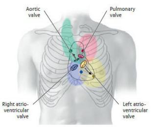
—The sounds, carried by the blood as it flows into the next vessel or chamber, are best distinguished at auscultation sites downstream from the valves
(Table 7.3).
Table 7.3 Position and Auscultation Sites of Cardiac Valves
(From Schuenke M, Schulte E, Schumacher U. THIEME Atlas of Anatomy, Vol 2. Illustrations by Voll M and Wesker K. 3rd ed. New York: Thieme Publishers; 2020.)
|
Anatomical |
|
Valve |
Anatomical Projection |
Auscultation Site |
Aortic valve |
Left sternal border (at |
Right 2nd intercostal |
|
level of 3rd rib) |
space (at sternal |
|
|
margin) |
|
|
|
Pulmonary valve |
Left sternal border (at |
Left 2nd intercostal |
|
level of 3rd costal |
space (at sternal |
|
cartilage) |
margin) |
|
|
|
Left atrioventricular |
Left 4th/5th costal |
Left 5th intercostal |
valve |
cartilage |
space (at midclavicular |
|
|
line) or cardiac apex |
|
|
|
Right atrioventricular |
Sternum (at level of 5th |
Left 5th intercostal |
valve |
costal cartilage) |
space (at sternal margin |
|
|
|
Note: Auscultation sites of the cardiac valves, indicated by colored dots.
Valvular heart disease causes turbulent blood flow through a valve; this produces a murmur that may be detected in the colored region around the valve.
Conduction System of the Heart (Fig. 7.14)
The conduction system of the heart generates and transmits impulses that modulate the contraction of the cardiac muscle. It consists of nodes, which initiate the impulses, and conducting fibers, which distribute the impulses to cardiac muscle to effect a coordinated contraction of the heart chambers.
—The sinoatrial (SA) node, the pacemaker of the heart, initiates and coordinates the timing of the contraction of the heart chambers.
•At a frequency of 60 to 70 beats per minute, the SA node transmits impulses to both atria and to the atrioventricu-lar node.
•It is subepicardial, located on the external surface of the heart, just within the myocardium of the right atrium at the junction with the superior vena cava.
•A branch of the right coronary artery usually supplies the SA node.
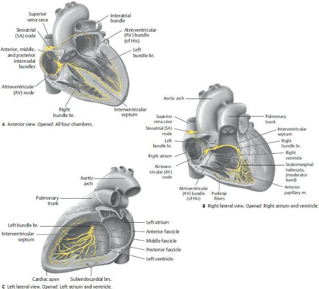
Fig. 7.14 Cardiac conduction system
(From Schuenke M, Schulte E, Schumacher U. THIEME Atlas of Anatomy, Vol 2. Illustrations by Voll M and Wesker K. 3rd ed. New York: Thieme Publishers; 2020.)
—The atrioventricular (AV) node is stimulated by the SA node and transmits impulses to the AV bundle.
•It is subendocardial, located at the base of the interatrial septum above the septal cusp of the tricuspid valve.
•The AV nodal artery, usually a branch of the right coronary artery, arises near the origin of the posterior interventric-ular artery at the crux of the heart.

—The atrioventricular (AV) bundle (of His) arises from cells of the AV node and transmits impulses to the walls of the ventricles.
•It runs first along the membranous part of the interven-tricular septum and then divides into right and left bundle branches that descend to the apex on either side of the muscular part of the septum.
•The bundle branches end as Purkinje fibers, modified cardiac fibers, which ascend within the muscular walls of the ventricles.
BOX 7.7: CLINICAL CORRELATION
ATRIOVENTRICULAR HEART BLOCK
AV heart block is a partial or complete blockage of the conduc-tion of electrical impulses between the atria and ventricles. This causes bradycardia (slow heartbeat) and arrhythmias (irregular heartbeat) and ultimately prevents the heart from effectively contracting and delivering blood to the body. Although heart block may be congenital, it is frequently the result of damage to the heart muscle after myocardial infarction (MI).
The Cardiac Cycle
The heart’s conduction system moderates the cardiac cycle, the coordinated systole (contraction) and diastole (relaxation) of the atria and ventricles. Fig. 7.15 summarizes the sequence of events of the cycle.

Fig. 7.15 The cardiac cycle
(From Schuenke M, Schulte E, Schumacher U. THIEME Atlas of Anatomy, Vol 2. Illustrations by Voll M and Wesker K. 3rd ed. New York: Thieme Publishers; 2020.)
1.In the initial phase of diastole, the atria and ventricles are relaxed, and the AV and semilunar valves are closed.
2.In late diastole, the atria fill, the AV valves open, and blood flows passively into the ventricles.
3.Stimulation from the SA node initiates the contraction of the atria, forcing the remaining atrial blood volume into the ventricles.
4.As pressure in the ventricles rises above that in the atria, the AV valves close.
5.Stimulation from the AV node and the AV bundles initiates contraction of the ventricles (ventricular systole).
6.Rising intraventricular pressure forces the semilunar valves to open. Blood
is ejected from the right ventricle into the pulmonary trunk and from the left ventricle into the aorta.
7.Relaxation of the ventricles (ventricular diastole) causes backward flow in the pulmonary trunk and aorta and closure of the pulmonary and aortic valves.
7.5 Middle Mediastinum: Neurovasculature of
the Heart
Coronary Arteries (Figs. 7.16 and 7.17; Table 7.4)
The right and left coronary arteries arise from the ascending aorta just superior to the right and left cusps of the aortic valve. In the initial phase of ventricular diastole, the local surge in aortic pressure caused by the backflow closes the aortic valve and drives blood into the coronary arteries. Blood flow in the arteries is greatest during diastole because of the compression of arteries within the myocardium during systole.
Fig. 7.16 Coronary arteries and cardiac veins (continued on page 112)
(From Gilroy AM, MacPherson BR, Wikenheiser JC. Atlas of Anatomy. Illustrations by Voll M and Wesker K. 4th ed. New York: Thieme Publishers; 2020.)
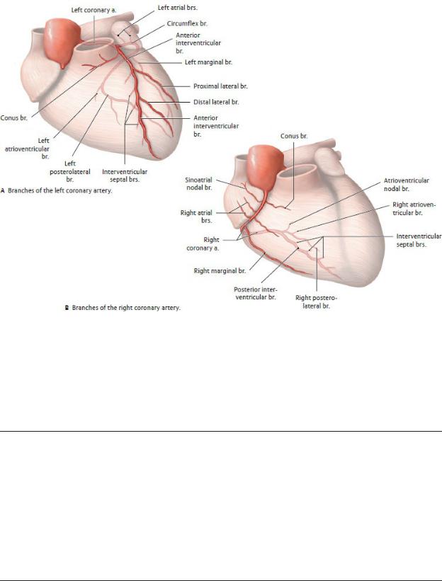
Fig. 7.17 Classification of the coronary arteries
Anterior view, sternocostal surface. (From Schuenke M, Schulte E, Schumacher U. THIEME Atlas of Anatomy, Vol 2. Illustrations by Voll M and Wesker K. 3rd ed. New York: Thieme Publishers; 2020.)
Table 7.4 Branches of the Coronary Arteries
Right coronary artery
—Sinoatrial nodal br.
—Right atrial brs.
—Right conus br.
—Anterior ventricular brs.
—Right marginal a.
—Atrioventricular nodal br.
—Posterior (inferior) interventricular br. (posterior descending a.)
—Interventricular septal brs.
Left coronary artery

—Anterior interventricular a. (left anterior descending a.)
•Left conus br.
•Proximal lateral (diagonal) br.
•Distal lateral (diagonal) br.
•Interventricular septal brs.
—Circumflex a.
•Sinoatrial nodal br. (in 40%)
•Left atrial brs.
•Left marginal a.
•Left posterolateral br.
•Posterior (inferior) interventricular a. (in about one third of cases)
—The right and left coronary arteries supply the myocardium and epicardium of the heart.
•The right coronary artery descends within the coronary sulcus around the right side of the heart. Its major branches and their distribution are
◦the branch to the sinoatrial node (SA nodal artery), which supplies the right atrium and the SA node;
◦the right marginal branch, which supplies the apex and part of the right ventricle;
◦the posterior (inferior) interventricular branch, which supplies the right and left ventricles and posterior third of the interventricular septum and sometimes anastomoses with the interventricular branch of the left coronary artery near the apex on the diaphragmatic surface; and
◦the AV nodal artery, which supplies the AV node.
•The left coronary artery, typically larger than the right coronary artery, arises from the aorta posterior to the pulmonary trunk. After a short but variable course, it divides into two large branches, the anterior interventric-ular (left anterior descending, LAD) artery, which descends in the anterior interventricular sulcus, and the circumflex artery, which runs around the left side of the heart in the coronary sulcus. Their branches and distributions include
◦the anterior interventricular artery, which supplies the anterior aspects of the right and left ventricles and the anterior two thirds of the interventricular septum, including the AV bundle of the conducting system; and
◦the circumflex artery, which supplies the left atrium and, via its left marginal branch, the left ventricle. In ~ 40% of the population, an SA nodal branch arises to supply the SA node.
—Variation in the coronary circulation is common but the descriptive language is misleading. The word dominant refers not to the artery that supplies the greater volume of cardiac tissue (that is almost always the left coronary artery), but to the artery that gives rise to the posterior interventricular branch.
•A right dominant circulation occurs in about two thirds of the population. The posterior interventricular branch arises from the right coronary artery and supplies the posterior third of the interventricular septum.
•A small percentage of people exhibit left dominance in which the posterior interventricular artery is a branch of the circumflex artery. In these cases, the entire interven-tricular septum and the AV node are supplied by the left coronary artery.
•In the shared or “balanced” dominance, seen in a small group of people, branches from both coronary arteries run in the interventricular groove and jointly supply the interventricular septum.
BOX 7.8: CLINICAL CORRELATION
ANGINA
Angina (angina pectoris), a sudden, crushing substernal pain, is a result of myocardial ischemia (insufficient blood supply) caused by a narrowing of the coronary arteries. Exercise following a heavy meal, stress, or even cold weather can trigger an episode. Although angina pain may be severe, it is relieved by a short rest and does not result in infarction of cardiac muscle.
BOX 7.9: CLINICAL CORRELATION
CORONARY ARTERY DISEASE
Coronary artery disease results in ischemia of the myocardium and is a leading cause of death in the United States. In atheroscle-rosis of the coronary arteries, lipid deposits build up on the inner wall of the vessel and gradually narrow the lumen. In acute disease a fragment of plaque breaks off and completely obstructs the vessel. This creates a necrotic (dead) area of myocardium known as a myocardial infarction. Chronic disease is characterized by a gradual narrowing (stenosis) of the vessels. Overtime a collateral circulation develops that circumvents the narrowed segment and may prevent, or limit, damage from other ischemic events.
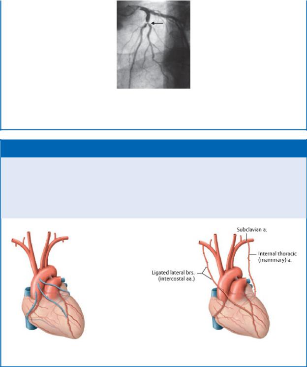
Severe stenosis (arrow) of the circumflex artery
(From Claussen CD, et al. Pareto Reihe Radiologie. Herz/Pareto Series Radiology. Heart. Stuttgart: Thieme; 2007.)
BOX 7.10: CLINICAL CORRELATION
CORONARY ARTERY BYPASS GRAFT
Coronary artery bypass graft (CABG) is a surgical procedure per-formed to bypass atherosclerotic narrowings of the coronary arteries that are the cause of anginal pain. If left untreated, these narrowings can eventually occlude the vessel and lead to myocar-dial infarction (MI). The internal thoracic artery and the great saphenous vein are most commonly used as the bypass vessel.
(From Schuenke M, Schulte E, Schumacher U. THIEME Atlas of Anatomy, Vol 2. Illustrations by Voll M and Wesker K. 3rd ed. New York: Thieme Publishers; 2020.)
Coronary Veins (see Fig. 7.18)
—The coronary sinus, which receives most of the venous return from the heart, runs in the posterior coronary sulcus between the left atrium and ventricle. The thebesian valve guards the orifice of the coronary sinus
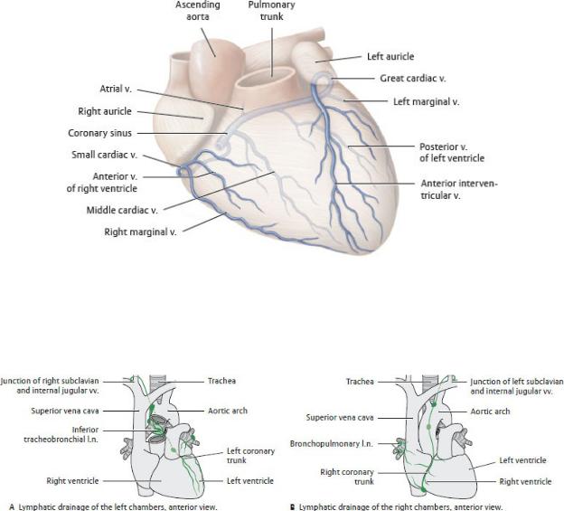
where it drains into the right atrium near the opening of the inferior vena cava (Figs. 7.9 and 7.12).
—The large veins of the heart are tributaries of the coronary sinus.
•The great cardiac vein receives the anterior interventricular, left marginal, and posterior left ventricular veins, which drain the left atrium and both ventricles.
•The posterior interventricular (middle cardiac) vein runs in the posterior interventricular groove with the posterior interventricular artery and drains the posterior part of the interventricular septum.
Fig. 7.18 Classification of cardiac veins
Anterior view, sternocostal surface. (From Schuenke M, Schulte E, Schumacher U. THIEME Atlas of Anatomy, Vol 2. Illustrations by Voll M and Wesker K. 3rd ed. New York: Thieme Publishers; 2020.)
Fig. 7.19 Lymphatic drainage of the heart
A unique “crossed” drainage pattern exists in the heart: lymph from the left
atrium and ventricle drains to the right venous junction, whereas lymph from the right atrium and ventricle drains to the left venous junction. (From Schuenke M, Schulte E, Schumacher U. THIEME Atlas of Anatomy, Vol 2. Illustrations by Voll M and Wesker K. 3rd ed. New York: Thieme Publishers; 2020.)
•The small cardiac vein, which drains the posterior right atrium and the right ventricle, accompanies the right coronary artery in the atrioventricular groove.
—Anterior cardiac veins drain the anterior surface of the right ventricle and open directly into the right atrium.
Lymphatic Drainage of the Heart
—Lymphatic vessels of the heart have a crossed drainage pattern. Lymph from the left atrium and ventricle drains via a left coronary trunk to inferior tracheobronchial nodes. Efferents from these nodes usually drain to the right venous junction via the bronchomediastinal trunk. Lymph from the right ventricle and atrium drains via a right coronary trunk that runs along the ascending aorta to brachiocephalic nodes near the left venous junction (Fig. 7.19).
—The pericardium usually drains to the right and left venous junctions via superior phrenic nodes and bronchomediastinal trunks, but it may also drain superiorly to the brachiocephalic nodes.
Innervation of the Heart
The autonomic nerves of the cardiac plexus innervate the conduc-tion system of the heart (Fig. 7.20, also see Section 5.2); they therefore regulate the heart rate but do not initiate the heartbeat.
—Sympathetic innervation increases the rate and force of contractions by increasing the response of the SA and AV nodes. It also allows dilation of the coronary arteries.
—Parasympathetic innervation decreases the rate of contractions and causes vasoconstriction of the coronary arteries.
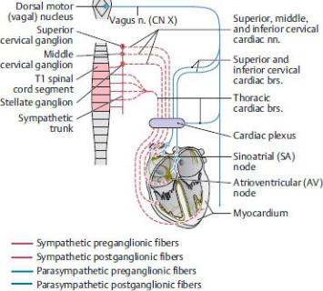
Fig. 7.20 Autonomic innervation of the heart
Schematic. (From Gilroy AM, MacPherson BR, Wikenheiser JC. Atlas of Anatomy. Illustrations by Voll M and Wesker K. 4th ed. New York: Thieme Publishers; 2020.)
—Visceral sensory fibers innervating the baroreceptors (receptors that measure blood pressure) and chemoreceptors (receptors that measure blood CO2) in the heart and aortic arch travel with the parasympathetic fibers of the vagus nerve.
—Visceral sensory fibers carrying pain sensation travel with sympathetic fibers to the T1–T5 spinal cord.
7.6 Prenatal and Neonatal Circulation
Prenatal Circulation
Fetal shunts that direct the flow of blood through the liver, heart, and lungs create a prenatal circulation that differs from that of the adult. The numbered steps in Fig. 7.21 illustrate the blood flow in the fetal circulation:
1.Fetal blood supplied with oxygen and nutrients in the placenta courses through the umbilical vein toward the liver of the fetus.
2.Although a portion of the blood is distributed to the liver, over half bypasses the liver and is redirected through a shunt, the ductus venosus, which empties directly into the inferior vena cava. This blood mixes with smaller
amounts from the liver and lower parts of the body before entering the right atrium of the heart.
3.The eustachian valve (see Fig. 7.12A) at the orifice of the inferior vena cava directs this well-oxygenated mixture across the right atrium into the left atrium through the oval foramen on the interatrial septum. The higher systolic pressure in the right atrium relative to that in the left creates this right-to-left shunt.
From the left atrium, the flow continues into the left ventricle, the aorta, and the systemic circulation of the head and neck. Thus the most highly oxygenated and nutrient-rich blood from the placenta is directed into the coronary, carotid, and subclavian arteries to supply the upper body, especially the heart and developing brain.
4.Oxygen-depleted blood entering the right atrium from the superior vena cava is directed downward through the tricuspid valve into the right ventricle and out through the pulmonary trunk.
Because the high vascular resistance in the lungs prevents much blood from entering the pulmonary arteries, most is diverted to the descending aorta through the ductus arteriosus, a connection between the pulmonary trunk and the aortic arch.
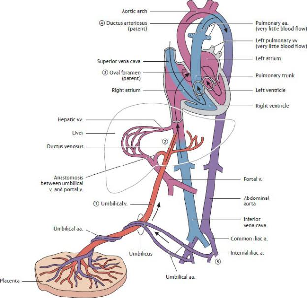
Fig. 7.21 Prenatal circulation
(After Fritsch H, Kuhnel W. Taschenatlas der Anatomie. Bd.2.7. Aufl. Stuttgart: Thieme Publishers; 2001.)
5.The blood entering the aorta from the ductus arteriosus mixes with some blood from the aortic arch. This partially oxygenated blood flows into the descending aorta and is distributed to the lower body, or back to the placenta by way of the paired umbilical arteries.
Cardiovascular Changes at Birth
At birth, a series of changes occurs in the cardiovascular system (Fig. 7.22;
Table 7.5).
1.Pressure in the right atrium decreases as a result of
a ligation of the umbilical vein, which cuts off blood flow from the placenta; and
b the onset of pulmonary respiration, which dramatically decreases the pulmonary blood pressure and increases blood flow to the lungs.
Table 7.5 Derivatives of Fetal Circulatory Structures
Fetal Structure |
Adult Remnant |
Ductus arteriosus |
Ligamentum arteriosum |
|
|
Oval foramen (foramen ovale) |
Oval fossa (fossa ovalis) |
|
|
Ductus venosus |
Ligamentum venosum |
|
|
Umbilical v. |
Round lig. of the liver (ligamentum |
|
teres) |
|
|
Umbilical a. |
Medial umbilical lig. |
|
|
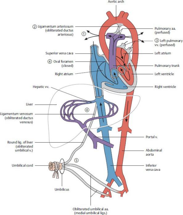
Fig. 7.22 Postnatal circulation
(After Fritsch H, Kuhnel W. Taschenatlas der Anatomie. Bd.2.7. Aufl. Stuttgart: Thieme Publishers; 2001.)
2.As a result of increased flow through the lungs, there is a decreased blood flow through the ductus arteriosus, which constricts within 10 to 15 hours of

birth. In the adult, the remnant of this structure is the ligamentum arteriosum.
3.Blood returning to the heart through the pulmonary veins increases pressure in the left atrium.
4.Increased pressure in the left atrium, coupled with a corresponding decrease in pressure in the right atrium, causes a functional closure of the oval foramen (foramen ovale) within hours of birth. Complete closure of the oval foramen usually occurs after several months, forming the oval fossa (fossa ovalis) in the adult heart.
5.Functional closure of the umbilical arteries, umbilical vein, and ductus venosus occurs within minutes of birth, although the obliteration of the lumen in each vessel may take several months. The remnants of these vessels in the adult are ligamentous structures.
BOX 7.11: DEVELOPMENTAL CORRELATION
VENTRICULAR SEPTAL DEFECT
Ventricular septal defects (VSDs), the most common of congen-ital heart defects, usually involve the membranous part of the interventricular septum and are associated with Down syndrome, tetralogy of Fallot, and Turner syndrome. VSDs may also occur traumatically from rupture of the membranous septum follow-ing a myocardial infarction. When the VSD is large, the resulting left-to-right shunt increases blood flow through the pulmonary circulation, causing pulmonary hypertension and cardiac failure (Fig. 7.23B).
BOX 7.12: DEVELOPMENTAL CORRELATION
PATENT DUCTUS ARTERIOSUS
The opening of the pulmonary circulation at birth causes the duc-tus arteriosus to constrict, probably in response to an increase in local oxygen tension. If the ductus remains open, as a patent duc-tus arteriosus (PDA), deoxygenated blood continues to enter the descending aorta (Fig. 7.23C). There may be no symptoms if the defect is small, but larger defects may cause failure to thrive, dys-pnea (shortness of breath), fatigue, tachycardia (increased heart rate), and cyanosis. Because prostaglandins maintain patency of the ductus during fetal life, premature infants whose ductus does not constrict spontaneously at birth may be treated with prosta-glandin inhibitors.
BOX 7.13: DEVELOPMENTAL CORRELATION
ATRIAL SEPTAL DEFECT
Atrial septal defects (ASDs) (Fig. 7.23D) are among the most common congenital cardiac anomalies and are particularly associ-ated with Down syndrome. Most ASDs result from a failure of the oval foramen to close at birth, but they may also result from incomplete development of the septal components. ASDs result in left-to-right shunting and an increase in blood volume through the pulmonary circulation. Small ASDs are often asymptomatic, but larger ASDs will cause hypertrophy of the right atrium, right ventricle, and pulmonary arteries
due to the fluid overload.
Fig. 7.23 Congenital heart defects.
(From Schuenke M, Schulte E, Schumacher U. THIEME Atlas of Anatomy, Vol 2. Illustrations by Voll M and Wesker K. 3rd ed. New York: Thieme Publishers; 2020.)
BOX 7.14: DEVELOPMENTAL CORRELATION
COARCTATION OF THE AORTA
Coarctation, or stenosis, of the aorta usually occurs in proximity to the ligamentum arteriosum, where it limits (or obstructs) the normal blood flow from the aortic arch through the descending aorta. When the narrowing is distal to the ligamentum (postductal stenosis), an effective collateral circulation links the proximal and distal aortic segments via the internal thoracic arteries and their intercostal branches. The intercostal arteries may become large and tortuous enough to form notches along the lower margins of the ribs.
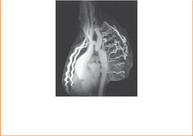
MRI of coarctation in a young child
Note the large internal thoracic arteries and tortuous intercostal arteries that provide collateral pathways between the thoracic and abdominal parts of the aorta. (From Gunderman R. Essential Radiology, 3rd ed. New York: Thieme; 2014)
7.7 Superior and Posterior Mediastina
Many of the large arteries and veins of the thorax—superior vena cava (SVC), inferior vena cava (IVC), aorta, common carotid and subclavian arteries—pass through the superior and posterior mediastinum en route to the neck or the abdomen or both. They are accompanied by the vagus, phrenic, and cardiac nerves. These structures are listed in Table 7.1 and described in detail in Section 5.2. Only viscera of the superior and posterior mediastina will be discussed in this section.
Esophagus
The esophagus, the thoracic segment of the gastrointestinal tract, is a narrow, but highly distensible, muscular tube. It connects the pharynx in the neck to the stomach in the abdomen (Fig. 7.24).
—The esophagus descends anterior to the thoracic vertebral bodies in the posterior mediastinum. Superiorly, it lies posterior to the trachea; inferiorly, it lies posterior to the left atrium of the heart.
—In the upper thorax, the esophagus descends on the right side of the aorta.
Before passing through the esophageal aperture of the diaphragm, the esophagus passes first anterior to, and then to the left of, the aorta.
—The upper esophagus is composed mostly of striated muscle arranged in inner circular and outer longitudinal layers. Striated muscle is gradually replaced by smooth muscle fibers inferiorly.
—Three constrictions narrow the lumen of the esophagus (Fig. 7.25):
1.The upper esophageal sphincter, created by the crico pharyngeus muscle (part of the inferior constrictor muscle of the pharynx), which surrounds the upper esophageal opening in the neck
2.The middle esophageal constriction, created by the aortic arch and left main bronchus
3.The lower esophageal constriction, or cardiac sphinc-ter, created by circular muscles of the distal esophagus, folds in the mucosa formed by a submucosal venous plexus, and the muscular esophageal aperture of the diaphragm
—The blood supply to the upper, middle, and lower esophagus arises from vessels in the neck (inferior thyroid), thorax (esophageal branches of the descending aorta), and abdomen (left gastric and left inferior phrenic), respectively.
—The veins of upper and middle esophageal segments drain into the azygos system. Veins of the lower esophageal seg-ment drain inferiorly along inferior phrenic veins into the hepatic portal system (the venous drainage for organs of the abdominal gastrointestinal tract). When portal venous flow is obstructed in the liver (such as from cirrhosis), blood can be diverted through the esophageal veins to the azygos system and superior vena cava
(Fig. 7.26).
—The esophageal plexus, formed by the right and left vagus nerves with contributions from thoracic splanchnic nerves, innervates the esophagus.
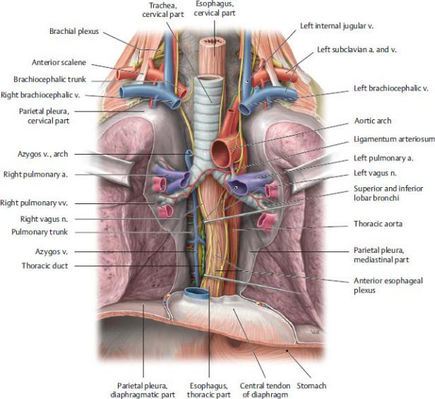
Fig. 7.24 Esophagus in situ
Anterior view. (From Gilroy AM, MacPherson BR, Wikenheiser JC. Atlas of Anatomy. Illustrations by Voll M and Wesker K. 4th ed. New York: Thieme
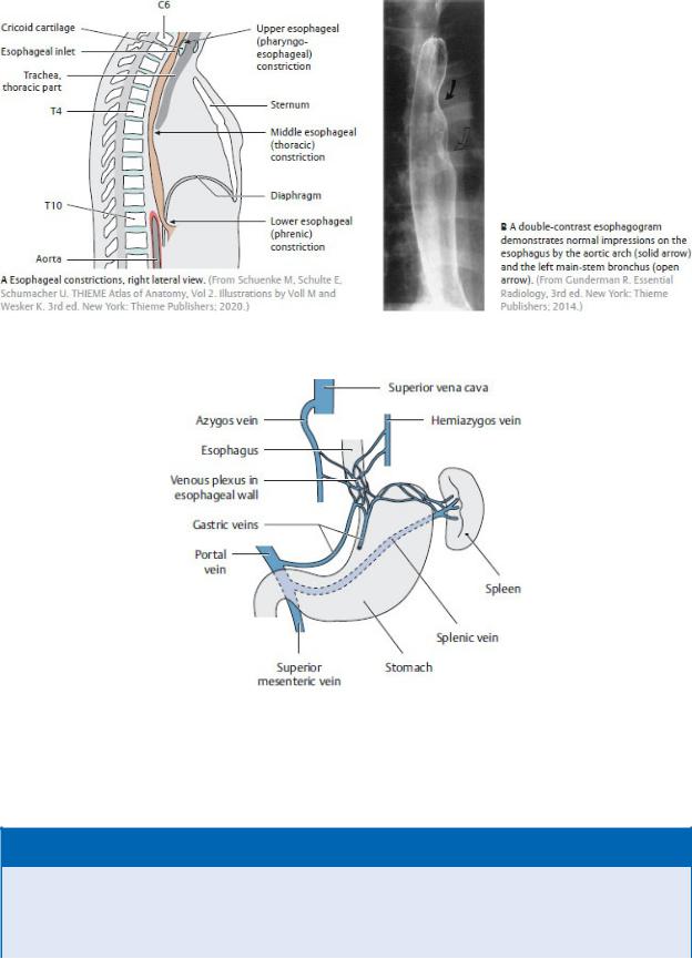
Fig. 7.25 Esophagus: location and constrictions
Fig. 7.26 Esophageal venous collaterals
(From Schuenke M, Schulte E, Schumacher U. THIEME Atlas of Anatomy, Vol 2. Illustrations by Voll M and Wesker K. 3rd ed. New York: Thieme Publishers; 2020.)
BOX 7.15: CLINICAL CORRELATION
ACHALASIA
Achalasia is a deficiency of inhibitory neurons in the lower part of the esophagus. These neurons are responsible for overriding the normal resting tonic contraction of the smooth muscle cells in the lower esophageal sphincter. Their deficiency results in fail-ure of the
sphincter to relax during swallowing. As food accumu-lates above the sphincter, there is an increased risk of aspiration pneumonia.
Trachea and Bronchi
The trachea, located in the superior mediastinum, is the proximal part of the tracheobronchial tree, a passageway for air between the lungs and the external environment. The distal part of this passageway, the bronchial tree, extends into the lungs and is dis-cussed with the pulmonary cavities in Chapter 8.
—As the trachea descends through the superior mediastinum, slightly to the right of the midline, it lies anterior to the esophagus and posterior to the great vessels.
—C-shaped cartilaginous rings form the skeleton of the trachea and prevent collapse of the lumen. A muscular membrane connects the ends of the rings posteriorly (Fig. 7.27).
Fig. 7.27 Trachea
(From Schuenke M, Schulte E, Schumacher U. THIEME Atlas of Anatomy, Vol 2. Illustrations by Voll M and Wesker K. 3rd ed. New York: Thieme Publishers;
2020.)
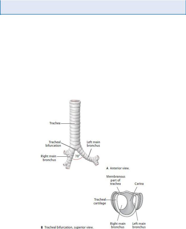
—The carina, a wedge-shaped cartilage, marks the bifurcation of the trachea into a right and left bronchus at the T4-T5 vertebral level.
—Of the two main bronchi, the right is shorter, wider, and more vertical than the left, and therefore is more prone to obstruc-tion by foreign objects.
—Descending branches of the inferior thyroid artery (a branch of the thyrocervical trunk) in the neck as well as bronchial arteries that arise from the thoracic aorta, supply the trachea. Venous blood drains to the inferior thyroid veins.
—Thoracic splanchnic nerves and parasympathetic fibers from the vagus (cranial nerve X) nerve innervate the trachea via the pulmonary plexus.