


3Back
3.1The Vertebral Column
The back includes the vertebral column, the spinal cord and spinal nerves, and the overlying muscles and skin.
General Features
—The vertebral column
•encloses and protects the spinal cord,
•supports the head and trunk,
•provides an attachment for the limbs, and
•transfers the weight of the body to the lower limbs. —— The vertebral column, which extends from its articulation with the skull to the coccyx, comprises 33 vertebrae and the intervening intervertebral disks, which are divided among five regions (Fig. 3.1):
•7 cervical vertebrae
•12 thoracic vertebrae
•5 lumbar vertebrae
•5 fused sacral vertebrae
•4 (3–5) fused coccygeal vertebrae
—Within each region, individual vertebrae are identified by number, a designation often referred to as a vertebral level (such as T8 vertebral level).
—Vertebrae increase in size from the cervical to the lumbar regions and decrease in size from the top of the sacrum to the coccyx.
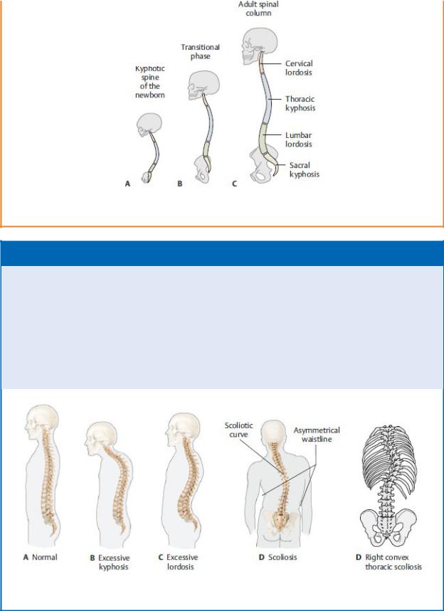
—When the vertebral column is viewed laterally, two types of curvatures are evident (Fig. 3.2):
•The kyphotic curvatures of the thoracic and sacral regions, known as primary curvatures, are curved posteriorly and are present before birth.
•The lordotic curvatures of the cervical and lumbar regions, curved anteriorly, are secondary curvatures that develop postnatally.
—A vertebral canal passes through the center of the vertebral column and encloses the spinal cord, the spinal meninges (membranes surrounding the
spinal cord), and the roots of the spinal nerves, and associated vasculature (see Section 3.2).
—Intervertebral foramina, openings between vertebrae, allow the passage of spinal nerves.
—Strong vertebral ligaments support the joints of the vertebral column while allowing flexibility of the trunk.
—Fibrocartilaginous intervertebral disks lie between the vertebral bodies of all vertebrae, except between C1 and C2. They act as shock absorbers for the spine and allow flexibility between vertebrae. With the vertebral bodies, they form the anterior wall of the vertebral canal.
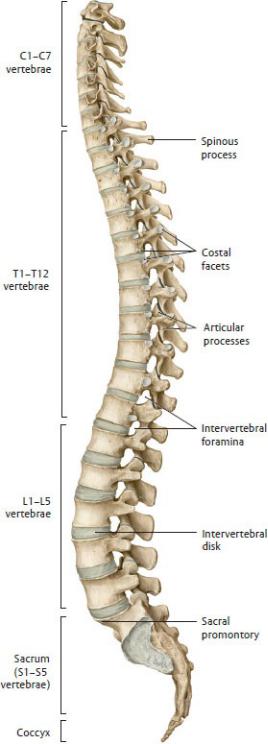
Fig. 3.1 Vertebral column
Left lateral view. (From Schuenke M, Schulte E, Schumacher U. THIEME Atlas of Anatomy, Vol 1. Illustrations by Voll M and Wesker K. 3rd ed. New York: Thieme Publishers; 2020.)
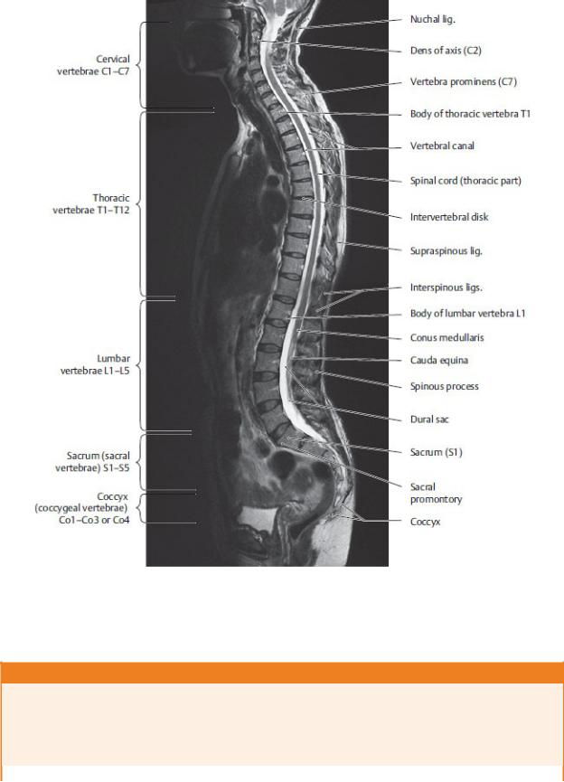
Fig. 3.2 MRI of the spine
Sagittal view. (From Moeller TB, Reif E. Pocket Atlas of Sectional Anatomy: The Musculoskeletal System. New York: Thieme Publishers; 2009.)
BOX 3.1: DEVELOPMENTAL CORRELATION
SPINAL DEVELOPMENT
The characteristic curvatures of the adult spine appear over the course of postnatal development, being only partially present in a newborn. The newborn has a ”kyphotic” spinal curvature (A); lumbar lordosis develops later and becomes stable at puberty (C).
(From Gilroy AM, MacPherson BR, Wikenheiser JC. Atlas of Anatomy. Illustrations by Voll M and Wesker K. 4th Edition. New York: Thieme Publishers; 2020.)
BOX 3.2: CLINICAL CORRELATION
ABNORMAL CURVATURES OF THE VERTEBRAL COLUMN: KYPHOSIS, LORDOSIS, AND SCOLIOSIS
Kyphosis (“hunchback”), an excessive anterior curvature of the thoracic spine, is often seen in elderly women. Although it may be congenital or posture related, it is usually secondary to degenera-tive changes (collapse) of the vertebral bodies. Lordosis (“swayback”), an excessive posterior curvature of the lumbar spine, fre-quently develops as a temporary side effect during pregnancy, but in nonpregnant individuals it may have pathologic or even weight-related causes. Scoliosis is a lateral curvature of the spine and may be congenital or neuromuscular, caused by diseases such as cerebral palsy and muscular dystrophy.
(From Gilroy AM, MacPherson BR, Wikenheiser JC. Atlas of Anatomy. Illustrations by Voll M and Wesker K. 4th Edition. New York: Thieme Publishers; 2020.)

BOX 3.3: CLINICAL CORRELATION
OSTEOPOROSIS
The spine is the primary target for degenerative diseases of the skeleton, such as osteoporosis, in which the rate of reabsorption by osteoclasts exceeds that of bone formation by osteoblasts. The resulting loss of bone mass predisposes the individual to com-pression fractures of the spine.
Regional Characteristics of Vertebrae
Most vertebrae share a typical form (Fig. 3.3), although specific features vary by region.
—Most vertebrae have the following:
•An anterior vertebral body
•A posterior vertebral arch formed by paired pedicles and paired laminae (the pedicles attach to the vertebral body, and the paired laminae join to form a spinous process)
•Paired transverse processes that project laterally from the vertebral arch
•Superior and inferior articular processes that articulate with the adjacent vertebrae
•A vertebral foramen encircled by the vertebral body and vertebral arch (the combined vertebral foramina of all vertebrae form the vertebral canal)
—Cervical vertebrae, the smallest of all of the vertebrae, support the head and form the posterior skeleton of the neck (Fig. 3.4). The seven cervical vertebrae are characterized as typical or atypical.
•Typical cervical vertebrae (Fig. 3.5A)
◦C3–C6 have a small body, a large vertebral foramen, and often bifid (two-pronged) spinous processes.
•Atypical cervical vertebrae
◦C1, the atlas, lacks a vertebral body and spinous process (Fig. 3.5B). It has anterior and posterior vertebral arches that are connected on each side by lateral masses. C1 articulates superiorly with the occipital bone of the skull and inferiorly with C2.
◦C2, the axis, has a peglike dens projecting superiorly from its body that articulates with the anterior arch of C1 (Fig. 3.5C).
◦C7, the vertebra prominens, has a long, palpable spinous process.
Fig. 3.3 Structural elements of a vertebra
Left posterosuperior view. With the exception of the atlas (C1) and axis (C2), all vertebrae consist of the same structural elements. (From Schuenke M, Schulte E, Schumacher U. THIEME Atlas of Anatomy, Vol 1. Illustrations by Voll M and Wesker K. 3rd ed. New York: Thieme Publishers; 2020.)
Fig. 3.4 Cervical spine
Bones of the cervical spine, left lateral view. (From Gilroy AM, MacPherson
BR, Wikenheiser JC. Atlas of Anatomy. Illustrations by Voll M and Wesker K. 4th ed. New York: Thieme Publishers; 2020.)
•All cervical vertebrae have paired transverse foramina, openings formed by the anterior and posterior tubercles of each transverse process.
—Paired vertebral arteries ascend in the neck through the transverse foramina of C1–C6, pass through a groove on the posterior arch of C1, and enter the base of the skull through a large opening, the foramen magnum.
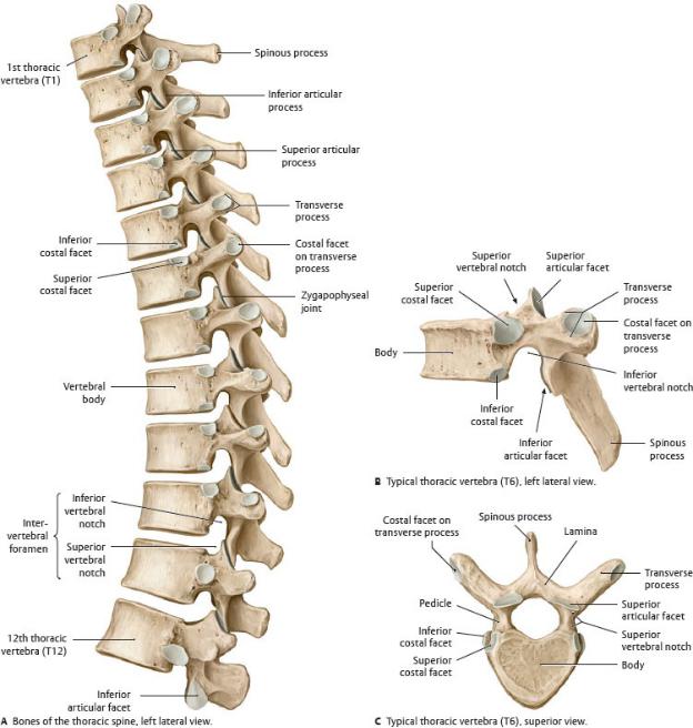
—Thoracic vertebrae (Fig. 3.6) have
•long spinous processes that project inferiorly,
•heart-shaped vertebral bodies,
•superior and inferior articular facets that are oriented in the coronal plane, and
•costal facets that articulate with the ribs.
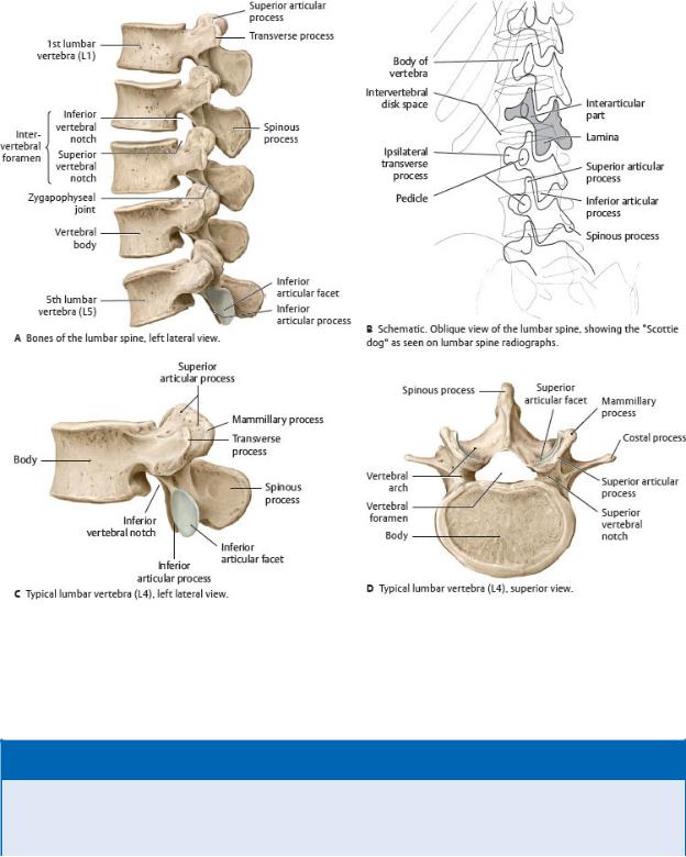
—Lumbar vertebrae (Fig. 3.7), the largest vertebrae, have
•large bodies;
•short, broad spinous processes; and
•an interarticular part (pars interarticularis), part of the lamina between the superior and inferior articular facets, that forms the neck of the “Scottie dog” seen on oblique views of lumbar spine radiographs. It is a common site of vertebral fractures.
—The five sacral vertebrae are fused into a single bone, the sacrum (Fig. 3.8), which forms the posterosuperior wall of the pelvis and articulates laterally with the hip bones. The sacrum contains
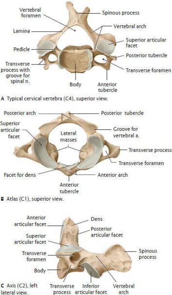
Fig. 3.5 Cervical vertebrae
(From Schuenke M, Schulte E, Schumacher U. THIEME Atlas of Anatomy, Vol 1. Illustrations by Voll M and Wesker K. 3rd ed. New York: Thieme Publishers; 2020.)
•the sacral canal, a continuation of the vertebral canal, which is open inferiorly at the sacral hiatus;
•the median sacral crest, the fused spinous processes of the sacral vertebrae;
•paired medial sacral crests that end inferiorly as the sacral cornua on either side of the sacral hiatus;
•four pairs of anterior and posterior sacral foramina for the passage of spinal nerve branches; and
•the promontory, formed by the anterior lip of the S1 vertebral body.
—The small coccygeal vertebrae, usually four (but this can vary from three to five), fuse into a single triangularly shaped bone, the coccyx, which articulates superiorly with the sacrum at the sacrococcygeal joint.
Fig. 3.6 Thoracic spine
(From Schuenke M, Schulte E, Schumacher U. THIEME Atlas of Anatomy, Vol 1. Illustrations by Voll M and Wesker K. 3rd ed. New York: Thieme Publishers; 2020.)
Fig. 3.7 Lumbar spine
(From Gilroy AM, MacPherson BR, Wikenheiser JC. Atlas of Anatomy. Illustrations by Voll M and Wesker K. 4th ed. New York: Thieme Publishers; 2020.)
BOX 3.4: CLINICAL CORRELATION
SPONDYLOLYSIS AND SPONDYLOLISTHESIS
Spondylolysis is a fracture across the interarticular part of the lamina, usually at L5, and appears as a collar on the “Scottie dog” seen on lumbar radiographs. When the defect is
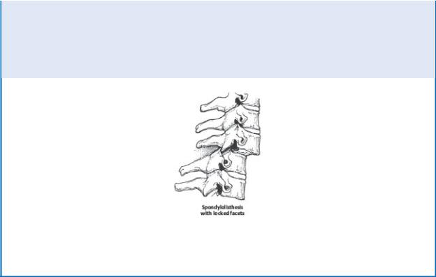
bilateral, the verte-bral body may separate from its vertebral arch and shift anteriorly relative to the vertebra below it, a condition known as spondylolisthesis. Mild cases can be asymptomatic, but more severe cases compress the spinal nerves and cause pain in the lower limbs and back. Note: Spondylosis, a simi-lar-sounding name but unrelated condition, refers to age-related degeneration and the formation of osteophytes (see Box 3.7 Age-Related Changes in Vertebrae).
A 50% anterolisthesis of one vertebral body on another, with the inferior articular facets of the upper vertebra locked in front of the superior articular facets of the lower vertebra. Naturally, this anterolisthesis tends to compromise the vertebral canal and threatens the cord. (From Gunderman R. Essential Radiology, 3rd ed. New York: Thieme; 2014)
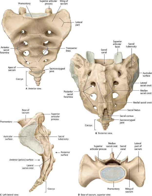
Fig. 3.8 Sacrum and coccyx

(From Schuenke M, Schulte E, Schumacher U. THIEME Atlas of Anatomy, Vol 1. Illustrations by Voll M and Wesker K. 3rd ed. New York: Thieme Publishers; 2020.)
Joints of the Vertebral Column
Joints of the vertebral column include articulations between adja-cent vertebral bodies and articulations between adjacent verte-bral arches. Joints also form between the vertebral column and the skull (Table 3.1). Individual vertebral joints allow small local movements, but the combination of these movements over mul tiple vertebral levels accounts for the considerable flexibility of the vertebral column.
—Craniovertebral joints (Fig. 3.9) are synovial joints between the skull and C1, and between C1 and C2:
•Paired atlanto-occipital joints between the occipital bone of the skull and the atlas (C1) allow flexion and extension of the head (as when nodding “yes”).
•Atlantoaxial joints, which include one median and two lateral articulations between the atlas and axis (C1 and C2), allow rotation of the head from side to side (as when saying “no”).
BOX 3.5: CLINICAL CORRELATION
INJURIES OF THE CERVICAL SPINE
The laxity of the cervical spine makes it prone to hyperextension injuries, such as “whiplash,” the excessive and often violent back-ward movement of the head, resulting in fractures of the dens of the axis and traumatic spondylolisthesis (see Box 3.4 Spondyloly-sis and Spondylolisthesis). Patient prognosis is largely dependent on the spinal level of the injuries.
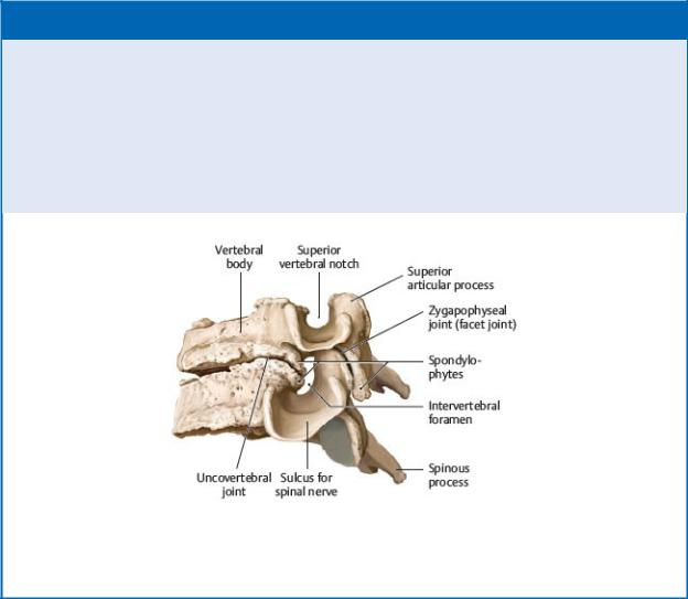
—Uncovertebral joints form between the uncinate processes (lateral lips on the superior edges of the vertebral bodies) of C3–C7 vertebrae and the vertebral bodies immediately superior to them.
•These joints, which are not present at birth, form during childhood, probably as a result of a fissure in the cartilage of the intervertebral disk that then assumes a joint-like character.
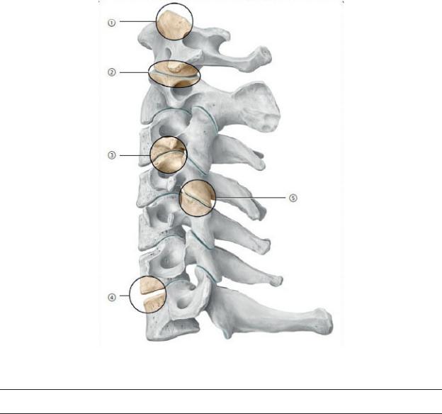
Table 3.1 Joints of the Vertebral Column
Craniovertebral joints
|
Atlanto-occipital joints |
Occiput-CI |
|
Atlantoaxial joints |
C1–C2 |
Joints of the vertebral bodies |
|
|
|
|
|
|
Uncovertebral joints |
C3–C7 |
|
Intervertebral joints |
C2–S1 |
Joints of the vertebral arch |
|
|
|
Zygapophyseal joints |
C1–S1 |
|
|
|
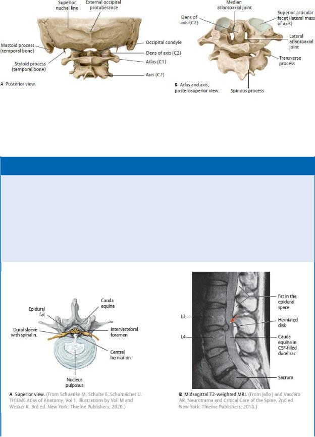
Fig. 3.9 Craniovertebral joints
(From Gilroy AM, MacPherson BR, Wikenheiser JC. Atlas of Anatomy. Illustrations by Voll M and Wesker K. 4th ed. New York: Thieme Publishers; 2020.)
BOX 3.6: CLINICAL CORRELATION
HERNIATION OF INTERVERTEBRAL DISKS
As elasticity of the anulus fibrosus declines with age, compressive forces can cause the nucleus pulposus to protrude through weak-ened areas. If the fibrous ring of the anulus ruptures posteriorly, the herniated material may compress the contents of the dural sac, but posterolateral herniations that compress spinal nerves are most common, particularly at the L4–L5 or L5–S1 level. In the lumbar region, where spinal nerves exit the vertebral canal above the IV disk, the hernia is likely to compress the spinal nerve inferior to that level (e.g., a herniation of the L4–L5 disk will impact the L5 spinal nerve), and pain is felt along the corresponding dermatome.
Posterior herniation (A,B). In the MRI a conspicuously herniated disk at the level of L3-L4 protrudes posteriorly. The dural sac is deeply indented at that level. CSF, cerebrospinal fluid.
Posterolateral herniation (C,D). A posterolateral herniation may compress the spinal nerve as it passes through the intervertebral foramen. If more medially positioned, the herniation may spare the nerve at that level but impact nerves at inferior levels.
Fig. 3.10 Intervertebral disk
Fourth lumbar vertebra, superior view. (From Schuenke M, Schulte E, Schumacher U. THIEME Atlas of Anatomy, Vol 1. Illustrations by Voll M and Wesker K. 3rd ed. New York: Thieme Publishers; 2020.)
—Intervertebral joints form between intervertebral (IV) disks and the articular surfaces of vertebral bodies. There is no IV disk between C1 and C2, and those between sacral and coccygeal vertebrae are rudimentary.
•The IV disks act as shock absorbers and are composed of an outer fibrous ring, the anulus fibrosus, and a gelatinous core, the nucleus pulposus (Fig. 3.10).
•The height of the IV disk relative to the height of the vertebral body determines the degree of mobility of the joint; mobility is greatest in the
cervical and lumbar regions.
•The differences between anterior and posterior heights of the cervical and lumbar disks contribute to the lordotic curvatures.
—Zygapophyseal joints, also known as facet joints, are synovial joints that join the superior and inferior articular facets of adjacent vertebrae. The orientation of these joints differs among regions and influences the degree and direction of movement of the vertebral column.
•In the cervical region, the joints are mostly in the horizontal plane and allow movement in most directions.
•In the thorax, the joints largely lie in the coronal plane, limiting movement to lateral flexion.
•In the lumbar region, the joints are in the sagittal plane, facilitating flexion and extension.
BOX 3.7: CLINICAL CORRELATION
AGE-RELATED CHANGES IN VERTEBRAE
With advancing age, a decrease in bone density and aging of the IV disks can lead to an increase in compressive forces on the ver-tebral joints. Subsequent degenerative changes can include the depletion of articular cartilage and the formation of osteophytes (bony spurs). Osteophyte formation at the periphery of the verte-bral bodies where they join the IV disks is known as spondylosis. Similar degenerative changes of the zygapophyseal joints indicate osteoarthritis, common in the cervical and lumbar spine but also manifested in the joints of the hand, hip, and knee.
Advanced uncovertebral arthrosis of the cervical spine. (Drawing based on specimen from the Anatomical Collection at Kiel Univer-sity.) (From Schuenke M, Schulte E, Schumacher U. THIEME Atlas of Anatomy, Vol 1. Illustrations by Voll M and Wesker K. 3rd ed. New York: Thieme Publishers; 2020.)
Vertebral Ligaments
Vertebral ligaments support the joints of the vertebral column.
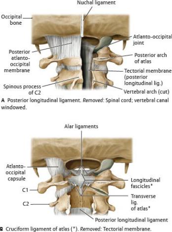
—Ligaments that support the cranial vertebral joints (Fig. 3.11) include
•the atlanto-occipital membranes, which connect the occipital bone of the skull to the anterior and posterior arches of the atlas (C1);
•the alar ligaments, which secure the dens of C2 to the skull; and
Fig. 3.11 Dissection of the craniovertebral joint ligaments
Posterior view. (From Gilroy AM, MacPherson BR, Wikenheiser JC. Atlas of Anatomy. Illustrations by Voll M and Wesker K. 4th ed. New York: Thieme Publishers; 2020.)
•the cruciform ligament, formed by longitudinal fascicles (fibers) and a transverse ligament, which secures the dens against the anterior arch of the atlas.
—Two longitudinal ligaments (Figs. 3.12 and 3.13) join all of the vertebral bodies:
1.The anterior longitudinal ligament, a broad fibrous band extending from the occipital bone of the skull to the sacrum, attaches to the anterior and lateral surfaces of the vertebral bodies and IV disks and prevents hyperextension.
2.The posterior longitudinal ligament, a thin fibrous band extending from C2 to the sacrum along the anterior aspect of the vertebral canal, attaches primarily to the IV disks and offers weak resistance to hyperflexion. Superiorly, this ligament extends into the skull as the tectorial membrane (see Fig. 2.15A).
—Ligaments that join the vertebral arches of adjacent vertebra include the following:
• the paired ligamenta flava, which join the laminae of adjacent vertebrae on the posterior wall of the vertebral canal. They limit flexion and provide postural support of the vertebral column (Fig. 3.14).
• the supraspinous ligament, which connects the posterior ridge of the spinous processes (Fig. 3.15)
• the nuchal ligament, a finlike expansion of the supraspi-nous ligament in the neck that extends from the occipital bone to the spinous process of C7 (Fig. 3.15)
—Additional vertebral ligaments connect elements of the vertebral arches and spinous processes (see Fig. 3.12).
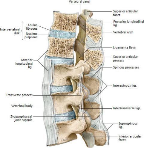
Fig. 3.12 Ligaments of the vertebral column: thoracolumbar junction
Left lateral view of T11–L3, with T11–T12 sectioned in the midsagittal plane. (From Gilroy AM, MacPherson BR, Wikenheiser JC. Atlas of Anatomy. Illustrations by Voll M and Wesker K. 4th ed. New York: Thieme Publishers; 2020.)
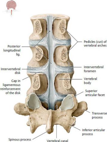
Fig. 3.13 Posterior longitudinal ligament
Posterior view of opened vertebral canal at level of L2–L5. Removed: L2–L4 vertebral arches at pedicular level. (From Gilroy AM, MacPherson BR, Wikenheiser JC. Atlas of Anatomy. Illustrations by Voll M and Wesker K. 4th ed. New York: Thieme Publishers; 2020.)
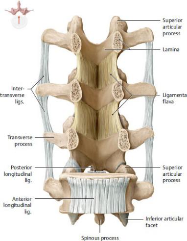
Fig. 3.14 Ligamenta flava and intertransverse ligaments Anterior view of opened vertebral canal at level of L2-L5. Removed: L2-L4 vertebral bodies. (From Gilroy AM, MacPherson BR, Wikenheiser JC. Atlas of Anatomy. Illustrations by Voll M and Wesker K. 4th Edition. New York: Thieme Publishers; 2020.)
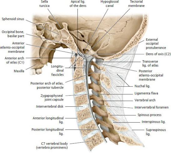
Fig. 3.15 Ligaments of the cervical spine
Midsagittal section, left lateral view. The nuchal ligament is the broadened, sagittally oriented part of the supraspinous ligament that extends from the vertebra prominens (C7) to the external occipital protuberance. (From Gilroy AM, MacPherson BR, Wikenheiser JC. Atlas of Anatomy. Illustrations by Voll M and Wesker K. 4th ed. New York: Thieme Publishers; 2020.)
Neurovasculature of the Vertebral Column
—The following arteries (Fig. 3.16) supply the vertebrae, vertebral ligaments, meninges, and spinal cord:
•The segmental arteries, paired branches of the descending aorta such as the posterior intercostal and lumbar arteries, that arise in the thoracic and lumbar regions
•Branches of the subclavian artery in the neck, including the supreme intercostal artery (which supplies the first and second intercostal arteries), and the vertebral and ascending cervical arteries

•The median sacral artery, arising near the aortic bifurcation, and the iliolumbar and lateral sacral arteries, branches of the internal iliac artery in the pelvis
—The vertebral venous (Batson) plexus surrounds the vertebral bodies and drains the spinal cord, meninges, and vertebrae (Fig. 3.17).
•Anterior and posterior external plexuses surround the vertebrae, and anterior and posterior internal plexuses lie within the epidural space in the vertebral canal.
•Both the internal and external plexuses drain into intervertebral veins, which in turn drain to vertebral veins of the neck and segmental veins (paired tributaries of the inferior vena cava and azygos system) in the thoracic, lumbar, and sacral regions.
•The veins of the vertebral venous plexus have few valves; thus, there is free venous communication between the skull, neck, thorax, abdomen, and pelvis.
—Lymphatic drainage from the vertebrae and vertebral ligaments generally follows the arteries that supply each region and end in cervical, thoracic, lumbar, and sacral lymph nodes.
—Posterior rami, and meningeal branches of anterior rami, of spinal nerves innervate the vertebrae, vertebral ligaments, and spinal meninges.
BOX 3.8: CLINICAL CORRELATION
METASTASIS AND THE VERTEBRAL VENOUS PLEXUS
The vertebral venous plexus links the venous drainages of viscera in the thorax, abdomen, and pelvis and the venous sinuses of the brain. These communications have been identified as a likely route of metastases of carcinoma of the prostate (commonly), breast, and lung (less commonly) to the central nervous system and bone.
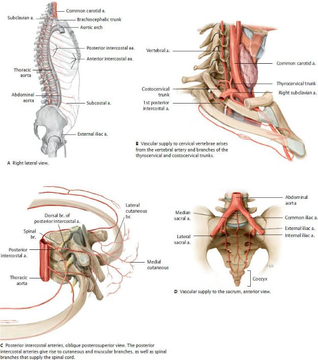
Fig. 3.16 Arteries of the trunk
(From Gilroy AM, MacPherson BR, Wikenheiser JC. Atlas of Anatomy. Illustrations by Voll M and Wesker K. 4th ed. New York: Thieme Publishers; 2020.)
Fig. 3.17 Vertebral venous plexus
The intervertebral and basivertebral veins connect the internal and external venous plexuses, which drain into the azygos system. (From Gilroy AM, MacPherson BR, Wikenheiser JC. Atlas of Anatomy. Illustrations by Voll M and Wesker K. 4th ed. New York: Thieme Publishers; 2020.)
3.2 The Spinal Cord
The spinal cord is the part of the central nervous system that relays information between the brain and the body. The spinal cord, along with its spinal nerves, surrounding membranes (the meninges), and associated vasculature, is enclosed within the vertebral canal.
Structure of the Spinal Cord
—The spinal cord, a slightly flattened cylindrical structure, is continuous with the brainstem. Within the vertebral canal, it extends from the skull base to a tapered end, the conus medullaris, at the level of L1 or L2 vertebra (Fig. 3.18).
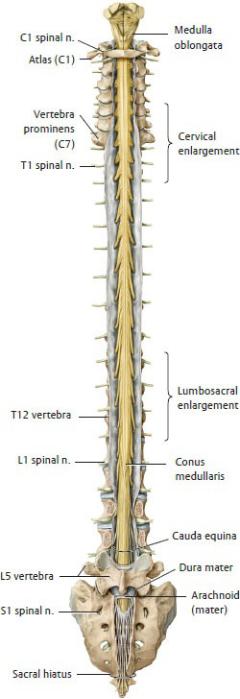
Fig. 3.18 Spinal cord in situ
Posterior view with vertebral canal windowed. (From Gilroy AM, MacPherson BR, Wikenheiser JC. Atlas of Anatomy. Illustrations by Voll M and Wesker K. 4th ed. New York: Thieme Publishers; 2020.)
— Along its length, two swellings occur in the regions of the spinal cord that
innervate the limbs:
•The cervical enlargement at C4–T1 is related to the brachial plexus, a plexus of nerves that innervate the upper limb.
•The lumbosacral enlargement at T11–S1 is related to the lumbar and sacral plexuses, nerve plexuses that innervate the abdominal wall and lower limb.
—The spinal cord consists of 31 segments (8 cervical, 12 thoracic, 5 lumbar, 5 sacral, and 1 coccygeal), each of which innervates a specific area of the trunk or limbs. Each spinal cord segment is associated with a pair of spinal nerves that pass to and from the vertebral canal through the intervertebral foramina at the corresponding vertebral level. Both spinal cord segments and spine nerves are identified by region and number (e.g., T4).
—The adult spinal cord is considerably shorter than the vertebral column, occupying only the superior two thirds of the vertebral canal. As a result, most spinal cord segments do not lie adjacent to the vertebral level of the same number although the spinal nerves arising from those segments still exit through the corresponding intervertebral foramina (Fig. 3.19).
—The spinal cord is surrounded by three layers, or meninges, and is suspended within cerebrospinal fluid.
Meninges of the Spinal Cord
The spinal meninges are membranes that surround the spinal cord and nerve roots and that contain the cerebrospinal fluid (a fluid that cushions and nourishes the brain and spinal cord) (Fig. 3.20); see also Section 26.2).
—The three layers of spinal meninges are continuous with the meninges that surround the brain:
1.Dura mater, a tough outer layer that forms the dural sac enclosing the spinal cord and extending along the nerve roots to the intervertebral foramina. The dural sac begins at the foramen magnum of the skull and ends at the level of S2.
2.Arachnoid mater, a delicate middle layer that lines the dural sac and is connected to the underlying membrane by arachnoid trabeculae (strands of connective tissue).
3.Pia mater, a thin layer that adheres to the surface of the spinal cord. Denticulate ligaments, transverse extensions of the pia mater, attach to the dura mater and suspend the spinal cord within the dural sac.
—The filum terminale, a thin cord of connective tissue invested by pia mater, extends from the conus medullaris to the apex of the dural sac. There it is surrounded by spinal dura mater and extends to the end of the vertebral
canal, where it anchors both membranes to the coccyx.
—Three spaces separate the layers of meninges (Fig. 3.21):
•The epidural space lies between the bony wall of the vertebral canal and the dura mater. It contains fat and the vertebral venous plexus.
•The subdural space, a potential space between the dura and arachnoid layers, contains a thin film of lubricating fluid.
•The subarachnoid space lies deep to the arachnoid layer and contains the cerebrospinal fluid which bathes the spinal cord and roots of the spinal nerves. This space expands inferiorly as the lumbar cistern between the end of the spinal cord at the L1/L2 vertebra, and the end of the arachnoid-lined dural sac at the S2 vertebra.
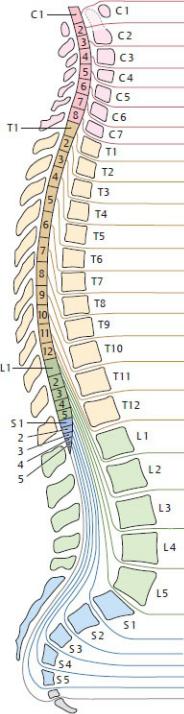
Fig. 3.19 Spinal cord segments and vertebral levels
The spinal cord is divided into four major regions: cervical, thoracic, lumbar, and sacral. Spinal cord segments are numbered by the exit points of their associated spinal nerves. (From Schuenke M, Schulte E, Schumacher U. THIEME Atlas of Anatomy, Vol 1. Illustrations by Voll M and Wesker K. 3rd ed.
New York: Thieme Publishers; 2020.)
Fig. 3.20 Spinal cord and its meningeal layers
Posterior view. The dura mater is opened, and the arachnoid mater is sectioned. (From Gilroy AM, MacPherson BR, Wikenheiser JC. Atlas of Anatomy. Illustrations by Voll M and Wesker K. 4th ed. New York: Thieme Publishers; 2020.)
BOX 3.9: CLINICAL CORRELATION
LUMBAR PUNCTURE, SPINAL ANESTHESIA, AND EPIDURAL ANESTHESIA
A lumbar puncture, used to extract cerebro spinal fluid from the spinal subarachnoid space, is administered by inserting a needle between the spinous process of L3 and L4 (sometimes between L4 and L5). The nee-dle pierces the ligamentum flavum and wall of the dural sac before entering the lumbar cistern (2). The injection of a local anes-thetic for spinal anesthesia is also admin-istered in this manner. A similar approach may be used for epidural anesthesia (1), to anesthetize emerging spinal nerves, but the anesthetic is injected into the epidural space without entering the dural sac. A cau-dal approach through the sacral hiatus also allows
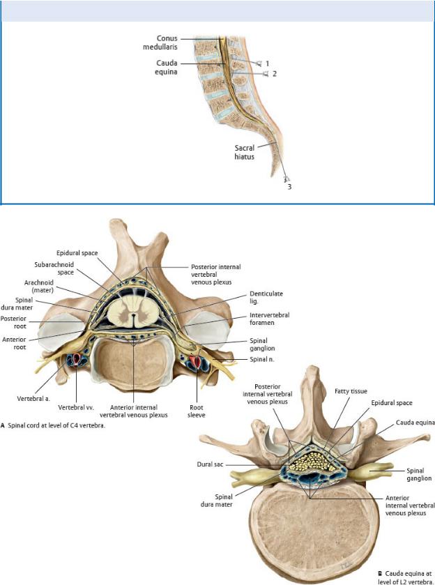
access to the epidural space (3).

Fig. 3.21 Spinal cord in situ: transverse section
Superior view. (From Gilroy AM, MacPherson BR, Wikenheiser JC. Atlas of Anatomy. Illustrations by Voll M and Wesker K. 4th ed. New York: Thieme Publishers; 2020.)
BOX 3.10: DEVELOPMENTAL CORRELATION
DEVELOPMENTAL CHANGES OF THE SPINAL CORD, DURAL SAC, AND VERTEBRAL COLUMN
During postnatal development, the longitudinal growth of the vertebral column exceeds that of the spinal cord. At birth, the conus medullaris is at the level of the L3 vertebra, but in the average adult it lies at the level of the L1 or L2 vertebra. At all ages, the lumbar cistern of the dural sac extends into the sacral canal.
(From Gilroy AM, MacPherson BR, Wikenheiser JC. Atlas of Anatomy. Illustrations by Voll M and Wesker K. 4th ed. New York: Thieme Publishers; 2020.)
Blood Supply to the Spinal Cord
Arterial blood supply to the spinal cord arises from the vertebral arteries as well as from branches of the subclavian arteries and descending aorta (Fig. 3.22).
—Longitudinal spinal arteries supply the superior part of the spinal cord.
•A single anterior spinal artery arises from the two vertebral arteries (branches of the subclavian arteries) and supplies the anterior two thirds
of the spinal cord.
•Paired posterior spinal arteries arise from the vertebral arteries (or one of their branches, the posterior cerebellar artery) and supply the posterior third of the spinal cord.
—Anterior and posterior segmental medullary arteries are large, irregularly spaced vessels that communicate with the spinal arteries.
•They arise from branches of the subclavian artery and segmental arteries in the thoracic and lumbar regions.
•The medullary arteries enter the vertebral canal through the intervertebral foramina and are found mainly at the cervical and lumbar enlargements.
—The great anterior segmental medullary artery (of Adamkiewicz), a single large, usually left-sided vessel, can provide an important contribution to the circulation of the lower two thirds of the spinal cord.
•It arises as a branch of a lower thoracic or lumbar segmental artery.
•It enters the vertebral canal through an intervertebral foramen in the lower thorax or upper lumbar region.
—The anterior and posterior radicular arteries are small arteries that supply the roots of the spinal nerves and the superficial gray matter of the spinal cord. They do not communicate with the spinal arteries.
Veins of the spinal cord, which are more numerous than the arter-ies, have the same distribution, anastomose freely with one another, and drain into the internal vertebral plexus (Fig. 3.23).
Fig. 3.22 Arteries of the spinal cord
The unpaired anterior and paired posterior spinal arteries typically arise from the
vertebral arteries. As they descend within the vertebral canal, the spinal arteries are reinforced by anterior and posterior segmental medullary arteries. Depending on the spinal level, these reinforcing branches may arise from the vertebral, ascending or deep cervical, posterior intercostal, lumbar, or lateral sacral arteries. (From Gilroy AM, MacPherson BR, Wikenheiser JC. Atlas of Anatomy. Illustrations by Voll M and Wesker K. 4th ed. New York: Thieme Publishers; 2020.)
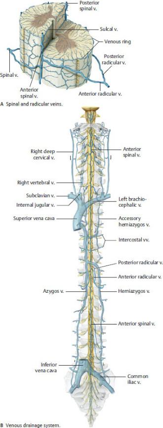
Fig. 3.23 Veins of the spinal cord
The interior of the spinal cord drains via venous plexuses into an anterior and a posterior spinal vein. The radicular and spinal veins connect the veins of the spinal cord with the internal vertebral venous plexus. The intervertebral and basivertebral veins connect the internal and external vertebral venous plexuses, which drain into the azygos system. (From Gilroy AM, MacPherson BR, Wikenheiser JC. Atlas of Anatomy. Illustrations by Voll M and Wesker K. 4th ed. New York: Thieme Publishers; 2020.)
3.3 Spinal Nerves
Spinal nerves transmit information between peripheral body tis-sues and the spinal cord. A single pair of spinal nerves arises from each spinal cord segment.
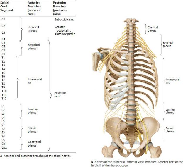
—There are 31 pairs of spinal nerves: 8 cervical, 12 thoracic, 5 lumbar, 5 sacral, and 1 coccygeal. Each pair is named for the spinal cord segment from which it arises.
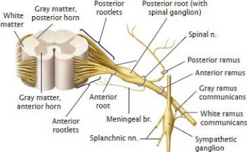
—Spinal nerves are formed by the merging of (Fig. 3.24)
•an anterior root carrying motor (efferent) fibers whose cell bodies are located in the anterior horn of the spinal cord, and
•a posterior root carrying sensory (afferent) fibers whose cell bodies are located in a spinal ganglion located outside the spinal cord.
—Spinal nerves pass through the intervertebral foramina at the corresponding vertebral level.
•Cervical nerves C1–C7 exit the vertebral canal superior to the vertebra of the same number (e.g., C4 spinal nerve exits between C3 and C4 vertebrae).
•C8 spinal nerve exits below the C7 vertebra (between the C7 and T1 vertebrae).
•Spinal nerves T1–Co1 exit the canal inferior to the corresponding vertebra.
—Because the spinal cord is shorter than the vertebral column, nerve roots from the lower spinal cord (L2–Co1) must descend below the conus medullaris within the lumbar cistern of the dural sac before exiting through the respective intervertebral foramina. This loose group of nerve roots within the dural sac is called the cauda equina (see Figs. 3.19 and 3.21).
Peripheral Nerve Pathways of the Somatic Nervous system
The somatic nervous system innervates structures under conscious control
including the skin and skeletal muscles.
—As spinal nerves emerge from the intervertebral foramen, they split to form rami (branches) that contain both sensory and motor fibers (except C1, which contains only motor fibers) (Fig. 3.24).
•Posterior rami innervate skin and muscles of the posterior trunk, head, and neck.
•Anterior rami form peripheral nerves and plexuses that innervate the rest of the body.
—Posterior rami do not form plexuses, and most are referred to by the spinal cord segment from which they originate (e.g., T4 posterior ramus). The suboccipital n. (C1), greater occipital n. (C2), and third occipital n. (C3) that innervate the scalp are the only individually named posterior rami (Fig. 3.25A).
—Anterior rami have a larger distribution than posterior rami, and most have a more complex course (Fig. 3.25).
•Anterior rami of thoracic spinal nerves do not form plexuses but become intercostal nerves, which run in the spaces between ribs to innervate the thoracic and anterolateral abdominal walls.
•Anterior rami of the cervical, lumbar, and sacral regions form plexuses that give rise to multi-segmental nerves that may carry only sensory or only motor fibers, or they may be mixed nerves, carrying both sensory and motor fibers. These nerves are usually given specific names (e.g., radial n., femoral n.). These somatic plexuses include:
◦Cervical plexus (C1–C4) innervates muscles of the neck and skin over the neck and scalp.
◦Brachial plexus (C5–T1) innervates the pectoral girdle and upper limb.
◦Lumbar plexus (L1–L4) innervates the lower anterior abdominal wall and anterior thigh.
◦Sacral plexus (L4–S3) innervates the gluteal region, posterior thigh, and leg.
—In somatic nerve plexuses, sensory nerve fibers associated with a single spinal cord segment are distributed among several peripheral nerves. Traveling from the periphery toward the spinal cord, the sensory fibers converge to enter the spinal cord via the posterior root. There they synapse with sensory neurons in the posterior horn of the spinal cord (Fig. 3.26).
—Sensory roots from each spinal cord segment correspond to the sensory (cutaneous) innervation of a particular area of skin, known as a dermatome (Fig. 3.27). Because sensory fibers from each segment are distributed
among multiple peripheral nerves, there is a large area of overlap between adjacent dermatomes. Thus, the lesion of a single sensory nerve root (such as by the impingement of a herniated disk) would have a minimal effect.
—Typically peripheral sensory nerves carry fibers from several spinal cord levels. Lesions of a sensory nerve, therefore, would affect a larger cutaneous territory covering multiple derma tomes (Fig. 3.28).
—Cutaneous sensations transmitted by somatic sensory nerves include those of pain, pressure (touch), and temperature. Sensory nerves also transmit a sense of position, or proprioception, that provides information about the spatial position of the limbs.
—Similar to the distribution of sensory fibers in a plexus, motor fibers from multiple spinal cord levels may combine in a peripheral nerve to innervate a single skeletal muscle. In other cases, muscles may be innervated by a single spinal cord segment. Muscles are divided into two groups based on their innervation patterns (Fig. 3.29):
•Monosegmentally innervated muscles are innervated by motor neurons from a single spinal cord segment.
•Polysegmentally innervated muscles are innervated by neurons whose nuclei extend over several spinal cord segments.
—Myotomes represent the muscle mass that is innervated by a single spinal cord segment. For example, although the femoral n. and the obturator n. both contain anterior rami of L2–L4, they innervate different muscles. The L2 myotome would comprise all of the muscle fibers innervated by the L2 spinal segment, whether carried in the femoral n. or obturator n.
Fig. 3.24 Structure of a spinal cord segment
Anterior view. Spinal cord segments are defined as a section of the spinal cord that is associated with a single pair of spinal nerves. The nerves arise as anterior
(motor) and posterior (sensory) rootlets that combine to form anterior and posterior roots, respectively. The two roots fuse within the intervertebral foramen to form a mixed spinal nerve. Subsequent branches of the spinal nerve contain both motor and sensory fibers (except the meningeal branch, which is only sensory). (From Gilroy AM, MacPherson BR, Wikenheiser JC. Atlas of Anatomy. Illustrations by Voll M and Wesker K. 4th ed. New York: Thieme Publishers; 2020.)
Fig. 3.25 Nerves of the trunk wall
(From Schuenke M, Schulte E, Schumacher U. THIEME Atlas of Anatomy, Vol 1. Illustrations by Voll M and Wesker K. 3rd ed. New York: Thieme Publishers; 2020.)
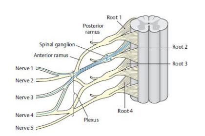
Fig. 3.26 Principles of plexus formation: sensory nerves
The axons, which form the afferents from a dermatome, extend from that dermatome to a single root in the spinal cord. (From Gilroy AM, MacPherson BR, Wikenheiser JC. Atlas of Anatomy. Illustrations by Voll M and Wesker K. 4th Edition. New York: Thieme Publishers; 2020.)
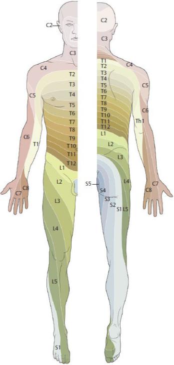
Fig. 3.27 Dermatomes of the head, trunk, and limbs
Each spinal cord segment innervates a particular skin area (dermatome). Dermatomes are band-like areas of skin innervated by a pair of spinal nerves arising from a single spinal cord segment. Since spinal nerve C1 carries only motor fibers, there is no corresponding dermatome. (From Schuenke M, Schulte
E, Schumacher U. THIEME Atlas of Anatomy, Vol 1. Illustrations by Voll M and Wesker K. 3rd ed. New York: Thieme Publishers; 2020.)
—Muscles that are innervated by a single segment of the spinal cord (monosegmental) can be evaluated clinically by testing a corresponding reflex. Reflexes, such as the patellar tendon reflex, are mediated by motor (efferent limb) and sensory (afferent limb) neurons that are located within a single seg-ment of the spinal cord.

Fig. 3.28 Pattern of peripheral sensory cutaneous sensation
Sensory loss patterns that are associated with a peripheral nerve lesion overlap multiple dermatomes. (From Schuenke M, Schulte E, Schumacher U. THIEME Atlas of Anatomy, Vol 1. Illustrations by Voll M and Wesker K. 3rd ed. New York: Thieme Publishers; 2020.)
Fig. 3.29 Monosegmental and polysegmental innervation
(From Gilroy AM, MacPherson BR, Wikenheiser JC. Atlas of Anatomy. Illustrations by Voll M and Wesker K. 4th ed. New York: Thieme Publishers; 2020.)
Peripheral Nerve Pathways of the Autonomic Nervous System
The autonomic nervous system, the visceral part of the peripheral nervous system regulates the internal environment in response to both internal and external stimuli.
—Its two divisions often have antagonistic effects on the same organ and coordinate these responses to maintain a homeo-static internal environment
(Fig. 3.30; Table 3.2):
•The sympathetic division allows the body to respond to stress (“fight or flight”).
•The parasympathetic division allows the body to maintain, or return to, a state of homeostasis (“rest and digest”).
—Nerve pathways of both the sympathetic and parasympathetic systems
consist of a two-neuron chain between the central nervous system (CNS) and the target organ: a proximal preganglionic (presynaptic) neuron and distal postganglionic (postsynaptic) neuron that synapse with each other in an intervening ganglion (Fig. 3.31).
—Autonomic nerves often form dense plexuses, which travel to their target organs along arterial routes. Plexuses are named for the organs they innervate, such as cardiac or hepatic, or for the artery they follow, such as internal carotid, celiac, or renal.
—The sympathetic division is known as the thoracolumbar component because its nerves arise from the lateral horn of the spinal cord in the thoracic and lumbar regions (T1–L2). They exit through the intervertebral foramina with the motor fibers of the corresponding spinal nerves.
—In addition to sympathetic nerves, the sympathetic system includes paired sympathetic trunks, chains of paravertebral ganglia that contain the cell bodies of postganglionic sympa-thetic nerves. The trunks run along each side of the vertebral bodies from C1 to S5. The paravertebral ganglia are connected to spinal nerves via
•gray rami communicans, which connect paravertebral ganglia at all levels (C1–S5) to the corresponding spinal nerve; and
•white rami communicans, which connect only paraver tebral ganglia between T1 and L2 to the corresponding spinal nerve.
—Shortly after emerging from the vertebral column, the pregangli-onic sympathetic fibers leave the spinal nerve via white rami communicans to enter the paravertebral ganglion. From this point the sympathetic fibers can take three different routes:
•They can synapse with the postganglionic neuron in the paravertebral ganglia at that level and return to the spinal nerve via the gray rami communicans. As part of the spinal nerve, these fibers will follow the sensory and motor fibers of the anterior and posterior rami to innervate structures in the dermatome.
•They can course up or down within the trunk to synapse with ganglia at other levels. The postganglionic fibers then join the spinal nerves at those levels. This route allows sympathetic fibers to be distributed to spinal nerves along the entire length of the spinal cord, and thus to all regions of the body, even though they originate only from the thoracolumbar segment.
•They can pass through the paravertebral ganglia without synapsing to form thoracic, lumbar, or sacral splanchnic nerves. These synapse in prevertebral ganglia, such as the celiac ganglion (see Section 11.2).
Postganglionic fibers contribute to autonomic plexuses in the thorax, abdomen, and pelvis that travel along periarterial routes to innervate viscera in those regions.
—The parasympathetic division is known as the craniosacral component because its nerves arise from the brain and the S2–S4 segments of the sacral spinal cord.
•Preganglionic parasympathetic fibers that arise from the brain travel with cranial nerves III, VII, IX, and X and synapse in parasympathetic ganglia of the head or, in the case of the vagus nerve, ganglia near the target organ. The neural pathways involved are discussed in detail in Section 26.3.
◦The vagus nerve is the only cranial nerve that extends inferior to the neck. Its parasympathetic fibers reach as far as the transverse colon in the abdomen. Thus, it provides parasympathetic innervation to all thoracic viscera and most of the abdominal viscera.
•Similar to sympathetic fibers in the thoracolumbar region, sacral parasympathetic nerves leave the spinal cord with the motor nerves of the corresponding somatic spinal nerves (S2–S4). These preganglionic parasympathetic fibers are known as pelvic splanchnic nerves. They con-tribute to autonomic plexuses in the pelvis and abdomen and synapse in small ganglia located close to, or within, their target organ.
—Smooth muscle in the skin and body wall (important for vasoconstriction, piloerection, gland secretion) does not receive any parasympathetic innervation. Sympathetic innervation is responsible for constriction of blood vessels; vasodilation results when sympathetic stimulation ceases.
—Visceral sensory fibers convey sensations from physiologic processes, such as distension of the bladder. Nociceptive (pain) fibers have been shown to accompany both sympa-thetic and parasympathetic nerves.
•Nociceptive fibers from viscera travel in the splanchnic nerves to sympathetic ganglia and reach the spinal nerve by way of the white rami communicans. Like somatic sensory neurons, their cell bodies are located in spinal ganglia. They enter the posterior horn of the spinal cord through the posterior roots.
•Nociceptive fibers traveling with cranial parasympathetic fibers have their cell bodies in the inferior or superior ganglia of the vagus nerve (cranial nerve X). Those traveling with sacral parasympathetic nerves have their cell bodies in the sacral spinal ganglia of S2–S4.
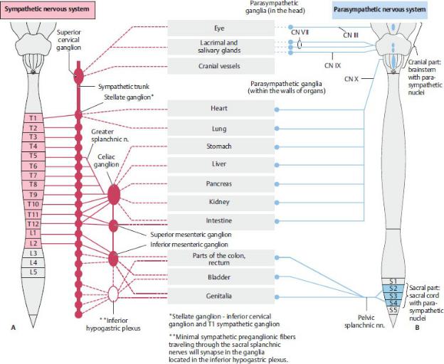
Fig. 3.30 Autonomic nervous system
The autonomic nervous system is subdivided into (A) sympathetic (thoracolumbar) and (B) parasympathetic (craniosacral) divisions. Each utilizes a two -neuron pathway in which preganglionic neurons synapse with postganglionic neurons in peripheral autonomic ganglia. (From Gilroy AM, MacPher-son BR, Wikenheiser JC. Atlas of Anatomy. Illustrations by Voll M and Wesker K. 4th ed. New York: Thieme Publishers; 2020.)
Table 3.2 Effects of the Sympathetic and Parasympathetic Nervous
Systems
Organ |
Sympathetic Nervous |
Parasympathetic |
|
System |
Nervous System |
|
|
|
Eye |
Pupillary dilation |
Pupillary constriction and |
|
|
increased curvature of the |
|
|
lens |
|
|
|
Salivary glands |
Decreased salivation |
Increased salivation |
|
(scant, viscous) |
(copious, watery) |
|
|
|
Heart |
Elevation of the heart rate |
Slowing of the heart rate |
|
|
|
Lungs |
Decreased bronchial |
Increased bronchial |
|
secretions; bronchial |
secretions; bronchial |
|
dilation |
constriction |
|
|
|
Gastrointestinal |
Decreased secretions and |
Increased secretions and |
tract |
motor activity |
motor activity |
|
|
|
Pancreas |
Decreased secretion from |
Increased secretion |
|
the endocrine part of the |
|
|
gland |
|
|
|
|
Male sex |
Ejaculation |
Erection |
organs |
|
|
|
|
|
Skin |
Vasoconstriction, sweat, |
No parasympathetic |
|
secretion, piloerection |
innervation |
|
|
|
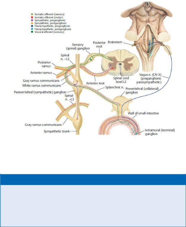
Fig. 3.31 Circuitry of the autonomic nervous system
(From Gilroy AM, MacPherson BR, Wikenheiser JC. Atlas of Anatomy. Illustrations by Voll M and Wesker K. 4th ed. New York: Thieme Publishers; 2020.)
BOX 3.11: CLINICAL CORRELATION
REFERRED PAIN
Referred pain is a sensation that originates from viscera but is perceived as if coming from an overlying or nearby somatic structure. It occurs because the somatic and visceral sensory fibers converge onto the same spinal cord segment. Diaphragmatic irritation from a splenic abscess, for example, is typically referred to the shoulder because both the diaphragm and the skin over the shoulder convey sensory information to C3–C5 segments of the spinal cord
(Fig. 3.32).
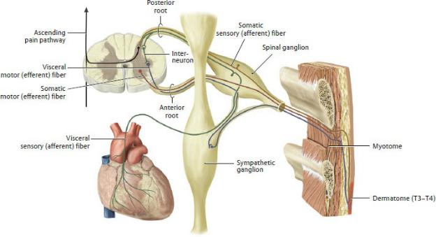
Fig. 3.32 Referred pain
It is believed that nociceptive sensory fibers from dermatomes (somatic pain) and internal organs (visceral pain) terminate on the same relay neurons in the spinal cord. The convergence of somatic and visceral sensory fibers confuses the relationship between the perceived and actual sites of pain, a phenomenon known as referred pain. The pain is typically perceived at the somatic site given that somatic pain is well localized while visceral pain is not. (From Gilroy AM, MacPherson BR, Wikenheiser JC. Atlas of Anatomy. Illustrations by Voll M and Wesker K. 4th ed. New York: Thieme Publishers; 2020.)
3.4 Muscles of the Back and Suboccipital Region
—Extrinsic muscles, the most superficial muscles that overlie the back, stabilize and move the upper limb. (See Chapter 19 for a discussion of the muscles of the upper limb.)
•Extrinsic muscles include the trapezius, latissimus dorsi, levator scapulae, and rhomboid major and minor.
—Intrinsic muscles, which attach to vertebrae or ribs, move and support the vertebral column (Table 3.3).
•They are arranged in superficial, intermediate, and deep layers
(Figs. 3.33 and 3.34).
•The superficial layer includes the splenius muscle group that covers
the deeper neck muscles laterally and posteri-orly. These muscles extend and rotate the head and neck. They extend superolaterally from the spinous processes of cervical and upper thoracic vertebrae to the occipital bone and transverse processes of C1 and C2.
•The intermediate layer includes the erector spinae muscle group that extends from the midline of the back to the angle of the ribs laterally. These large muscles are the main extensors and stabilizers of the thoracic and lumbar vertebral column. They include:
◦the iliocostalis, the most lateral column that arises from the thoracolumbar fascia, the sacrum, iliac crest, and ribs and extends superolaterally to the ribs and to cervical and lumbar vertebrae.
◦the longissimus, the middle column that arises from the sacrum, iliac crest, spinous processes of lumbar vertebrae, and transverse processes of thoracic and cervical vertebrae. It inserts superiorly on the temporal bone of the skull, to cervical, thoracic and lumbar vertebrae and to the ribs.
◦the spinalis, the most medial column that extends between the spinous processes of cervical and thoracic vertebrae.
•The deep layer includes short muscles at multiple verte-bral levels that produce small movements along the entire vertebral column. They are divided into a transversospina-lis muscle group and a deep segmental muscle group. The transversospinalis muscles extend between the transverse and spinous processes of the vertebrae. They include:
◦the semispinalis muscles, the most superficial group ◦◦ the multifidus, most prominent in the lumbar region ◦◦ the rotatores, the deepest muscles of the transversospinalis group, best developed in the thoracic region
•The deep segmental muscles are minor muscles of the back. They include the interspinales and intertransversarii that connect adjacent vertebrae, and the levatores costarum that connect vertebrae to ribs.
Table 3.3 Muscles of the Back and Suboccipital Region
Muscle Group |
Innervation |
Action |
Intrinsic muscles of the back |
|
|
|
|
|
Superficial layer |
Posterior rami of |
Extend, rotate, and |
Splenius capitis |
cervical spinal nn. |
laterally flex the head |
Splenius cervicis |
|
and cervical spine |
Intermediate layer |
Posterior rami of |
Extend and laterally flex |
(erector spinae) Spinalis |
spinal nn. |
the spine |
Longissimus Iliocostalis |
|
|
|
|
|
Deep layer |
Posterior rami of |
Extend, rotate, and |
Transversospinalis |
spinal nn. |
laterally flex the head |
group Rotatores (brevis |
|
and spine |
and longus) Multifidus |
|
|
Semispinalis Deep |
|
|
segmental group |
|
|
Interspinals |
|
|
Intertransversarii |
|
|
Levatores costarum |
|
|
|
|
|
Muscles of the suboccipital region |
|
|
Rectus capitis posterior |
Suboccipital n. (C1, |
Extend and rotate the |
major Rectus capitis |
posterior ramus) |
head |
posterior minor |
|
|
Obliquus capitis |
|
|
superior Obliquus |
|
|
capitis inferior |
|
|
|
|
|
Fig. 3.33 Superficial and intermediate muscles of the back
Posterior view. Removed: Thoracolumbar fascia (left). (From Gilroy AM, MacPherson BR, Wikenheiser JC. Atlas of Anatomy. Illustrations by Voll M and Wesker K. 4th ed. New York: Thieme Publishers; 2020.)
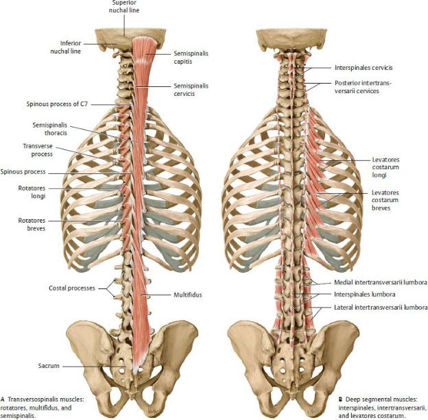
Fig. 3.34 Deep intrinsic back muscles
Posterior view. (From Gilroy AM, MacPherson BR, Wikenheiser JC. Atlas of Anatomy. Illustrations by Voll M and Wesker K. 4th ed. New York: Thieme Publishers; 2020.)
•A deep fascia that encloses the intrinsic muscles runs laterally from the posterior midline to the cervical and lumbar transverse processes and to the ribs. This thoraco-lumbar fascia continues into the neck as the deep layer of the nuchal fascia, the posterior extension of the cervical fascia (see Section 25.2).
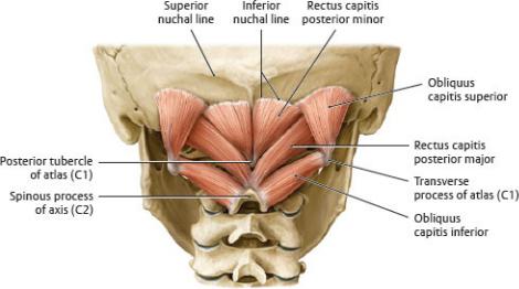
—Muscles of the posterior neck occupy the small suboccipital compartment (Fig. 3.35) that is inferior to the base of the skull and deep to the trapezius and intrinsic back muscles that extend into the neck. The suboccipital muscles arise from C1 or C2 and extend upward to insert on the occipital bone or transverse process of C1. All assist in the positioning of the head and are innervated by the suboccipital nerve, the posterior ramus of C1. They include the rectus capitis posterior major, rectus capitis posterior minor, obliquus capitis inferior, and obliquus capitis superior.
—Posterior intercostal and lumbar arteries (branches of the descending aorta and subclavian artery) supply the skin and muscles of the back.
—Posterior intercostal and lumbar veins accompany the corre-sponding arteries to drain muscles of the back. These veins are tributaries of the azygos system, which communicates with both the vertebral venous plexus and the vena cavae.
—Posterior rami of intercostal and lumbar spinal nerves supply the skin and intrinsic muscles of the back (Figs. 3.36 and 3.37).
Fig. 3.35 Short nuchal and craniovertebral joint muscles
Suboccipital muscles, posterior view. (From Schuenke M, Schulte E, Schumacher U. THIEME Atlas of Anatomy, Vol 1. Illustrations by Voll M and Wesker K. 3rd ed. New York: Thieme Publishers; 2020.)
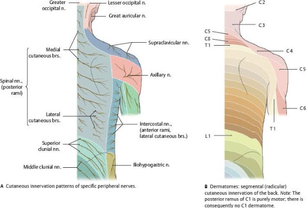
Fig. 3.36 Cutaneous innervation of the back
(From Gilroy AM, MacPherson BR, Wikenheiser JC. Atlas of Anatomy. Illustrations by Voll M and Wesker K. 4th ed. New York: Thieme Publishers; 2020.)
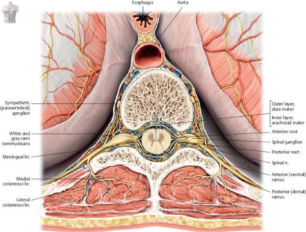
Fig. 3.37 Nerves of the back
Cross section through the vertebral column and spinal cord with surrounding musculature, superior view. (From Gilroy AM, MacPherson BR, Wikenheiser JC. Atlas of Anatomy. Illustrations by Voll M and Wesker K. 4th ed. New York: Thieme Publishers; 2020.)