


4 Clinical Imaging Basics of the Spine
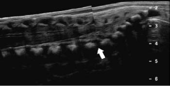
Radiographs provide a fast initial assessment of overall alignment of the spine and are often the first imaging study obtained in trauma patients. However, nondisplaced fractures may be invisi-ble on x-ray and only detected with the high resolution and detail of computed tomography (CT). Because x-ray and CT do not show soft tissues well, magnetic resonance imaging (MRI) is used for optimal evaluation of the spinal cord, nerve roots, ligaments, and intervertebral disks (Table 4.1). The fully developed adult spine is not suitable for ultrasound analysis as the bones of the verte-brae block the sound waves. In infants, the posterior aspect of the vertebral bodies are not yet ossified, allowing for sound transmis-sion into the spinal canal from the infant’s back (Fig. 4.1). This anatomic consideration, the infant’s small size, and the lack of radiation make ultrasound ideal for evaluation of the spinal cord and spinal canal in infants in the evaluation of developmental spi-nal anomalies, such as a tethered cord.
Table 4.1 Suitability of Imaging Modalities for the Back and Spine
Modality |
Clinical Notes |
Radiographs (X-rays) |
Excellent for first-line evaluation of alignment |
|
of the vertebral column and assessment for |
|
fractures in trauma patients; vertebral |
|
anatomy is well depicted |
|
|
CT (computed |
Although important soft structures are not well |
tomography) |
seen, other anatomic relationships are seen |
|
with exceptional detail; more sensitive than |
|
radiographs for the evaluation of fractures |
|
(especially nondisplaced fractures) and |
|
injuries to the posterior structures |
|
|
MRI (magnetic |
Best for evaluation of the disks, nerve roots, |
resonance imaging) |
spinal cord, ligaments, and other soft tissues |
|
|
Ultrasound |
Used only in young infants whose spinal |
|
column is not fully ossified, allowing for |
|
excellent imaging of the infant spinal cord |
|
|
Radiographic examination of the spine should include both frontal and lateral views. This is especially important for assessing alignment. Note that in both views the vertebral bodies appear as rectangles, the result of the “summation shadow” (Figs. 4.2 and 4.3). The spinous processes and the pedicles of each vertebra, superimposed on the vertebral bodies, are easily seen on the fron-tal radiographs because of the brightness of the cortical edges. The facet joints are best assessed on the lateral view. CT provides supe-rior details of the bony anatomy and vertebral column. Addition-ally, images can be reconstructed into multiple planes and into three-dimensional images to optimize the evaluation of patho-logic conditions (Fig. 4.4). MRI is extremely valuable in spine/back imaging. The superior soft tissue detail and contrast are invaluable for evaluation of the spinal cord, nerve roots, intervertebral disks, bone marrow of the vertebral bodies, and surrounding soft tissues (Figs. 4.5 and 4.6).
Fig 4.1 Infant spinal ultrasound.
Longitudinal view of a normal lower spine/spinal canal in an infant. Note the two successive images are “stitched” together at the L5-S1 level (arrow) to create this panoramic view. The probe is placed on the lower back of the infant and oriented as to center the spinal canal. (From Beek E, Van Rijn R, ed. Diagnostic Pediatric Ultrasound. 1st ed. New York: Thieme; 2015)
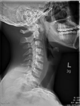
Fig. 4.2 Radiograph of the cervical spine
Left lateral view.
In a normal radiograph of the spine, the vertebral bodies appear roughly rectangular in shape and the corners of each vertebral body line up with the corners of the vertebral body above and below it. The intervertebral disk spaces are uniform from level to level and the spinous process of each vertebra is easily identified. (Courtesy of Joseph Makris, MD, Baystate Medical Center.)
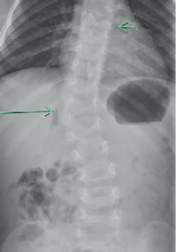
Fig. 4.3 Frontal view of the lower thoracic and lumbar spine in an infant
The inferior ribs can be seen articulating with lower thoracic vertebrae; the lower-most vertebral body with an adjacent rib is T12. Note the abnormal shape of the T12 vertebral body and that there are two pedicles on its right side (long arrow). Also note the abnormal shape of the T7 vertebral body (short arrow) with a central vertical cleft—this a “butterfly” vertebrae. Vertebral bodies should be rectangular, and each should have a small rounded pedicle visible on each side (small white circles due to the cortical bone). (Courtesy of Joseph Makris, MD, Baystate Medical Center.)
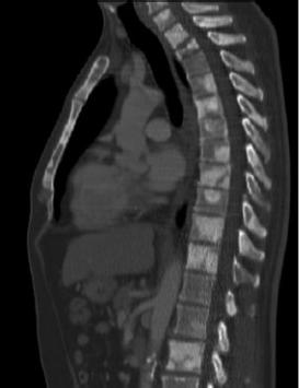
Fig. 4.4 CT reconstruction of the spine
Midsagittal section.
This midsagittal section represents a single slice of a digitally recon-structed CT scan. This image is a single slice of the midline that was digitally “reconstructed” in the sagittal plane from the original CT images. The spine is easier to evaluate in this plane because the relationships between vertebrae are better depicted. This image is presented in the “bone window,” which is best for evaluating the osseous structures. The multiple densities (whiter spots) throughout many of the vertebral bodies are metastatic lesions from prostate cancer, referred to as sclerotic or blastic lesions because there is increased bone at the metastatic focus. Although these may be visible on an x-ray of the spine, they are more obvious with CT. As with radiographs, the edges of each vertebral body should line up with the edges of the vertebral body above and below. (From Gunderman R. Essential Radiology, 3rd ed. New York: Thieme; 2014.)
Fig. 4.5 MRI of the lumbar spine
Parasagittal section through the lateral edges of the vertebral bodies and the intervertebral foramina. Left lateral view.
(Fat = white, muscles = black, nerve roots = light gray, bone = dark gray.) This image demonstrates MRI’s superior ability to show soft tissues, an advantage over radiographs and CT. The intervertebral foramina are clearly visualized with the dark nerve roots surrounded by fat within the intervertebral foramina. If this patient had a disk herniation, the disk tissue would be clearly visible intruding into the spinal canal. Note the white nucleus pulposus in the middle of the intervertebral disks. (From Moeller TB, Reif E. Pocket Atlas of Sectional Anatomy, Vol 3, 2nd ed. New York: Thieme; 2017.)

Fig. 4.6 MRI of the lumbar spine, herniated disc
Sagittal section through the mid-lumbar spine showing protrusion of the intervertebral disks into the spinal canal, most pronounced at the L4/L5 level. (Courtesy of Joseph Makris, MD, Baystate Medical Center.)

Unit II Review Questions: Back
1.During the surgical repair of an aortic aneurysm in a 62-year-old
patient, the left T10 intercostal artery that supplied the great anterior segmental artery (of Adam-kiewicz) was inadvertently ligated. A likely consequence of this would be a disruption of the blood supply to
A.the cervical enlargement of the spinal cord
B.the lumbosacral enlargement of the spinal cord
C.the lower thoracic vertebrae
D.the deep intrinsic muscles of the back
E.the posterior one third of the spinal cord
2.A 57-year-old woman who runs a 4-mile route three times a week
complained to her primary care physician of low back pain and an irritating paresthesia (tingling) along the inner aspect of her right leg. Electromyographic studies identified a lesion of her L4 spinal nerve on the right side. The L4 spinal nerve
A.carries only sensory fibers
B.contributes to both the lumbar and sacral nerve plexuses
C.contains sympathetic nerve fibers that innervate pelvic viscera
D.exits the intervertebral foramen between the L3 and L4 vertebrae
E.innervates intrinsic muscles and skin of the back via its anterior ramus
3.The filum terminale is best described as
A.an extension of the arachnoid mater that connects to the pia mater
B.a transverse ligament that suspends the spinal cord within the dural sac
C.an extension of the pia mater that descends within the lumbar cistern
D.a ligament that connects the spinous processes of the vertebrae
E.an extension of the pia mater that anchors the conus medullaris to the L2 vertebra

4.The nerves that descend within the lumbar cistern of the dural sac are
called
A.lumbar plexus
B.sacral plexus
C.cauda equina
D.posterior rami
E.sacral spinal nerves
5.An 87-year-old man sees his primary care physician with a complaint
of mental confusion and back pain. Although the physician considers that these symptoms may be a normal consequence of the patient’s age, examination reveals that the patient has advanced prostate cancer that has spread to his brain and vertebral column. The probable route of metastasis is identified as the vertebral venous plexus, which
A.drains the vertebrae and intervertebral disks but does not drain the spinal cord
B.lies within the subarachnoid space
C.consists of veins with multiple valves along their length
D.consists of paired longitudinal veins within the vertebral canal
E.is a valveless system of veins
6.A 47-year-old construction worker presented to his physician with
debilitating pain in his lower back and lower limb. Radiographic studies showed a herniated intervertebral disk at the L4–L5 vertebral level. Which of the following is true?
A.The herniated disk most likely signals a weakness of the anterior longitudinal ligament.
B.A posterior herniation of this disk could result in compression of the adjacent spinal cord.
C.Pain from this hernia would most likely be felt along the L4 dermatome.
D.The herniation is the result of a loss of elasticity of the anulus fibrosus, which allowed protrusion of the nucleus pulposus.
E.None of the above

7.The splenius muscle group
A.lies deep to the trapezius
B.is enclosed by the thoracolumbar fascia
C.extends the cervical spine and head
D.is innervated by posterior rami of cervical spinal nerves
E.All of the above
8.A 92-year-old woman remains mentally alert and physi-cally active but
has lost several inches in height, and her posture is stooped forward. Her geriatrician explains that this exaggerated curvature is due to degeneration of the bodies of two of her thoracic vertebrae that often occurs in older women as a result of decreased bone density. The curvature exhibited by this patient is known as
A.scoliosis
B.spondylolysis
C.lordosis
D.kyphosis
E.spondylosis
9.The spinal cord terminates caudally as the
A.denticulate ligament
B.filum terminale
C.conus medullaris
D.lumbar cistern
E.sacral hiatus
10.Which of the following ligaments would prevent hyper extension of the
vertebral column?
A.Anterior longitudinal ligament
B.Posterior longitudinal ligament
C.Ligamentum flavum
D.Alar ligament

E.Cruciform ligament
11.Which of the following vertebral characteristics is paired with the
correct vertebral region?
A.Promontory—sacral
B.Vertebral prominens—thoracic
C.Dens—thoracic
D.Transverse foramen—lumbar
E.Costal facet—cervical
12.Parasympathetic stimulation is responsible for responses such as
contraction of the pupil of the eye, slowing of the heart rate, and erection of the penis in the male. Which of the following statements accurately describes characteris-tics of the parasympathetic system?
A.Preganglionic fibers synapse in large sacral spinal ganglia.
B.Pelvic splanchnic nerves are parasympathetic nerves that arise from levels S2–S4 to innervate viscera of the pelvis.
C.Preganglionic fibers that arise from the brain travel with cranial nerves III, IV, V, and X.
D.Parasympathetic nerves in the skin innervate smooth muscle responsible for constriction of blood vessels.
E.Nociceptive (pain) fibers travel only with nerves of the sacral component of the parasympathetic system.
13.During the last month of her pregnancy, Janice confided to her
obstetrician that she was very anxious about mother-hood and was particularly concerned about the pain that she knew accompanied the delivery. Her physician suggested she undergo a procedure that would provide a degree of anesthesia to lessen the discomfort. Her recommendation most likely involved
A.an injection of anesthetic into the epidural space
B.a lumbar puncture
C.an injection for spinal anesthesia at the T12–L1 level
D.an extraction of cerebrospinal fluid from the subarachnoid space

E.an extraction of cerebrospinal fluid from the lumbar cistern
14.The evaluation of reflexes is part of most standard physical exams. It
measures the integrity of the nerve supply to muscles. A reflex, such as the patellar reflex, involves all of the following except:
A.primarily autonomic nerves that arise from the sympathetic trunk
B.a sensory (afferent) and a motor (efferent) limb that are located within a single spinal cord segment
C.only monosegmentally innervated muscles
D.only skeletal muscles
E.nerves that likely arise from a somatic nerve plexus
15.Valerie is an active 55-year-old executive who has recently experienced
some pain and “cracking” in her neck. Although she can relieve the pain with ibuprofen, she’s noticed that the rotation of her head from side to side is a bit more limited than previously. During her yearly physical, an x-ray of her cervical spine showed a slight loss of bone density and the formation of osteocytes. Her physician explained that this was a common result of aging and recommended continued use of mild pain relievers and regular exercise. Which of the following best describes her condition?
A.a fracture of the dens of her second cervical vertebra
B.spondylolisthesis
C.spondylosis
D.spondylolysis
E.increased laxity of the cervical spine
16.Which statement correctly distinguishes between a dermatome and a
myotome?
A.A dermatome carries only sensory fibers; a myotome carries only motor fibers.
B.Dermatomes are composed of somatic muscle fibers; myotomes are composed of smooth muscle.
C.Sensory nerves of dermatomes transmit sensation of pain, pressure, and temperature; sensory nerves of myotomes carry sensation of

proprioception (sense of position).
D.Each dermatome is innervated by a pair of spinal nerves from a single spinal cord segment; each myotome is innervated by a pair of spinal nerves from multiple spinal cord segments.
E.Lesions of a single spinal root would have a major impact on the corresponding dermatome; it would have minimal effect on the corresponding myotome.
17.A 45-year-old man has severe back pain and pain radiating down his left
leg (sciatica). Your physical exam reveals relative weakness and decreased sensation on the left side. Which imaging modality would be best suited for evaluat-ing your suspicion that the patient has a herniated intervertebral disk?
A.MRI
B.CT
C.Ultrasound
D.Radiography (X-ray)
18.A teenager is complaining of neck pain after being in a motor vehicle
accident. He is otherwise doing well. The cervical spine has been stabilized by a collar by the EMTs on the scene. Which imaging modality would you choose as a first-line assessment of the cervical spine?
A.MRI
B.CT
C.Ultrasound
D.Radiography (X-ray)
Answers and Explanations
1.B. The great anterior segmental artery (of Adamkiewicz) supplies the
inferior two thirds of the spinal cord, which includes the region of the lumbosacral enlargement (T11–S1) (Section 3.2).
A. The great anterior segmental artery (of Adam-kiewicz) supplies the

inferior two thirds of the spinal cord, which does not include the region of the cervical enlarge-ment (C4–T1).
C.Vertebrae are supplied by segmental arteries of the descending aorta, as well as by branches of the subclavian arteries and arteries of the pelvis.
D.Intrinsic muscles of the back get their blood supply from posterior branches of the intercostal and lumbar arteries.
E.The posterior spinal arteries that usually arise from the vertebral arteries in the neck supply the posterior third of the spinal cord.
2.B. The lumbar plexus includes anterior rami of spinal nerves L1–L4,
and the sacral plexus includes anterior rami of spinal nerves L4–S4 (Section 3.3).
A. All spinal nerves carry both sensory and motor fibers
C. Sympathetic fibers are carried only in spinal nerves between T1 and
L2.
D.Except for nerves in the cervical region, spinal nerves exit below the vertebra of the corresponding number; thus, L4 exits between the L4 and L5 vertebrae.
E.Intrinsic muscles and skin of the back are innervated by posterior rami of spinal nerves.
3.C. The filum terminale is a filament of pia mater that runs within the
lumbar cistern with the cauda equina, from the conus medullaris to the end of the dural sac. Inferior to the dural sac, it is surrounded by spinal dura mater and extends to the coccyx (Section 3.2).
A.The arachnoid trabeculae connect the arachnoid mater to the pia
mater.
B.Denticulate ligaments suspend the spinal cord within the dural sac.
D.The supraspinous ligament connects the spinous processes of all
thoracic, lumbar, and sacral vertebrae. In the cervical region it expands as the nuchal ligament, a finlike ligament that attaches superiorly to the occipital bone.
E. The end of the spinal cord, the conus medullaris, lies adjacent to the L2 vertebra but is not attached to it.

4.C. Because the spinal cord is shorter than the vertebral column, spinal
nerves L2–Co1 descend as a group (cauda equina) within the dural sac before exiting at the appropri-ate intervertebral foramen (Section 3.3).
A.The lumbar plexus forms outside the vertebral canal on the posterior abdominal wall and contains only anterior rami of lumbar spinal nerves (L1– L4).
B.The sacral plexus forms outside the vertebral canal on the posterior wall of the pelvis and contains only anterior rami of L4–S4 spinal nerves.
D.The nerves of the cauda equina are spinal nerves, which contain both anterior and posterior rami.
E.The cauda equina contains both lumbar and sacral spinal nerves.
5.E. The vertebral venous plexus is a valveless system that allows
communication between the caval and azygos systems, which drain the trunk, and the venous sinuses of the brain (Section 3.1).
A.The vertebral venous plexus drains the vertebrae, meninges, and spinal cord.
B.The internal vertebral venous plexus lies in the epidural space. The external plexus surrounds the outside of the vertebral column.
C.The vertebral venous plexus is a valveless system that allows communication between the caval and azygos systems, which drain the trunk, and the venous sinuses of the brain.
D.The venous plexus consists of interconnecting veins that form an internal plexus within the vertebral canal and an external plexus that surrounds the vertebrae.
6.D. Loss in elasticity of the fibrous ring, which may occur with aging,
allows herniation of the nucleus pulposus (Section 3.1).
A.The anterior longitudinal ligament supports the vertebral bodies and disks anteriorly. The posterior longi-tudinal ligament supports the disks posteriorly where herniation normally occurs.
B.The spinal cord ends at L2 and is not present in this part of the vertebral canal.
C.The L4 spinal nerve exits the intervertebral foramen superior to the
intervertebral disk and is usually unaffected by the herniation. The hernia would compress the next inferior spinal nerve, L5, and pain would be felt along that dermatome.
E.Not applicable
7.E. All of the above (Section 3.4)
A.The splenius muscles are superficial intrinsic back muscles that lie deep to the trapezius, an extrinsic muscle of the upper back. B through D are also correct (E).
B.All intrinsic back muscles, including the splenius group, are enclosed by the deep fascia of the back, the thoracolumbar fascia. A, C, and D are also correct (E).
C.Splenius muscles extend the cervical spine and head when working bilaterally. Unilaterally, they flex and rotate the head to the same side. A, B, and D are also correct (E).
D.The splenius muscles are innervated by the posterior rami of C1–C6 spinal nerves. A through C are also correct (E).
8.D. Kyphosis is an abnormal posterior curvature of the thoracic spine
often seen in older women (Section 3.1).
A.Scoliosis is a lateral curvature of the spine.
B.Spondylolysis refers to a fracture or defect across the interarticular part of the lamina of the lumbar vertebrae.
C.Lordosis is an exaggerated anterior curvature of the lumbar spine often seen in pregnant women.
E.Spondylosis is a degeneration of the intervertebral disk and corresponding vertebral body that results in osteophyte formation.
9.C. The spinal cord terminates caudally as the conus medullaris. This
usually corresponds to the L1–L2 vertebral level in an adult (Section 3.2).
A.Denticulate ligaments are transverse extensions of the pia mater that attach to dura mater and suspend the spinal cord within the dural sac.
B.Pia mater terminates caudally as the filum terminale.
D. The lumbar cistern is part of the subarachnoid space that lies between

the conus medullaris and the inferior end of the dural sac.
E. The sacral hiatus is the inferior opening of the sacral canal, which is a continuation of the vertebral canal.
10.A. The anterior longitudinal ligament attaches the anterior and lateral
surfaces of the vertebral bodies and interverte-bral disks and prevents hyperextension (Section 3.1).
B.The posterior longitudinal ligament attaches primarily to the intervertebral disks and produces weak resistance to hyperflexion.
C.The ligamenta flava join the laminae of adjacent vertebrae. They limit flexion and provide postural support of the vertebral column.
D.The alar ligaments secure the dens of C2 to the skull.
E.The cruciform ligament, formed by longitudinal fibers and a transverse ligament, secure the dens against the anterior arch of the atlas.
11.A. The anterior lip of S1 forms the promontory of the sacrum (Section
3.1).
B.The vertebral prominens is the C7 vertebra, named for its long palpable spinous process.
C.Only the C2 vertebra has a dens, the peg-like process that articulates with C1.
D.Only cervical vertebrae have transverse foramina.
E.Only thoracic vertebrae have costal facets where they articulate with the ribs.
12.B. Pelvic splanchnic nerves (S2–S4) form the parasympa-thetic
component of the autonomic plexuses that innervate pelvic viscera (Section 3.3).
A. Preganglionic nerves synapse in small ganglia near, or within, their target organ.
C.Only cranial nerves III, VII, IX, and X carry parasym-pathetic
nerves.
D.Vasoconstriction occurs through sympathetic stimulation. Blood vessels receive no parasympathetic innervation.

E. Nociceptive fibers travel with both cranial and sacral parts of the parasympathetic system, as well as with sympathetic splanchnic nerves.
13.A. Epidural anesthesia, often used during delivery, involves an injection
of anesthetic into the epidural space (Section 3.2).
B.Lumbar puncture is used to extract cerebrospinal fluid and does not involve injection of anesthetic.
C.Injections into the spinal canal should be performed inferior to L2, below the level of the conus medullaris in order to avoid damage to the spinal cord.
D and E. Anesthesia of the relevant spinal nerves requires an injection of anesthetic, not the extraction of cerebrospi-nal fluid.
14.A. Autonomic nerves innervate only involuntary muscle and are not
involved in reflexes (Section 3.3).
B.An intact reflex involves both sensory and motor limbs that are mediated at the spinal cord level.
C.Both the sensory and motor limbs of the reflex are located within a single segment of the spinal cord.
D.Only skeletal muscles are involved in reflexes. Smooth or cardiac muscles are innervated by autonomic nerves and not involved in reflexes.
E.Reflexes are conducted by somatic nerves, which most often (except intercostal nerves) form somatic nerve plexuses.
15.C. Spondylosis is an age-related condition characterized by loss of bone
density and the formation of osteocytes (Section 3.1).
A.Only a traumatic incident, such the violent “whiplash” resulting from a car accident, normally results in the fracture of the dens.
B.Spondylolisthesis describes a condition in which a vertebral body is displaced anteriorly relative to the vertebral body below it.
C.Spondylolysis describes a fracture of one or both lamina of the vertebra. When bilateral, it may result in spondylolisthesis.
E.Increased laxity of the cervical spine makes the spine more prone to injury but is not a result of aging or cause of bone loss and osteocyte

formation.
16.A. A dermatome is a band of skin, which is innervated by a sensory
nerve from a single spinal cord segment. A myotome refers to the muscle mass that’s innervated by a motor nerve from a single spinal cord segment (Section 3.3).
B.Dermatomes are bands of skin; myotomes refer to a group of skeletal (somatic) muscle fibers.
C.Sensory nerves of dermatomes transmit sensations of pain, pressure, temperature, and proprioception; myo-tomes are groups of muscle fibers innervated by motor nerves, which do not transmit sensory information.
D.Both dermatomes and myotomes are innervated by nerves from a single spinal cord segment.
E.Because of the considerable overlap among der-matomes, the loss of a single spinal nerve root would have minimal impact on sensation in that dermatome.
17.A. MRI is best for evaluation of the intervertebral disks, nerve roots,
spinal canal, and surrounding soft tissues. In this case, MRI would show the herniated portion of the intervertebral disk extending into the neuroforamina and impinging on the exiting nerve root.
B.CT is highly sensitive for fractures and malalignment of the spine but does not have sufficient soft tissue contrast to reliably diagnose nerve root, spinal cord, and disk pathology.
C.Ultrasound has no role in the evaluation of the adult spine. Unlike in infants, the adult trunk is too large and the adult spinal column is too ossified to allow for an adequate sonographic window.
D.Radiography is best for evaluating overall spinal alignment and for displaced fractures but would not be helpful in identifying disk or other soft tissue pathology.
18.D. Radiographs would be the best choice here as they provide excellent
evaluation of spinal alignment, are good for screening for fractures, and can be obtained quickly. If the x-ray is normal, you could then “clear” the
cervical spine by a thorough physical examination of the neck after removing the collar.
A.MRI would be reserved for evaluating a patient with an abnormal physical exam or if the pain is persistent, or if physical exam cannot be reliably performed (unconscious).
B.Although CT is more sensitive for fractures and subtle malalignment, its use would be reserved for cases where the radiographs are abnormal or suboptimal in quality (e.g., very large patients), especially in children to reduce overall radiation exposure. CT is also often used in unconscious patients who cannot have a reliable physical examination.
C.Ultrasound has no role in the evaluation of spinal trauma.

Unit III Thorax
5Overview of the Thorax
5.1General Features
5.1Major Structures of the Thoracic Cavity
5.2Neurovasculature of the Thorax
5.2Branches of the Thoracic Aorta
5.3Peripheral Sympathetic Nervous System
5.4Peripheral Parasympathetic Nervous System
5.1Clinical Correlation: Superior Vena Cava Syndrome
6Thoracic Wall
6.1The Breast
6.1Clinical Correlation: Carcinoma of the Breast
6.2The Thoracic Skeleton
6.3Muscles of the Thorax
6.1Muscles of the Thoracic Wall
6.4Neurovasculature of the Thoracic Wall
6.2Clinical Correlation: Insertion of a Chest Tube
7Mediastinum
7.1Regions of the Mediastinum
7.1Contents of the Mediastinum
7.2Anterior Mediastinum
7.3Middle Mediastinum: Pericardium and Pericardial Cavity
7.1Clinical Correlation: Pericarditis
7.2Clinical Correlation: Cardiac Tamponade
7.3Clinical Correlation: Surgical Significance of the Transverse Pericardial Sinus
7.4Middle Mediastinum: The Heart
7.2Borders of the Heart
7.3Position and Auscultation Sites of Cardiac Valves
7.4Developmental Correlation: Tetralogy of Fallot
7.5Clinical Correlation: Mitral Valve Prolapse
7.6Clinical Correlation: Aortic Valve Stenosis
7.7Clinical Correlation: Atrioventricular Heart Block
7.5Middle Mediastinum: Neurovasculature of the Heart
7.4Branches of the Coronary Arteries
7.8Clinical Correlation: Angina
7.9Clinical Correlation: Coronary Artery Disease
7.10Clinical Correlation: Coronary Artery Bypass Graft
7.6Prenatal and Neonatal Circulation
7.5Derivatives of Fetal Circulatory Structures
7.11Developmental Correlation: Ventricular Septal Defect
7.12Developmental Correlation: Patent Ductus Arteriosus
7.13Developmental Correlation: Atrial Septal Defect
7.14Developmental Correlation: Coarctation of the Aorta
7.7Superior and Posterior Mediastina
7.15Clinical Correlation: Achalasia
8Pulmonary Cavities
8.1The Pleura and Pleural Cavity
8.1Clinical Correlation: Pleuritis
8.2Clinical Correlation: Pneumothorax
8.3Clinical Correlation: Tension Pneumothorax
8.4Clinical Correlation: Pleural Effusion
8.2The Lungs
8.1Structure of the Lungs
8.3The Tracheobronchial Tree
8.5Clinical Correlation: Foreign Body Aspiration
8.6Clinical Correlation: Atelectasis
8.7Developmental Correlation: Neonatal Respiratory Distress Syndrome
8.8Clinical Correlation: Chronic Obstructive Pulmonary Disease
8.4Mechanics of Respiration
8.5Neurovasculature of the Lungs and Bronchial Tree.
8.2Autonomic Innervation of the Lungs and Bronchial Tree
8.9Clinical Correlation: Pulmonary Embolism
8.10Clinical Correlation: Carcinoma of the Lung
9Clinical Imaging Basics of the Thorax
9.1Suitability of Imaging Modalities for the Thorax
Review Questions: Thorax