


13 Clinical Imaging Basics of the Abdomen
In patients with acute abdominal pain, radiographs are a quick and inexpensive first imaging step that give an overview of the bowel gas pattern and can identify abdominal emergencies such as a bowel obstruction or a perforated bowel. However, x-rays usually lack the specificity to identify specific pathology.
Computed tomography (CT) on the other hand shows anatomic detail of all of the internal organs and can usually provide a diag-nosis of most intraabdominal pathology. The speed and overall ease of obtaining CT scans make it ideal for emergent conditions. Magnetic resonance imaging (MRI) also shows high detail within the abdomen but is usually reserved for non-emergent situations due to long scanning time. When an abnormality of the biliary system or urinary tract is considered, ultrasound is often the best imaging test. Lack of radiation, easy access and overall low cost of ultrasound make it suitable for both emergent and non -emergent evaluation (Table 13.1). In pediatrics, ultrasound offers additional utility as a first-line imaging tool in the evaluation of children with abdominal pain, specifically for evaluation of the appendix (Fig. 13.1).
Standard radiographic (x-rays) views of the abdomen include frontal (anteroposterior, AP) views in supine and upright positions (Fig. 13.2). Changes in position should redistribute bowel gas in a normal patient. When evaluating abdominal radiographs, a sys-tematic approach is important and should include
•evaluating the pattern of gas distribution in the bowel,
•gross estimation of organ size and location, assessment for abnormal calcifications (only the bones should be calcified, i.e., white), and
•assessment for abnormal air (outside of the bowel).
An abnormal bowel gas pattern can be a sign of a serious underly-ing condition, and the ability to recognize such patterns is an important skill to develop. All students should become familiar with both normal and abnormal patterns as a speedy diagnosis can be critical (Figs. 13.2 and 13.3). Details of abdominal struc-tures can be enhanced by the use of barium (ingested by the patient), used in fluoroscopic studies (Fig. 13.4), and by oral or intravenous contrast used for abdominal CT scans (Fig. 13.5). In MRI studies, intra-abdominal fat can act as a natural “contrast agent” by outlining the organs (Fig. 13.6). Ultrasound often
utilizes adjacent structures to better see specific target organs (Fig. 13.7).
Table 13.1 Suitability of Imaging Modalities for the Abdomen
Modality |
Clinical Uses |
|
Radiographs |
|
|
• |
X-ray |
Abdominal x-ray (“KUB”: kidneys, ureter, and |
|
|
bladder) is often the first choice in patients with |
|
|
acute abdominal pain to assess for bowel |
|
|
obstruction and/or bowel perforation. |
|
|
Radiography is readily available and a radiograph |
|
|
can be obtained very quickly. Although many |
|
|
patients will require more advanced imaging |
|
|
and/or other testing, the abdominal radiograph |
|
|
can provide crucial information and lead to |
|
|
therapeutic decisions. |
• |
Fluoroscopy |
“Real-time” radiography: x-rays viewed |
|
|
dynamically on a computer screen as the image |
|
|
is obtained; most often used to image the |
|
|
gastrointestinal (GI) tract with the aid of |
|
|
intraluminal contrast material (usually barium). |
|
|
The advent of direct visualization with endoscopy |
|
|
has minimized the use of this modality. |
CT (computed |
Provides cross-sectional anatomic detail not |
|
tomography) |
possible with plain radiographs. Most accurate |
|
|
|
imaging for evaluation of solid organs, ducts, and |
|
|
blood vessels. |
|
|
|
MRI (magnetic |
Very useful for evaluating solid organs and is |
|
resonance imaging) |
becoming increasingly more useful for evaluation |
|
|
|
of the bowel. The drawback of MRI is the long |
|
|
exam time and expense. |
|
|
|
Ultrasound |
Usually first line of imaging for evaluation of the |
|
|
|
biliary system and the kidneys. |
|
|
|
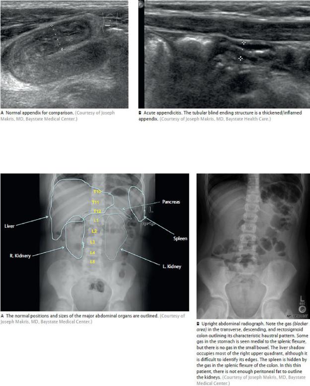
Fig. 13.1 Abdominal Ultrasound
Focused ultrasound of the right lower quadrant of the abdomen in children with abdominal pain.
Fig. 13.2 Abdominal radiographs
Anterior view.
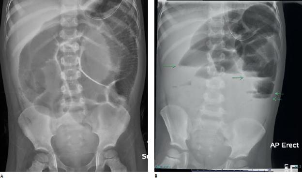
Fig. 13.3 Bowel obstruction in a young child
Supine (A) and upright (B) radiographs show several loops of gas-filled and dilated bowel. Notice that the abnormal bowel loops form C shapes and appear to be stacked upon each other. Also, note the straight horizontal lines that form in the upright position at the inferior aspect of the gas-filled bowel (arrows). These are air-fluid levels. (Courtesy of Joseph Makris, MD, Baystate Medical Center.)
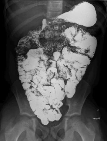
Fig. 13.4 Abdominal radiograph with barium
Anterior view.
The barium has progressed through the bowel, coating its walls, to the level of the ascending colon. Note that the loops of small bowel in the left upper quadrant (jejunum) have many more folds per length of bowel than does the bowel in the lower abdomen (ileum). This beautifully illustrates the physiologic and anatomic properties relating to absorption of nutrients in the proximal and distal small bowel (form follows function). (Courtesy of Joseph Makris, MD, Baystate Medical Center.)

Fig. 13.5 CT of the abdomen at the level of the upper kidneys
Inferior view.
Oral contrast is used to highlight the bowel (bright white); intravenous contrast is used to enhance the appearance of vessels and organs. This image demonstrates the cortical phase of enhancement; the renal cortex is whiter than the remainder of the kidneys. The full capability of CT is realized when scrolling through a full set of axial images on a computer screen and cross-referencing the axial images with the full data set of sagittal and coronal reformatted images. (From Moeller TB, Reif E. Pocket Atlas of Sectional Anatomy, Vol 2, 3rd ed. New York: Thieme Publishers; 2007.)
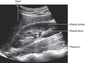
Fig. 13.6 MRI of the abdomen
Coronal section.
In this image fat is bright, air is black, and the soft tissues are shades of gray. Fluid is darker gray. Note the architecture of the normal kidney, with darker pyramids (darker because fluid content is greater there than in the cortex). The kidneys are surrounded by retroperitoneal fat. (From Moeller T, et al. Pocket Atlas of Sectional Anatomy, Vol. II: Thorax, Abdomen, Heart, and Pelvis, 3rd ed. Stuttgart: Thieme; 2007.)
Fig. 13.7 Ultrasound of the right kidney
Sagittal section.
The probe is positioned on the anterior abdominal wall and the liver is used as a “window” to view the kidney. The echogenic (whiter) renal sinus is easily seen within the renal cortex. (From Block B. Color Atlas of Ultrasound Anatomy, 2nd ed. New York: Thieme; 2012.)

Unit IV Review Questions: Abdomen
1.An arteriogram performed on one of your patients revealed significant
arterial disease (atherosclerosis) of the proximal 3 cm of the superior mesenteric artery, which narrowed the angle between the artery and the aorta. What structure normally crosses the aorta below the artery and is at risk of compression in this patient?
A.Left renal vein
B.Second part of the duodenum
C.Jejunum
D.Transverse colon
E.Pancreas
2.A 12-year-old girl is seen in the emergency department for the suspicion
of appendicitis. However, her pain is vague, and she does not complain when you gently press on her abdominal wall in the lower right quadrant. You are undeterred in your diagnosis, however, because you know that pain from an inflamed appendix refers first to another area of the abdomen based on its embryonic origins. Which of the following is true regarding referred pain from the appendix?
A.As part of the embryonic foregut, pain is referred to the epigastric region.
B.As part of the embryonic midgut, pain is referred to the epigastric region.
C.As part of the embryonic midgut, pain is referred to the periumbilical region.
D.As part of the embryonic hindgut, pain is referred to the hypogastric region.
E.As part of the embryonic hindgut, pain is referred to the periumbilical region.
3.A renal abscess can irritate the nerves of the posterior abdominal wall.

This is often referred to the dermatome that runs just above the inguinal ligament from the iliac crest to the pubis. Which nerves transmit this irritation?
A.Lateral femoral cutaneous nerve
B.Ilioinguinal and iliohypogastric nerves
C.Femoral nerve
D.Inferior phrenic nerve
E.T10 intercostal nerve
4.Which of the following is true regarding the renal vessels?
A.The right renal artery passes posterior to the inferior vena cava.
B.Both renal veins receive tributaries from the suprare-nal glands.
C.The left renal vein is shorter than the right renal vein.
D.Renal arteries are the most anterior structure in the renal hilum.
E.Renal arteries arise from the aorta at the L4 vertebral level.
5.During a colectomy on a patient with colon cancer, you ask a medical
student to describe characteristics of the descending colon. Her correct answer would include that it
A.is supplied primarily by branches of the superior mesenteric artery
B.receives parasympathetic innervation by the vagus nerve
C.is marked by three teniae coli on its outer surface
D.is primarily retroperitoneal
E.is derived from the embryonic midgut
6.The external oblique muscle and aponeurosis contribute to the
formation of all except which of the following structures?
A.Umbilical ring
B.Linea alba
C.Conjoined tendon
D.Inguinal ligament
E.Superficial inguinal ring

7.Which of the following is true regarding the relations of the pancreas?
A.The splenic artery runs along its inferior border.
B.The portal vein forms anterior to the neck and body.
C.The neck crosses the midline slightly superior to the transpyloric plane.
D.The accessory pancreatic duct drains inferiorly to the horizontal part of the duodenum.
E.It lies on the posterior wall of the omental bursa.
8.The artery that supplies blood directly to the pyloric region of the
stomach is the
A.left gastric artery
B.short gastric artery
C.right gastric artery
D.left gastro-omental artery
E.superior pancreaticoduodenal artery
9.Inferior to the arcuate line, the posterior layer of the rectus sheath is
composed of the
A.aponeurosis of the external oblique muscle
B.aponeurosis of the internal oblique muscle
C.aponeurosis of the transversus abdominis muscle
D.transversalis fascia
E.All of the above
10.One of your elderly patients has lost significant weight and complains
of abdominal pain following meals. You know that his inferior mesenteric artery was ligated several years ago as part of an aortic aneurysm repair, and imaging reveals that his superior mesenteric artery is now severely narrowed at its origin. As a result, anastomosing vessels between the celiac trunk and the superior mesenteric artery are enlarged. What vessels are involved in this anastomosis?

A.Marginal artery
B.Pancreaticoduodenal arteries
C.Gastro-omental artery
D.Proper hepatic arteries
E.Left gastric artery
11.A 46-year-old woman is admitted to the emergency department with
acute abdominal pain due to peritonitis from a ruptured duodenal ulcer. Imaging reveals an abscess in one of the peritoneal recesses. Which of the following is the lowest space within the peritoneal cavity where fluid accumulation and abscess formation are most likely to occur in a bedridden patient?
A.Omental bursa
B.Infracolic compartment
C.Left paracolic gutter
D.Subphrenic recess
E.Hepatorenal recess
12.A young mother is determined to get into shape following the birth of
her first child. She was humiliated in aerobics class when she could no longer do the required sit-ups. Strengthening which of the following muscles would help her accomplish this exercise?
A.External oblique
B.Internal oblique
C.Rectus abdominis
D.Psoas major
E.All of the above
13.Your 45-year-old brother was taken to the emergency department for
severe pain that radiated along his lower abdominal wall, inguinal region, and scrotum. Ultrasound revealed a large calculus lodged in his right ureter. From your understanding of ureteric anatomy, select the INCORRECT statement.
A. The calculus is likely to be lodged in one of the ureter’s normal
anatomic constrictions, which include the ureterovesical and ureteropelvic junctions.
B.Pain associated with the calculus is relayed to the spinal cord via sympathetic routes.
C.Pain from the ureter is felt along the T11–L2 dermatomes.
D.Ureters convey urine from the kidneys to the bladder through peristaltic action of its muscular walls.
E.Ureteric branches of the renal arteries are the primary blood supply to the pelvic ureter.
14.In patients who have conditions that affect the peritoneal cavity, such as
an inflammation due to a perforated gastric ulcer, the abdominal wall muscles, in a “defense” reflex, become rigid and can be encountered as such during physical examination. Which of the following nerves contribute sensory and motor branches to this defense mechanism?
A.Phrenic nerve
B.Vagus nerve
C.Intercostal nerves
D.Lumbar splanchnic nerves
E.Greater splanchnic nerve
15.A pediatric surgeon is performing an appendectomy on a 10-year-old
child, but upon entering the abdomen he finds a healthy appendix. With further exploration he finds an inflamed finger of bowel originating from the ileum ~ 2 feet from the ileocecal valve. It is connected to the umbilicus by a fibrous stalk. What embryonic structure failed to degenerate in this patient?
A.Ductus venosus
B.Umbilical vein
C.Umbilical artery
D.Omphalomesenteric duct
E.Urachus
16.One of your patients suffers from multisystem disease as a result of his

poor diet and long-term alcoholism. He exhibits many symptoms of chronic portal hypertension, but you suspect that some of his other symptoms have a different underlying cause. Which of the following symptoms are probably not associated with his portal hypertension?
A.Esophageal varices
B.Enlarged spleen
C.Rectal varices
D.Renal calculi
E.Ascites (fluid in the peritoneal cavity)
17.A 6-month-old boy underwent surgery for an indirect inguinal hernia.
The surgeon opened the superficial inguinal ring and located the hernia sac, which protruded through the abdominal wall
A.below the inguinal ligament
B.medial to the inferior epigastric vessels
C.within the inguinal triangle
D.at the deep inguinal ring
E.above the conjoined tendon
18.Which of the following is a characteristic of the suprarenal gland?
A.It’s a secondarily intraperitoneal organ.
B.Its cortex is composed of nervous tissue derived from neural crest cells.
C.It’s supplied by a single suprarenal artery, which arises from the renal artery.
D.Its medulla is innervated by preganglionic sympa-thetic neurons that synapse directly on the chromaf-fin cells of the medulla.
E.It sits on the superior pole of each kidney but remains outside of the perirenal fat and renal fascia that encompass the kidney.
19.Your neighbor was recently diagnosed with liver cancer. His doctor
explained that, because the primary tumor was in the bare area, it metastasized quickly to the posterior mediastinum and supraclavicular nodes. You recall that, although most lymph from the liver drains toward

the celiac nodes and intestinal trunks, the bare area drains to bronchomediastinal trunks in the thorax. What is the bare area?
A.An area on the diaphragmatic surface bounded by coronary and triangular ligaments
B.The area of liver that lines the gallbladder fossa
C.An area on the visceral surface surrounding the porta hepatis
D.The space between the leaflets of the falciform ligament
E.A subperitoneal fibrous capsule that covers the surface of the liver
20.A 58-year-old postal worker complained to his physician that he noticed
a swelling in his scrotum that felt similar to “a bag of worms,” that was present during the day but disappeared in the morning. On examination you are able to diagnose a varicocele of the pampiniform plexus that drains his left testis. What is the venous drainage of the testes?
A.The right testis drains to the inferior vena cava; the left testis drains to the left common iliac vein.
B.The right testis drains to the inferior vena cava; the left testis drains to the left renal vein.
C.The right testis drains to the right renal vein; the left testis drains to the inferior vena cava.
D.Both testes drain to ipsilateral renal veins.
E.Both testes drain to the inferior vena cava.
21.In portal hypertension portocaval anastomoses allow portal blood to
divert into the systemic system. These anastomoses might involve the
A.pancreaticoduodenal vein
B.periumbilical veins
C.renal vein
D.testicular vein
E.None of the above
22.The bile duct is most often formed by the
A.right and left hepatic ducts
B.cystic duct and common hepatic duct

C.main pancreatic duct and common hepatic duct
D.hepatopancreatic duct and cystic duct
E.main pancreatic duct and cystic duct
23.Within the testis, sperm develop in the
A.epididymis
B.tunica albuginea
C.ductus deferens
D.rete testis
E.seminiferous tubules
24.A 34-year-old male with a remote history of abdominal surgery presents
to the emergency department with severe abdominal pain, vomiting, and lethargy. What should be the first imaging study performed?
A.CT
B.Ultrasound
C.Chest x-rays
D.Abdominal x-rays
E.MRI
25.A man presented to his physician’s office with the com-plaint of severe
intermittent pain in the upper right quadrant of his abdomen. You recognize his condition as biliary colic due to gallstones lodged at the entrance to the gallbladder. What valve or sphincter maintains the opening of the cystic duct?
A.Spiral valve
B.Ileocecal valve
C.Pyloric sphincter
D.Sphincter of Oddi
E.Major duodenal papilla
26.You routinely perform vasectomies at the free clinic in your area. You

make a small incision at the top of the scrotum to access the spermatic cord. Ligation of which of the cord structures would be most effective in preventing the transmission of sperm?
A.Testicular artery
B.Pampiniform plexus
C.Urachus
D.Ductus deferens
E.Epididymis
27.What anatomic feature of the gallbladder makes it highly suitable for
ultrasound evaluation?
A.It lies on the surface of the liver.
B.It stores bile
C.A and B
D.It is pear shaped
E.It is retroperitoneal.
28.Which of the following structures forms as an intraperito-neal organ but
becomes secondarily retroperitoneal during later development?
A.Aorta
B.Pancreas
C.Spleen
D.Transverse colon
E.Kidney
29.Which of the following are reflections or remnants of peritoneum?
A.Gerota’s fascia
B.Tunica albuginea
C.Glisson’s capsule
D.Lesser omentum
E.Medial umbilical ligament

30.Which of the following are tributaries of the inferior vena cava?
A.Inferior mesenteric vein
B.Lumbar vein
C.Left gastric vein
D.Left colic vein
E.Superior rectal vein
Answers and Explanations
1.A. The left renal vein passes under the superior mesenteric artery as it
crosses the aorta and can be compressed in the narrow angle (Section 12.3).
B.The second part of the duodenum lies on the right side of the vertebral column and does not cross the aorta.
C.The jejunum is suspended by the mesentery that contains the superior mesenteric artery.
D.The transverse colon is suspended from the trans-verse mesocolon and lies anterior to the superior mesen-teric artery.
E.The pancreas lies anterior to the superior mesenteric artery.
2.C.The appendix is derived from the embryonic midgut, which refers
pain to the periumbilical region (Section 12.1).
A.The appendix is derived from the embryonic midgut, not the foregut.
B.Midgut structures refer pain to the periumbilical region; foregut structures refer pain to the epigastric region.
D.The appendix is derived from the embryonic midgut, not the hindgut.
E.Pain from the appendix is referred to the periumbilical region, but it is not part of the embryonic hindgut.
3.B. The pain is felt in the L1 dermatome innervated by the ilioinguinal
and iliohypogastric nerves (Section 10.3).
A. The lateral femoral cutaneous nerve transmits sensation from the

lateral thigh.
C.The femoral nerve transmits sensation from the anterior thigh.
D.The inferior phrenic nerve transmits sensation from the inferior surface of the diaphragm.
E.The T10 intercostal nerve transmits sensation from the T10 dermatome at the level of the umbilicus.
4.A. The right renal artery is longer than the left and passes posterior to
the inferior vena cava (Section 12.3).
B.The left suprarenal gland drains into the left renal vein, but the right suprarenal gland drains into the inferior vena cava.
C.The left renal vein crosses the aorta anteriorly and is longer than the right renal vein.
D.At the renal hilum the renal veins are anterior to the renal arteries. The renal pelvis is the most posterior structure.
E.Renal arteries arise from the aorta at the L1/L2 vertebral level.
5.C. The teniae coli, three longitudinal bands of muscle, are
characteristics of the entire large intestine (Section 12.1).
A.Branches of the inferior mesenteric artery supply the descending
colon.
B.Pelvic splanchnic nerves provide parasympathetic innervation to the descending colon.
D.The descending colon forms with the gastrointestinal tract as an intraperitoneal organ and loses its mesentery during later development, becoming secondarily retroperitoneal.
E.The descending colon is part of the embryonic hindgut.
6.C. The conjoined tendon is formed by the aponeuroses of the internal
oblique and transversus abdominis muscles (Section 10.2).
A.The umbilical ring, a remnant of the opening for the umbilical cord, interrupts the linea alba at the L4 vertebral level.
B.The linea alba is a tendinous raphe formed in the midline by the aponeuroses of the three anterior abdomi-nal wall muscles.

D.The lower edge of the external oblique muscle is thickened and curved inward to form the inguinal ligament.
E.The superficial inguinal ring is a defect in the aponeurosis of the external oblique muscles that allows passage of the spermatic cord.
7.E. The pancreas lies behind the stomach on the posterior wall of the
omental bursa (Section 12.2).
A.The splenic artery runs along the superior border of the pancreas.
B.The portal vein forms by the union of the splenic and superior mesenteric veins posterior to the neck of the pancreas.
C.The neck and body cross the midline slightly below the transpyloric plane at approximately the L2 vertebral level.
D.The accessory pancreatic duct drains into the descending part of the duodenum, superior to the drainage of the main pancreatic duct.
8.C. The right gastric artery, a branch of the proper hepatic artery,
supplies the pyloric region (Sections 11.2 and 12.1).
A.The left gastric artery supplies blood to the cardiac part of the stomach and the gastroesophageal sphincter.
B.The short gastric arteries supply blood to the fundus of the stomach.
D.The left gastro-omental artery supplies blood to the greater curvature
of the stomach and the greater omentum.
E. The superior pancreaticoduodenal artery supplies blood to the descending duodenum and the head of the pancreas.
9.D. Inferior to the arcuate line, the posterior wall of the rectus sheath is
composed of transversalis fascia (Section 10.2).
A.The external oblique muscle only contributes to the anterior layer of the rectus sheath.
B.The internal oblique muscle forms part of the posterior layer of the rectus sheath above the arcuate line and part of the anterior layer of the rectus sheath below the arcuate line.
C.The transversus abdominis forms part of the anterior layer of the rectus sheath below the arcuate line.

E.Not applicable.
10.B. The superior pancreaticoduodenal arteries arise from the
gastroduodenal artery (a secondary branch of the celiac trunk). The inferior pancreaticoduodenal arteries arise from the superior mesenteric artery. These vessels anastomose within the head of the pancreas and can enlarge significantly to form an effective collateral pathway (Section 11.2).
A. The marginal artery establishes a collateral circu lation between the superior mesenteric and inferior mesenteric arteries but does not communicate directly with branches of the celiac trunk.
C.The gastro-omental arteries anastomose with the gastroduodenal and splenic arteries but do not communi-cate with the superior mesenteric artery.
D.The proper hepatic artery anastomoses with the left gastric artery through its right gastric branch but it does not communicate directly with the superior mesenteric artery.
E.The left gastric artery anastomoses with the hepatic and splenic arteries but does not communicate directly with the superior mesenteric artery.
11.E.The hepatorenal recess, which is continuous with the subphrenic
recess, is the lowest and most gravitydependent space in the peritoneal cavity. Therefore, it is a common site for fluid collection and abscess formation (Section 11.1).
A.Fluid from the omental bursa flows into the hepato-renal recess.
B.The infracolic compartment lies below the transverse mesocolon and is separated into right and left sides by the mesentery of the small intestine. Fluid in this space can drain to the paracolic gutters and the pelvis.
C.Fluid in the left paracolic gutter would likely drain to the pelvis.
D.Fluid in the subphrenic recess drains to the more gravity-dependent hepatorenal recess in the supine patient.
12.E. The external oblique, internal oblique, and rectus abdominis muscles
flex the trunk when acting bilaterally and help stabilize the pelvis. The psoas major assists in raising the trunk from the supine position (Section

10.2).
A.The external oblique muscles flex the trunk when acting bilaterally and help stabilize the pelvis. B through D are also correct (E).
B.The internal oblique muscles flex the trunk when acting bilaterally and help stabilize the pelvis. A, C, and D are also correct (E).
C.The rectus abdominis flexes the trunk, compresses the abdomen, and stabilizes the pelvis. A, B, and D are also correct (E).
D.The psoas major flexes the hip and assists in raising the trunk from the supine position. A through C are also correct (E).
13.E. The pelvic ureter is supplied by superior vesical, inferior vesical and
uterine arteries (Section 12.3).
A.Calculi can be lodged in several natural constrictions of the ureter, including the where it passes behind the gonadal vessels or across the common iliac artery, as well as at the ureterovesical and ureteropelvic junctions.
B.Distension of the ureteric walls sends pain signals to the T11–L2 spinal cord via sympathetic nerves.
C.Pain is felt first in the lower lumbar region and moves downward to the inguinal region and medial thigh, the areas that represent the T11–L2 dermatomes.
D.The muscular walls of the ureter function through peristaltic action.
14.C. The intercostal nerves are instrumental in the ability to sense, and
react to, abdominal pain as they innervate the parietal peritoneum (sensory branches) and the abdominal wall muscles (motor branches). Inflammation of the parietal peritoneum can cause severe pain. The visceral peritoneum is not very sensitive (Section 11.1).
A.The phrenic nerve innervates the diaphragm but does not contribute to the innervation of the abdominal wall muscles.
B.The vagus nerve innervates neither the peritoneum nor the abdominal wall muscles.
D.Lumbar splanchnic nerves carry sympathetic fibers that innervate abdominal viscera.
E.The greater splanchnic nerve carries only sympathetic fibers, which
innervate abdominal viscera.
15.D. The omphalomesenteric duct (yolk stalk) has failed to regress and
remains as an ileal (Meckel’s) diverticulum (Section 12.1).
A.The ductus venosus diverts blood in the umbilical vein into the inferior vena cava in the fetus.
B.The remnant of the umbilical vein, the round ligament (ligamentum teres), runs in the inferior edge of the falciform ligament, which connects the liver to the anterior abdominal wall.
C.The medial umbilical ligament on the anterior abdominal wall is the remnant of the umbilical artery.
E.The urachus, the remnant of the fetal allantois, extends superiorly on the anterior abdominal wall from the apex of the bladder to the umbilicus, as the median umbilical ligament.
16.D. Renal calculi form in the kidney from concentrated urine and are
associated with inflammatory bowel disease and other pathology but are not a symptom of portal hyperten-sion (Section 12.2).
A.Esophageal veins that drain superiorly to the azygos (systemic) system and inferiorly to the portal system form an important portocaval anastomosis. Varices of these veins are a typical symptom of portal hypertension.
B.In portal hypertension, flow through the splenic vein slows, causing the spleen to enlarge abnormally (splenomegaly).
C.Rectal veins drain superiorly to the portal system and inferiorly to the systemic system. In portal hypertension they enlarge (forming varices) to accommodate the greater flow into the systemic system.
E.Ascites is a typical symptom of portal hypertension due to liver
disease.
17.D. Indirect inguinal hernias pass through the deep inguinal ring, which
is lateral to the inferior epigastric vessels and superior to the inguinal ligament (Section 10.4).
A. Inguinal hernias are located above the inguinal ligament; femoral

hernias are located below the ligament.
B.Indirect inguinal hernias are located lateral to the inferior epigastric vessels; direct inguinal hernias are located medial to the vessels.
C.Direct inguinal hernias protrude through the inguinal triangle; indirect inguinal hernias protrude through the deep inguinal ring lateral to the triangle.
E.The aponeuroses of the internal oblique and transversus abdominis muscles form the conjoined tendon where they attach to the pubic ramus. This is not a com-mon site for hernias.
18.D. The medulla contains chromaffin cells that function as sympathetic
ganglia where preganglionic sympathetic fibers from the celiac plexus synapse (Section 12.3).
A.The suprarenal glands develop within the retroperi-toneum and therefore are primary retroperitoneal organs.
B.The cortex is derived from mesoderm; the medulla is derived from neural crest cells.
C.Each suprarenal gland is supplied by multiple arteries arising from the aorta, and inferior phrenic and renal arteries.
E.The suprarenal glands are enclosed by the renal fascia and perirenal fat, separated from the kidney only by a thin septum.
19.A. The bare area is an area devoid of peritoneum adjacent to the inferior
surface of the diaphragm bounded by the coronary and triangular ligaments (Section 12.2).
B.The fossa of the gallbladder on the visceral surface of the liver is also devoid of peritoneum but is not known as the bare area.
C.Peritoneum covers the surface of the liver around the porta hepatis, the entry site for the structures of the portal triad.
D.The leaflets of the falciform ligament do not contain the bare area, but on the surface of the liver the leaflets separate to form the coronary ligaments, which surround the bare area.
E.The subperitoneal fibrous capsule of the liver is Glisson’s capsule, not the bare area.

20.B. The right testicular vein drains into the inferior vena cava, but the
left vein drains into the left renal vein. The angle at which the left testicular vein enters the left renal vein increases its susceptibility to obstruction. This is probably the reason why varicoceles are most commonly found on the left side (Section 10.4).
A. Neither testis drains to the common iliac vein.
C.The right testis drains to the inferior vena cava, and the left drains to the left renal vein.
D.Although the left testis drains to the ipsilateral renal vein, the right testis drains directly to the inferior vena cava.
E.Although the right testis drains directly to the inferior vena cava, the left testis drains to the ipsilateral renal vein.
21.B. Periumbilical veins anastomose with veins on the anterior abdominal
wall and act as a portocaval shunt in severe portal hypertension (Section 11.2).
A. Pancreaticoduodenal veins drain to the portal vein but do not anastomose with veins of the systemic system.
C.Renal veins drain into the inferior vena cava and do not connect to the portal system.
D.Testicular veins drain to the systemic system and do not anastomose with the portal system.
E.Not applicable.
22.B. The common hepatic duct of the liver joins the cystic duct of the
gallbladder to form the bile duct (Section 12.2).
A. The right and left hepatic ducts join to form the common hepatic duct.
C.The main pancreatic duct joins the common hepatic duct to form the hepatopancreatic ampulla.
D.The cystic duct does not join the hepatopancreatic duct. It joins the common hepatic duct to form the common bile duct.
E.The main pancreatic duct does not join the cystic duct.

23.E. Seminiferous tubules are highly coiled structures within the lobes of
the testes where sperm develop (Section 10.4).
A.The epididymis is a site of sperm storage and maturation.
B.The tunica albuginea is the tough connective tissue capsule of the
testis.
C.The ductus deferens transports sperm along the spermatic cord to the deep pelvis.
D.The rete testis is a network of ducts through which sperm exit the
testis.
24.D. Abdominal x-rays are the easiest and fastest way to get an overview
of the bowel gas pattern and assess for bowel obstruction and/or bowel perforation, both intra-abdomi-nal emergencies that may require immediate surgical intervention (Chapter 13).
A.CT may be a secondary choice in this patient.
B.Ultrasound may be a secondary tool if additional clinical signs point to a liver or biliary abnormality.
C.The patient is not having cardiopulmonary symptoms.
E.MRI is generally too time-consuming in the emergent setting.
25.A. The spiral valve in the neck of the gallbladder maintains the opening
of the cystic duct (Section 12.2).
B.The ileocecal valve is located between the ileum and cecum.
C.The pyloric sphincter is located between the pylorus of the stomach and the first part of the duodenum.
D.The sphincter of Oddi surrounds the hepatopan creatic ampulla as it enters the descending part of the duodenum at the major duodenal papilla.
E.The major duodenal papilla is an elevation on the medial wall of the descending part of the duodenum where the hepatopancreatic ampulla (formed by the union of the bile duct and pancreatic duct) enters.
26.D. The ductus deferens is the structure within the spermatic cord that

transmits sperm (Section 10.4).
A.The testicular artery does not transmit sperm.
B.The pampiniform plexus is a venous plexus that drains the testis.
C.The urachus connects the apex of the bladder to the umbilicus and transmits urine in the fetus.
E.The epididymis is a site for sperm maturation and storage located within the scrotal sac on the posterior surface of the testis.
27.C. (A and B) The liver is an excellent acoustic “window” and the
gallbladder’s position within a fossa on the visceral surface of the liver, as well as physiologically being filled with liquid bile, make ultrasound ideal for its evaluation (Chapter 13).
D. The pear shape is not a factor.
E.The gallbladder is not retroperitoneal.
28.B. Most of the pancreas is secondarily retroperitoneal. The tail of the
pancreas lies within the splenorenal ligament and is considered intraperitoneal (Sections 11.1 and 12.2).
A. The abdominal aorta is retroperitoneal and lies on the left side of the vertebral bodies.
C.The spleen is completely intraperitoneal, supported by the gastrosplenic ligament and the splenorenal ligament.
D.The transverse colon is intraperitoneal, supported by its transverse mesocolon.
E.The kidney is a primary retroperitoneal organ, surrounded by perirenal fat and covered by peritoneum only on its anterior surface.
29.D. The lesser omentum is a two-layer peritoneal mem-brane that
connects the liver to the stomach and duode-num (Section 11.1).
A.Gerota’s fascia is the renal fascia that surrounds the kidney, suprarenal glands, renal vessels, and perirenal fat.
B.The tunica albuginea is the tough outer fibrous membrane that surrounds the testis and invaginates to form the testicular lobes.
C.Glisson’s capsule is a subperitoneal fibrous capsule that surrounds the

liver.
E. The medial umbilical ligament is the remnant of the umbilical artery on the anterior abdominal wall.
30.B. The four lumbar veins on each side drain the posterior abdominal
wall and terminate in the inferior vena cava (Section 11.2).
A. The inferior mesenteric vein terminates either in the superior mesenteric or in the splenic veins, which are tributaries of the portal vein.
C.The left gastric vein drains into the portal vein.
D.The left colic vein drains to the inferior mesenteric vein, which is a tributary of the portal vein.
E.The superior rectal vein drains superiorly to the inferior mesenteric vein, which is a tributary of the portal

Unit V Pelvis and Perineum
14Overview of the Pelvis and Perineum
14.1General Features
14.1Divisions of the Pelvis and Perineum
14.1Clinical Correlation: Pelvic Diameters: True Conjugate, Diagonal Conjugate
14.2The Bony Pelvis
14.2Clinical Correlation: Laxity of Ligaments and Inceased Mobility During Pregnancy
14.3Pelvic Walls and Floor
14.2Muscles of the Pelvic Floor
14.4Pelvic Fascia
14.5Pelvic Spaces
14.6Neurovasculature of the Pelvis and Perineum
14.3Branches of the Internal Iliac Artery
14.4Lymph Nodes of the Pelvis
14.3Clinical Correlation: Pudendal Nerve Block
14.4Clinical Correlation: Pain Transmission During Labor and Delivery
15Pelvic Viscera
15.1Male Genital Structures
15.1Clinical Correlation: Prostatectomy
15.2Clinical Correlation: Prostatic Carcinoma and Hypertrophy
15.2Female Genital Structures
15.3Clinical Correlation: Ectopic Pregnancy
15.4Developmental Correlation: Bicornuate Uterus
15.5Clinical Correlation: Culdocentesis
15.3Pelvic Urinary Organs
15.6Clinical Correlation: Urethral Rupture in Males
15.4The Rectum
15.7Clinical Correlation: Rectal Examination
16The Perineum
16.1Perineal Spaces
16.2Muscles of the Perineum
16.1Muscles of the Perineum
16.1Clinical Correlation: Episiotomy
16.2Clinical Correlation: Prolapse of Pelvic Organs
16.3Male Urogenital Triangle
16.3Clinical Correlation: Retrograde Ejaculation
16.4Female Urogenital Triangle
16.5Anal Triangle
16.4Clinical Correlation: Hemorrhoids
16.5Clinical Correlation: Anal Fissures
17 Clinical Imaging Basics of the Pelvis and Perineum
7.1Suitability of Imaging Modalities for the Pelvis
Review Questions: Pelvis and Perineum