

10 The Abdominal Wall and Inguinal Region
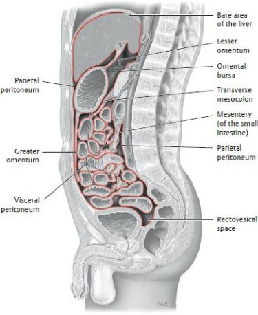
The abdomen, the region of the trunk between the thorax and the pelvis, contains the largest portion of the abdominopelvic cavity, a peritoneal-lined space that it shares with the pelvis (Fig. 10.1). The abdomen houses the primary organs of the gastrointestinal and urinary systems, although some abdominal viscera (i.e., small intestine) typically overflow the boundaries of the abdomen to occupy pelvic spaces, and pelvic viscera, when distended (i.e., bladder and uterus), can extend superiorly into the abdomen.
The abdominal wall, composed of skin, subcutaneous tissue, and muscles, is supported by its attachments to the ribs, lumbar vertebrae, and bony pelvis. It moves and stabilizes the trunk, supports the abdominal viscera, and creates intraabdominal pressure that is crucial in digestion and respiration. The muscular abdominal wall provides little protection for underlying viscera, but much of the upper abdominal viscera lies under the dome of the diaphragm, where the viscera are protected by the thoracic skeleton. The bony pelvis protects most viscera in the lower abdomen.
10.1 Regions and Planes of the Abdominal Wall
—In order to describe the location of abdominal viscera, we divide the abdomen into four quadrants or nine regions, using vertical reference lines and standard transverse planes (Fig. 10.2).
—The transpyloric plane, a transverse plane at the level of T12-L1, measured halfway between the jugular notch and pubic crest, is a useful horizontal plane that provides orientation to the internal anatomy of the abdomen (Fig. 10.3). The T12–L1 plane passes through (or very close to)
•the pylorus of the stomach,
•the ampulla of the duodenum,
•the origin of the celiac trunk,
•the origin of the superior mesenteric artery,
•the origin of the portal vein,
•the neck of the pancreas, and
•the left colic flexure of the large intestine.
Fig. 10.1 Peritoneal relationships
Midsagittal section through abdominopelvic cavity in the male, viewed from the left side. The peritoneum is shown (red). (From Gilroy AM, MacPherson BR, Wikenheiser JC. Atlas of Anatomy. Illustrations by Voll M and Wesker K. 4th ed. New York: Thieme Publishers; 2020.)
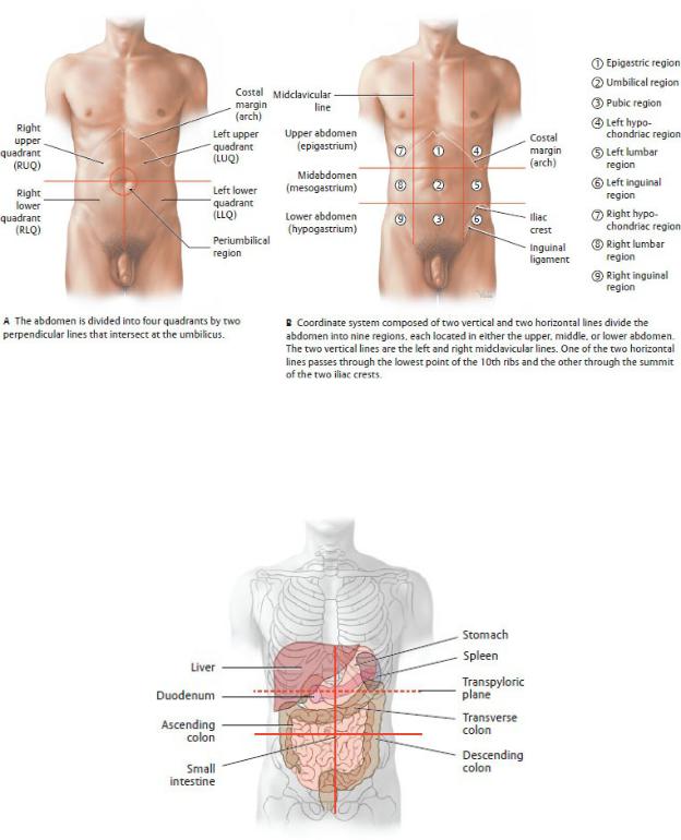
Fig. 10.2 Criteria for dividing the abdomen into regions
(From Schuenke M, Schulte E, Schumacher U. THIEME Atlas of Anatomy, Vol 1. Illustrations by Voll M and Wesker K. 3rd ed. New York: Thieme Publishers; 2020.)
Fig. 10.3 Transpyloric plane (dashed red line) and its relationship to abdominal viscera
Anterior view. (From Schuenke M, Schulte E, Schumacher U. THIEME Atlas of Anatomy, Vol 2. Illustrations by Voll M and Wesker K. 3rd ed. New York:
Thieme Publishers; 2020.)
10.2 Structure of the Abdominal Wall
Subcutaneous Layer
—The subcutaneous layer, sometimes referred to as the “superficial fascia,” of the abdominal wall, lies deep to the skin and superficial to the muscular layer. It has two components (see Fig. 10.5):
•The superficial fatty layer (Camper’s fascia), a subcutane-ous layer of fat whose thickness varies among individuals and that is continuous with the superficial fascia of the thorax, back, and lower limb
•The deep membranous layer (Scarpa’s fascia), a tough fibrous sheet that lies deep to the superficial fatty layer, covers the lower anterior abdominal wall, and extends inferiorly into the perineum, where it is continuous with the superficial perineal (Colles’) fascia.
Muscular Layer: Anterior and Posterior Walls
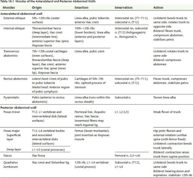
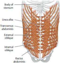
—Three flat muscles make up most of the muscular layer of the lateral and anterior walls of the abdomen: the external oblique, internal oblique, and transversus abdominis. Their large aponeuroses constitute the most anterior part of the abdominal wall (Fig. 10.4; Table 10.1).
•The thickened inferior edge of the external oblique aponeurosis forms the inguinal ligament, which attaches laterally to the anterior superior iliac spine and medially to the pubic tubercle of the pubis. Some fibers of the medial end of the ligament reflect downward as the lacunar ligament to attach to the superior edge of the pubis (see Fig. 10.14).
•Inferiorly, the aponeuroses of the internal oblique and transversus abdominis muscles join to form the conjoined tendon, where they attach to the pubis.
•In the anterior midline, the aponeuroses of the three muscles overlap those of the contralateral muscles, forming the linea alba, a tendinous raphe (junction) that extends from the xiphoid process to the pubis. The umbilical ring, a remnant of the opening for the umbilical cord, interrupts the raphe at its midpoint.
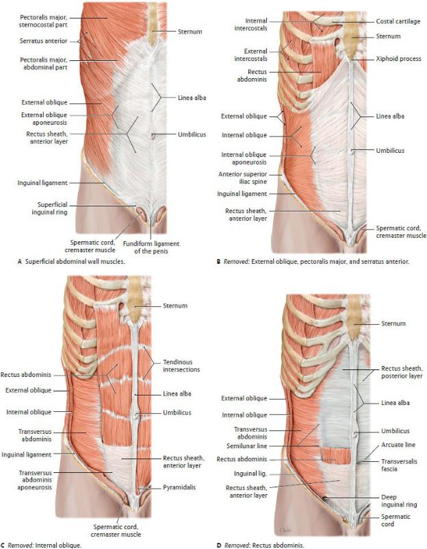
Fig. 10.4 Muscles of the anterolateral abdominal wall
Right side, anterior view. (From Schuenke M, Schulte E, Schumacher U.
THIEME Atlas of Anatomy, Vol 1. Illustrations by Voll M and Wesker K. 3rd ed. New York: Thieme Publishers; 2020.)
—On either side of the anterior midline, a rectus abdominis and pyramidalis muscle is enclosed in a rectus sheath, whose lateral edges are visible externally as the semilunar lines. The sheath has anterior and posterior layers, formed by the aponeuroses of the anterolateral muscles as they pass around the rectus muscles to decussate at the linea alba in the midline (Fig. 10.5 and 10.6).
•The anterior layer extends the length of the rectus muscle, but the posterior layer lines only its upper two thirds. The inferior end of the posterior layer is marked by a curved horizontal arcuate line, located at
a point one third of the distance between the umbilicus and pubis.
•Above the arcuate line, the anterior layer of the sheath is formed by the aponeurosis of the external oblique and an anterior leaf of the aponeurosis of the internal oblique. The posterior layer of the sheath is formed by the aponeurosis of the transversus abdominis and posterior leaf of the aponeurosis of the internal oblique.
•Below the arcuate line, the aponeuroses of all three muscles pass anterior to the rectus muscle to form the anterior rectus sheath. In this area, the posterior surface of the rectus muscle is lined only by transversalis fascia (a component of the endoabdominal fascia) and peritoneum.
—The posterior abdominal wall is the posterior boundary of the abdominal cavity. Although continuous with the region designated as the “back,” which contains the vertebral column and paravertebral muscles, the posterior abdominal wall is conceptually considered a separate region.
—Five muscles form most of the posterior abdominal wall: the psoas major, psoas minor (sometimes absent), quadratus lumborum, iliacus, and diaphragm (Fig. 10.7).
•The psoas major and iliacus muscles unite to form the iliopsoas muscle, which passes into the thigh and acts on the hip joint.
•The thoracic diaphragm forms part of the superior portion of the posterior abdominal wall.
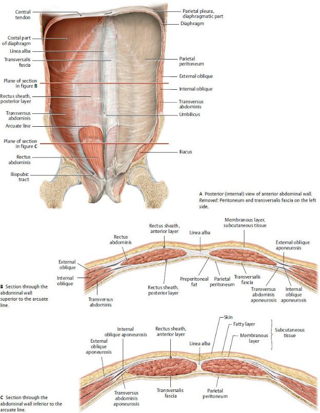
Fig. 10.5 Anterior abdominal wall and rectus sheath
(From Gilroy AM, MacPherson BR, Wikenheiser JC. Atlas of Anatomy. Illustrations by Voll M and Wesker K. 4th Edition. New York: Thieme Publishers; 2020.)
Fig. 10.6 Arrangement of the abdominal wall muscles and rectus sheath
(From Schuenke M, Schulte E, Schumacher U. THIEME Atlas of Anatomy, Vol 1. Illustrations by Voll M and Wesker K. 3rd ed. New York: Thieme Publishers; 2020.)
•The transversus abdominis muscle contributes to the lateral part of the posterior abdominal wall.
•Of the posterior wall muscles, only the quadratus lumborum is enclosed by the thoracolumbar fascia (anterior layer), which also encloses the paraspinal muscles of the back. This layer passes posterior to the psoas major and laterally fuses with the aponeurosis of the transversus abdominis.
—Endoabdominal fascia is a deep fascial layer that lines the internal surface of the abdominal wall muscles. It lies superficial to (outside of) the parietal peritoneum and in most places is separated from it by a layer of fat called preperito-neal fat.
•Each part of the endoabdominal fascia is named for the muscle it lines: transversalis fascia (Fig. 10.5), diaphrag-matic fascia, psoas fascia.
•In the inguinal region (groin), a thickened line of the transversalis fascia, the iliopubic tract, attaches to the inner edge of the inguinal ligament, where it supports the posterior wall of the inguinal canal (Fig. 10.5).
•On the posterior wall, the psoas fascia attaches to the lumbar vertebrae medially, and superiorly it blends with the medial arcuate ligament of the diaphragm. It extends inferiorly into the thigh with the tendon of the iliopsoas. This fascia separates the psoas muscle and the lumbar plexus (see Section 11.2) from viscera in the retroperito-neum of the abdominal cavity.
Internal Surface of the Anterior Abdominal Wall
The internal surface of the anterior abdominal wall is lined with transversalis fascia and parietal peritoneum, with a variable amount of intervening preperitoneal fat (Figs. 10.8 and 10.9).
—Peritoneal folds form where structures tent the peritoneum as they course between it and the transversalis fascia. The folds include
•the median umbilical fold, a single midline fold created by the median umbilical ligament, a remnant of the urachus (a fetal connection between the bladder and umbilicus);
•the medial umbilical folds, paired folds created by the medial umbilical ligaments, remnants of the umbilical arteries in the fetus; and
•the lateral umbilical folds, paired folds created by the inferior epigastric vessels.
Fig. 10.7 Muscles of the posterior abdominal wall
Coronal section with the diaphragm in the intermediate position, anterior view. (From Gilroy AM, MacPherson BR, Wikenheiser JC. Atlas of Anatomy. Illustrations by Voll M and Wesker K. 4th ed. New York: Thieme Publishers; 2020.)
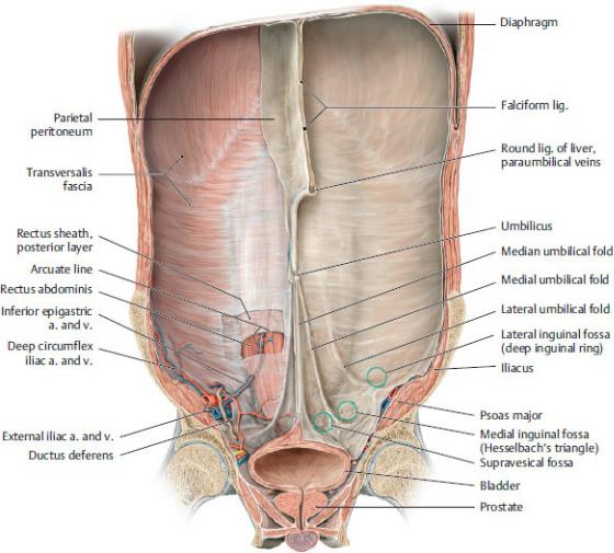
Fig. 10.8 Internal surface anatomy of the anterior abdominal wall in the male
Coronal section through the abdominal and pelvic cavity at the level of the hip joints, posterior view. (From Gilroy AM, MacPherson BR, Wikenheiser JC. Atlas of Anatomy. Illustrations by Voll M and Wesker K. 4th ed. New York: Thieme Publishers; 2020.)

Fig. 10.9 Inferior anterior abdominal wall: structure and fossae
Coronal section, posterior (internal) view of left inferior portion of the anterior abdominal wall. (From Gilroy AM, MacPherson BR, Wikenheiser JC. Atlas of Anatomy. Illustrations by Voll M and Wesker K. 4th ed. New York: Thieme Publishers; 2020.)
—Peritoneal fossae are formed between the peritoneal folds and are potential sites of herniation (protrusion of viscera through a wall or tissue). The fossae include
•the supravesical fossa between the median and medial umbilical folds;
•the medial inguinal fossa, commonly known as the inguinal triangle
(Hesselbach’s triangle) between the medial and lateral umbilical folds; and
•the lateral inguinal fossa, lateral to the lateral umbilical folds.
—The falciform ligament is a double-layered peritoneal reflection between the liver and the anterior abdominal wall that extends superiorly from the umbilicus to the roof of the abdominal cavity. It encloses the round ligament (remnant of the umbilical vein) and paraumbilical veins.
10.3 Neurovasculature of the Abdominal Wall
Arteries of the Abdominal Wall
Arteries of the abdominal wall, which anastomose extensively with one another, arise from the internal thoracic artery, the abdominal aorta, the external iliac artery, and the femoral artery (Fig. 10.10).
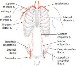
Fig. 10.10 Arteries of the abdominal wall
The superior and inferior epigastric arteries form a potential anastomosis between the subclavian and femoral arteries, which can allow blood to bypass the abdominal aorta. (From Schuenke M, Schulte E, Schumacher U. THIEME Atlas of Anatomy, Vol 2. Illustrations by Voll M and Wesker K. 2nd ed. New York: Thieme Publishers; 2016.)
—The branches of each internal thoracic artery are
•the musculophrenic artery and
•the superior epigastric artery, which descends within the rectus sheath posterior to the rectus abdominis muscle, where it anastomoses with the inferior epigastric artery.
—The paired segmental branches of the abdominal aorta are
•the intercostal, subcostal, and lumbar arteries.
—The branches of the external iliac artery are
•the inferior epigastric artery and deep circumflex iliac artery.
—The branches of the femoral artery in the thigh that supply the abdominal wall are
•the superficial epigastric artery and
•the superficial circumflex iliac artery.
Veins of the Abdominal Wall
—The deep veins of the abdominal wall accompany the arteries of similar name and drain to the superior and inferior venae cavae via the brachiocephalic, azygos, hemiazygos, and common iliac veins (Fig 10.11).
—An extensive subcutaneous venous network drains superiorly to the internal thoracic and lateral thoracic veins of the thorax and inferiorly to the inferior and superficial epigastric veins.
—Obstruction of the superior or inferior vena cava may alter the venous flow across the abdominal wall, resulting in the development or enlargement of a superficial anastomosis between the axillary and femoral veins through the thoraco-epigastric vein (see Section 6.4).
Lymphatic Drainage of the Abdominal Wall
—Lymphatic drainage of the abdominal wall is divided into upper and lower regions by a curved line (“watershed”) located between the umbilicus and costal margin (Fig. 10.12).
•From the upper region, lymph drains superiorly to ipsilateral axillary and parasternal nodes before draining superiorly to the right and left jugulosubclavian junctions (venous angles).
•From the lower region, lymph drains inferiorly to ipsilateral superficial inguinal nodes. These drain to external iliac and common iliac nodes and eventually to the thoracic duct.
Nerves of the Abdominal Wall
—Nerves of the abdominal wall arise from thoracic and lumbar spinal nerves
(Fig. 10.13) and include
•the lower intercostal nerves (T7–T11) and the subcostal nerve (T12) of the thorax, and
•the iliohypogastric nerve of the lumbar plexus.
—Dermatomes of the abdominal wall follow the slope of the ribs. Landmark dermatomes that correspond to visible surface features of the abdominal wall include T10 at the umbilicus and L1 at the inguinal ligament and top of the pubis.
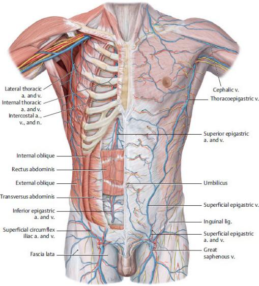
Fig. 10.11 Neurovascular structures of the anterior trunk wall
Anterior view.
Left side: superficial dissection. Right side: deep dissection. Removed: pectoralis major and minor. Partially removed: external oblique, internal oblique, transversus abdominis, rectus abdominis, and intercostal muscles. (From Schuenke M, Schulte E, Schumacher U. THIEME Atlas of Anatomy, Vol 1. Illustrations by Voll M and Wesker K. 3rd ed. New York: Thieme Publishers; 2020.)
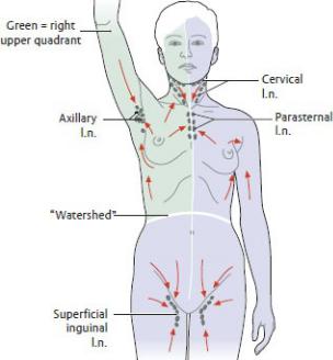
Fig. 10.12 Lymphatic pathways and regional lymph nodes of the anterior trunk wall
Anterior view. Arrows indicate the direction of lymph flow. (From Schuenke M, Schulte E, Schumacher U. THIEME Atlas of Anatomy, Vol 1. Illustrations by Voll M and Wesker K. 3rd ed. New York: Thieme Publishers; 2020.)
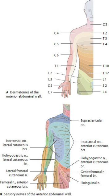
Fig. 10.13 Cutaneous innervation of the anterior abdominal wall
(From Schuenke M, Schulte E, Schumacher U. THIEME Atlas of Anatomy, Vol 1. Illustrations by Voll M and Wesker K. 3rd ed. New York: Thieme Publishers; 2020.)
10.4 The Inguinal Region
The inguinal region describes the inferolateral region of the anterior abdominal wall; the inguinal canal; and in males, the spermatic cord.
—The skin and subcutaneous tissue of the abdominal wall continues inferiorly into the thigh below the inguinal ligament and inferomedially into the perineum (see Sections 16.3 and 16.4 for discussions of the perineum).
•In the female, the skin and both the fatty and the membranous layers of the subcutaneous tissue form the labia majora.
•In the male, the skin extends into the perineum as the scrotum. The fatty layer of the subcutaneous tissue is absent, but the membranous layer continues over the penis as the superficial penile fascia and lines the scrotum as the superficial perineal fascia (Colles’ fascia).
—The anterolateral muscles of the abdominal wall and their fasciae form the inguinal canal and contribute to the coverings of the spermatic cord.
Inguinal Canal
The inguinal canal is an oblique passage through the abdominal wall that allows structures to pass between the abdominal and pelvic cavities and the perineum. Deficiencies in the anterolateral abdominal muscles, their aponeuroses, and their deep fascia cre-ate the inguinal canal (Table 10.2). The canal is present in both males and females, although it is more pronounced in the male.
—The boundaries of the canal are
•the anterior wall, formed by the aponeurosis of the external oblique muscle;
•the posterior wall, formed by transversalis fascia and conjoined tendon;
Table 10.2 Structures and Relations of the Inguinal Canal
(From Gilroy AM, MacPherson BR, Wikenheiser JC. Atlas of Anatomy. Illustrations by Voll M and Wesker K. 4th ed. New York: Thieme Publishers; 2020.)
Structures |
Formed by |
|
Wall |
Anterior wall |
External oblique aponeurosis |
|
Roof |
Internal oblique m. |
|
|
Transversus abdominis m. |
|
Posterior wall |
Transversalis fascia |
|
|
Parietal peritoneum |
|
Floor |
Inguinal lig. (densely interwoven fibers |
|
|
of the lower external oblique aponeurosis |
|
|
and adjacent fascia lata of thigh) |
|
|
|
Openings Superficial |
Opening in external oblique aponeurosis; |
|
|
inguinal ring |
bounded by medial and lateral crura, |
|
|
intercrural fibers, and reflected inguinal |
|
|
lig. |
|
|
|
|
Deep inguinal |
Outpouching of the transversalis fascia |
|
ring |
lateral to the lateral umbilical fold (inferior |
|
|
epigastric vessels) |
|
|
|
•the floor, formed by the inguinal ligament; and
•the roof, formed by the arching fibers of the aponeuroses of the internal oblique and transversus abdominis muscles.
—The canal has two openings:
•At the medial end of the canal, fibers of the external oblique aponeurosis split to create an opening known as the superficial inguinal ring. This ring lies in the anterior wall of the inguinal triangle.
•At the lateral end of the inguinal canal, immediately lateral to the origin of the inferior epigastric vessels, the transversalis fascia evaginates into the canal and creates the deep inguinal ring. This ring lies in the lateral inguinal fossa (see Fig. 10.9).
Fig. 10.14 Male inguinal
Right side, anterior view. (From Schuenke M, Schulte E, Schumacher U. THIEME Atlas of Anatomy, Vol 1. Illustrations by Voll M and Wesker K. 3rd ed. New York: Thieme Publishers; 2020.)
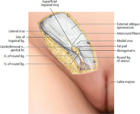
Fig. 10.15 Female inguinal region
Right side, anterior view. (From Gilroy AM, MacPherson BR, Wikenheiser JC. Atlas of Anatomy. Illustrations by Voll M and Wesker K. 4th ed. New York: Thieme Publishers; 2020.)
The Spermatic Cord
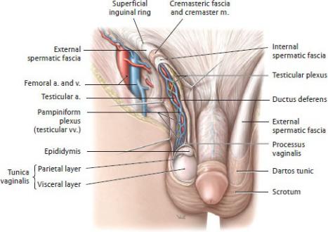
The spermatic cord forms at the deep inguinal ring, traverses the inguinal canal, and exits through the superficial inguinal ring. It enters the scrotum and descends to the posterior surface of the testis (Fig. 10.16).
—The structures in the spermatic cord include
•the ductus deferens;
•processus vaginalis;
•the testicular artery and pampiniform plexus of veins, the artery and vein of the ductus deferens, and the cremasteric artery and vein;
•lymphatic vessels of the testis and spermatic cord; and
•sympathetic and parasympathetic fibers of the testicular plexus and the genital branch of the genitofemoral nerve.
—Derivatives of the muscles and fascia of the abdominal wall sur-round the contents of the spermatic cord as they pass through the inguinal canal. The layers formed by the muscles and fascia are the same as those surrounding
the testis (Table 10.3):
•Internal spermatic fascia derived from the transversalis fascia
•Cremaster muscle and cremasteric fascia derived from the internal oblique muscle and fascia
•External spermatic fascia derived from the external oblique aponeurosis and fascia.
—The ilioinguinal nerve is not contained within the spermatic cord. However, as it traverses the layers of the abdominal wall, it travels within the inguinal canal next to the cord and is enclosed by the external spermatic fascia as it exits the superficial ring.
Fig. 10.16 Spermatic cord
Male pelvis, anterior view. Opened: Inguinal canal and coverings of the spermatic cord. (From Gilroy AM, MacPherson BR, Wikenheiser JC. Atlas of Anatomy. Illustrations by Voll M and Wesker K. 4th ed. New York: Thieme Publishers; 2020.)
Table 10.3 Coverings of the Testis
(From Gilroy AM, MacPherson BR, Wikenheiser JC. Atlas of Anatomy. Illustrations by Voll M and Wesker K. 4th ed. New York: Thieme Publishers; 2020.)
Covering layer |
Derived from |
|
|
Scrotal skin |
Abdominal skin |
Dartos m. and fascia |
Membranous layer, subcutaneous |
|
tissue |
|
|
External spermatic fascia |
External oblique aponeurosis and |
|
superficial investing fascia |
|
|
Cremaster m. and cremasteric |
Internal oblique m. |
fascia |
|
|
|
Internal spermatic fascia |
Transversalis fascia |
Tunica vaginalis, parietal layer |
Peritoneum |
|
|
Tunica vaginalis, visceral layer |
|
|
|
The transversus abdominis has no contribution to the spermatic cord or covering of the testis.
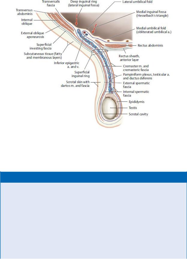
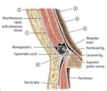
Fig 10.17 Schematic of the male inguinal canal and its relation to structures of the abdominal wall
Right side, anterior view. (From Gilroy AM, MacPherson BR, Wikenheiser JC. Atlas of Anatomy. Illustrations by Voll M and Wesker K. 4th ed. New York: Thieme Publishers; 2020.)
BOX 10.1: CLINICAL CORRELATION
INGUINAL HERNIA
Inguinal hernias account for the large majority of abdominal wall hernias, and of those, most occur in males. A hernia is the protrusion of a visceral structure into a space that it does not normally occupy. Inguinal hernias involve the protrusion of parietal peritoneum, peri-toneal fat, or the small intestine. Of the two types of inguinal her-nias, the indirect hernia can be acquired or congenital and is com-mon in young males, whereas the direct hernia is always acquired and results from a weakening of the abdominal wall and generally occurs in middle-aged males.
During development, a tongue of peritoneum, the processus vaginalis, evaginates into the newly formed inguinal canal and accompanies the testis in its descent into the scrotum. Before birth most of the processus vaginalis obliterates, closing the communication between it and the peritoneal cavity. If the processus vagi-nalis fails to obliterate, however, abdominal contents can herniate (indirect hernia) through its opening at the deep inguinal ring in the lateral inguinal fossa (lateral to the inferior epigastric vessels) and extend into the scrotum (or
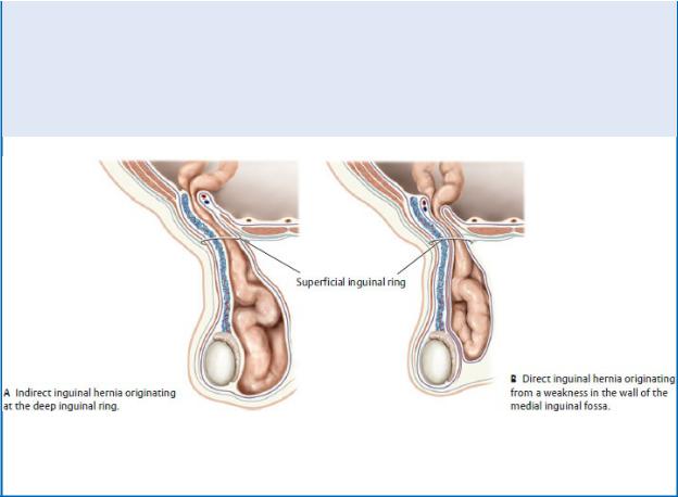
labia in females). Herniated viscera travel within the spermatic cord and therefore are covered by the layers of the cord in addition to peritoneum and transversalis fascia.
Direct hernias occur where weakening of the anterior abdominal wall in the medial inguinal triangle (medial to the inferior epigastric vessels) allows viscera to protrude through the medial end of the canal, then through an enlarged superficial ring, and into the scro-tum. Since these herniated viscera travel outside the spermatic cord, they are not covered by layers of the spermatic cord but are covered by peritoneum and transversalis fascia of the abdominal wall.
(From Schuenke M, Schulte E, Schumacher U. THIEME Atlas of Anatomy, Vol 1. Illustrations by Voll M and Wesker K. 3rd ed. New York: Thieme Publishers; 2020.)
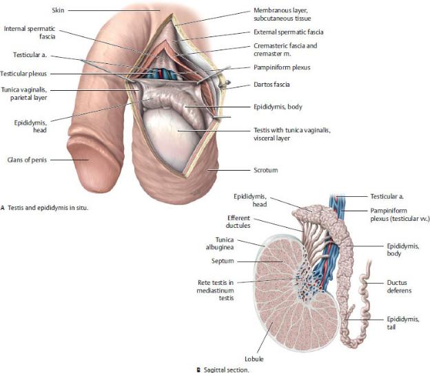
The Testes
The testes are paired ovoid reproductive organs, 4 to 5 cm long and 3 cm wide, located in separate compartments within the scro-tum. They produce spermatozoa and MRI of the testessecrete the male hormone testosterone (Figs. 10.18 and 10.19; see also Table 10.3).
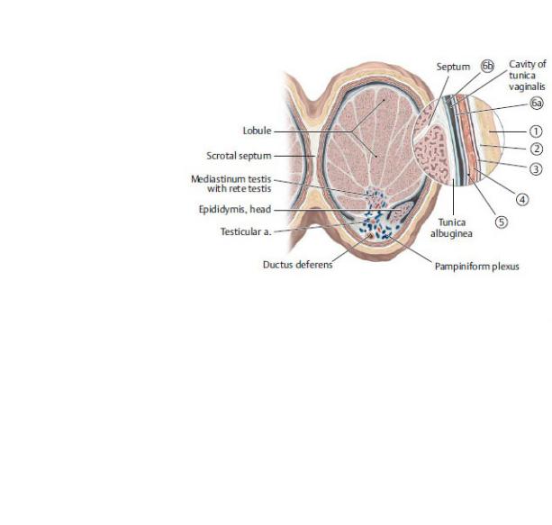
—An extension of the peritoneum known as the tunica vaginalis forms a closed sac that folds around the testis, surrounding it on all sides except on its posterior edge. The tunic has an outer parietal layer and an inner visceral layer that is adherent to the surface of the testis.
—Each testis is enveloped by the tunica albuginea, a tough capsule of connective tissue that thickens along the posterior border as the mediastinum of the testis and invaginates to divide the testis into over 200 lobules.
—Sperm develop in the seminiferous tubules, highly coiled tubules within the lobules. They exit the testes through a ductal network, the rete testis in the mediastinum, and then pass through efferent ductules to the epididymis.
—The testicular artery, a branch of the abdominal aorta, supplies the testis. A rich collateral blood supply arises from anastomoses with the artery of the ductus deferens, the cremasteric artery (a branch of the inferior epigastric artery), and the external pudendal artery (a branch of the femoral artery)
(Fig. 10.20).
—The pampiniform plexus of veins drains the testis and converges to form the testicular vein. The testicular veins drain to the inferior vena cava on the right and to the renal vein on the left.
Fig. 10.18 Testis and epididymis
Left lateral view. (From Schuenke M, Schulte E, Schumacher U. THIEME Atlas of Anatomy, Vol 2. Illustrations by Voll M and Wesker K. 3rd ed. New York: Thieme Publishers; 2020.)
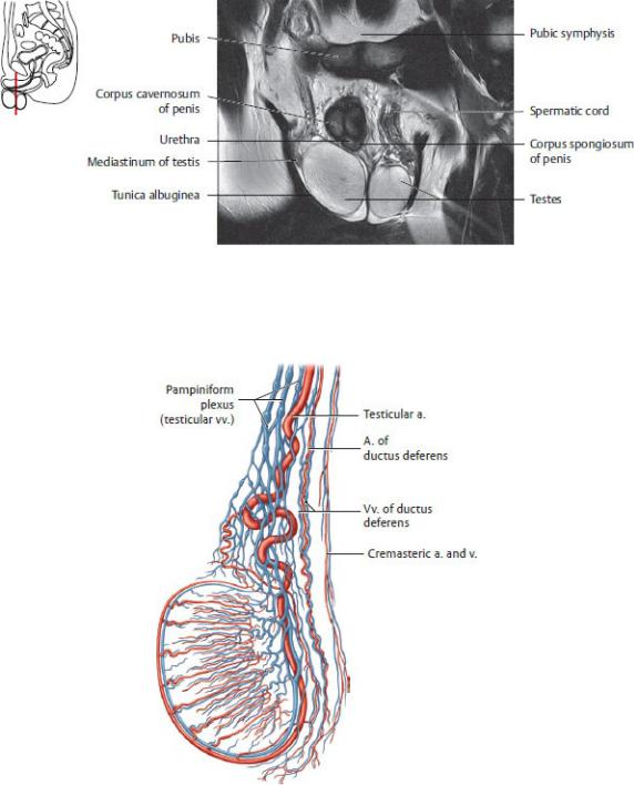
Fig 10.19 MRI of the testes
Coronal section, anterior view. (From Moeller TB, Reif E. Pocket Atlas of Sectional Anatomy, Vol 2, 3rd ed. New York: Thieme Publishers; 2007.)
Fig. 10.20 Blood vessels of the testis
Left lateral view. (From Schuenke M, Schulte E, Schumacher U. THIEME Atlas of Anatomy, Vol 1. Illustrations by Voll M and Wesker K. 3rd ed. New York: Thieme Publishers; 2020.)
— The lymph vessels of the testes drain directly to lateral aortic and preaortic
lymph nodes.
—The cremasteric reflex, initiated by stroking of the inner thigh, contracts the cremaster muscle and elevates the testis. The ilioinguinal nerve provides the sensory limb; the genital branch of the genitofemoral nerve provides the motor limb.
—The testicular nerve plexus arises from the aortic plexus and travels along with the testicular artery. It contains sympa-thetic fibers from the T7 spinal cord level, as well as visceral afferent and vagal parasympathetic fibers.
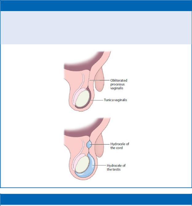
BOX 10.2: CLINICAL CORRELATION
HYDROCELE
The opening into a persistent processus vaginalis may be small enough to prevent herniation but large enough to form a hydro-cele, the accumulation of excess peritoneal fluid. The hydrocele can be confined to the scrotum (hydrocele of the testis) or to the cord (hydrocele of the cord). Ultrasound or transillumination of the scrotum confirms the presence of excess fluid.
BOX 10.3: CLINICAL CORRELATION
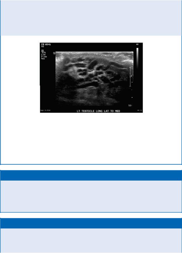
VARICOCELE
The pampiniform plexus from each testis surrounds the testicular artery and converges to form a testicular vein. If the venous valves become incompetent, the plexus can become dilated and tortu-ous, forming a varicocele that is often reported to feel like “a bag of worms.” Varicoceles are predominantly on the left side. This is generally attributed to the abrupt termination of the left testicu-lar vein at the left renal vein, which may slow venous return.
This 14-year-old boy presented with scrotal discomfort and a palpable mass. This scrotal ultrasound image shows a mass consisting of multiple serpiginous tubules, which indicate a varicocele.
(From Gunderman R. Essential Radiology, 3rd ed. New York: Thieme Publishers; 2014.)
BOX 10.4: CLINICAL CORRELATION
TESTICULAR TORSION
An absent or reduced cremasteric reflex that is accompanied by sudden testicular pain, inflammation and elevation of one testis, nausea, and vomiting may indicate testicular torsion (twisting of a testis). Prompt surgery for testicular torsion (to untwist the affected testis and anchor both testes) may prevent loss of a testis.
BOX 10.5: CLINICAL CORRELATION
TESTICULAR CANCER
Testicular cancer is the most common cancer in males between 15 and 34 years of age. The vast majority of these cancers are seminomas or germ cell tumors that arise in the germ cells that produce immature sperm. Symptoms include a lump in the affected testis (usually only
one testis is affected), a feeling of heaviness in the scrotum, pain in the affected testis or scrotum, a sudden collection of fluid in the scrotum, and the development of excess breast tissue (gynecomastia). Testicular cancer commonly metastasizes via lymph nodes to the lungs or via the bloodstream, commonly to the liver, lungs, brain, and spine.
Fig. 10.21 Asymmetric venous drainage of the right and left testes
(From Schuenke M, Schulte E, Schumacher U. THIEME Atlas of Anatomy, Vol 2. Illustrations by Voll M and Wesker K. 3rd ed. New York: Thieme Publishers; 2020.)
The Epididymis and Ductus Deferens
The epididymis and ductus deferens are parts of the male ductal system that transport sperm from the testis to the genital struc-tures in the pelvis (see Fig. 10.18).
—The epididymis, a highly coiled tubule where sperm are stored and mature, hugs the posterior surface of the testis. Its expanded head contains the lobules with the efferent ductules, its body is made up of a long convoluted duct, and its tail is continuous with the ductus deferens.
—The ductus deferens is a muscular tube that transmits sperm from the scrotum to the pelvis.
•It begins at the tail of the epididymis and continues as part of the spermatic cord through the inguinal canal.
•At the deep inguinal ring, the ductus deferens descends into the pelvis posterior to the bladder, where, near its termination, it enlarges as the ampulla of the ductus deferens (see Fig. 15.2).
•The ampulla joins with the duct of the seminal gland to form the ejaculatory duct within the prostate gland (see Section 15.1).

11 The Peritoneal Cavity and Neurovasculature
of the Abdomen
11.1 The Peritoneum and Peritoneal Cavity
The peritoneum, a thin, transparent serous membrane, lines the abdominopelvic cavity. Parietal and visceral layers of the perito-neum enclose the peritoneal cavity, which contains a thin film of serous fluid that facilitates the movement of the viscera during digestion and respiration (see Fig. 11.1).
Peritoneal Relations
—Structures in the abdomen are defined with respect to their relationship to the peritoneum (Table 11.1; Figs. 11.1 and 11.2).
•Intraperitoneal organs, almost completely enclosed by the visceral layer of the peritoneum, are suspended within the peritoneal cavity by mesenteries, double layers of peritoneum that attach to the body wall.
•Extraperitoneal structures lie posterior or inferior to the peritoneal cavity.
◦Primarily retroperitoneal structures lie posterior to the peritoneal cavity, are not suspended by a mesentery, and are covered by peritoneum only on their anterior surface.
◦Secondarily retroperitoneal structures were previously intraperitoneal structures that became fixed to the posterior abdominal wall when their mesentery fused with the parietal peritoneum of the posterior abdominal wall during development.
◦Subperitoneal structures include pelvic organs that lie below the peritoneum.
—Organs associated with the gastrointestinal tract are intraperitoneal or secondarily retroperitoneal. Organs of the urinary system are retroperitoneal.
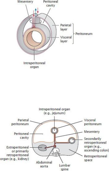
Fig. 11.1 Peritoneum and mesentery
Red and blue arrows indicate location of blood vessels. (From Gilroy AM, MacPherson BR, Wikenheiser JC. Atlas of Anatomy. Illustrations by Voll M and Wesker K. 4th ed. New York: Thieme Publishers; 2020.)
Fig. 11.2 Peritoneal relations of the organs in the abdomen
Transverse section through the abdomen showing the peritoneal relationships of abdominal organs. Viewed from above. (From Gilroy AM, MacPherson BR, Wikenheiser JC. Atlas of Anatomy. Illustrations by Voll M and Wesker K. 4th ed. New York: Thieme Publishers; 2020.)
Table 11.1 Organs of the Abdomen
Location |
Organs |
|
|
Intraperitoneal organs: These organs have a mesentery and are almost completely covered by the peritoneum.
Abdominal peritoneal cavity |
• |
Stomach |
|
|
|
|
|
• |
Small intestine |
(jejunum, |
|||
|
|
ileum, |
some |
of |
the |
|
|
|
superior |
part |
of |
the |
|
|
• |
duodenum) |
|
|
|
|
|
Spleen |
|
|
|
|
|
|
• Liver (with |
the |
exception |
|||
|
• |
of the bare area) |
|
|
||
|
Gallbladder |
|
|
|
||
|
• |
Cecum |
with |
vermiform |
||
|
|
appendix |
|
(portions |
of |
|
|
|
variable |
size |
may |
be |
|
|
• |
retroperitoneal) |
|
|
||
|
Transverse |
and sigmoid |
||||
|
|
colon |
|
|
|
|
Extraperitoneal organs: These organs either have no mesentery or lost it during development.
Retroperitoneum |
Primarily |
• |
Kidneys |
|
|
retroperitoneal |
• |
Suprarenal glands |
|
|
Secondarily |
• |
Duodenum |
(descending, |
|
retroperitoneal |
• |
horizontal, and ascending) |
|
|
|
Pancreas |
|
|
|
|
• |
Ascending |
and |
|
|
|
descending colons |
|
|
|
|
|
|
Peritoneal Structures
Most of the abdominal viscera are somewhat mobile during digestion and respiration. Reflections of the peritoneum that connect organs to the body wall or to other organs prevent excessive movement (i.e., twisting), which can compromise normal function. These reflections form mesenteries, omenta, and peritoneal ligaments (Figs. 11.3, 11. 4, 11.5, 11.6).
—A mesentery is a double layer of peritoneum that connects intraperitoneal organs to the posterior abdominal wall and transmits vessels and nerves (see Fig. 11.1). There are three major mesenteries in the abdomen (Fig. 11.4):
•The mesentery of the small intestine, or the “mesentery,” is a fanshaped apron of peritoneum that suspends the second and third parts (jejunum and ileum) of the small intestine.
•The transverse mesocolon suspends the transverse section of the large intestine.
•The sigmoid mesocolon suspends the sigmoid colon of the large intestine in the left lower quadrant.
—An omentum is a double layer of peritoneum that connects the stomach and duodenum to another organ. There are two omenta (Figs. 11.3, 11.5, and 11.6):
•The greater omentum , a four-layered apron of peritoneum, originates as a double layer of peritoneum from the greater curvature of the stomach and proximal duodenum. It drapes inferiorly, anterior to the coils of small intestine, before looping upward to its distal attachment on the posterior abdominal wall.
◦The gastrocolic ligament is a portion of the greater omentum that adheres to the transverse colon.
◦The gastrosplenic ligament is a lateral extension of the greater omentum that connects the stomach to the spleen and is traversed by branches of the splenic artery.
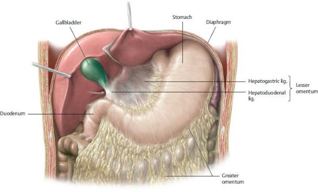
•The lesser omentum , a double layer of peritoneum, extends from the liver to the stomach and proximal duodenum. It is formed by
◦the hepatogastric ligament , between the liver and stomach, and
◦the hepatoduodenal ligament , between the liver and duodenum, which encloses the structures of the portal triad (the portal vein, hepatic artery, and bile duct) in its free edge.
—Peritoneal ligaments are reflections of peritoneum that connect organs to each other or to the body wall. They support the organ in position and may convey their neurovasculature. Individual ligaments are discussed in Sections 9.2 to 9.4 with the specific abdominal organs.
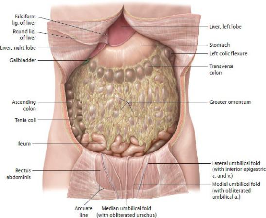
Fig. 11.3 Greater omentum in situ
(From Schuenke M, Schulte E, Schumacher U. THIEME Atlas of Anatomy, Vol 2. Illustrations by Voll M and Wesker K. 3rd ed. New York: Thieme Publishers; 2020.)
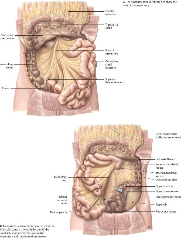
Fig. 11.4 Overview of the mesenteries
Anterior view. Transverse colon and greater omentum have been reflected superiorly. (From Schuenke M, Schulte E, Schumacher U. THIEME Atlas of Anatomy, Vol 2. Illustrations by Voll M and Wesker K. 3rd ed. New York: Thieme Publishers; 2020.)
Fig. 11.5 The lesser omentum
Anterior view with liver retracted superiorly. The arrow points to the omental foramen, the opening into the omental bursa, posterior to the lesser omentum. (From Gilroy AM, MacPherson BR, Wikenheiser JC. Atlas of Anatomy. Illustrations by Voll M and Wesker K. 4th ed. New York: Thieme Publishers; 2020.)
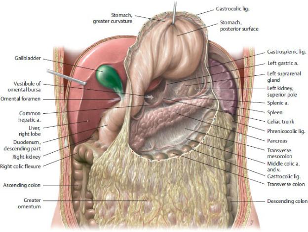
Fig. 11.6 Omental bursa in situ
Anterior view. Divided: Gastrocolic ligament. Retracted: Liver. Reflected: Stomach. (From Gilroy AM, MacPherson BR, Wikenheiser JC. Atlas of Anatomy. Illustrations by Voll M and Wesker K. 4th ed. New York: Thieme Publishers; 2020.)
Subdivisions of the Peritoneal Cavity
—The peritoneal cavity is divided into two spaces:
•The greater sac, which includes the entire peritoneal cavity except that space defined as the lesser sac
•The omental bursa (lesser sac), which is a small exten-sion of the peritoneal cavity that lies behind the stomach and lesser omentum (Table 11.2; Figs. 11.6, 11.7, 11.8). It communicates with the greater sac through a single opening, the omental (epiploic) foramen (Table 11.3).
—Attachments of the peritoneum to the body wall that form during development of the gastrointestinal tract further subdivide the greater sac. These attachments can influence the flow of fluid within the cavity (Fig.
11.9).
•The subphrenic recess between the diaphragm and liver is limited by the coronary ligaments and separated into right and left spaces by the falciform ligament.
•The subhepatic space lies between the liver and the transverse colon. A posterior extension of this space, the hepatorenal recess (hepatorenal pouch, Morison’s pouch), lies between the visceral surface of the liver and the right kidney and suprarenal gland. The hepatorenal recess communicates with the right subphrenic recess.
•The supracolic and infracolic compartments are defined by the attachment of the transverse mesocolon on the posterior abdominal wall —with the supracolic compart-ment above the attachment site and the infracolic compartment below it. The root of the mesentery of the small intestine further divides the infracolic compartment into right and left spaces.
•The paracolic gutters, which lie adjacent to the ascending and descending colons, allow communication between the supracolic and infracolic compartments.
Fig. 11.7 Structure of the greater and lesser omenta and their relation to the omental bursa
Sagittal section, left lateral view. (From Gilroy AM, MacPherson BR, Wikenheiser JC. Atlas of Anatomy. Illustrations by Voll M and Wesker K. 4th ed. New York: Thieme Publishers; 2020.)
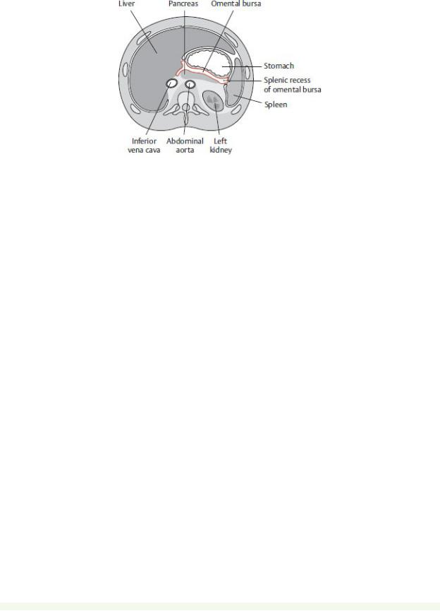
Fig. 11.8 Location of the omental bursa
Transverse section, inferior view. (From Gilroy AM, MacPherson BR, Wikenheiser JC. Atlas of Anatomy. Illustrations by Voll M and Wesker K. 4th ed. New York: Thieme Publishers; 2020.)
Table 11.2 Boundaries of the Omental Bursa (Lesser Sac)
Direction |
Boundary |
Recess |
Anterior |
Lesser omentum, gastrocolic ligament |
— |
|
|
|
Inferior |
Transverse mesocolon |
Inferior |
|
|
recess |
|
|
|
Superior |
Liver (with caudate lobe) |
Superior |
|
|
recess |
|
|
|
Posterior |
Pancreas, aorta (abdominal part), celiac trunk, |
— |
|
splenic a. and v., gastrosplenic fold, left |
|
|
suprarenal gland, left kidney (superior pole) |
|
|
|
|
Right |
Liver, duodenal bulb |
— |
|
|
|
Left |
Spleen, gastrosplenic ligament |
Splenic |
|
|
recess |
|
|
|
Table 11.3 Boundaries of the Omental Foramen
The communication between the greater and lesser sacs is the omental (epiploic) foramen (see arrow in Figs. 11.5 and 11.6).
Direction |
Boundary |
Anterior |
Hepatoduodenal ligament with the portal v., proper |
|
hepatic a., and bile duct |
|
|
Inferior |
Duodenum (superior part) |
|
|
Posterior |
Inferior vena cava, diaphragm (right crus) |
|
|
Superior |
Liver (caudate lobe) |
|
|
BOX 11.1: CLINICAL CORRELATION
PERITONEAL INFECTIONS AND ABSCESSES
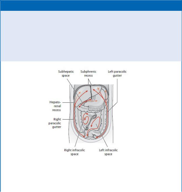
The flow of fluid in the peritoneal cavity can spread intraperito-neal infections and determine the sites of peritoneal abscess for-mation. Fluid commonly collects in the right and left subphrenic recesses, although abscesses are more likely to form on the right side due to duodenal or appendiceal ruptures. Fluid in the supra-colic compartment, such as subphrenic recesses and the omental bursa, can drain to the hepatorenal recess, the lowest part of the abdominal cavity in the supine patient. Therefore, this is a com-mon site of pus accumulation and abscess formation. In the infracolic compartment, the paracolic gutters direct peritoneal fluid and infections toward the pelvis (see Figs. 11.9 and 11.10).
Drainage spaces within the peritoneal cavity
Anterior view. (From Schuenke M, Schulte E, Schumacher U. THIEME Atlas of Anatomy, Vol 2. Illustrations by Voll M and Wesker K. 3rd ed. New York: Thieme Publishers; 2020.)

BOX 11.2: CLINICAL CORRELATION
PERITONITIS AND ASCITES
Bacterial contamination of the peritoneum following surgery or rupture of an inflamed organ (duodenum, gallbladder, appen-dix) results in peritonitis, inflammation of the peritoneum. It is accompanied by severe abdominal pain, tenderness, nausea, and fever and can be fatal when generalized throughout the perito-neal cavity. It often results in ascites, the accumulation of excess peritoneal fluid due to a change in concentration gradients that results in loss of capillary fluid. Ascites can also accompany other pathologic conditions, such as metastatic liver cancer and portal hypertension. In these cases, many liters of ascitic fluid can accumulate in the peritoneal cavity. The fluid is aspirated by paracen-tesis. The needle is carefully inserted into the abdominal wall so as to avoid the urinary bladder and inferior epigastric vessels.
Posterior Wall and Retroperitoneum
—The posterior wall of the abdominal cavity is continuous with the area designated as the “back” but is generally accepted as a separate defined area that is composed of the posterior abdominal wall muscles and their fascia. The retroperitoneum is considered part of the posterior wall.
—The retroperitoneum is a space, or compartment, on the anterior surface of the posterior wall that contains specific retroperitoneal viscera. It is bound anteriorly by the parietal peritoneum and superiorly by the diaphragm. Laterally it’s continuous with the extraperitoneal space of the anterior and lateral abdominal wall, and inferiorly it’s continuous with the subperitoneal space of the pelvis.
•Retroperitoneal organs include the kidneys, ureters, and suprarenal glands, along with their neurovasculature.
•Some components of the gastrointestinal tract have become retroperitonealized during development by losing part of their peritoneal covering. These include the second to fourth parts of the duodenum, the pancreas, and ascending and descending colons.
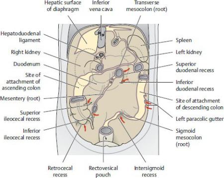
Fig. 11.9 Recesses within the peritoneal cavity
Posterior wall of the peritoneal cavity, anterior view. The mesenteric roots and sites of organ attachment create bounded spaces (recesses or sulci) where peritoneal fluid can flow freely. (From Gilroy AM, MacPherson BR, Wikenheiser JC. Atlas of Anatomy. Illustrations by Voll M and Wesker K. 4th ed. New York: Thieme Publishers; 2020.)

Fig. 11.10 Posterior wall of the peritoneal cavity
Anterior view. Removed: All intraperitoneal organs. (From Schuenke M, Schulte E, Schumacher U. THIEME Atlas of Anatomy, Vol 2. Illustrations by Voll M and Wesker K. 3rd ed. New York: Thieme Publishers; 2020.)
•The retroperitoneum contains the major neurovascular structures of the abdomen, including the aorta and its branches, inferior vena cava and its tributaries, the azygos and hemiazygos veins, the preand para-aortic lymph nodes and thoracic duct, and the lumbar plexus and abdominal autonomic plexuses of the abdomen.
•Mesenteries of the small and large intestines, as well as the peritoneal ligaments associated with the liver and spleen, attach to the retroperitoneum.
•The retroperitoneum is divided into a series of compartments defined by fascia that is variable and often difficult to discern. The perirenal and pararenal spaces around the kidneys are examples of these. The importance of this organization is appreciated when considering the containment of disease process or hemorrhage.
Neurovasculature of the Peritoneum
The parietal and visceral layers of the peritoneum derive their blood supply, lymphatic drainage, and innervation from different sources.
—Parietal peritoneum derives its neurovasculature from vessels and nerves of the body wall.
•Its sensitivity to pain, pressure, and temperature is well localized (felt acutely) through somatic nerves of the overlying muscles and skin.
—Visceral peritoneum derives its neurovasculature from the underlying organs.
•Autonomic nerves mediate sensitivity to stretching and chemical irritation, but the visceral peritoneum lacks sensitivity to touch and temperature.
•Sensation is poorly localized and is usually referred to regions that reflect the embryologic origins of the underlying organ.
◦Sensation from foregut structures is referred to the epigastric region.
◦Sensation from midgut structures is referred to the umbilical region.
◦Sensation from hindgut structures is referred to the pubic region.
11.2 Neurovasculature of the Abdomen
Arteries of the Abdomen
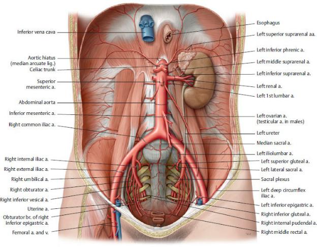
—The abdominal aorta supplies abdominal viscera and most of the anterior abdominal wall (Fig. 11.11).
•It enters the abdomen at T12 through the aortic hiatus of the diaphragm and descends along the vertebral column to the left of the midline.
•It terminates at the L4 vertebral level, where it bifurcates into two common iliac arteries.
•A single median sacral artery arises near the bifurcation.
—Table 11.4 lists major branches of the abdominal aorta.
•Paired parietal (segmental) branches supply the structures of the
posterior abdominal wall. These include the inferior phrenic and lumbar arteries.
•Paired visceral branches supply organs of the retroperitoneum. These are the middle suprarenal, testicular or ovarian, and renal arteries.
•Three unpaired visceral branches supply the intestines and accessory organs of the gastrointestinal tract:
1.The celiac trunk, a short trunk that arises at T12/L1 and supplies the abdominal foregut. Its branches, the splenic, left gastric, and common hepatic arteries, anastomose extensively with each other
(Figs. 11.12, 11.13, 11.14, 11.15).
2.The superior mesenteric artery (SMA), which arises at L1, posterior to the neck of the pancreas. It supplies midgut structures, and its major branches include the inferior pancreaticoduodenal, middle colic, right colic, and ileocolic arteries, as well as a series of jejunal and ileal branches (Figs. 11.16 and 11.17).

Fig. 11.11 Abdominal aorta
Female abdomen, anterior view. Removed: Abdominal organs and peritoneum. The abdominal aorta is the distal continuation of the thoracic aorta. It enters the abdomen at the T12 level and bifurcates into the common iliac arteries at L4. (From Schuenke M, Schulte E, Schumacher U. THIEME Atlas of Anatomy, Vol 2. Illustrations by Voll M and Wesker K. 3rd ed. New York: Thieme Publishers; 2020.)
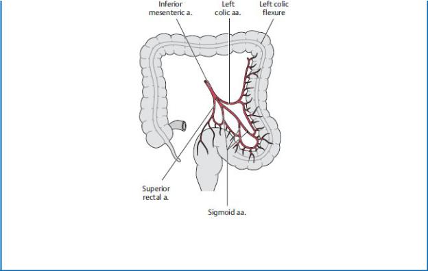
3.The inferior mesenteric artery (IMA), which arises at L3 and has the smallest caliber of the three visceral trunks. It supplies the hindgut through its left colic, sigmoidal, and superior rectal branches (Figs. 11.18 and 11.19).
•Common iliac arteries pass along the brim of the pelvis and terminate by bifurcating into two major branches (see Fig. 11.11):
◦ The internal iliac artery, which descends into the pelvis.
◦ The external iliac artery, which gives off the inferior epigastric and deep circumfex iliac arteries before passing into the lower limb as the femoral artery.
—Important anastomoses connect the three unpaired visceral branches of the aorta and provide a collateral blood supply to the intestinal organs.
•The celiac trunk and superior mesenteric artery anastomose in the head of the pancreas through the pancreaticoduodenal arteries and in the body and tail of the pancreas through dorsal pancreatic and inferior pancreatic arteries (Fig. 11.15).
•The superior mesenteric and inferior mesenteric arteries anastomose near the junction of the transverse and descending colons through the middle and left colic arteries. The marginal artery runs along the mesenteric border of the large intestine and connects the ileocolic, right colic, middle colic, and left colic arteries.
•The inferior mesenteric artery anastomoses with arteries of the rectum through its superior rectal artery (see Fig. 14.19).
BOX 11.3: CLINICAL CORRELATION
ABDOMINAL AORTIC ANEURYSM
Abdominal aortic aneurysms (AAAs) most commonly occur between the renal arteries and the bifurcation of the aorta. When small they can remain asymptomatic, but large aneurysms can be palpated through the abdominal wall to the left of the midline. Ruptured AAAs present with severe abdominal pain that radiates to the abdomen or back. Mortality rates for ruptured aneurysms approach 90% due to overwhelming hemorrhage.

Table 11.4 Branches of the Abdominal Aorta
The abdominal aorta gives rise to three major unpaired trunks (bold) and the unpaired median sacral artery, as well as six paired branches.
Branch from Abdominal |
Branches |
|
Aorta |
|
|
Inferior phrenic aa. |
Superior suprarenal aa. |
|
(paired) |
|
|
|
|
|
Celiac trunk |
Left gastric a. |
|
|
|
|
|
Splenic a. |
|
|
|
|
|
Common hepatic a. |
Proper hepatic |
|
|
a. |
|
|
|
|
|
Right gastric a. |
|
|
|
|
|
Gastroduodenal |
|
|
a. |
Middle suprarenal aa. (paired)
Superior mesenteric a. |
Inferior pancreaticoduodenal a. |
|
Middle colic a. |
|
|
|
Right colic a. |
|
|
|
Jejunal and ileal aa. |
|
|
|
Ileocolic a. |
|
|
Renal aa. (paired) |
Inferior suprarenal aa. |
Lumbar aa. (1st–4th, paired)
Testicular/ovarian aa. (paired)
Inferior mesenteric a. |
Left colic a. |
|
Sigmoidal aa. |
|
|
|
Superior rectal a. |
|
|
Common iliac aa. (paired) External iliac a.
Internal iliac a.
Median sacral a.
Fig. 11.12 Celiac trunk: Stomach,
Anterior view. Opened: Lesser omentum. Incised: omentum. The celiac trunk arises from the abdominal aorta at about the level of L1. (From Schuenke M, Schulte E, Schumacher U. THIEME Atlas of Anatomy, Vol 2. Illustrations by Voll M and Wesker K. 3rd ed. New York: Thieme Publishers; 2020.)
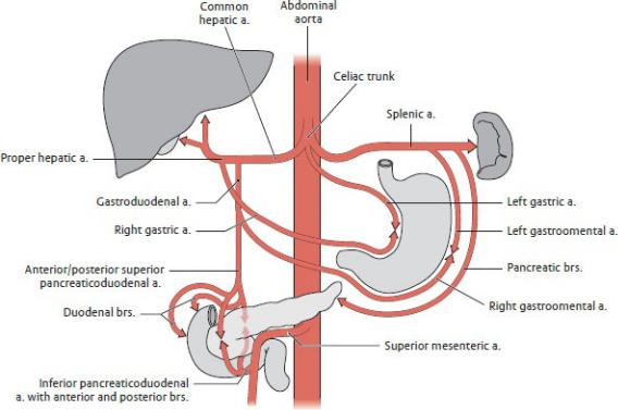
Fig. 11.13 Distribution of the celiac trunk and anastomoses between its branches
(From Schuenke M, Schulte E, Schumacher U. THIEME Atlas of Anatomy, Vol 2. Illustrations by Voll M and Wesker K. 3rd ed. New York: Thieme Publishers; 2020.)
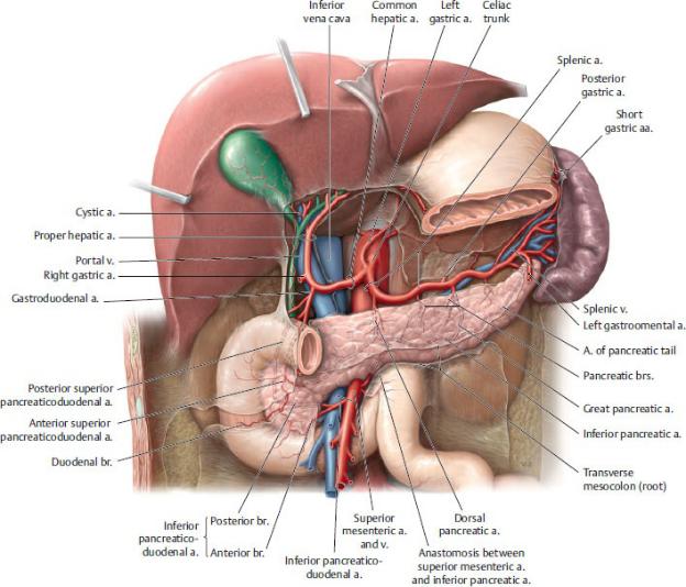
Fig. 11.14 Celiac trunk: Pancreas, duodenum, and spleen
Anterior view. Removed: Stomach (body) and lesser omentum. (From Schuenke M, Schulte E, Schumacher U. THIEME Atlas of Anatomy, Vol 2. Illustrations by Voll M and Wesker K. 3rd ed. New York: Thieme Publishers; 2020.)
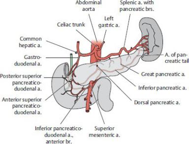
Fig. 11.15 The pancreaticoduodenal arcade, an anastomosis between branches of the celiac trunk and superior mesenteric artery
(From Gilroy AM, MacPherson BR, Wikenheiser JC. Atlas of Anatomy. Illustrations by Voll M and Wesker K. 4th ed. New York: Thieme Publishers; 2020.)
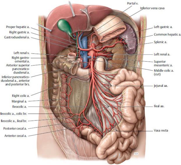
Fig. 11.16 Superior mesenteric artery
Anterior view. Partially removed: Stomach and peritoneum. Note: The middle colic artery has been truncated. The superior mesenteric artery arises from the aorta opposite L2. (From Schuenke M, Schulte E, Schumacher U. THIEME Atlas of Anatomy, Vol 2. Illustrations by Voll M and Wesker K. 3rd ed. New York: Thieme Publishers; 2020.)
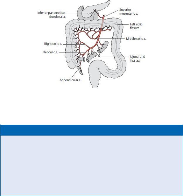
Fig. 11.17 Distribution of the superior mesenteric artery
(From Gilroy AM, MacPherson BR, Wikenheiser JC. Atlas of Anatomy. Illustrations by Voll M and Wesker K. 4th ed. New York: Thieme Publishers; 2020.)
BOX 11.4: CLINICAL CORRELATION
MESENTERIC ISCHEMIA
A decrease in blood flow to the intestine (ischemia) can result from occlusion of the superior mesenteric artery (SMA) by a thrombus or embolus (acute) or may be secondary to severe atherosclerosis (chronic). In the acute condition, the embolus can obstruct the SMA at its origin or, if small enough, may travel further to obstruct a more peripheral branch. Acute ischemia results in necrosis of the affected part of the intestine. Chronic ischemia is less threatening since obstruction of the vessels occurs gradually, allowing the formation of collateral vessels that will supply the affected intestine. Because of the extensive anastomoses between intestinal arteries, chronic vascular isch-emia is rare. Symptoms occur only if two of the three major ves-sels (celiac trunk or superior or inferior mesenteric arteries) are compromised.
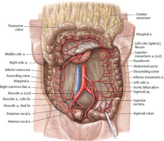
Fig. 11.18 Inferior mesenteric artery
Anterior view. Removed: Jejunum and ileum. Reflected: Transverse colon. The inferior mesenteric artery arises from the aorta opposite L3. (From Schuenke M, Schulte E, Schumacher U. THIEME Atlas of Anatomy, Vol 2. Illustrations by Voll M and Wesker K. 3rd ed. New York: Thieme Publishers; 2020.)

Fig. 11.19 Distribution of the inferior mesenteric artery
(From Gilroy AM, MacPherson BR, Wikenheiser JC. Atlas of Anatomy. Illustrations by Voll M and Wesker K. 4th ed. New York: Thieme Publishers; 2020.)
BOX 11.5: CLINICAL CORRELATION
ANATOMOSES BETWEEN ARTERIES OF THE LARGE INTESTINE
Anastomoses between branches of the superior mesenteric and inferior mesenteric arteries can compensate for abnormally low blood flow in either of the arteries. Two of these anastomoses, although variable, are of significant value:
Riolan’s arcade (arc of Riolan) – connects the middle colic and left colic arteries close to their origins from the superior and inferior mesenteric arteries, respectively.
Marginal artery (of Drummond) – connects all arteries of the colon that run along the periphery of the mesentery close to the intestinal tube.
(From Gilroy AM, MacPherson BR, Wikenheiser JC. Atlas of Anatomy. Illustrations by Voll M and Wesker K. 4th ed. New York: Thieme Publishers; 2020.)
Veins of the Abdomen
Venous drainage of the abdomen and pelvis is accomplished through two systems, the systemic (caval) system and the hepatic portal system (Fig. 11.20).
1.Organs that drain directly into the inferior vena cava or its tributaries make up the systemic (caval) venous system.
—The inferior vena cava (IVC) receives blood from retroperi-toneal and pelvic organs, walls of the abdomen and pelvis, and the lower limbs
(Fig. 11.21).
•It originates at the L5 vertebral level where the common iliac veins merge.
•It ascends along the right side of the vertebral column, passes posterior to the liver, and pierces the central tendon of the diaphragm at the T8 vertebral level where it enters the right atrium of the heart.
—Table 11.5 lists the direct tributaries of the inferior vena cava.
•Paired common iliac veins drain the external iliac and internal iliac veins.
•Paired inferior phrenic and lumbar veins drain the posterior abdominal wall and diaphragm and accom-pany the arteries of
similar name.
•Veins of the retroperitoneal organs include the right and left renal veins, the right suprarenal vein, and the right testicular or ovarian
(gonadal) vein. The suprarenal and gonadal veins on the left side drain to the left renal vein.
•Typically three hepatic veins enter the IVC from the liver immediately below the diaphragm.
—Paired ascending lumbar veins communicate with the lumbar veins and are continuous with the azygos and hemiazygos veins of the thorax. These communications between the lumbar, ascending lumbar, azygos, and hemiazygos veins function as a collateral pathway between the inferior and superior venae cavae.
2.Organs that drain into the portal vein or its tributaries and pass through the liver before entering the inferior vena cava make up the hepatic portal system.
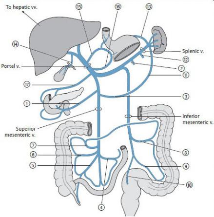
—The portal vein shunts nutrient-rich venous blood from the capillary beds of the gastrointestinal tract and its associated organs (liver, gallbladder, pancreas, and spleen) to sinusoids of the liver (Fig. 11.22). This blood eventually enters the inferior vena cava through the hepatic veins.
—Tributaries of the portal vein are listed in Table 11.6 and include the following:
•The splenic vein, which drains the spleen, and the superior mesenteric vein, which drains the small intestine and most of the large intestine. These two veins unite behind the neck of the pancreas to form the portal vein.
•The inferior mesenteric vein, which drains the hindgut portion of the gastrointestinal tract. It usually joins the splenic vein but may empty directly into the portal vein.
•Veins from the lower esophagus, stomach, pancreas, duodenum, and gallbladder
—Normal connections between the systemic (caval) venous system and portal venous system, called portosystemic pathways (Fig. 11.23), can become abnormally dilated when there is an obstruction of the portal or systemic circulations (i.e., cirrhosis of the liver or pregnancy). These dilations are most prominent in
1. esophageal veins,
2. periumbilical veins through the superior and inferior epigastric veins of the abdominal wall,
3.colic veins in the retroperitoneum, and
4.rectal veins of the rectum and anal canal.
Fig. 11.20 Schematic of systemic and portal venous systems
(From Schuenke M, Schulte E, Schumacher U. THIEME Atlas of Anatomy, Vol 2. Illustrations by Voll M and Wesker K. 3rd ed. New York: Thieme Publishers; 2020.)
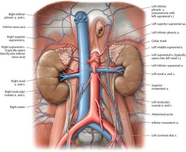
Fig. 11.21 Inferior vena cava
Anterior view. Removed: All organs except the kidneys and suprarenal glands. (From Schuenke M, Schulte E, Schumacher U. THIEME Atlas of Anatomy, Vol 2. Illustrations by Voll M and Wesker K. 3rd ed. New York: Thieme Publishers; 2020.)
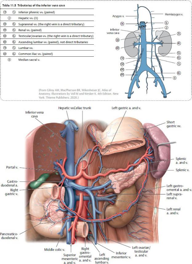

Fig. 11.22 Portal vein: In situ
Anterior view. Partially removed: Stomach, pancreas, and peritoneum. (From Schuenke M, Schulte E, Schumacher U. THIEME Atlas of Anatomy, Vol 2. Illustrations by Voll M and Wesker K. 3rd ed. New York: Thieme Publishers; 2020.)
Table 11.6 Tributaries of the Portal Vein
•Superior mesenteric vein with its tributaries:
Pancreaticoduodenal vv.
Pancreatic vv.
Right gastro-omental v.Jejunal and ileal vv.
Ileocolic v.Right colic v.
Middle colic v.
•Inferior mesenteric vein with its tributaries:
Left colic v.Sigmoid vv.
Superior rectal v.
•Splenic vein with its tributaries:Left gastro-omental v.
Pancreatic vv.Short gastric vv.
•Direct tributaries
Cystic v.
Left gastric v. with esophageal vv.Right gastric v.
Posterior superior pancreaticoduodenal v.
— Paraumbilical vv. (see Fig. 11.23)
(From Schuenke M, Schulte E, Schumacher U. THIEME Atlas of Anatomy, Vol 2. Illustrations by Voll M and Wesker K. 3rd ed. New York: Thieme Publishers; 2020.)
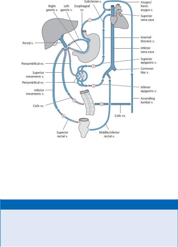
Fig. 11.23 Portosystemic pathways
When the portal system is compromised, the portal vein can divert blood away from the liver back to its supplying veins, which return this nutrient-rich blood to the heart via the venae cavae. Red arrows indicate flow reversal in the (1) esophageal veins, (2) paraumbilical veins, (3) colic veins, and (4) middle and inferior rectal veins. (From Schuenke M, Schulte E, Schumacher U. THIEME Atlas of Anatomy, Vol 2. Illustrations by Voll M and Wesker K. 3rd ed. New York: Thieme Publishers; 2020.)
BOX 11.6: CLINICAL CORRELATION
ESOPHAGEAL VARICES
Submucosal veins of the esophagus drain superiorly to the sys-temic system (through the azygos veins) and inferiorly to the por-tal system. When flow through the portal vein is obstructed (as in portal hypertension), these portosystemic anastomoses allow blood in the lower esophagus to drain to the systemic veins. The esophageal varices, enlarged veins that result from this increased flow, bulge into the esophageal lumen and can rupture, causing severe hemorrhaging.

BOX 11.7: CLINICAL CORRELATION
PORTAL HYPERTENSION AND SURGICAL PORTOCAVAL SHUNTS
Portal hypertension occurs secondary to liver disease (e.g., cirrho-sis) or thrombosis of the portal vein. Increased resistance to the flow of blood in the portal system forces portal blood through portosystemic (portocaval) anastomoses to the systemic circula-tion, reversing flow in some venous pathways. Symptoms of por-tal hypertension include ascites, caput medusa (enlargement of periumbilical veins on the anterior abdominal wall), varices of the rectal veins (hemorrhoids), and esophageal varices. Symptoms may be relieved by surgically creating a portocaval shunt between the portal and systemic circulations (portal vein to the inferior vena cava or splenic vein to the left renal vein).
Lymphatic Drainage of the Abdomen
Lymph from abdominal and pelvic regions drains through lym-phatic vessels that usually accompany the arteries supplying those regions. The lymph passes through one or more lymph node groups that can include primary, or regional, nodes and secondary, or col-lecting, nodes. These latter groups receive lymph from multiple regions and in the abdomen and pelvis and are known as lumbar lymph nodes. They surround the aorta and inferior vena cava and are subdivided by location (Fig. 11.24). Lymph drains from these nodes into either the lumbar or intestinal lymphatic trunks, which converge in the upper abdomen to form the cisterna chyli and the thoracic duct.
—Groups of lumbar lymph nodes drain all of the abdominal viscera (except a small hepatic segment, which can drain to nodes of the diaphragm) and most of the abdominal wall (Fig. 11.25; Table 11.7). They include:
•Preaortic nodes, which lie anterior to the abdominal aorta. They receive lymph from the gastrointestinal tract (as far as the midrectum) and associated organs. Nodes surrounding the base of the major arteries form collecting nodal groups, such as the superior and inferior mesenteric nodes. These drain to celiac nodes, which drain to intestinal lymph trunks.
•Right and left lumbar nodes (lateral aortic and caval nodes), which lie along the medial border of the psoas muscles, the crura of the diaphragm, the aorta, and the inferior vena cava. They drain the abdominal and pelvic walls and the viscera of the retroperitoneum, including the ovaries and testes. They also receive lymph from the common iliac nodes, which drain the pelvic viscera and the lower limb. Drainage from these lumbar nodes forms a lumbar trunk on each side.
—Common iliac nodes drain organs of the pelvis and the lower limbs. Lymph
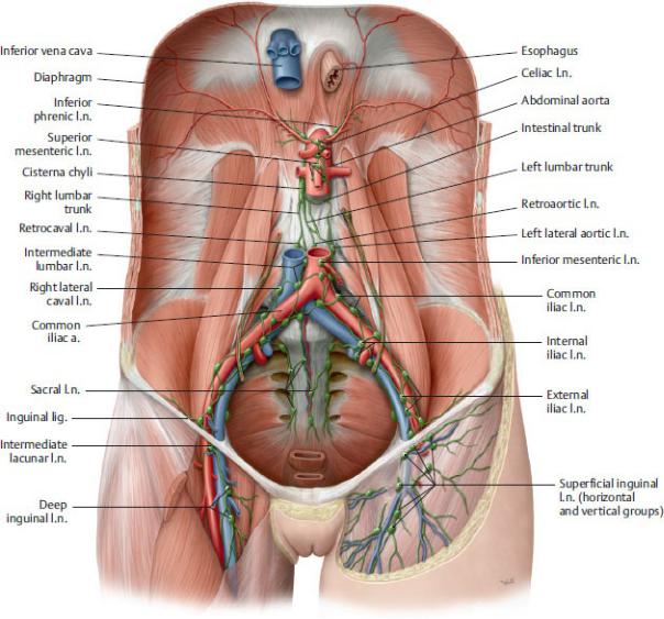
from these nodes drain to the right and left lumbar nodes.
—The cisterna chyli is an elongated, lobulated, thin-walled dilation that, when present, gives rise to the thoracic duct. It lies to the right of the T12 vertebral body and receives the lumbar and intestinal trunks.
Fig. 11.24 Parietal lymph nodes in the abdomen and pelvis
Anterior view. Removed: All visceral structures except vessels (From Schuenke M, Schulte E, Schumacher U. THIEME Atlas of Anatomy, Vol 2. Illustrations by Voll M and Wesker K. 3rd ed. New York: Thieme Publishers; 2020.)
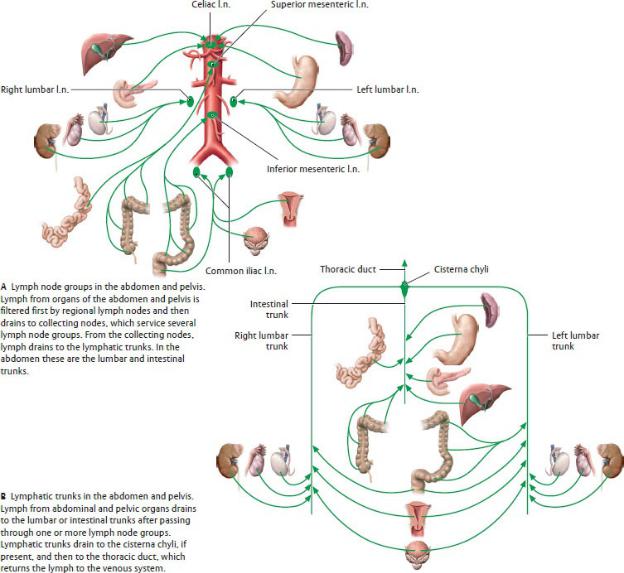
Fig. 11.25 Lymphatic trunks and lymph nodes of the abdomen
(From Schuenke M, Schulte E, Schumacher U. THIEME Atlas of Anatomy, Vol 2. Illustrations by Voll M and Wesker K. 3rd ed. New York: Thieme Publishers; 2020.)
Table 11.7 Lymph Node Groups and Tributary Regions
Lymph |
|
Organs or Organ Segments that |
Node |
|
Drain to These |
Groups and |
|
|
Collecting |
Location |
Organs or Organ Segments that |
|
|
|
Lymph |
|
Drain to These Lymph Node Groups |
Nodes |
|
(Tributary Regions) |
Celiac l.n. |
Around the |
Distal third of esophagus, stomach, |
|
celiac trunk |
greater omentum, duodenum (superior |
|
|
and descending parts), pancreas, |
|
|
spleen, liver, and gallbladder |
|
|
|
Superior |
At the origin of |
Second through fourth parts of |
mesenteric |
the superior |
duodenum, jejunum and ileum, cecum |
l.n. |
mesenteric a. |
with vermiform appendix, ascending |
|
|
colon, transverse colon (proximal two- |
|
|
thirds) |
|
|
|
Inferior |
At the origin of |
Transverse colon (distal third), |
mesenteric |
the inferior |
descending colon, sigmoid colon, |
l.n. |
mesenteric a. |
rectum (proximal part) |
|
|
|
Lumbar l.n. |
Around the |
Diaphragm (abdominal side), kidneys, |
(right, |
abdominal aorta |
suprarenal glands, testis and |
intermediate, |
and inferior vena |
epididymis, ovary, uterine tube, uterine |
left) |
cava |
fundus, ureters, retroperitoneum |
|
|
|
Iliac l.n. |
Around the iliac |
Rectum (anal end), bladder and |
|
vessels |
urethra, uterus (body and cervix), |
|
|
ductus deferens, seminal vesicle, |
|
|
prostate, external genitalia (via |
|
|
inguinal l.n.) |
|
|
|
Nerves of the Abdomen
—Lower intercostal nerves (T7–T11) and the subcostal nerve (T12) continue anteroinferiorly from their position on the thoracic wall to innervate most of the muscles and skin of the anterolateral abdominal wall.
—The lumbar plexus is a somatic nerve plexus formed by the anterior rami of spinal nerves T12–L4. Its branches pass laterally through the psoas major muscle and onto the posterior abdominal wall (Fig. 11.26). Most nerves of this plexus innervate the lower limb (see Section 21.4 and Table 21.1). Branches that innervate the abdominal wall and inguinal region include
Fig. 11.26 Nerves of the lumbar plexus
Anterior view. (From Gilroy AM, MacPherson BR, Wikenheiser JC. Atlas of Anatomy. Illustrations by Voll M and Wesker K. 4th ed. New York: Thieme Publishers; 2020.)
•iliohypogastric and ilioinguinal nerves (L1), which innervate the skin
and muscles of the inferior anterior abdominal wall and the skin over the inguinal and pubic regions;
•genitofemoral nerve (L1-L2), whose genital branch innervates the cremaster muscle surrounding the spermatic cord and the skin over the scrotum and labia; and
•short muscular branches (T12–L4) that innervate the muscles of the posterior abdominal wall.
—Lumbar sympathetic trunks, the continuations of the sympathetic trunks in the thorax, descend along the lateral aspect of the lumbar vertebral bodies and give off three to four lumbar splanchnic nerves, which join the autonomic plexuses of the abdomen.
—Autonomic plexuses form along the aorta and travel with the major abdominal arteries to innervate the abdominal viscera (Figs. 11.27, 11.28, 11.29, 11.30, 11.31, 11.32; Tables 11.8 and 11.9). The plexuses contain combinations of
•preganglionic sympathetic nerves that synapse in the ganglia associated with the plexuses. (Note that the sympathetic nerves innervating the adrenal medulla are an exception and do not synapse in these ganglia.) The preganglionic sympathetic nerves arise from
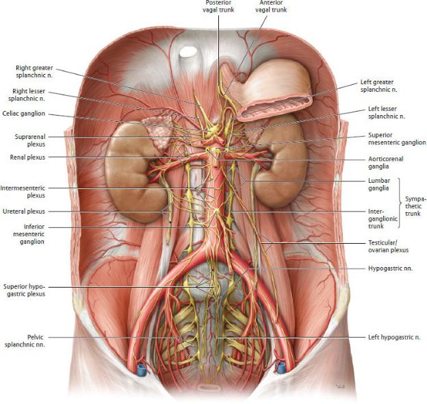
Fig. 11.27 Autonomic plexuses in the abdomen and pelvis
Anterior view of the male abdomen. Removed: Peritoneum and majority of the stomach. (From Schuenke M, Schulte E, Schumacher U. THIEME Atlas of Anatomy, Vol 2. Illustrations by Voll M and Wesker K. 3rd ed. New York: Thieme Publishers; 2020.)
◦thoracic splanchnic nerves (T5–T12), which contribute to the celiac, superior mesenteric and renal plexuses
◦lumbar splanchnic nerves (T11–L2), which contribute to the inferior mesenteric, superior hypogastric, and inferior hypogastric plexuses
•preganglionic parasympathetic nerves, which pass through the plexuses
and synapse in ganglia near their target organ. They arise from either
◦the vagus nerves (cranial nerve X), which enter the abdomen as anterior and posterior vagal trunks from the esophageal plexus. They supply most of the abdominal viscera, including the digestive tract, except for its most distal segment (the descending colon to the anal canal). They contribute to all of the abdominal plexuses except the inferior mesenteric, superior hypogastric, and inferior hypogastric plexuses.
Or
◦the pelvic splanchnic nerves (S2–S4), which ascend from the pelvis to innervate the descending and sigmoid colons in the abdomen. They also innervate viscera of the pelvis. These fibers contribute to the inferior hypogastric plexuses.
—Although most abdominal plexuses contain both sympathetic and parasympathetic nerves, the inferior mesenteric and superior hypogastric plexuses contain only sympathetic fibers. Viscera supplied by these plexuses receive parasym pathetic innervation via pelvic splanchnic nerves and the inferior hypogastric plexus.
—When pain arising from viscera (visceral pain) and pain arising from somatic structures (somatic pain) are conveyed to the same area of the spinal cord, the convergence of these visceral and somatic fibers confuses the relationship between the visceral pain’s actual origin and its perceived origin. This phenomenon is known as referred pain. The perceived origin of visceral pain from a specific organ is consistently projected to a welldefined area of the skin. Thus, a knowledge of cutaneous zones of referred pain is instrumental in identify-ing underlying problems.

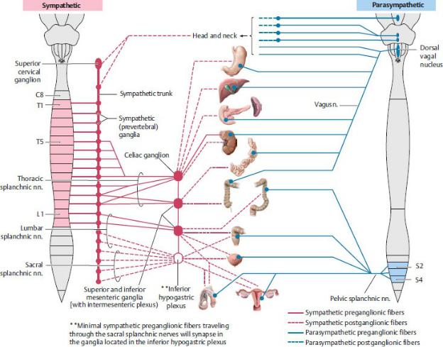
Fig. 11.28 Sympathetic and parasympathetic nervous systems in the abdomen and pelvis
(From Gilroy AM, MacPherson BR, Wikenheiser JC. Atlas of Anatomy. Illustrations by Voll M and Wesker K. 4th ed. New York: Thieme Publishers; 2020.)
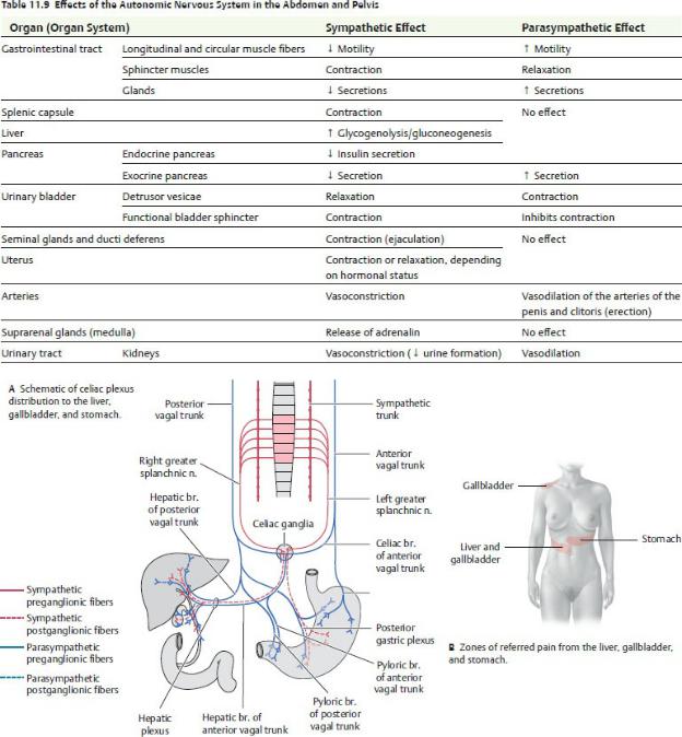
Fig. 11.29 Autonomic innervation of the liver, gallbladder, and stomach
(From Gilroy AM, MacPherson BR, Wikenheiser JC. Atlas of Anatomy. Illustrations by Voll M and Wesker K. 4th ed. New York: Thieme Publishers; 2020.)
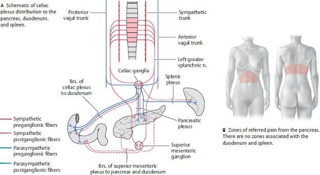
Fig. 11.30 Autonomic innervation of the pancreas, duodenum, and spleen
(From Gilroy AM, MacPherson BR, Wikenheiser JC. Atlas of Anatomy. Illustrations by Voll M and Wesker K. 4th ed. New York: Thieme Publishers; 2020.)
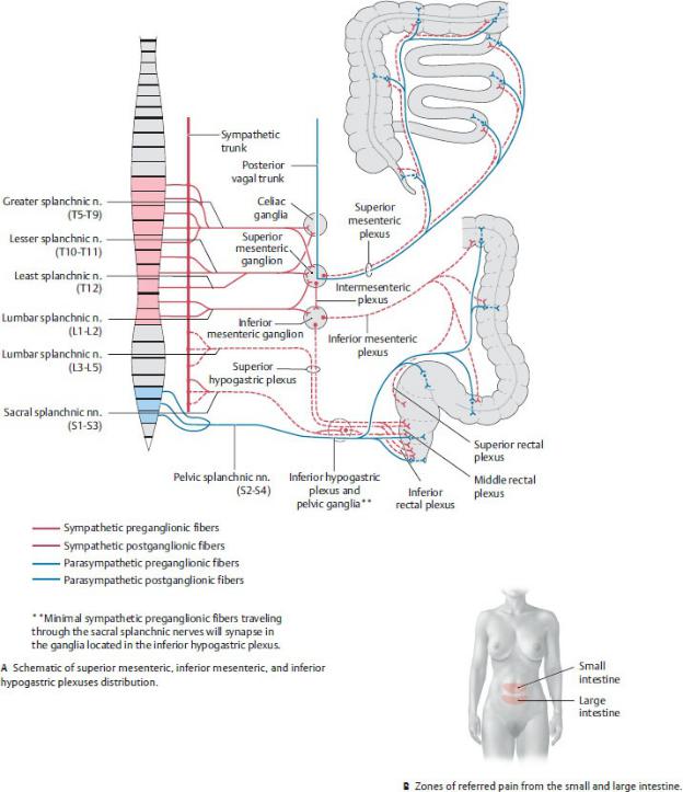
Fig. 11.31 Autonomic innervation of the intraperitoneal organs
(From Gilroy AM, MacPherson BR, Wikenheiser JC. Atlas of Anatomy. Illustrations by Voll M and Wesker K. 4th ed. New York: Thieme Publishers; 2020.)
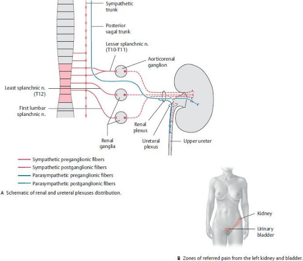
Fig. 11.32 Autonomic innervation of the kidneys and upper ureters
(From Gilroy AM, MacPherson BR, Wikenheiser JC. Atlas of Anatomy. Illustrations by Voll M and Wesker K. 4th ed. New York: Thieme Publishers; 2020.)