


22 Functional Anatomy of the Lower Limb
The strong muscles and joints of the lower limb are adapted for bipedal locomotion and, in conjunction with the muscles of the trunk, maintain the body’s center of gravity.
Schematics of lower limb muscles accompany the muscle tables (origin, attachment, innervation, and action) in the appropriate chapter sections. To see the muscles in situ, view the gallery of topographic images in Section 22.9 at the end of the chapter.
22.1 The Pelvic Girdle
The hip (coxal) bones and the sacrum form the pelvic girdle (bony pelvis), which is related anatomically and functionally to the pelvis and the lower limb (Fig. 22.1). (See Section 14.2 for a discussion of the pelvic girdle.)
—Articulations at the sacroiliac joint and pubic symphysis, supported by strong sacroiliac, sacrotuberous, and sacrospinous ligaments, create a stable framework that
•supports and encloses the pelvic viscera,
•transfers the weight of the trunk to the lower limb, and
•forms part of the hip joint and provides attachment sites for lower limb muscles.
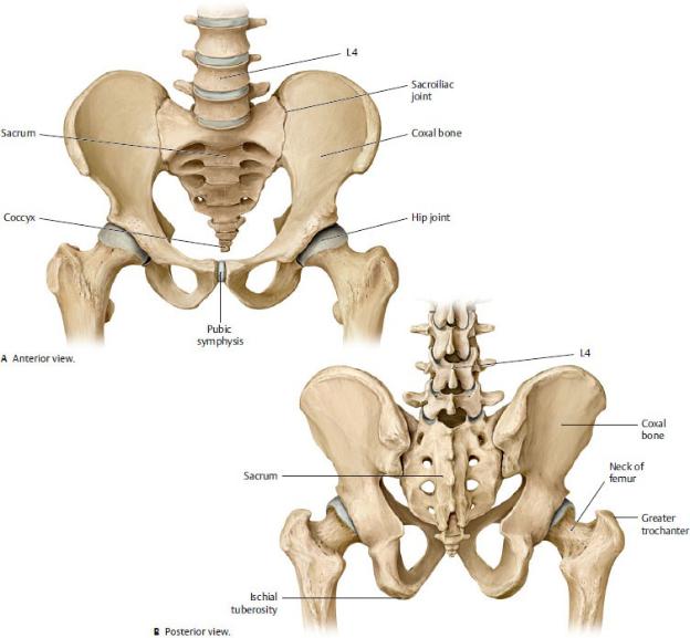
Fig. 22.1 The coxal bones and their relation to the vertebral column.
(From Gilroy AM, MacPherson BR, Wikenheiser JC. Atlas of Anatomy. Illustrations by Voll M and Wesker K. 4th ed. New York: Thieme Publishers; 2020.)
22.2 The Gluteal Region
The gluteal region includes the buttock, which overlies the posterior part of the pelvic girdle, and the lateral hip region, which covers the hip joint and extends as far anteriorly as the anterior superior iliac spine.
— The muscular compartment of the gluteal region contains
•muscles that laterally rotate, abduct, adduct, extend, and flex the hip joint ( Table 22.1);
•the superior gluteal, inferior gluteal, and sciatic nerves; and
•the superior and inferior gluteal arteries and veins.

Fig. 22.2 Sciatic foramina
Right gluteal region. (From Schuenke M, Schulte E, Schumacher U. THIEME Atlas of Anatomy, Vol 1. Illustrations by Voll M and Wesker K. 3rd ed. New York: Thieme Publishers; 2020.)
BOX 22.1: CLINICAL CORRELATION
PIRIFORMIS SYNDROME
The sciatic nerve normally passes into the gluteal region inferior to the piriformis muscle. Tightening or shortening of the muscle can compress and irritate the sciatic nerve, causing pain and paresthesia (tingling and numbness) in the buttocks and posterior thigh. In some cases the sciatic nerve, or its common fibular component, is compressed as it passes through the muscle rather than inferior to it. Piriformis syndrome should be distinguished from sciatica in which the pain and paresthesia result from compression of lumbar nerve roots by a herniated intervertebral disk.


The Hip Joint
The hip joint is a highly mobile, yet stable, ball-and-socket joint between the proximal femur and the acetabulum of the hip bone (Figs. 22.3 and 22.4). Muscles crossing the joint, particularly in the gluteal region, provide additional stability as well as mobility.
—More than half of the large femoral head is seated in the bony acetabulum of the hip bone, which is oriented slightly anteriorly and inferiorly.
—The axis through the neck of the femur is laterally rotated with respect to the condylar axis of the distal femur; thus, when the femoral head is centered in the hip joint, the distal femur and knee joint are directed slightly inward (Figs. 22.5 and 22.6).
—A strong fibrous capsule surrounds the hip joint (Fig. 22.7).
•Iliofemoral, pubofemoral, and ischiofemoral ligaments are extracapsular ligaments that strengthen the capsule. The iliofemoral ligament is the strongest and most supportive of these. The ligaments are arranged in a spiral around the joint. When the hip is extended, the spiral tightens, pushing the femoral head more firmly into the acetabulum and further stabilizing the joint. In hip flexion, the spiral unwinds, allowing a greater degree of joint mobility as well as vulnerability (Fig. 22.8).
—A fibrocartilaginous acetabular labrum attached to the acetabular rim deepens the joint (Fig. 22.9).
—A weak ligament of the head of the femur attaches to the acetabulum within the joint space but provides little support.
—Inferiorly, a transverse ligament of the acetabulum completes the circle of the C-shaped acetabular labrum.
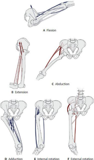
—Muscles of the gluteal region and thigh move the hip joint ( Table 22.2).
BOX 22.2: CLINICAL CORRELATION
CONGENITAL HIP DISLOCATION
Congenital hip dislocation (also known as hip dysplasia) is a common problem that occurs when the femoral head is not properly seated in the acetabulum. Hip abduction is impaired, and since the femoral head sits higher than normal, the affected limb is shorter than the contralateral limb. In routine neonatal screenings, a dislocated hip will “click” when it is adducted and pushed posteriorly.

Fig. 22.3 Right hip joint
(From Schuenke M, Schulte E, Schumacher U. THIEME Atlas of Anatomy, Vol 1. Illustrations by Voll M and Wesker K. 3rd ed. New York: Thieme Publishers; 2020.)
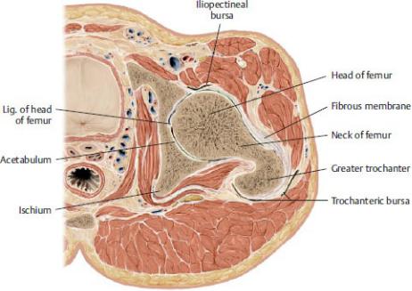
Fig. 22.4 Hip joint: transverse section
Right hip joint, superior view. (From Gilroy AM, MacPherson BR, Wikenheiser JC. Atlas of Anatomy. Illustrations by Voll M and Wesker K. 4th ed. New York: Thieme Publishers; 2020.)


1. Illustrations by Voll M and Wesker K. 3rd ed. New York: Thieme Publishers; 2020.)
BOX 22.3: CLINICAL CORRELATION
ACQUIRED HIP DISLOCATION
Acquired hip dislocation usually occurs as a result of trauma that causes the femoral head to be displaced out of the acetabulum; anterior dislocations are rare, but posterior dislocations are common. Typically, in a head-on motor vehicle accident, the knees strike the dashboard, forcing the femoral head posteriorly through the joint capsule and onto the lateral surface of the ilium. The affected limb appears shortened, adducted, and internally rotated. The sciatic nerve is particularly vulnerable to injury in these cases.
Fig. 22.7 Ligaments of the hip joint and pelvic girdle
(From Schuenke M, Schulte E, Schumacher U. THIEME Atlas of Anatomy, Vol 1. Illustrations by Voll M and Wesker K. 3rd ed. New York: Thieme Publishers; 2020.)
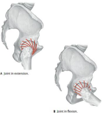
Fig. 22.8 Hip Ligaments as a function of joint position
Right hip, lateral view. (From Schuenke M, Schulte E, Schumacher U. THIEME Atlas of Anatomy, Vol 1. Illustrations by Voll M and Wesker K. 3rd ed. New York: Thieme Publishers; 2020.)
Fig. 22.9 Joint capsule of the hip joint
Lateral view. The capsule has been divided and the femoral head dislocated to expose the cut ligament of the head of the femur. (From Schuenke M, Schulte E, Schumacher U. THIEME Atlas of Anatomy, Vol 1. Illustrations by Voll M and Wesker K. 3rd ed. New York: Thieme Publishers; 2020.)
Table 22.2 Movements of the Hip Joint
Action |
Primary Muscles |
Flexion |
Iliopsoas |
|
Tensor fascia latae |
|
Sartorius |
|
Rectus femoris |
|
|
Extension |
Gluteus maximus |
|
Adductor magnus Biceps femoris, |
|
long head |
|
Semimembranosus |
|
Semitendinosus |
|
|
Abduction |
Gluteus medius |
|
Gluteus minimus |
|
Tensor fascia latae |
|
|
Adduction |
Gluteus maximus |
|
Pectineus |
|
Adductor longus |
|
Adductor brevis |
|
Adductor magnus |
|
|
Internal rotation |
Gracilis |
|
Gluteus medius |
|
Gluteus minimus |
|
Tensor fascia latae |
|
|
External rotation |
Gluteus maximus |
|
Pectineus |
|
Sartorius |
|
Quadratus femoris |
|
Piriformis |
|
Obturator externus |
|
Obturator internus |
|
|
(From Schuenke M, Schulte E, Schumacher U. THIEME Atlas of Anatomy, Vol 1. Illustrations by Voll M and Wesker K. 3rd ed. New York: Thieme Publishers; 2020.)
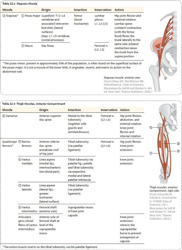
Muscles of the Thigh
The powerful muscles of the thigh move the hip and knee joints and are separated into three compartments (see Fig. 22.45A).
—The anterior compartment contains
• muscles that primarily flex the hip joint and extend the knee joint

(Tables 22.3 and 22.4);
•the femoral nerve; and
•branches of the femoral artery, the deep artery of the thigh, and their accompanying veins.
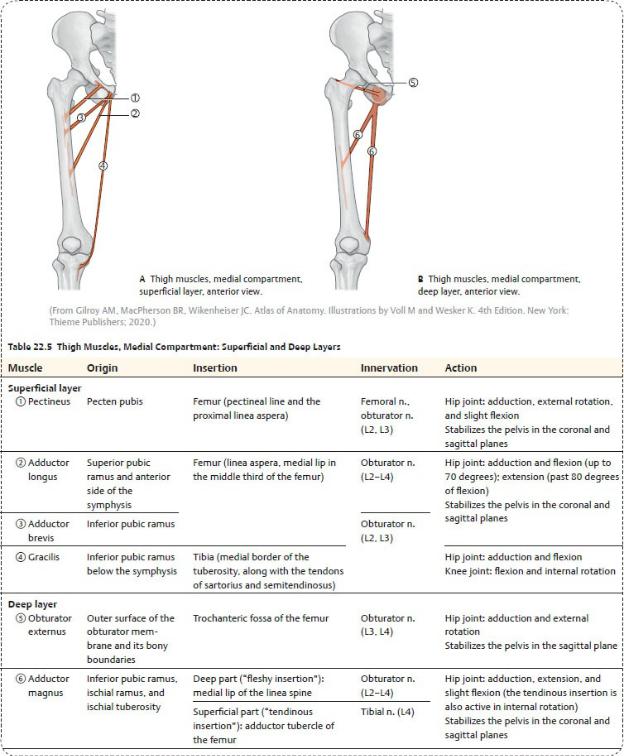
—The medial compartment contains
•muscles that primarily adduct, flex, and extend the hip joint ( Table 22.5);
•the obturator and femoral nerves; and
•the obturator artery and vein and deep artery and vein of the thigh.
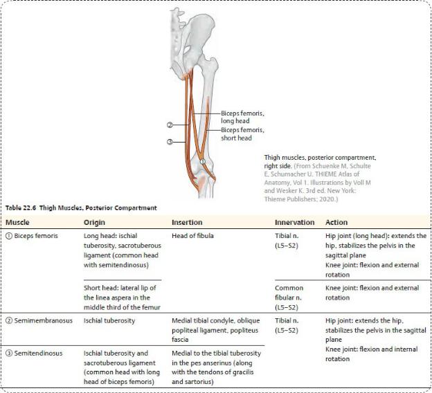
—The posterior compartment contains
•muscles that extend the hip joint and flex the knee joint ( Table 22.6),
•the sciatic nerve, and
•branches of the deep artery and vein of the thigh.
—Three muscles of the thigh, the sartorius, gracilis, and semitendinosus, cross the knee medially and join to form the pes anserinus, a common tendon that overlies the anserine bursa inferomedial to the knee joint (see
Fig. 22.37).
BOX 22.4: CLINICAL CORRELATION
HAMSTRING STRAINS
Hamstring strains are tears of the hamstring (posterior thigh) muscles at their proximal attachment to the pelvic girdle. This is a common injury in individuals who participate in sports that involve sprinting with sudden starts and stops. Forced high kicks, especially with an extended knee, may avulse the muscle tendons from their origin at the ischial tuberosity. Symptoms include sudden, sharp pain in the back of the thigh during physical activity, a popping or tearing feeling in the muscle, swelling, muscle weakness, and an inability to bear weight on the affected leg.
Spaces of the Thigh
The femoral nerve, artery, and vein descend through narrow passages in the thigh that are created by the inguinal ligament and anterior and medial thigh muscles.
—Anteriorly, structures pass from the abdomen into the thigh through the retroinguinal space, deep to the inguinal ligament. The retroinguinal space is divided into a lateral muscular compartment and a medial vascular compartment (Fig. 22.10).
•The muscular compartment contains the femoral nerve and iliopsoas muscle.
•The vascular compartment contains the femoral vessels enclosed within the femoral sheath.
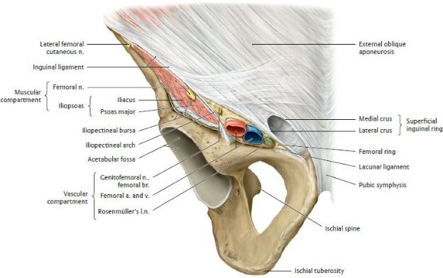
•The femoral sheath, formed by extensions of the transversalis and psoas fasciae, surrounds the femoral vessels as they pass under the inguinal ligament. Inferiorly, the sheath merges with the outer layer of the vessel walls (adventitia). Septa divide the sheath into compartments:
◦Lateral and central compartments contain the femoral artery and femoral vein, respectively.
◦A medial compartment within the sheath, the femoral canal, contains loose connective tissue, fat, and often a deep inguinal lymph node. The femoral ring defines the superior opening of the canal.
—The femoral triangle is an area of the anterior thigh (Fig. 22.11).
•It contains the femoral artery and vein and their branches and the terminal branches of the femoral nerve.
•Its boundaries are
◦superiorly, the inguinal ligament;
◦medially, the adductor longus muscle;
◦laterally, the sartorius muscle;
◦the floor, formed by the iliopsoas and pectineus muscles; and
◦the apex, formed by the inferior junction of the medial and lateral borders.
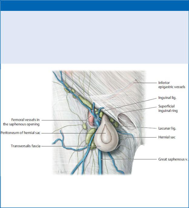
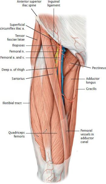
Fig. 22.11 Anterior thigh
Right thigh, anterior view. Revealed: Femoral triangle. Removed: Skin, subcutaneous tissue, and fascia lata. Partially transparent: Sartorius. (From Schuenke M, Schulte E, Schumacher U. THIEME Atlas of Anatomy, Vol 1. Illustrations by Voll M and Wesker K. 3rd ed. New York: Thieme Publishers; 2020.)
—The adductor canal is an intermuscular passage between the anterior and medial thigh muscles.
• It contains the femoral vessels and the saphenous branch of the femoral
nerve.
•The canal begins at the inferior apex of the femoral triangle and ends at the adductor hiatus, an opening in the tendon of the adductor magnus muscle.
22.4 The Knee and Popliteal Region
The popliteal region connects the thigh and leg. It contains the knee joint and the popliteal fossa and its contents.
The Knee Joint
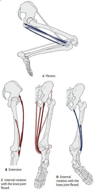
The knee joint is a modified hinge joint that includes the medial and lateral articulations between the condyles of the femur and tibia, and the articulation between the femur and patella (Fig. 22.12). Flexion and extension are the primary movements at the knee joint, but some rotation and sliding also occur.
—The patella articulates with the patellar surface of the femur between the medial and lateral condyles and protects the knee joint anteriorly. The patella is embedded in the tendon of the quadriceps femoris muscle, increasing the leverage of the muscle by holding the tendon away from the joint.
—Although the articular surfaces of the femur and tibia are extensive, there is little congruity between the bones, and stability depends chiefly on
•ligaments that connect the tibia and femur; and
•muscles that surround the joint, most importantly the quadriceps femoris (Figs. 22.13 and 22.14).
—The fibrous capsule of the knee joint is thin and incomplete and derives additional support from patellar retinacula (capsular ligaments that attach anteriorly to the quadriceps tendon), the patella, and extracapsular ligaments around the joint (Fig. 22.13).
—Extracapsular (external) ligaments support the fibrous capsule (Figs. 22.15, 22.16, 22.22).
•The patellar ligament, the distal part of the quadriceps tendon that supports the knee joint anteriorly, extends from the patella to the tibial tuberosity.
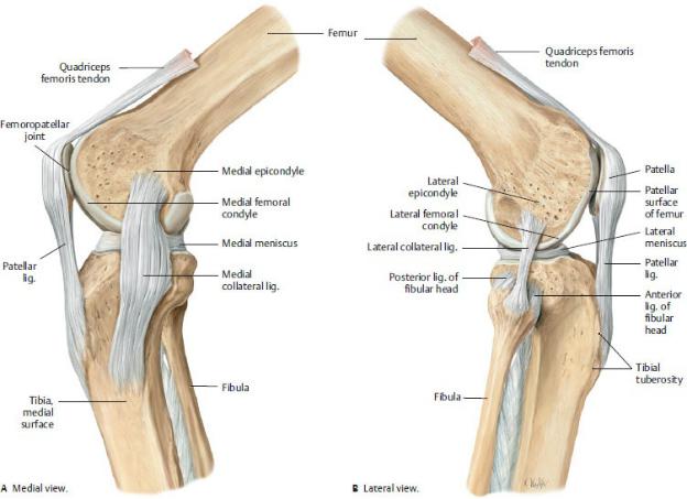
•The two collateral ligaments limit rotation and prevent medial and lateral dislocation of the knee joint. They are tightest in extension and slacken during flexion.
◦The lateral (fibular) collateral ligament is cordlike and remains unattached to the joint capsule. It extends from the lateral

epicondyle of the femur to the head of the fibula.
◦The medial (tibial) collateral ligament is flat and ribbonlike and is attached to the joint capsule and medial meniscus. It extends from the medial epicondyle of the femur to the medial condyle and anteromedial aspect of the tibia.
BOX 22.6: CLINICAL CORRELATION
PATELLAR TENDON REFLEX
The patellar tendon reflex is initiated by tapping the patellar tendon to elicit contraction of the quadriceps femoris muscle (extension of the knee). It tests the integrity of the L2–L4 spinal cord segments carried by the femoral nerve.
•The oblique popliteal ligament is an expansion of the semimembranosus tendon, which supports the joint capsule posteriorly and laterally.
•The arcuate popliteal ligament extends from the fibular head to the posterior knee joint and reinforces the joint capsule posteriorly and laterally.
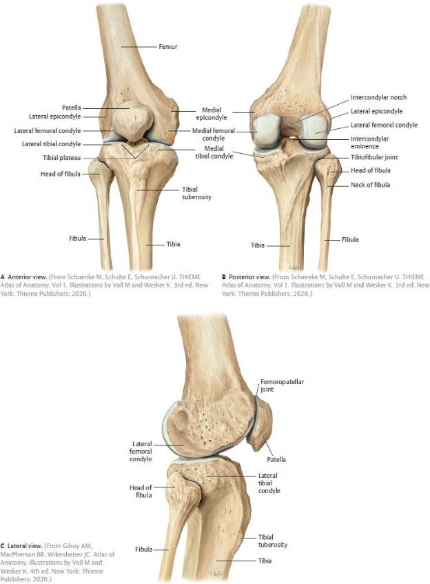
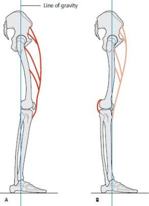
Fig. 22.12 Right knee joint
Fig. 22.13 Ligaments of the knee joint
Right knee, anterior view. (From Schuenke M, Schulte E, Schumacher U. THIEME Atlas of Anatomy, Vol 1. Illustrations by Voll M and Wesker K. 3rd ed. New York: Thieme Publishers; 2020.)
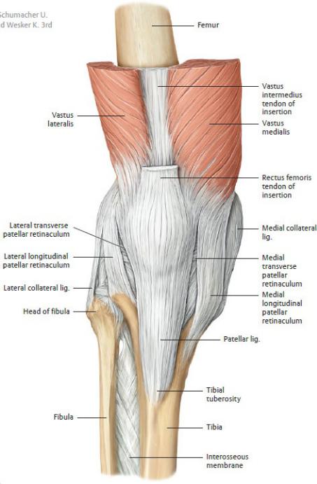
Fig. 22.14 Deficient stabilization of the knee joint due to weakness or paralysis of quadriceps femoris
Right lower limb, lateral view. (From Schuenke M, Schulte E, Schumacher U. THIEME Atlas of Anatomy, Vol 1. Illustrations by Voll M and Wesker K. 3rd ed. New York: Thieme Publishers; 2020.)
A When the quadriceps femoris is intact and the knee is in slight flexion, the line
of gravity falls behind the transverse axis of the knee motion. As the only extensor muscle of the knee joint, the quadriceps femoris keeps the body from tipping backward and ensures stability.
B With the weakness or paralysis of the quadriceps femoris, the knee joint can no longer be actively extended. In order to stand upright, the patient must hyperextend the knee so that the line of gravity, and thus the whole-body center of gravity, is shifted forward, in front of the knee, to utilize gravity as the extending force. The joint is stabilized in this situation by the posterior capsule and ligaments of the knee.
Fig. 22.15 Collateral and patellar ligaments of the knee joint
Right knee joint. Each knee joint has medial and lateral collateral ligaments. The medial collateral ligament is attached to both the capsule and the medial meniscus, whereas the lateral collateral ligament has no direct contact with either the capsule or the lateral meniscus. Both collateral ligaments are taut when the knee is in extension and stabilize the joint in the coronal plane. (From Schuenke M, Schulte E, Schumacher U. THIEME Atlas of Anatomy, Vol 1. Illustrations by
Voll M and Wesker K. 3rd ed. New York: Thieme Publishers; 2020.)
—Intra-articular ligaments provide stability during movements of the joint (Figs. 22.16 and 22.17A).
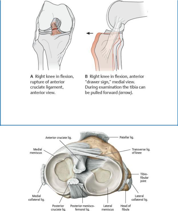
•Two cruciate ligaments are located within the joint capsule but lie external to the synovial layer. They provide stability in all positions, in addition to limiting rotation and preventing anterior and posterior dislocation of the joint.
◦The anterior cruciate ligament arises from the anterior intercondylar part of the tibia and extends posterolaterally to the medial aspect of the lateral femoral condyle.
◦The posterior cruciate ligament arises from the posterior intercondylar part of the tibia and extends anteromedially to the lateral aspect of the medial femoral condyle.
•A transverse ligament connects the menisci to each other along their anterior edges.
•A posterior meniscofemoral ligament joins the lateral meniscus to the posterior cruciate ligament and medial femoral condyle.
—Menisci, crescent-shaped fibrocartilaginous pads, deepen the articular surfaces of the tibial plateau. Wedge-shaped in cross section, they are tallest at the outer rim, where they are attached to the joint capsule and the intercondylar ridge (Figs. 22.17B, 22.18, 22.19).
•The medial meniscus is C-shaped and relatively immobile due to its additional attachment to the tibial collateral ligament.
•The lateral meniscus is nearly circular and more mobile during flexion and extension of the joint than its medial counterpart.
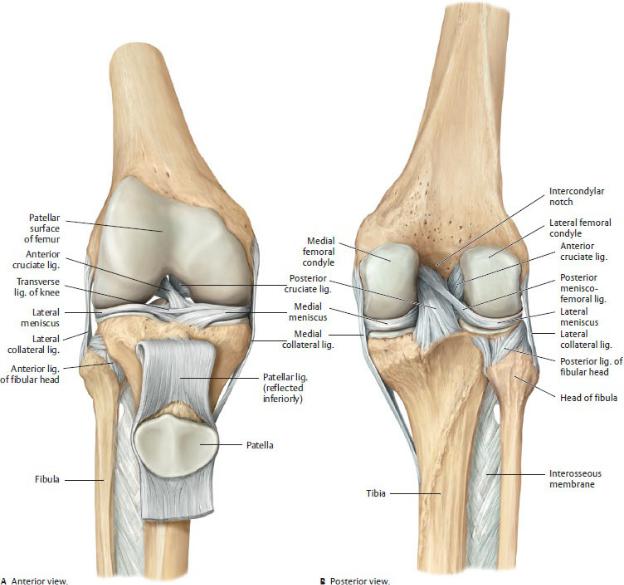
Fig. 22.16 Cruciate and collateral ligaments of the knee joint
Right knee. The cruciate ligaments keep the articular surfaces of the femur and tibia in contact, while stabilizing the knee joint primarily in the sagittal plane. Portions of the cruciate ligaments are taut in every joint position. (From Schuenke M, Schulte E, Schumacher U. THIEME Atlas of Anatomy, Vol 1. Illustrations by Voll M and Wesker K. 3rd ed. New York: Thieme Publishers; 2020.)
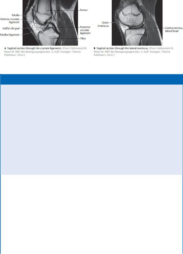
Fig. 22.17 MRI of the knee joint
BOX 22.7: CLINICAL CORRELATION
LIGAMENTOUS INJURIES OF THE KNEE
Most knee injuries occur during physical activity and involve rupture or strain of the knee ligaments. A forceful blow to the lateral side of the knee can strain the medial collateral ligament and, because of their intimate relationship, tear the medial meniscus as well. A similar injury can result from excessive lateral rotation of the knee and is often accompanied by rupture of the anterior cruciate ligament. The Lachman test is used to demonstrate instability of the knee joint resulting from rupture of the cruciate ligaments. Excessive anterior translation of the free-hanging tibia from under the stabilized femur is a positive anterior drawer sign, indicating anterior cruciate rupture. Posterior displacement of the tibia is a positive posterior drawer sign, indicating rupture of the posterior cruciate ligament.
Thieme Publishers; 2020.)
Fig. 22.19 Movements of the menisci
The medial meniscus, which is anchored more securely than the lateral meniscus, undergoes less displacement during knee flexion. As a result, it is more susceptible to injury. (From Schuenke M, Schulte E, Schumacher U. THIEME Atlas of Anatomy, Vol 1. Illustrations by Voll M and Wesker K. 3rd ed. New York: Thieme Publishers; 2020.)
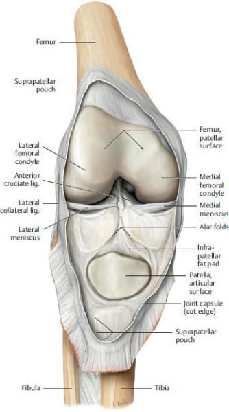
Fig. 22.20 Opened joint capsule of the knee
Right knee, anterior view, with patella reflected downward. (From Schuenke M, Schulte E, Schumacher U. THIEME Atlas of Anatomy, Vol 1. Illustrations by Voll M and Wesker K. 3rd ed. New York: Thieme Publishers; 2020.)
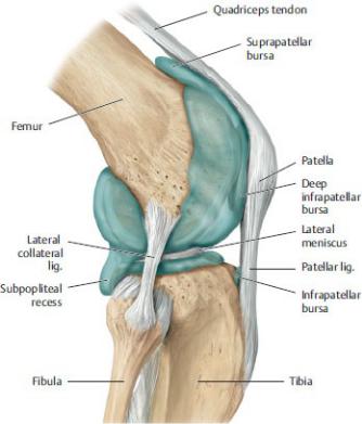
Fig. 22.21 Joint cavity of the knee
Right knee, lateral view. The joint cavity was demonstrated by injecting liquid plastic into the knee joint and later removing the capsule. (From Schuenke M, Schulte E, Schumacher U. THIEME Atlas of Anatomy, Vol 1. Illustrations by Voll M and Wesker K. 3rd ed. New York: Thieme Publishers; 2020.)
—An extensive synovial layer lines the internal surface of the joint capsule. Its posterior aspect extends into the intercondylar space of the joint cavity and reflects around the intracapsular ligaments, dividing most of the joint space into medial and lateral parts (Figs. 22.20 and 22.21).
—Muscles of the thigh and leg move the knee joint ( Table 22.7).
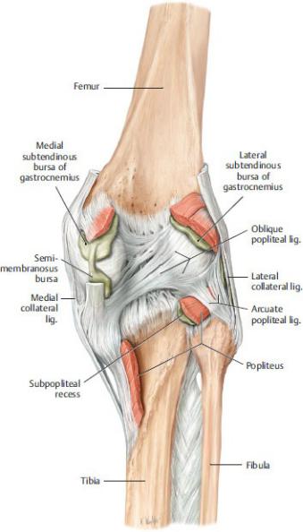
Fig. 22.22 Capsule, ligaments, and periarticular bursae of the knee joint
Right knee, posterior view. (From Schuenke M, Schulte E, Schumacher U. THIEME Atlas of Anatomy, Vol 1. Illustrations by Voll M and Wesker K. 3rd ed. New York: Thieme Publishers; 2020.)
—In addition to the support provided by extracapsular ligaments, the capsule is strengthened by tendinous attachments of muscles that cross the joint (semitendinosus, semimembranosus, biceps femoris, gastrocnemius, and quadriceps femoris). Bursae associated with these muscular attachments are numerous and include
BOX 22.8: CLINICAL CORRELATION
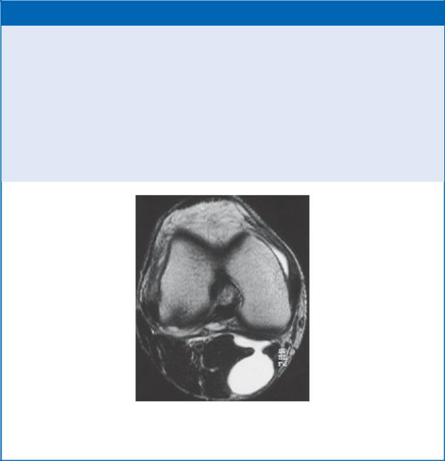
POPLITEAL (BAKER’S) CYST
Popliteal (Baker’s) cysts, palpable fluid-filled synovial sacs that protrude into the popliteal fossa, are usually a consequence of chronic effusion of the knee joint. Often they result from a herniation of the bursae deep to the gastrocnemius or semimembranosus muscles. The sacs protrude through the fibrous layer of the joint capsule but maintain a communication with the synovial cavity. Some cysts may be asymptomatic, but others are painful and can impair flexion and extension of the knee joint.
MRI of a Baker’s cyst in the popliteal fossa
Transverse section, inferior view. (From Vahlensieck M, Reiser M. MRT des Bewegungsapparates. 4. Aufl. Stuttgart: Thieme Publishers; 2014.)
•the suprapatellar bursa (pouch), which lies deep to the quadriceps femoris tendon and communicates with the cavity of the knee joint;
•the prepatellar bursa, which is subcutaneous over the patella;
•the superficial infrapatellar bursa, which is subcutaneous over the patellar ligament;
•the deep infrapatellar bursa, which lies deep to the patellar ligament; and
•the anserine bursa, which lies between the pes anserinus and tibial
collateral ligament.
—Additional bursae around the knee communicate with the joint cavity. They include the subpopliteal recess, the semimembranosus bursa, and the medial subtendinous bursa of the gastrocnemius (Fig. 22.22).
(From Schuenke M, Schulte E, Schumacher U. THIEME Atlas of Anatomy, Vol 1. Illustrations by Voll M and Wesker K. 3rd ed. New York: Thieme Publishers; 2020.)
Table 22.7 Movements of the Knee Joint

Action |
Primary Muscles |
Flexion |
Biceps femoris—short and long heads |
|
Semimembranosus |
|
Semitendinosus |
|
Sartorius |
|
Gracilis |
|
Gastrocnemius |
|
Popliteus |
|
|
Extension |
Quadriceps femoris |
|
|
Internal rotation |
Semimembranosus |
|
Semitendinosus |
|
Sartorius |
|
Gracilis |
|
Popliteus (of nonbearing leg) |
|
|
External rotation |
Biceps femoris |
|
|
BOX 22.9: CLINICAL CORRELATION
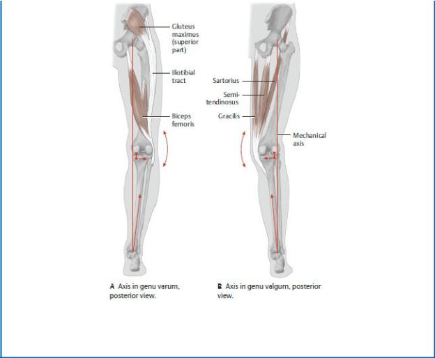
GENU VARUM AND GENU VALGUM
Although the femur lies diagonally within the thigh, the tibia is nearly vertical in the leg. This creates a Q angle at the knee between the long axes of the two bones. The angle varies with developmental stage and sex, but it can also be altered by disease. Normally, the head of the femur sits over the center of the knee joint, distributing weight evenly on the tibial plateau. In genu varum (bowleg), the Q angle is smaller than normal because the femur is more vertical. This increases the weight on the medial side of the knee, putting additional stress on the medial meniscus and medial (tibial) collateral ligament. When a person is standing upright with feet and ankles together, the knees are wide apart. In genu valgum (knock knee), the Q angle is larger because the femur is more diagonal. Greater weight is put on the lateral side of the knee, stressing the lateral meniscus and lateral (fibular) collateral ligament. In the upright position, the knees touch, but the ankles do not.
(From Schuenke M, Schulte E, Schumacher U. THIEME Atlas of Anatomy, Vol 1. Illustrations by Voll M and Wesker K. 3rd ed. New York: Thieme Publishers; 2020.)
The Popliteal Fossa
The popliteal fossa is a diamond-shaped space posterior to the knee joint (Fig. 22.23).
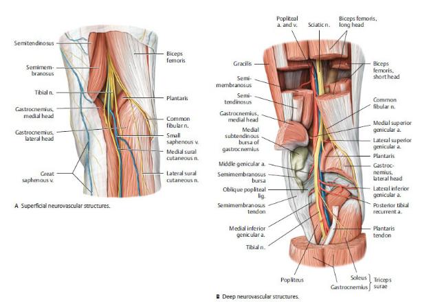
Fig. 22.23 Popliteal fossa
Right leg, posterior view. (From Schuenke M, Schulte E, Schumacher U. THIEME Atlas of Anatomy, Vol 1. Illustrations by Voll M and Wesker K. 3rd ed. New York: Thieme Publishers; 2020.)
—Its muscular boundaries are
•superomedially, the semimembranosus muscle;
•superolaterally, the biceps femoris muscle; and
•inferiorly, the medial and lateral heads of the gastrocnemius muscle.
—The contents of the fossa, which are embedded within popliteal fat, include
•the popliteal artery and vein and their genicular branches,
•popliteal lymph nodes, and
•tibial and common fibular nerves.
—At the superior apex of the popliteal fossa, the sciatic nerve splits into its two terminal branches.
•The tibial nerve descends into the posterior leg compartment with the popliteal vessels.
•The common fibular nerve courses laterally along the edge of the biceps
femoris muscle to enter the lateral leg compartment.
22.5 The Leg
The leg extends from the knee to the ankle; it contains the tibia and fibula and the muscles of the leg (crural) compartments.
Tibiofibular Joints
at the ankle. Unlike the extensive movement at the radioulnar joints in the forearm, only slight movement occurs at each tibiofibular joint, and no muscles are directly associated with them.
—The proximal tibiofibular joint is a plane-type synovial joint between the fibular head and an articular facet of the lateral condyle of the tibia.
Anterior and posterior ligaments of the fibular head secure the joint (see Fig. 22.16).
—The distal tibiofibular joint, a compound fibrous joint, creates a mortise formed by the medial and lateral malleoli that cups the talus and stabilizes the ankle joint. Ligaments of the tibiofibular joint (Fig. 22.24) include
•a deep interosseous tibiofibular ligament, the primary support of the joint, which is continuous with the interosseous membrane of the leg, and
•the external anterior tibiofibular and posterior tibiofibular ligaments.
—An interosseous membrane unites the shafts of the tibia and fibula and provides stability to the distal tibiofibular and ankle joints.
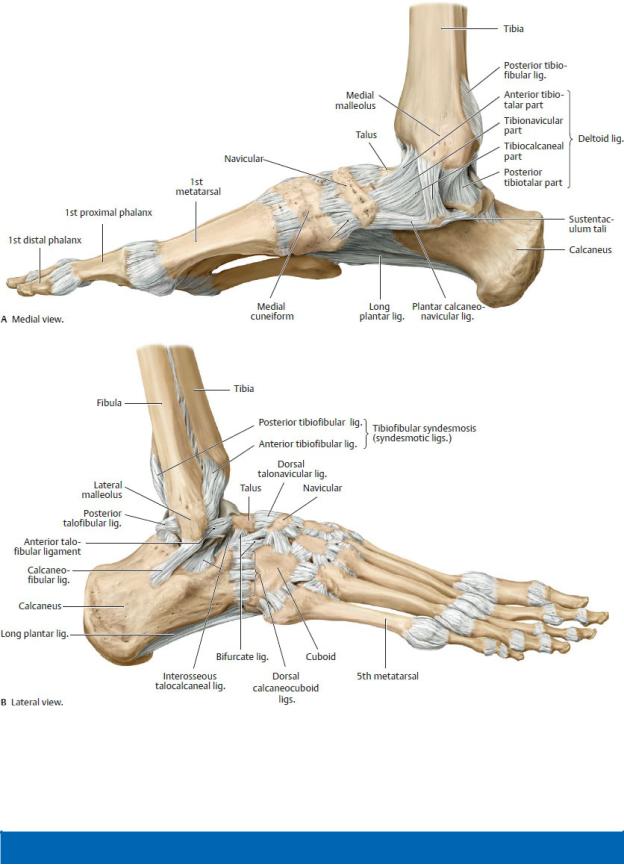
Fig. 22.24 Ligaments of the ankle and foot
Right foot. (From Schuenke M, Schulte E, Schumacher U. THIEME Atlas of Anatomy, Vol 1. Illustrations by Voll M and Wesker K. 3rd ed. New York: Thieme Publishers; 2020.)
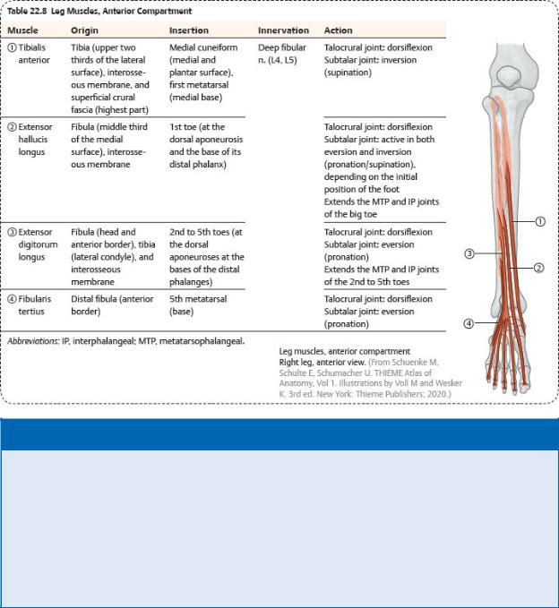
Muscles of the Leg
Muscles of the leg, which move the knee, ankle, and foot, are divided into four crural compartments (see Fig. 22.45B).
—The anterior compartment contains
•muscles that dorsiflex the foot and toes and invert the foot ( Table 22.8),
•the deep fibular nerve, and

•the anterior tibial artery and veins.
—The lateral compartment contains
•muscles that evert and plantar flex the foot ( Table 22.9),
•the superficial fibular nerve, and
•the muscular branches of the fibular artery and veins from the posterior compartment.
BOX 22.10: CLINICAL CORRELATION
SHIN SPLINTS
Shin splints result from chronic trauma of the tibialis anterior, usually incurred by overuse of the muscle during athletic activities. In what is considered a mild form of anterior compartment syndrome, small tears of the periosteum cause pain and swelling over the distal two thirds of the tibial shaft.
—The superficial posterior compartment contains
•muscles that plantar flex the foot, two of which, the gastrocnemius and soleus, form the triceps surae and share a common tendon of insertion, the calcaneal tendon ( Table 22.10);
•the tibial nerve; and
•the muscular branches of the posterior tibial artery and veins from the deep posterior compartment.
—The deep posterior compartment contains
•muscles that plantar flex the foot and toes and invert the foot ( Table 22.11) (only one muscle, the popliteus, crosses the knee and laterally rotates the femur),
BOX 22.11: CLINICAL CORRELATION
RUPTURE OF THE CALCANEAL TENDON
Rupture of the calcaneal tendon tends to occur following sudden forced plantar flexion of the foot, unexpected dorsiflexion of the foot, or violent dorsiflexion of a plantar-flexed foot in people unaccustomed to exercise or who exercise intermittently. The rupture disables the gastrocnemius, soleus, and plantaris muscles, rendering the patient unable to plantar flex the foot.
•the tibial nerve, and
•the posterior tibial and fibular artery and veins.
—Retinacula, thickened bands of the crural fascia, bind the tendons of the long extensor and flexor muscles as they cross the ankle (Fig. 22.25).
•Superior extensor and inferior extensor retinacula bind muscles of the anterior compartment.
•Medially, a flexor retinaculum binds muscles of the deep posterior

compartment.
•Superior fibular and inferior fibular retinacula bind muscles of the lateral compartment.
BOX 22.12: CLINICAL CORRELATION
COMPARTMENT SYNDROME
Compartment syndrome is a condition that may follow burns, hemorrhage, complex fractures, or crush injuries. Swelling, infection, or bleeding into the compartment can increase intracompartmental pressure and, when high enough, compress the small vascular structures that supply the compartment contents. Nerves are particularly vulnerable to ischemia, and permanent loss of motor and sensory functions distal to the compartment can result. Symptoms include severe pain that is out of proportion to the injury, paresthesia (tingling and numbness), pallor of the skin, paralysis of muscles affected, and loss of distal pulses in the affected limb. Treatment is prompt surgery to decompress the compartment by making long cuts through the fascia (fasciotomy).
—The tarsal tunnel is a passageway on the medial side of the ankle formed by the flexor retinaculum and its attachments to the medial malleolus and calcaneus. It transmits
•muscle tendons of the deep posterior compartment,
•posterior tibial artery and veins and their medial plantar and lateral plantar branches, and
•the tibial nerve and its medial plantar and lateral plantar branches.
BOX 22.13: CLINICAL CORRELATION
TARSAL TUNNEL SYNDR
Tarsal tunnel syndrome, similar to carpal tunnel syndrome of the hand, results from swelling of the synovial sheaths of the long flexor tendons within the tarsal tunnel. Compression of the tibial nerve can result in burning pain, numbness, and tingling that radiates to the heel.
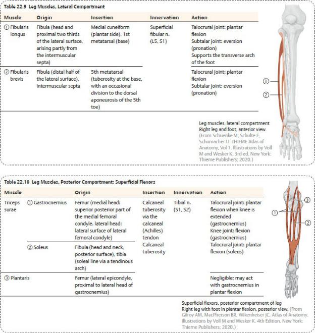
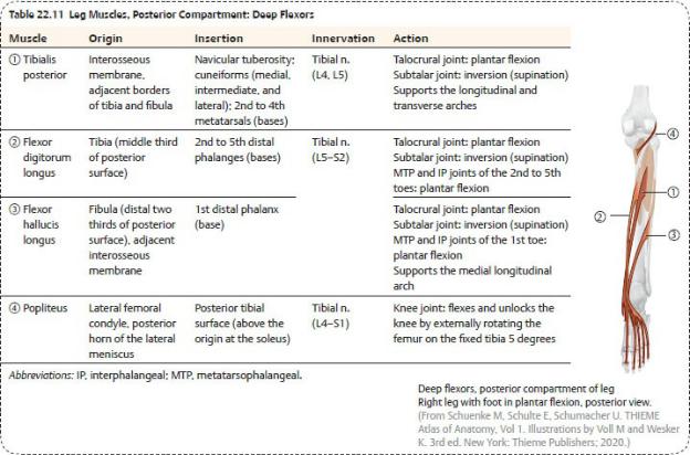
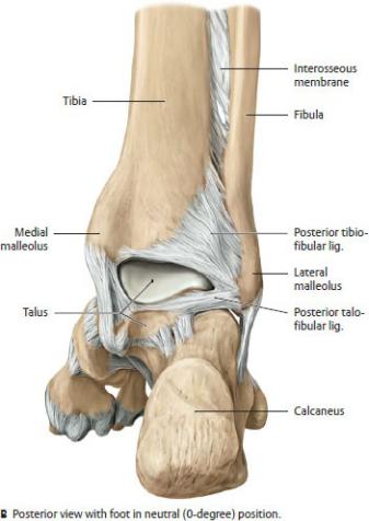
Fig. 22.25 Tendon sheaths and retinacula of the ankle
Right foot. The superior and inferior extensor retinacula retain the long extensor tendons, the fibularis retinacula hold the fibular muscle tendons in place, and the flexor retinaculum retains the long flexor tendons through the tarsal tunnel. (From Schuenke M, Schulte E, Schumacher U. THIEME Atlas of Anatomy, Vol 1. Illustrations by Voll M and Wesker K. 3rd ed. New York: Thieme Publishers; 2020.)
22.6 The Ankle (Talocrural) Joint
The weight of the body is transferred along the tibia of the leg, through the talus at the ankle joint, and onto the heel and ball of the foot. Strong bony and ligamentous supports stabilize the ankle and facilitate this transfer of weight.
The ankle joint includes articulations between the distal tibia and fibula and the talus (Figs. 22.26, 22.27, 22.28, 22.29).
— At the ankle joint, the ankle mortise, which is formed by the distal
tibiofibular joint, hugs the body of the talus. Tibiofibular ligaments that stabilize the ankle mortise also contribute to the stability of the ankle joint.
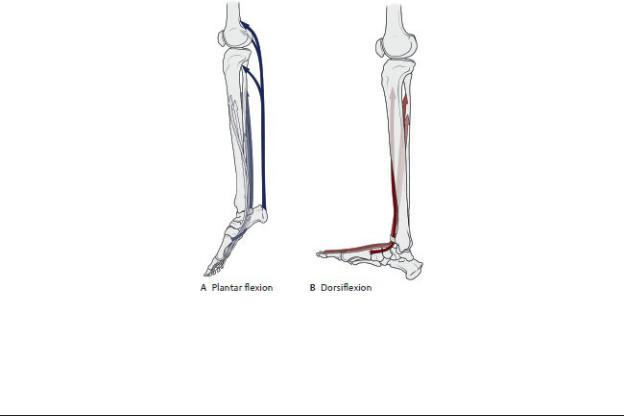
—The ankle joint is tighter and more stable with the foot in dorsiflexion, when the wider anterior part of the trochlea (of the talus) is wedged within the ankle mortise. Accordingly, the joint is looser and less stable in plantar flexion.
—Strong collateral talocrural ligaments connect the tibia and fibula to the tarsal bones and support the talocrural articulations (Fig. 22.30).
Fig. 22.26 Joints of the foot
Right foot with talocrural joint in plantar flexion, anterior view. (From Gilroy AM, MacPherson BR, Wikenheiser JC. Atlas of Anatomy. Illustrations by Voll M and Wesker K. 4th ed. New York: Thieme Publishers; 2020.)
•The medial deltoid ligament of the ankle extends from the medial malleolus of the tibia to the talus, calcaneus, and navicular. Its components are
◦ the anterior tibiotalar part,
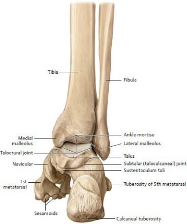
◦the posterior tibiotalar part,
◦the tibiocalcaneal part, and
◦the tibionavicular part.
•The lateral ligament of the ankle extends from the lateral malleolus of the fibula to the talus and calcaneus. Its parts are
◦the anterior talofibular ligament,
◦the posterior talofibular ligament, and
◦the calcaneofibular ligament.
•Movement at the ankle joint, provided by muscles of the leg, is limited primarily to dorsiflexion (extension) and plantar flexion ( Table 22.12).
Fig. 22.27 Talocrural and subtalar joints
Right foot with foot in neutral (0-degree) position, posterior view. (From Schuenke M, Schulte E, Schumacher U. THIEME Atlas of Anatomy, Vol 1. Illustrations by Voll M and Wesker K. 3rd ed. New York: Thieme Publishers; 2020.)
(From Schuenke M, Schulte E, Schumacher U. THIEME Atlas of Anatomy, Vol 1. Illustrations by Voll M and Wesker K. 3rd ed. New York: Thieme Publishers; 2020.)
Table 22.12 Movements of the Ankle Joint
Action |
Primary Muscles |
Plantar flexion |
Gastrocnemius |
|
Soleus |
|
Posterior tibialis |
|
Flexor digitorum longus |
|
Flexor hallucis longus |
|
|
Dorsiflexion |
Tibialis anterior |
|
Extensor hallucis longus |
|
Extensor digitorum longus |
|
Fibularis tertius |
|
|
Fig. 22.28 Talocrural joint
Right foot. The talocrural (ankle) joint is tighter and more stable with the foot in dorsiflexion, when the wider, anterior part of the trochlea (of the talus) is wedged within the ankle mortise. Accordingly, the joint is looser and less stable in plantar flexion. (From Gilroy AM, MacPherson BR, Wikenheiser JC. Atlas of Anatomy. Illustrations by Voll M and Wesker K. 4th ed. New York: Thieme Publishers; 2020.)
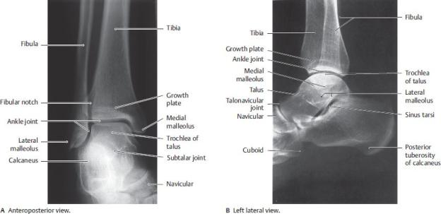
Fig. 22.29 Radiograph of the ankle
(From Moeller TB, Reif E. Pocket Atlas of Sectional Anatomy: The Musculoskeletal System. New York: Thieme Publishers; 2009.)

ANKLE SPRAINS
Ankle sprains (torn ligaments) are caused most often by forced inversion of the foot (e.g., walking on uneven ground). Damage to the lateral talocrural ligaments is correlated with the severity of the injury and proceeds from anterior to posterior: the anterior talofibular ligament is the most easily injured, followed by the calcaneofibular and the least frequently injured, the posterior talofibular ligament. Fracture of the lateral malleolus can also accompany inversion injuries.
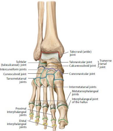
22.7 The Foot
The numerous joints of the foot create a flexible unit that effectively absorbs shock, distributes the weight of vertical loads, and participates in locomotion.
Joints of the Foot and Toes
Movements at the joints of the foot, particularly the intertarsal (subtalar and transverse tarsal), metatarsophalangeal, and interphalangeal joints, contribute to smooth locomotion and maintenance of balance (see Fig. 22.26).
—The subtalar joint is an articulation between the inferior surface of the talus and underlying calcaneus (Figs. 22.31 and 22.32; see also Fig. 22.29B).
•It is a compound joint that has anterior and posterior compartments.
◦The anterior compartment contains the talocalcaneal component of the talocalcaneonavicular articulation.
◦The posterior compartment contains the posterior talocalcaneal articulation.
•A strong interosseous talocalcaneal ligament joins the bones and separates the anterior and posterior parts of the joint.
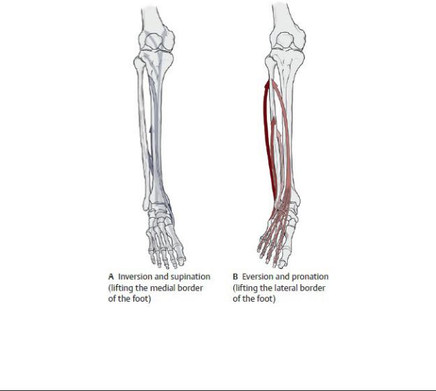
•This joint is the main articulation that allows inversion and eversion of the foot.
—The transverse tarsal joint is a compound joint that combines the talonavicular and calcaneocuboid articulations.
•Movement at this joint rotates the front of the foot relative to the heel, augmenting the inversion and eversion at the subtalar joint.
•This joint is a common site for surgical amputation of the foot.
—The tarsometatarsal and intermetatarsal joints are small and relatively immobile.
—Metatarsophalangeal joints are condyloid synovial joints between the heads of the metatarsals and base of the phalanges.
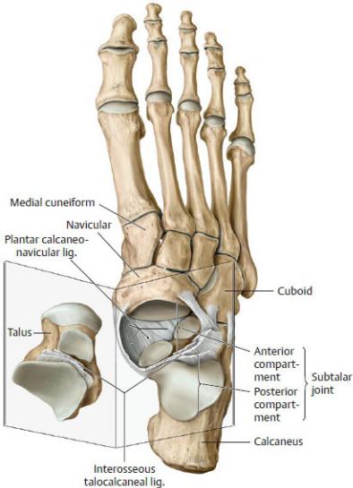
the posterior compartment (talocalcaneal joint) and the anterior compartment (talocalcaneonavicular joint). (From Schuenke M, Schulte E, Schumacher U. THIEME Atlas of Anatomy, Vol 1. Illustrations by Voll M and Wesker K. 3rd ed. New York: Thieme Publishers; 2020.)
Fig. 22.32 Ligaments of the plantar surface
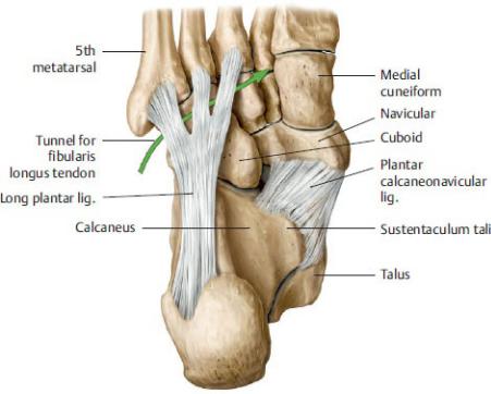
Plantar view. The plantar calcaneonavicular (“spring”) ligament completes the bony socket of the talocalcaneal joint. The long plantar ligament converts the tuberosity of the cuboid bone into a tunnel for the tibialis longus tendon (arrow). (From Gilroy AM, MacPherson BR, Wikenheiser JC. Atlas of Anatomy. Illustrations by Voll M and Wesker K. 4th ed. New York: Thieme Publishers; 2020.)


(From Schuenke M, Schulte E, Schumacher U. THIEME Atlas of Anatomy, Vol 1. Illustrations by Voll M and Wesker K. 3rd ed. New York: Thieme Publishers; 2020.)
Fig. 22.34 Stabilizers of the transverse arch
Right foot, plantar view. The transverse pedal arch is supported by both active and passive stabilizing structures (muscles and ligaments, respectively). Note: The arch of the forefoot has only passive stabilizers, whereas the arches of the metatarsus and tarsus have only active stabilizers. (From Schuenke M, Schulte E, Schumacher U. THIEME Atlas of Anatomy, Vol 1. Illustrations by Voll M and Wesker K. 3rd ed. New York: Thieme Publishers; 2020.)
Fig. 22.35 Stabilizers of the longitudinal arch
Right foot, medial view. (From Schuenke M, Schulte E, Schumacher U. THIEME Atlas of Anatomy, Vol 1. Illustrations by Voll M and Wesker K. 3rd ed. New York: Thieme Publishers; 2020.)
Fig. 22.35 (continued) Stabilizers of the longitudinal arch
Dorsum of the Foot
—Surface anatomy on the dorsum of the foot reveals
•skin that is thin and loose,
•a superficial dorsal venous arch that is the origin of the great saphenous and small saphenous veins, and
•tendons of extensor muscles of the anterior leg compartment.
—A dorsal muscular compartment of the foot contains two intrinsic muscles, the extensor digitorum brevis and the extensor hallucis brevis, which extend the toes ( Table 22.15).
Table 14.4 Radiographic Densities
Sole of the Foot
—Surface anatomy on the sole of the foot reveals
•tough, thickened skin, particularly on the heel, lateral side, and ball of the foot;
•skin that is firmly attached to the underlying plantar aponeurosis; and
•subcutaneous tissue divided by fibrous septa into fatfilled areas that act as shock absorbers, especially in the heel.
—The tough, longitudinal plantar aponeurosis (see Fig. 22.42)
•attaches tightly to the skin of the sole,
•originates at the calcaneus and is continuous distally with the fibrous digital sheaths that contain the flexor tendons of the toes,
•protects the sole of the foot from injury, and
•acts as a tie rod that supports the arches of the foot.
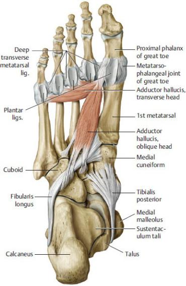
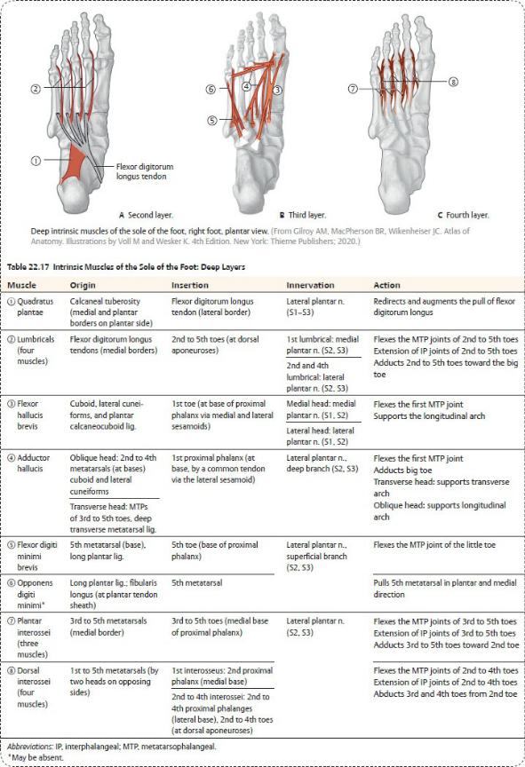
—The musculature of the sole is usually described as having four layers (Tables 22.16 and 22.17). However, as in the palm of the hand, the deep fascia separates the muscles of the sole into four fascial compartments:
•The medial compartment, which contains the abductor and flexors of the 1st digit (hallux)
•The central compartment, which contains the short and long flexors of the 2nd through 4th digits, the adductor hallucis, the lumbricals, and the quadratus plantae
•The lateral compartment, which contains the abductor and flexor of the 5th digit
•The interosseous compartment, which contains the interossei muscles

BOX 22.16: CLINICAL CORRELATION
PLANTAR FASCIITIS
Inflammation of the plantar aponeurosis is a common and painful affliction of runners that is characterized by tenderness on the sole, particularly on the calcaneus. Pain is usually greatest following rest and may dissipate with activity.
BOX 22.17: CLINICAL CORRELATION
THE PLANTAR REFLEX
The plantar reflex tests the integrity of the L4–S2 nerve roots. It is performed by firmly stroking the lateral aspect of the sole from the heel and across the foot to the base of the big toe. In the normal individual, this elicits flexion of the toes. Extension of the big toe with flaring of the 2nd through 5th digits is called the Babinski sign, an abnormal response in adults, indicating brain damage. Because of the immaturity of their central nervous system, the Babinski sign is a normal response in young children (under 4 years).

22.8 The Gait Cycle
Gait is a complex activity that requires the coordinated action of muscles of the hip, thigh, and leg. Table 22.18 provides a summary of muscle action during the gait cycle.
—Gait is described in two phases: in normal walking, the stance phase makes up ~ 60%, and the swing phase makes up ~ 40%.
—One cycle of gait consists of the action of one leg through each of the two phases.
Table 22.18 Muscle Action Sequence During the Gait Cycle
Activity |
Active Muscle |
|
Groups |
Stance phase |
Hip extensors |
This phase begins with the heel strike. |
Dorsiflexors |
|
|
The foot begins to accept the weight of the |
Hip adductors |
body, and the pelvis is stabilized. |
Knee extensors |
|
Plantar flexors |
|
|
At midstance, the pelvis, knee, and ankle are |
Hip abductors |
stabilized. |
Knee extensors |
|
Plantar flexors |
|
|
This phase ends with the push off that |
Hip abductors |
includes “heel lift” and “toe off.” Pelvis is |
Plantar flexors |
stabilized. |
|
|
|
Throughout the stance phase, the arches of |
Long tendons of the |
the foot are preserved. |
foot |
|
Intrinsic muscles of |
|
the foot |
|
|
Swing phase |
Hip flexors |
This phase begins with forward acceleration |
|
of the thigh. |
|
|
|
The foot needs to clear the ground as it |
Dorsiflexors |

swings forward.
The thigh decelerates in preparation for |
Hip extensors |
landing. |
|
|
|
As the foot prepares for heel strike, the knee |
Knee extensor |
extends and positions the foot. |
Dorsiflexors |
|
|
22.9 Topographic Anatomy of Lower Limb
Musculature
Thigh, Hip, and Gluteal Region
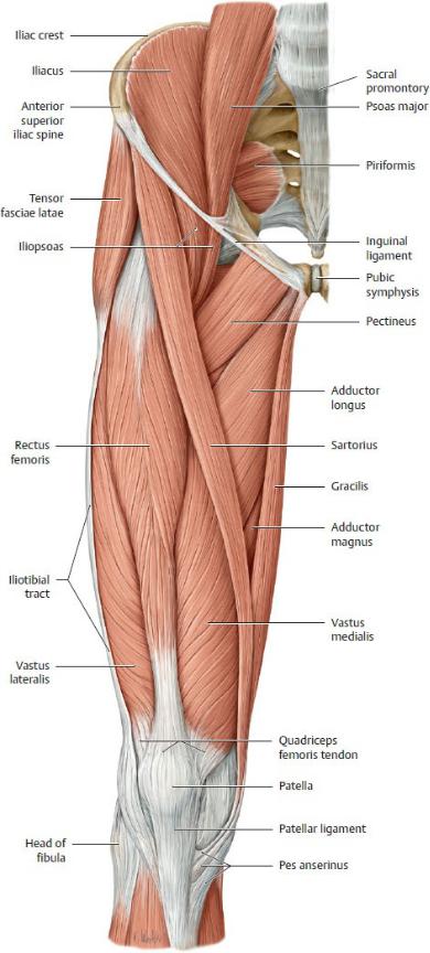
Fig. 22.36 Muscles of the hip and thigh
Right limb, anterior view. Muscle origins are shown in red, insertions in gray. Removed: Fascia lata of the thigh (to the lateral iliotibial tract). (From Schuenke M, Schulte E, Schumacher U. THIEME Atlas of Anatomy, Vol 1. Illustrations by Voll M and Wesker K. 3rd ed. New York: Thieme Publishers; 2020.)
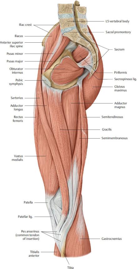
Fig. 22.37 Muscles of the hip, thigh, and gluteal region
Midsagittal section, medial view. (From Gilroy AM, MacPherson BR, Wikenheiser JC. Atlas of Anatomy. Illustrations by Voll M and Wesker K. 4th ed. New York: Thieme Publishers; 2020.)
Fig. 22.38 Muscles of the hip, thigh, and gluteal region
Right limb, posterior view.
Removed: Fascia lata (to iliotibial tract). (From Schuenke M, Schulte E, Schumacher U. THIEME Atlas of Anatomy, Vol 1. Illustrations by Voll M and Wesker K. 3rd ed. New York: Thieme Publishers; 2020.)
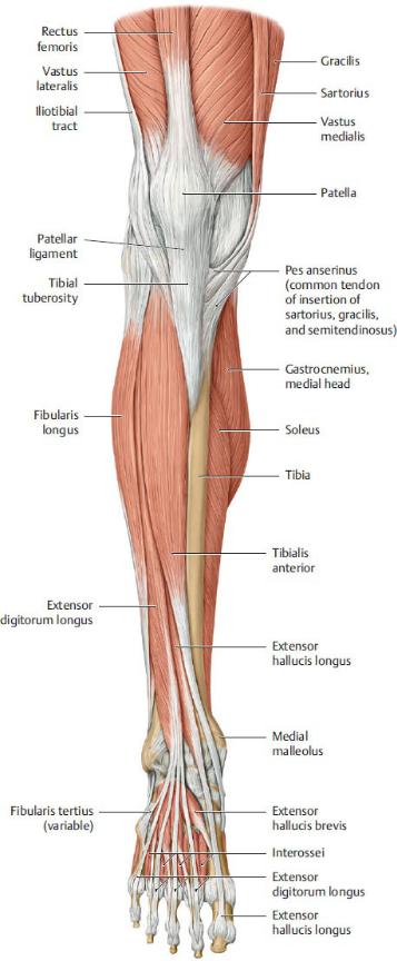
Fig. 22.39 Muscles of the leg
Right leg, anterior view. (From Schuenke M, Schulte E, Schumacher U. THIEME Atlas of Anatomy, Vol 1. Illustrations by Voll M and Wesker K. 3rd ed. New York: Thieme Publishers; 2020.)
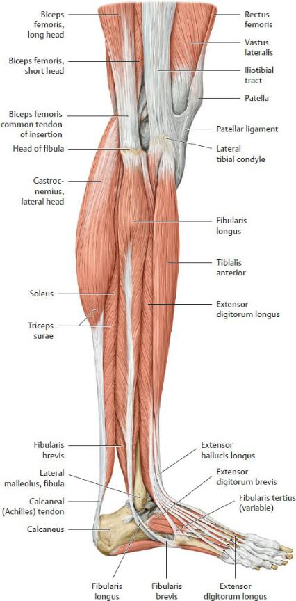
Fig. 22.40 Muscles of the leg
Right leg, lateral view.
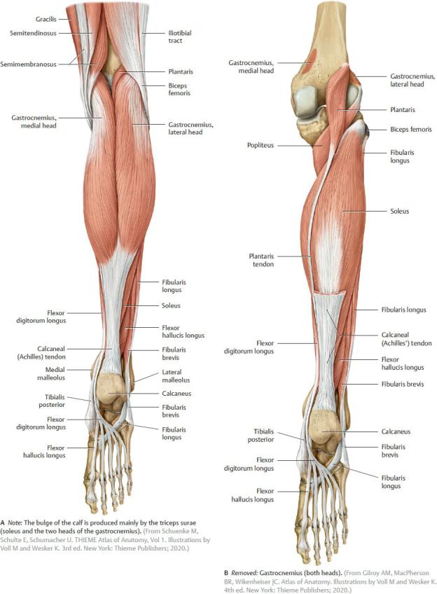
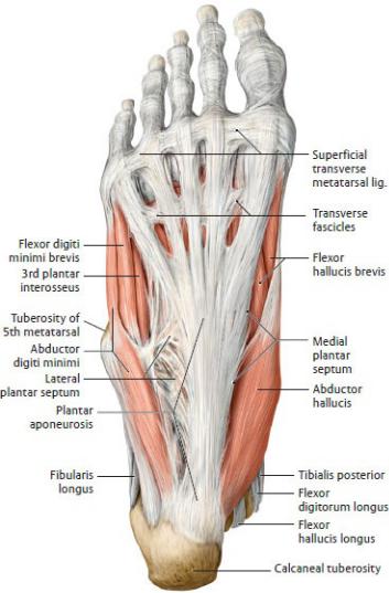
Fig. 22.41 Muscles of the leg
Right leg, posterior view.
The Foot
Fig. 22.42 Plantar aponeurosis
Right foot, plantar view. The plantar aponeurosis is a tough aponeurotic sheet, thickest at the center, that blends with the dorsal fascia (not shown) at the borders of the foot. (From Schuenke M, Schulte E, Schumacher U. THIEME Atlas of Anatomy, Vol 1. Illustrations by Voll M and Wesker K. 3rd ed. New York: Thieme Publishers; 2020.)
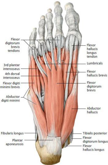
Fig. 22.43 Intrinsic muscles of the sole of the foot
Right foot, plantar view.
Superficial (first) layer. Removed: Plantar aponeurosis, including the superficial transverse metacarpal ligament. (From Schuenke M, Schulte E, Schumacher U. THIEME Atlas of Anatomy, Vol 1. Illustrations by Voll M and Wesker K. 3rd ed. New York: Thieme Publishers; 2020.)
Compartments of the Thigh and Leg
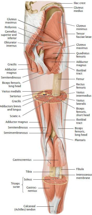
Fig. 22.44 Windowed dissection
Right limb, posterior view. (From Schuenke M, Schulte E, Schumacher U. THIEME Atlas of Anatomy, Vol 1. Illustrations by Voll M and Wesker K. 3rd ed. New York: Thieme Publishers; 2020.)

Wesker K. 3rd ed. New York: Thieme Publishers; 2020.)