

12 Abdominal Viscera
The peritoneal cavity of the abdomen contains the primary and accessory organs of the gastrointestinal tract. The primary organs are the stomach, small intestine, and large intestine; the accessory organs are the liver, gallbladder, pancreas, and spleen. The kidneys, proximal ureters, and suprarenal glands are found outside the peritoneal cavity within the retroperitoneum on the posterior abdominal wall
(Fig. 12.1).
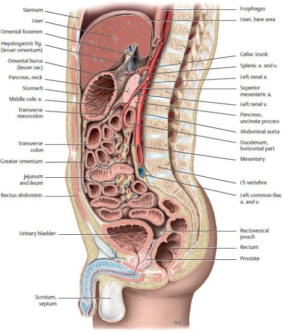
Fig. 12.1 Organs of the abdomen and pelvis
Midsagittal section through male pelvis, viewed from the left side. (From Schuenke M, Schulte E, Schumacher U. THIEME Atlas of Anatomy, Vol 2. Illustrations by Voll M and Wesker K. 3rd ed. New York: Thieme Publishers; 2020.)

12.1 Organs of the Peritoneal Cavity—
Gastrointestinal Tract
Divisions of the Gastrointestinal Tract
The three divisions of the embryonic gastrointestinal tract are retained in the adult system and reflected in its blood supply and innervation. These divisions are
—the foregut, which consists of the distal esophagus, the stomach, the proximal half of the duodenum, the liver, the gallbladder, and the superior part of the pancreas;
—the midgut, which includes the distal half of the duodenum, the jejunum, ileum, cecum, and appendix, and the ascending colon and proximal two thirds of the transverse colon; and
—the hindgut, which includes the distal third of the transverse colon, the descending and sigmoid colons, the rectum, and the upper part of the anal canal.
BOX 12.1: DEVELOPMENTAL CORRELATION
ROTATION OF THE MIDGUT TUBE
Development of the midgut is characterized by rapid elongation of the gut and its mesentery, resulting in the formation of the primary intestinal loop. The loop is attached anteriorly to the yolk sac by the omphaloenteric duct (yolk stalk) and posteriorly to the posterior abdominal wall by the superior mesenteric artery. As a result of this rapid elongation and expansion of the liver, the abdominal cavity becomes too small to contain all the intestinal loops, and they physiologically herniate into the proximal part of the umbilical cord. Coincident with growth in length, the primary intestinal loop rotates 270 degrees counterclockwise (if viewed from the front) around the axis established by the attachment of the superior mesenteric artery. Rotation occurs during herniation (approximately 90 degrees) and during the return of the intestinal loops into the abdominal cavity (remaining 180 degrees), which is thought to occur when the relative size of the liver and kidney decreases. Malrotation of the midgut can result in congenital abnormalities such as volvulus (twisting) of the intestine.
BOX 12.2: DEVELOPMENTAL CORRELATION
LOCALIZATION OF PAIN FROM FOREGUT, MIDGUT, AND HINDGUT DERIVATIVES
Pain from gastrointestinal organs follows pathways determined by embryologic origin. Pain from foregut structures is localized to the epigastric region, pain from structures of the midgut localizes in the periumbilical region, and pain arising from struc-tures of the hindgut localizes in the hypogastric region.
Stomach
The stomach, a hollow reservoir that stores, churns, and initiates digestion of food, communicates with the esophagus proximally and the duodenum of the small intestine distally (Figs. 12.2, 12.3, 12.4).
—It is normally J-shaped and lies in the left upper quadrant, although its shape and position vary among individuals and can change depending on its contents.
—The stomach has four parts:
1.The cardia, which surrounds the opening to the esophagus
2.The fundus, the superior portion that rises above and to the left of the opening between the esophagus and stomach (cardiac orifice)
3.The body, the large expanded portion below the fundus
4.The pyloric part, which is the outflow channel made up of the wide pyloric antrum, a narrow pyloric canal, and the pyloris or sphincteric region, which contains a muscular pyloric sphincter that surrounds the pyloric orifice into the first part of the duodenum.
—The stomach has a lesser curvature and a greater curvature.
• The lesser curvature makes up the superior concave border. An angular notch (or incisure) along the curvature marks the junction of the body and pyloric part.
• The greater curvature makes up the inferior convex border.
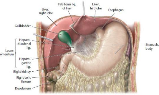
Fig 12.2 Stomach in situ
Anterior view of upper abdomen. The liver has been reflected superiorly to
expose the stomach and lesser omentum. (From Gilroy AM, MacPherson BR, Wikenheiser JC. Atlas of Anatomy. Illustrations by Voll M and Wesker K. 4th ed. New York: Thieme Publishers; 2020.)
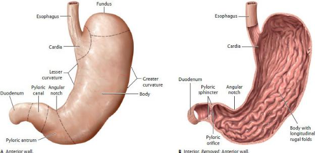
Fig. 12.3 Stomach
Anterior view. (From Schuenke M, Schulte E, Schumacher U. THIEME Atlas of Anatomy, Vol 2. Illustrations by Voll M and Wesker K. 3rd ed. New York: Thieme Publishers; 2020.)
—Although hollow organs of the gastrointestinal tract generally have walls composed of two muscular layers, the stomach is unique in having three layers: an outer longitudinal layer, a middle circular layer, and inner oblique layer. These enable the stomach to create powerful churning movements that break apart large food particles.
—The inner surface of the stomach is highly distensible; in adults, the stomach can accommodate 2 to 3 liters. Rugae (longitudinal folds) of the gastric mucosa, formed during contraction of the stomach, are most prominent in the pyloric part and along the greater curvature. Gastric filling causes the mucosal folds to disappear.
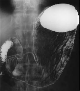
Fig. 12.4 Radiograph of double contrast barium enema of the stomach
Anterior view. (From Gunderman R. Essential Radiology, 3rd ed. New York: Thieme Publishers; 2014)
—The layers of peritoneum on the stomach’s anterior and posterior surfaces unite along the lesser curvature to form the lesser omentum; along the greater curvature they unite to form the greater omentum (see Figs. 12.1 and
12.2).
—Anteriorly, the stomach is in contact with the abdominal wall, the diaphragm, and the left lobe of the liver. Posteriorly, it forms the anterior wall of the omental bursa.
—When the body is supine, the stomach rests on the pancreas, the spleen, the left kidney, the left suprarenal gland, and the transverse colon and its mesentery.
—Right and left gastric arteries, right and left gastro-omental arteries, and short gastric arteries (all derived from branches of the celiac trunk) supply the stomach (see Figs. 11.12 and 11.14).
—Veins that accompany the arteries of the stomach drain to the hepatic portal venous system.
—Lymph vessels drain to gastric and gastro-omental nodes, which drain to the celiac lymph nodes.
—The celiac nerve plexus innervates the stomach (see Fig. 11.30).
•Sympathetic nerves promote vasoconstriction and inhibit peristalsis.
•Parasympathetic nerves stimulate gastric secretion.

BOX 12.3: CLINICAL CORRELATION
GASTRIC ULCERS
Gastric ulcers are open lesions of the mucosa that are believed to be initiated by increased acid secretion and exacerbated by the presence of the bacterium Helicobacter pylori. Gastric ulcers can cause hemorrhaging if they erode into gastric arteries. Posteriorly located ulcers can erode into the pancreas and splenic artery, causing severe hemorrhaging. Patients with chronic ulcers may undergo a vagotomy, the surgical sectioning of the vagus nerve, which may curtail the production of gastric acid.
Small Intestine
The small intestine extends from the pyloric orifice of the stom-ach to the ileocecal orifice at the ileocecal junction and is the pri-mary site of digestion and absorption of digested products. It is made up of three sections, the duodenum, which is mostly retro-peritoneal, and the jejunum and ileum, which are suspended by the mesentery of the small intestine.
—The duodenum, the first and shortest section, forms a C-shaped curve around the head of the pancreas and has four parts (Figs. 12.5, 12.6, 12.7):
1.The superior (first) part is at the level of the L1 vertebra.
◦The proximal 2-cm segment, called the duodenal bulb or ampulla, is suspended from a mesentery.
2.The descending (second) part extends along the right side of the L1–L3 vertebral bodies.
◦This is the site of the junction of the foregut and midgut.
◦The hepatopancreatic duct, formed by the common bile duct and main pancreatic duct, enters the duo-denum through the major duodenal papilla on the posteromedial wall. Superior to that, the accessory pancreatic duct enters through the minor duodenal papilla.
3.The horizontal (third) part crosses to the left, anterior to the inferior vena cava, the aorta, and the L3 vertebra, along the inferior border of the pancreas.
◦The root of the mesentery of the small intestine (mesenteric root) and superior mesenteric vessels cross anteriorly.
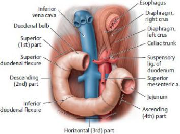
Fig. 12.5 Parts of the duodenum
Anterior view. (From Gilroy AM, MacPherson BR, Wikenheiser JC. Atlas of Anatomy. Illustrations by Voll M and Wesker K. 4th Edition. New York: Thieme Publishers; 2020.)
4.The ascending (fourth) part ascends along the left side of the aorta to the level of the L2 vertebra at the inferior border of the pancreas.
◦It joins the jejunum at the duodenojejunal flexure, which is suspended from the posterior abdominal wall by the suspensory ligament (of Treitz).
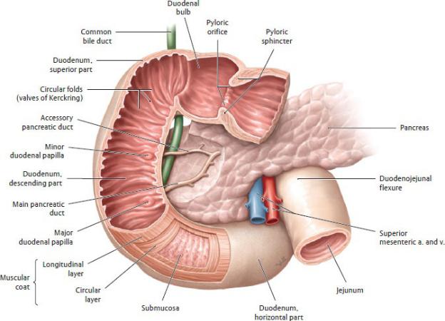
Fig. 12.6 Duodenum
Anterior view with the anterior wall opened. (From Schuenke M, Schulte E, Schumacher U. THIEME Atlas of Anatomy, Vol 2. Illustrations by Voll M and Wesker K. 3rd ed. New York: Thieme Publishers; 2020.)
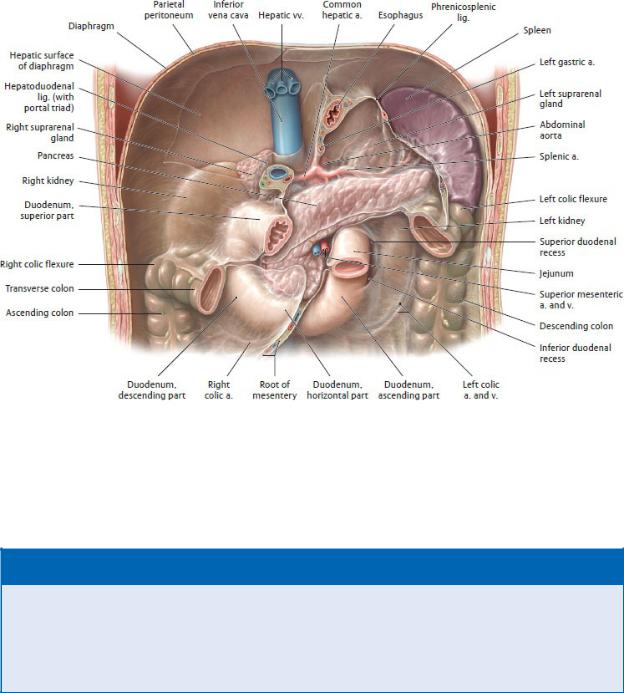
Fig. 12.7 Duodenum in situ
Anterior view. Removed: Stomach, liver, small intestine, and large portion of the transverse colon. (From Schuenke M, Schulte E, Schumacher U. THIEME Atlas of Anatomy, Vol 2. Illustrations by Voll M and Wesker K. 3rd ed. New York: Thieme Publishers; 2020.)
BOX 12.4: CLINICAL CORRELATION
DUODENAL (PEPTIC) ULCERS
Duodenal ulcers usually occur within a few centimeters of the pylorus on its posterior wall. Perforation of the duodenum can lead to peritonitis and ulceration of adjacent organs. Severe hem-orrhaging results if the ulcer erodes through the gastroduodenal artery, which runs along the posterior side of the duodenum.
—The jejunum, the proximal two fifths of the intraperitoneal portion of the small intestine, is suspended by the mesentery and is located predominantly in the left upper quadrant (Figs. 12.8, 12.9, 12.10, 12.11).
•It is thicker walled and larger in diameter than the ileum.
•Tall, closely packed circular folds (plicae circulares) line its inner surface, increasing its surface area for absorption.
•Widely spaced arterial arcades within its mesentery give rise to long, straight arteries, the vasa recta (see Fig. 11.16).
—The ileum, which makes up the distal three fifths of the intraperitoneal portion of the small intestine, is also sus-pended by the mesentery of the small intestine. The ileum extends from the end of the jejunum to its junction with the cecum (ileocecal junction) and resides in the lower left quadrant and the greater pelvis (Figs. 12.8, 12.9, 12.10, 12.11).
•It is longer in length than the jejunum.
•Lymphoid nodules (Peyer’s patches) bulge outward from the connective tissue layer underlying the epithelium (lamina propria).
•Circular folds (plicae circulares) are low and sparse.
•The ileum has more fat, denser arterial arcades, and shorter vasa recta in its mesentery than the jejunum.
—The blood supply, lymphatic drainage, and innervation of the parts of the small intestine reflect their development from the embryonic foregut and midgut (see Section 11.2).
•The section that extends from the pyloric sphincter to below the major duodenal papilla (foregut) is supplied by the superior pancreaticoduodenal branch of the gastroduodenal artery (supplied through the celiac trunk).
•The midgut (distal part of the descending duodenum, the jejunum, and the ileum) is supplied by the inferior pancreaticoduodenal, jejunal, and ileal arteries, branches of the superior mesenteric artery.
•Veins of similar names accompany the arteries and terminate in the hepatic portal system.
•Lymph vessels from the small intestine follow the course of the arteries and drain into the celiac and superior mesenteric nodes.
•The celiac (to foregut) and superior mesenteric (to midgut) plexuses innervate the small intestine.
◦Sympathetic innervation inhibits intestinal mobility, secretion, and vasodilation.
◦Parasympathetic innervation restores normal digestive activity following sympathetic stimulation.
◦Visceral sensory fibers transmit feelings of distension (often perceived as cramping), but the intestine is insensitive to most pain stimuli.
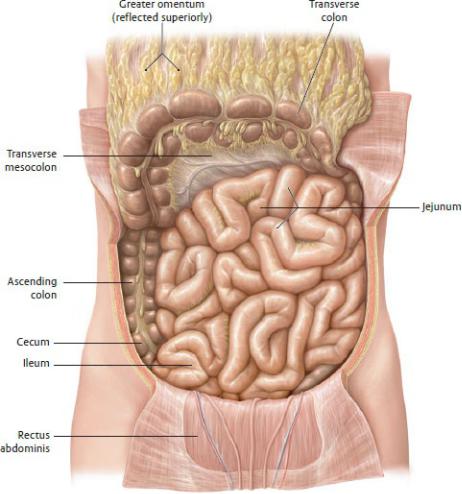
Fig. 12.8 Jejunum and ileum in situ
Anterior view. Reflected: Transverse colon and greater omentum. (From Schuenke M, Schulte E, Schumacher U. THIEME Atlas of Anatomy, Vol 2. Illustrations by Voll M and Wesker K. 3rd ed. New York: Thieme Publishers; 2020.)
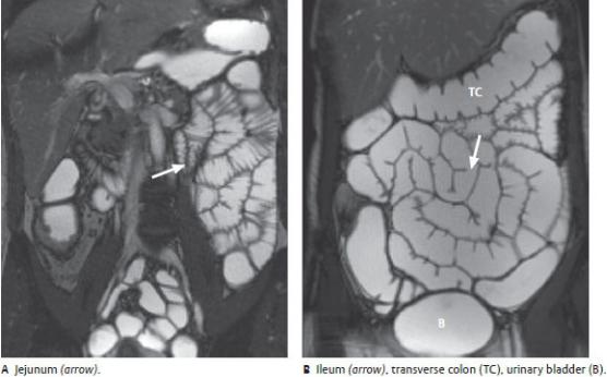
Fig. 12.9 MRI of the small intestine
Coronal view. Sectional imaging modalities like CT and MR have mostly replaced conventional radiographs in the evaluation of gastrointestinal disease. (From Krombach GA, Mahnken AH. Body Imaging: Thorax and Abdomen. New York: Thieme Publishers; 2015.)
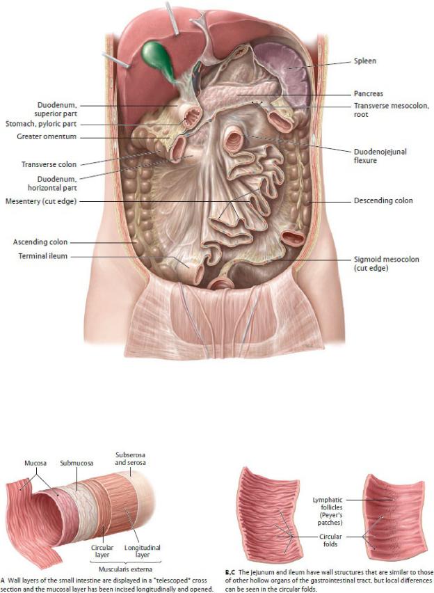
Fig. 12.10 Mesentery of the small intestine Anterior view. Removed: Stomach, jejunum, and ileum. Reflected: Liver. (From Schuenke M, Schulte E, Schumacher U. THIEME Atlas of Anatomy, Vol 2. Illustrations by Voll M and Wesker K. 3rd ed. New York: Thieme Publishers; 2020.)
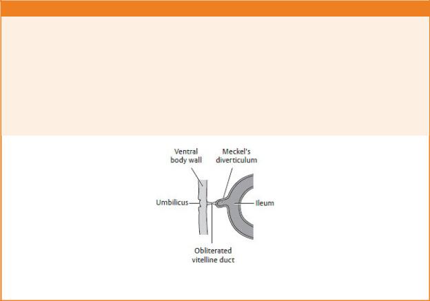
Fig. 12.11 Wall structure of the small intestine
(From Schuenke M, Schulte E, Schumacher U. THIEME Atlas of Anatomy, Vol 2. Illustrations by Voll M and Wesker K. 3rd ed. New York: Thieme Publishers; 2020.)
BOX 12.5: DEVELOPMENTAL CORRELATION
ILEAL DIVERTICULUM
Ileal diverticulum (also known as Meckel’s diverticulum), the most common congenital abnormality of the bowel, is an outpouching of the ileum that is a remnant of the omphalomesenteric duct (yolk stalk) that fails to resorb. The diverticulum may be unattached dis-tally or connect to the umbilicus via a fibrous cord or fistula. They are present in ~ 2% of the population, are located ~ 2 feet proximal to the ileocecal junction, and often contain two or more types of mucosa. The diverticulum can contain gastric, pancreatic, jejunal, or colonic tissue. An ileal diverticulum is usually asymptomatic but when inflamed may mimic acute appendicitis.
(From Schuenke M, Schulte E, Schumacher U. THIEME Atlas of Anatomy, Vol 2. Illustrations by Voll M and Wesker K. 3rd ed. New York: Thieme Publishers; 2020.)
Large Intestine
The large intestine extends from the cecum to the anal canal (Figs. 12.12, 12.13, 12.14). It converts liquid feces to a semisolid state through the absorption of water, electrolytes, and salts. It also stores and lubricates fecal matter. Although it consists of five parts, only the cecum, appendix, and colon reside in the abdo-men. The rectum and anal canal are described in Chapter 15, Pelvic Viscera.
—The cecum is a blind pouch located in the right lower quadrant.
•It is attached proximally in an end-to-side fashion to the terminal ileum and is continuous distally with the ascending colon.
•It lacks a mesentery but is surrounded by peritoneum and therefore is fairly mobile.
—The (vermiform) appendix is a blind muscular diverticulum (outpouching)
that opens into the posteromedial wall of the cecum below the ileocecal orifice.
•Its walls contain large masses of lymphoid tissue.
•Its mesoappendix (mesentery) suspends it from the ileum.
•Its position is highly variable, but it is often retrocecal (posterior to the cecum).
—The colon has four parts that frame the abdominal viscera:
1. The ascending colon ascends from the cecum in the right lower quadrant to the right colic (hepatic) flexure under the liver.
2. The transverse colon crosses the abdomen from the right colic flexure to the left upper quadrant, where it termi-nates at the left colic (splenic) flexure.
3. The descending colon descends along the left side of the abdomen to the left lower quadrant.
4. The sigmoid colon crosses the iliac fossa to join the rectum in the pelvis.
—The appendix, transverse colon, and sigmoid colon are intraperitoneal. Each is suspended by its respective meso colon (mesentery). The left colic flexure is attached to the diaphragm by the phrenicocolic ligament.
—The ascending and descending colons are secondarily retroperitoneal and therefore lack mesenteries.
—External features of the colon distinguish it from the small intestine:
•Taeniae coli, three longitudinal bands (mesocolic taenia, omental taenia, and free taenia), formed by the outer muscular layer,
•Haustra, outpouchings of the intestinal wall visible between the taeniae coli,
•Epiploic appendices, small sacs of fat aligned along the taeniae.
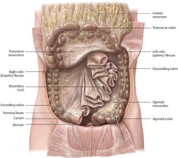
Fig 12.12 Large intestine in situ
Anterior view. Reflected: Transverse colon and greater omentum. Removed: Intraperitoneal small intestine. (From Schuenke M, Schulte E, Schumacher U. THIEME Atlas of Anatomy, Vol 2. Illustrations by Voll M and Wesker K. 3rd ed. New York: Thieme Publishers; 2020.)
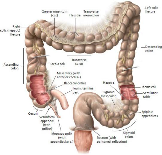
Fig. 12.13 Large intestine
Anterior view. (From Gilroy AM, MacPherson BR, Wikenheiser JC. Atlas of Anatomy. Illustrations by Voll M and Wesker K. 4th ed. New York: Thieme Publishers; 2020.)
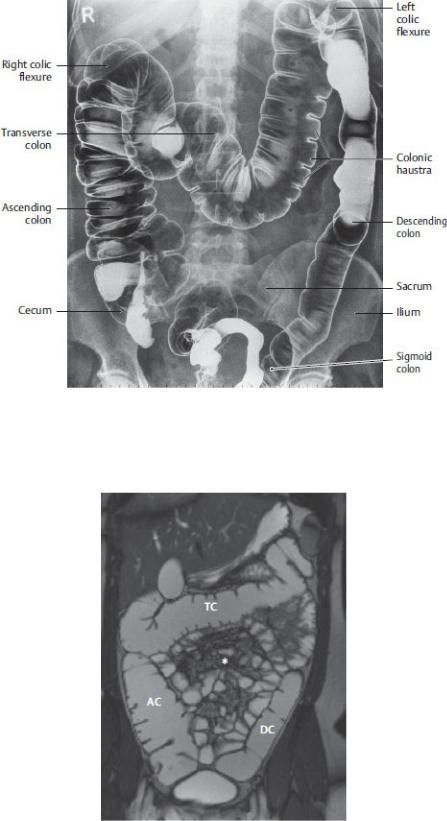
Fig. 12.14 Radiograph of double contrast barium enema of the large intestine
Anterior view. (From Moeller TB, Reif E. Pocket Atlas of Sectional Anatomy, Vol 2, 3rd ed. New York: Thieme; 2007.)
Fig. 12.15 MRI of the large intestine

Ascending colon (AC), descending colon (DC), transverse colon (TC), * small bowel and mesen-teric structures. (From Gilroy AM, MacPherson BR, Wikenheiser JC. Atlas of Anatomy. Illustrations by Voll M and Wesker K. 4th ed. New York: Thieme Publishers; 2020.)
—The blood supply, lymphatic drainage, and innervation of the parts of the large intestine reflect their development from the embryonic midgut and hindgut (see Section 11.2).
•The ileocolic, right colic, and middle colic branches of the superior mesenteric artery supply the cecum, ascending colon, and proximal two thirds of the transverse colon (midgut).
•The left colic and sigmoidal branches of the inferior mesenteric artery supply the distal third of the transverse colon, and the descending and sigmoid colons (hindgut). A superior rectal branch supplies the upper rectum in the pelvis.
•The marginal artery runs along the mesenteric border of the large intestine, anastomosing branches of the superior mesenteric artery with those of the inferior mesenteric artery. In turn, the superior rectal artery anastomoses with middle rectal and inferior rectal branches in the pelvis.
•Veins of the colon follow the arteries and drain into the hepatic portal system.
•Lymph vessels follow arterial pathways to drain into superior mesenteric or inferior mesenteric nodes.
•The superior mesenteric (midgut) and inferior mesenteric (hindgut) nerve plexuses innervate the large intestine.
BOX 12.6: CLINICAL CORRELATION
INFLAMMATORY BOWEL DISEASE
There are two types of inflammatory bowel disease (IBD): Crohn’s disease and ulcerative colitis. Crohn’s disease is a chronic inflam-matory disease that can affect the entire gastrointestinal (GI) tract but most commonly affects the terminal ileum and colon. It causes ulcers, fistulas (abnormal communications), and granulo-mata, producing symptoms such as fever, diarrhea, weight loss, and abdominal pain. Ulcerative colitis is a recurrent inflammatory disease of the colon and rectum that produces bloody diarrhea, weight loss, fever, and abdominal pain. These diseases are treated with drugs that reduce the inflammatory response.
BOX 12.7: CLINICAL CORRELATION
APPENDICITIS AND VARIABLE POSITION OF THE VERMIFORM APPENDIX
Anomalies in the rotation of the embryonic gut can result in several positional variations of the cecum and appendix. This can influence the accurate interpretation of symptoms of appendicitis. Inflammation of an appendix in the typical position is felt initially as vague pain in the periumbilical region, transmitted via visceral fibers from the T10 spinal cord segment. As the inflammation irritates the overlying parietal peritoneum, tenderness can be elicited by pressure at two points:
McBurney point: located one third of the distance along a line from the anterior superior iliac spine to the umbilicus.
Lanz point: located one third of the distance along a line from the right anterior superior iliac spine to the simi-lar landmark on the left side.
(From Schuenke M, Schulte E, Schumacher U. THIEME Atlas of Anatomy, Vol 2. Illustrations by Voll M and Wesker K. 3rd ed. New York: Thieme Publishers; 2020.)
BOX 12.8: CLINICAL CORRELATION
COLON CARCINOMA
Malignant tumors of the colon and rectum are among the most fre-quent solid tumors. More than 90% occur in patients over the age of 50. In early stages, the tumor may be asymptomatic; later symp-toms include loss of appetite, changes in bowel movements, and weight loss. Blood in the stools is particularly incriminating, necessi-tating a thorough examination. Hemorrhoids are not a sufficient explanation for blood in stools unless all other tests (including a colonoscopy) are negative.
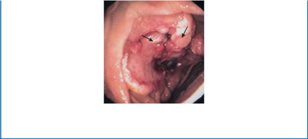
Colonoscopy of colon carcinoma. The tumor (black arrows) partially blocks the lumen of the colon. (From Gilroy AM, MacPherson BR, Wikenheiser JC. Atlas of Anatomy. Illustrations by Voll M and Wesker K. 4th Edition. New York: Thieme Publishers; 2020.)
12.2 Organs of the Peritoneal Cavity—Accessory Organs of the Gastrointestinal Tract
Liver
The liver is located in the right upper quadrant under the right hemidiaphragm and extends inferiorly to the costal margin (Fig. 12.16). It has a primary role in carbohydrate, protein, and fat metab-olism. It also produces and secretes bile and bile pigments; detox-ifies substances absorbed by the gastrointestinal tract; and stores vitamins and minerals, such as iron. In the fetus, the liver is the site of hematopoiesis (red blood cell production).
—Externally, ligaments and fissures divide the liver into four anatomic (topographic) lobes: the right, left, caudate, and quadrate lobes (Fig. 12.17).
—The liver’s diaphragmatic surface conforms to the shape of the diaphragm and is marked by the bare area, which lacks peritoneum and is in direct contact with the diaphragm.
—The liver’s visceral (inferior) surface has three prominent fissures:
1.The left sagittal fissure accommodates
◦the round ligament (ligamentum teres) of the liver anteriorly between the left and quadrate lobes. The round ligament is a remnant of the fetal umbilical vein.
◦the ligamentum venosum posteriorly between the left and caudate lobes. The ligamentum venosum is a remnant of the fetal ductus venosus.
2.The right sagittal fissure accommodates
◦the gallbladder anteriorly between the right and quadrate lobes and
◦the inferior vena cava posteriorly between the right and caudate lobes.
3.The transverse fissure accommodates
◦the porta hepatis, or hilum of the liver. Structures of the portal triad [proper hepatic artery, portal vein, and (common) bile duct] enter or exit here.
—The liver is intraperitoneal, covered by peritoneum except at the bare area, gallbladder fossa, and porta hepatis. Peritoneal reflections include
• the coronary and triangular ligaments, single-layered reflections between the liver and diaphragm that surround the bare area;
• the falciform ligament, a double layer of peritoneum, which attaches the liver to the anterior abdominal wall and contains the round ligament in its free edge; and
• the hepatogastric and hepatoduodenal ligaments (both parts of the lesser omentum), which attach the liver to the stomach and proximal duodenum.
—A subperitoneal fibrous capsule, Glisson’s capsule, covers the surface of the liver.
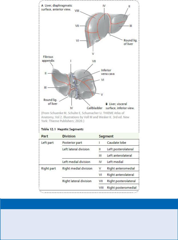
—Internally, branching of the intrahepatic blood vessels divides the liver into eight functional segments (designated as I through VIII) (Fig. 12.18, Table 12.1). This segmental arrangement of the blood supply facilitates the resection of individual diseased segments.
—The liver has a dual blood supply: the portal vein and the proper hepatic artery (see Section 11.2). Both vessels divide to form primary and secondary branches that supply the liver segments.
• The portal vein, carrying nutrient-rich blood from the digestive tract, provides 75 to 80% of the blood volume to the liver.
• The proper hepatic artery, supplied by the celiac trunk via the common hepatic artery, contributes 20 to 25% of the blood volume to the liver.
—Right, left, and intermediate (middle) hepatic veins run intersegmentally, draining adjacent segments, and open into the inferior vena cava immediately below the diaphragm.
—Right, left, and intermediate (middle) hepatic veins run intersegmentally, draining adjacent segments, and open into the inferior vena cava immediately below the diaphragm.
—The liver has superficial and deep lymphatic drainages.
• The superficial lymphatic plexus, found within the fibrous capsule,
drains the anterior liver surfaces to hepatic nodes (and eventually into the celiac nodes) and drains the posterior surfaces toward the bare area, which flow into phrenic or posterior mediastinal nodes.
•The deep plexus, which accompanies vessels within the liver segments, drains most of the liver, flowing first into the hepatic nodes in the porta hepatis and lesser omen-tum, before draining to the celiac nodes.
—The hepatic nerve plexus, a division of the celiac plexus, travels along the vessels of the portal triad to innervate the liver.
Fig. 12.16 Liver in situ
Anterior view. (From Schuenke M, Schulte E, Schumacher U. THIEME Atlas of Anatomy, Vol 2. Illustrations by Voll M and Wesker K. 3rd ed. New York: Thieme Publishers; 2020.)
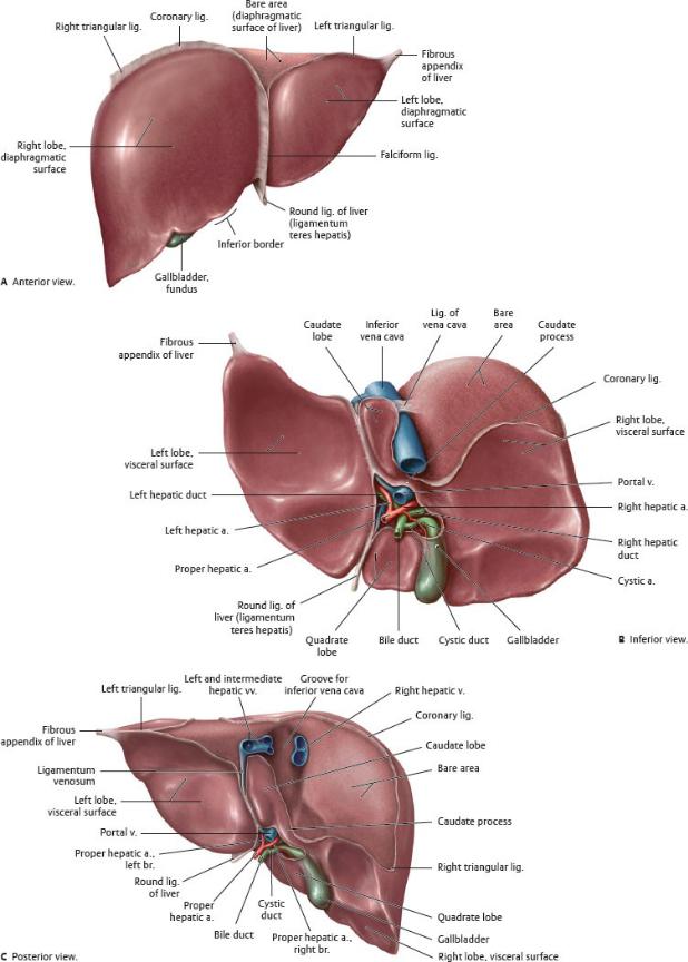

Fig. 12.17 Surfaces of the liver
The liver is divided by its ligaments into four lobes: right, left, caudate, and quadrate. (From Schuenke M, Schulte E, Schumacher U. THIEME Atlas of Anatomy, Vol 2. Illustrations by Voll M and Wesker K. 3rd ed. New York: Thieme Publishers; 2020.)
Fig. 12.18 Segmentation of the liver
Anterior view. Branches of the hepatic artery, portal vein, and hepatic ducts divide the liver into hepatic segments. (From Schuenke M, Schulte E, Schumacher U. THIEME Atlas of Anatomy, Vol 2. Illustrations by Voll M and Wesker K. 2nd ed. New York: Thieme Publishers; 2016.)
BOX 12.9: CLINICAL CORRELATION
CIRRHOSIS OF THE LIVER
Cirrhosis, most commonly caused by chronic alcoholism, is char-acterized by a progressive fibrosis of hepatic tissue around the intrahepatic vessels and biliary ducts that impedes blood flow. The liver becomes hard and nodular in appearance. Symptoms include ascites, splenomegaly, peripheral edema, esophageal var-ices, and other signs of portal hypertension.
Gallbladder and Extrahepatic Biliary System
The gallbladder is a pear-shaped sac that lies in a fossa on the vis-ceral surface of the liver (Figs. 12.17 and 12.19). It stores the bile produced and secreted by the liver and concentrates it by absorb-ing salts and water. Hormonal or neural stimulation causes the gallbladder to release bile into the extrahepatic biliary ducts (Fig. 12.20).
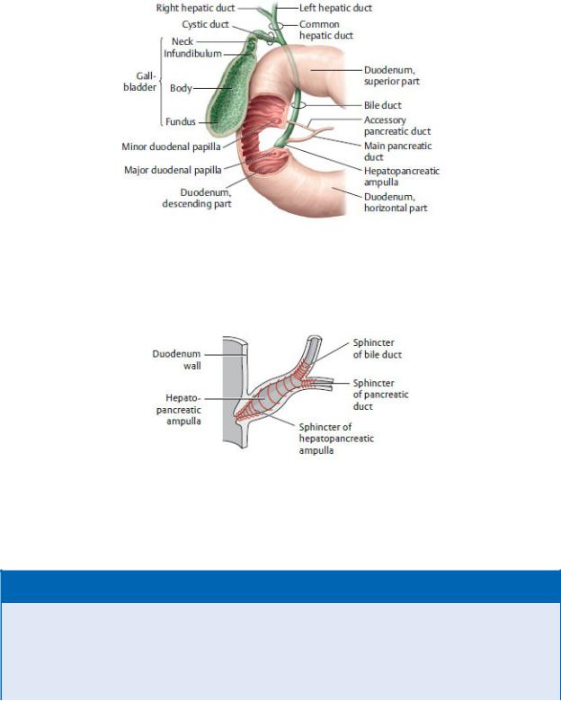
—The gallbladder has four parts (Fig. 12.21):
1.The fundus, the expanded distal end that is in contact with the anterior abdominal wall
2.The body, the main portion
3.The infundibulum between the body and neck
4.The neck, the narrow distal segment that joins with the cystic duct
—The extrahepatic biliary system of ducts transports bile from the liver and gallbladder to the duodenum. It consists of
• the common hepatic duct formed by the junction of the right and left hepatic ducts, which drain the liver;
• the cystic duct, which drains the gallbladder and com municates with the common hepatic duct from the liver (a spiral valve in the neck of the gallbladder keeps the cystic duct open); and
• the bile duct, formed by the junction of the common hepatic duct and cystic duct, which drains bile into the second part of the duodenum.
—The bile duct passes posterior to the first part of the duode-num and posterior to, or through, the head of the pancreas. It ends at the hepatopancreatic ampulla (of Vater), a dilation of the distal end of the duct, where it joins with the main pancreatic duct of the pancreas.
—A muscular sphincter (of Oddi) surrounds the hepatopancre-atic ampulla as it traverses the medial wall of the duodenum through the major duodenal papilla and opens into the descending part of the duodenum (Fig. 12.22).
—The cystic artery, usually a branch of the right hepatic artery, supplies the gallbladder.
—Venous blood from the gallbladder drains into hepatic veins in the liver, which drain into the inferior vena cava.
—The hepatic plexus innervates the gallbladder.
• Sympathetic stimulation inhibits bile secretion.
• Parasympathetic stimulation causes the gallbladder to contract and release bile.
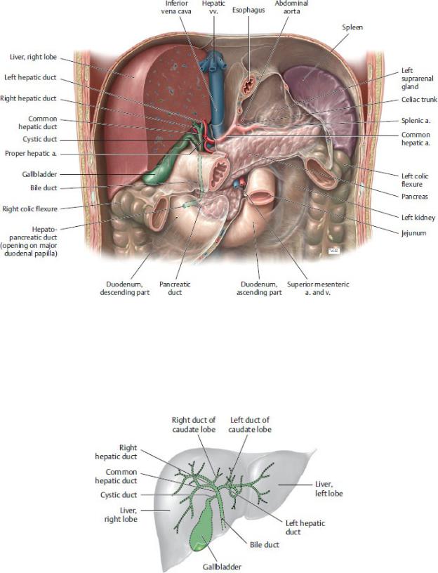
Fig. 12.19 Biliary tract in situ
Anterior view. Removed: Stomach, small intestine, transverse colon, and large portions of the liver. The gallbladder is intraperitoneal, covered by visceral peritoneum where it is not attached to the liver. (From Gilroy AM, MacPherson BR, Wikenheiser JC. Atlas of Anatomy. Illustrations by Voll M and Wesker K. 4th ed. New York: Thieme Publishers; 2020.)
Fig 12.20 Hepatic bile ducts
Projection onto the surface of the liver, anterior view. (From Schuenke M, Schulte E, Schumacher U. THIEME Atlas of Anatomy, Vol 2. Illustrations by Voll M and Wesker K. 3rd ed. New York: Thieme Publishers; 2020.)
Fig. 12.21 Extrahepatic bile ducts and gallbladder
Anterior view. Opened: Gallbladder and duodenum. (From Schuenke M, Schulte E, Schumacher U. THIEME Atlas of Anatomy, Vol 2. Illustrations by Voll M and Wesker K. 3rd ed. New York: Thieme Publishers; 2020.)
Fig. 12.22 Biliary sphincter system
Sphincters of the pancreatic and bile ducts. (From Schuenke M, Schulte E, Schumacher U. THIEME Atlas of Anatomy, Vol 2. Illustrations by Voll M and Wesker K. 3rd ed. New York: Thieme Publishers; 2020.)
BOX 12.10: CLINICAL CORRELATION
GALLSTONES
Gallstones are concretions of cholesterol crystals that lodge within the biliary tree. They occur most commonly in women over the age of 40. Gallstones can remain asymptomatic, but when they obstruct the hepatopancreatic ampulla, they impede the flow of bile and pancreatic secretions, which may cause bile to enter the pancreas, leading to pancreatitis. Gallstones
that obstruct the cystic duct leads to biliary colic, characterized by severe waves of pain, cholecystitis, and jaundice. Stones that accumulate in the fundus of a diseased gallbladder (also known as Hartmann’s pouch) may ulcerate through the wall of the fundus into the transverse colon. These can pass naturally via the rectum, or they may pass into the small intestine where they can obstruct the ile-ocecal valve, producing an intestinal obstruction (gallstone ileus). Pain arising from gallstones originates in the epigastric or right hypochondriac region but may also be referred to the posterior thoracic wall or right shoulder due to irritation of the diaphragm.
BOX 12.11: CLINICAL CORRELATION
CHOLECYSTECTOMY AND THE CYSTOHEPATIC TRIANGLE (OF CALOT)
Ninety-five percent of injuries to extrahepatic bile ducts are sus-tained intraoperatively, most commonly during removal of the gallbladder (cholecystectomy), which includes transection of the cystic artery and cystic duct. The cystohepatic triangle serves as a guide for accurate identification of these structures, whose anat-omy can be highly variable. The borders of this space are the cys-tic duct inferiorly, the common hepatic duct medially, and the visceral surface of the liver superiorly.
BOX 12.12: CLINICAL CORRELATION
IMPLICATIONS OF THE SHARED EMPTYING OF THE BILE DUCT AND PANCREATIC DUCT
The common bile duct and the pancreatic duct both empty into the major duodenal papilla. This has important clinical implica-tions (e.g., a tumor at the head of the pancreas may obstruct the common bile duct, causing biliary reflux into the liver and jaun-dice). Similarly, a gallstone that lodges in the common bile duct may obstruct the terminal part of the pancreatic duct, causing acute pancreatitis.
MR cholangiopancreatography
(From Moeller TB, Reif E. Pocket Atlas of Sectional Anatomy, Vol 2, 3rd ed. New York: Thieme Publishers; 2007.)
Pancreas
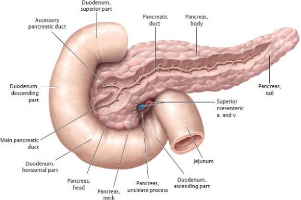
The pancreas is a lobulated gland that has dual functions. As an exocrine gland it synthesizes digestive enzymes, and as an endo-crine gland it produces and secretes the hormones insulin and glucagon (Figs. 12.19, 12.23, 12.24, 12.25).
—It is secondarily retroperitoneal and lies on the posterior wall of the omental bursa.
—It crosses the midline with its head within the “C” of the duo-denum and its tail touching the splenic hilum.
—The four parts of the pancreas are the head and uncinate pro-cess, the neck, the body, and the tail.
Fig. 12.23 Pancreas
Anterior view with dissection of the pancreatic duct. (From Schuenke M, Schulte E, Schumacher U. THIEME Atlas of Anatomy, Vol 2. Illustrations by Voll M and Wesker K. 3rd ed. New York: Thieme Publishers; 2020.)
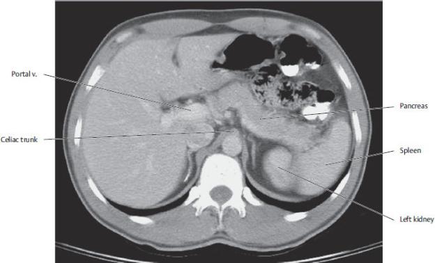
Fig 12.24 CT of the abdomen at the L1 vertebral level
(From Moeller TB, Reif E. Pocket Atlas of Sectional Anatomy, Vol 2, 3rd ed. New York: Thieme Publishers; 2007.)
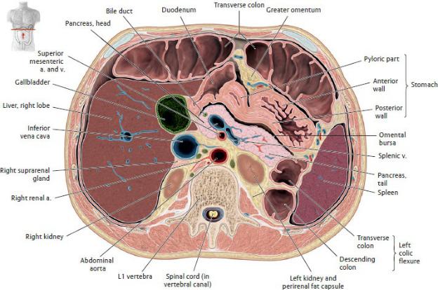
Fig. 12.25 Transverse section of the abdomen through the L1 vertebra
Inferior view. (From Schuenke M, Schulte E, Schumacher U. THIEME Atlas of Anatomy, Vol 2. Illustrations by Voll M and Wesker K. 3rd ed. New York: Thieme Publishers; 2020.)
—The main pancreatic duct (of Wirsung) traverses the length of the gland to join with the common bile duct at the hepa-topancreatic ampulla. Together they drain into the descending part of the duodenum at the major duodenal papilla (see Figs. 12.21 and 12.22).
—When present, an accessory pancreatic duct (of Santorini) may drain to the descending part of the duodenum at the minor duodenal papilla, 2 cm proximal to the drainage site of the main pancreatic duct.
—Because of its central location in the upper abdomen, the pancreas is related topographically to many of the major abdominal vascular structures (see
Fig. 11.23):
•The head lies anterior to the right renal vessels, left renal vein, and inferior vena cava.
•The neck and body cross anterior to the abdominal aorta, superior mesenteric vessels, and portal vein. The celiac trunk arises from the

aorta immediately superior to this.
•The tail crosses anterior to the left kidney and extends to the hilum of the spleen. The splenic artery runs along its superior border, and the splenic vein courses behind it.
—Branches of the celiac trunk and superior mesenteric artery supply the pancreas (see Section 11.2).
•Pancreaticoduodenal branches of the gastroduodenal artery (from the celiac trunk) and superior mesenteric artery supply the head.
•Branches of the splenic artery supply the neck, body, and tail.
—Venous blood drains into the splenic and superior mesenteric veins, which merge to form the hepatic portal system.
—Lymphatic drainage is as varied as its blood supply but generally follows arterial pathways, ultimately draining into celiac and superior mesenteric nodes.
—The celiac and superior mesenteric plexuses innervate the pancreas.
BOX 12.13: CLINICAL CORRELATION
CARCINOMA OF THE PANCREAS
Relations of the pancreas are of clinical significance in patients with pancreatic cancer. It metastasizes extensively to deep nodes and adjacent organs. Tumors of the head are most common and can obstruct the drainage of the bile and pancreatic ducts, result-ing in obstructive jaundice. Tumors of the neck and body can obstruct the portal vein or inferior vena cava.
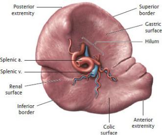
Spleen
The spleen is an intraperitoneal organ located in the hypochon-driac region of the left upper quadrant (Figs. 12.25, 12.26, 12.27). It functions as a lymphoid gland and as a filter for old or abnormal red blood cells.
—Nestled under the dome of the diaphragm, the spleen does not normally project below the costal margin and therefore is not palpable on examination.
—The convex surface admits the splenic vessels and nerves at the splenic hilum.
—The spleen is connected to adjacent organs by peritoneal ligaments.
•The splenorenal ligament, which contains the branches of the splenic vessels and the tail of the pancreas, connects the spleen to the kidney.
•The gastrosplenic ligament, which contains the short gastric and left gastro-omental vessels, connects the spleen to the stomach.
•The splenocolic ligament connects the spleen to the left colic flexure.
• The phrenicosplenic ligament connects the spleen to the diaphragm.
—Although it is sheltered by the 9th, 10th, and 11th ribs, the spleen is particularly vulnerable to lacerations from fractured ribs, and because of its dense vascularity, it bleeds profusely.
—Accessory spleens are common (20%) and are most often found within the gastrosplenic ligament, near the hilum or tail of the pancreas.
—The splenic artery, a large tortuous branch of the celiac trunk, divides within the splenorenal ligament at the splenic hilum (see Section 11.2).
—The spleen is vulnerable to infarction (interruption of the blood supply causing tissue death) because the splenic artery has no collateral arteries and is the gland’s sole blood supply.
—The splenic vein courses behind the pancreas, where it joins with the superior mesenteric vein to form the portal vein.
—The splenic plexus, a derivative of the celiac plexus, inner-vates the spleen.
Fig. 12.26 Spleen
Visceral surface. (From Schuenke M, Schulte E, Schumacher U. THIEME Atlas of Anatomy, Vol 2. Illustrations by Voll M and Wesker K. 3rd ed. New York: Thieme Publishers; 2020.)

Fig. 12.27 The spleen in situ: peritoneal relationships
Anterior view into the left upper quadrant (LUQ) with the stomach removed. (From Schuenke M, Schulte E, Schumacher U. THIEME Atlas of Anatomy, Vol 2. Illustrations by Voll M and Wesker K. 3rd ed. New York: Thieme Publishers; 2020.)
BOX 12.14: CLINICAL CORRELATION
SPLENIC TRAUMA AND SPLENECTOMY
Although the spleen appears to be well protected by the lower ribs on the posterior wall, it is the most frequently injured abdom-inal organ. It is particularly vulnerable to rupture from leftsided trauma that fractures the lower ribs. An enlarged spleen (spleno-megaly) that projects below the costal margin may be vulnerable during blunt abdominal trauma that can rupture the thin splenic capsule. Splenic rupture results in severe hemorrhage and may require a total or partial splenectomy. During a total splenectomy the tail of the pancreas is vulnerable to injury where it passes through the splenorenal ligament with the splenic vessels.
BOX 12.15: DEVELOPMENTAL CORRELATION
ACCESSORY SPLEENS
Accessory spleens are small nodules of splenic tissue, usually ~ 1 cm in diameter, that form separately from the main spleen. They are usually located at the splenic hilum near the tail of
the pancreas, in the gastrosplenic or splenorenal ligament, in the mesentery, or near the ovary or testis.
12.3 Organs of the Retroperitoneum
Kidneys
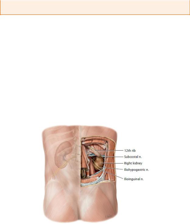
The kidneys are generally smooth-sided, reddish brown organs ~ 11 cm in length. They are retroperitoneal and lie anterior to the quadratus lumborum muscles on each side of the vertebral column between T12 and L3 vertebral levels (Figs. 12.28 and 12.29). The kidneys regulate blood pressure and the ionic balance and water content of the blood; they also eliminate metabolic waste and produce urine.
—The right kidney
•lies anterior to the 12th rib, slightly lower than the left, because of the presence of the large right lobe of the liver.
•lies posterior to the right suprarenal gland, liver, descend-ing part of the duodenum, and right colic flexure of the large bowel.
Fig. 12.28 Kidneys insitu
Posterior view with the trunk wall opened. (From Schuenke M, Schulte E, Schumacher U. THIEME Atlas of Anatomy, Vol 2. Illustrations by Voll M and Wesker K. 3rd ed. New York: Thieme Publishers; 2020.)
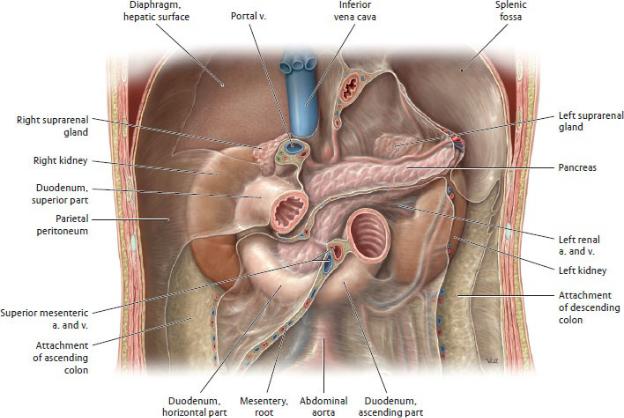
Fig. 12.29 Kidneys and suprarenal glands in the retroperitoneum
Anterior view. Both the kidneys and suprarenal glands are retroperitoneal. Removed: Intraperitoneal organs, along with portions of the ascending and descending colon. (From Schuenke M, Schulte E, Schumacher U. THIrenal artery, and renalEME Atlas of Anatomy, Vol 2. Illustrations by Voll M and Wesker K. 3rd ed. New York: Thieme Publishers; 2020.)
—The left kidney
•lies anterior to ribs 11 and 12.
•lies posterior to the left suprarenal gland, spleen, tail of the pancreas, and left colic flexure.
—A renal (Gerota’s) fascia surrounds each kidney and its asso-ciated suprarenal gland, renal vessels, ureter, and capsule of perirenal fat (Fig. 12.30). Pararenal fat lies outside this space and is thickest posteriorly.
—Deep to the renal fascia, a thin, fibrous renal capsule com-pletely invests each kidney (Fig. 12.31A).
—The lateral edge of the kidney is smooth and concave; the medial border has a vertical hilum that is pierced by the renal vein, renal artery, and renal pelvis. The hilum expands within the kidney as the renal sinus.
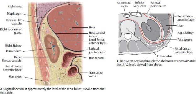
Fig. 12.30 Right kidney in the renal bed
(From Gilroy AM, MacPherson BR, Wikenheiser JC. Atlas of Anatomy. Illustrations by Voll M and Wesker K. 4th ed. New York: Thieme Publishers; 2020.)
—Internally, the kidney consists of cortical and medullary regions that contain up to 2 million nephrons, the renal functional units (Fig. 12.30).
•The cortex, the outer region, lies deep to the fibrous capsule and extends into the medullary region as renal columns.
•The medulla, the inner region, is arranged in renal pyramids with the broad base facing outward and the apex fitting into a cup-shaped minor calyx.
•Up to 11 minor calyces merge to form two or three major calyces, which combine to form the renal pelvis. At the hilum the renal pelvis narrows to form the ureter.
—A single renal artery, a direct branch of the abdominal aorta at L2, normally supplies each kidney (Figs. 12.31 and 12.32).
•The right renal artery is longer than the left artery and passes posterior to the inferior vena cava.
•The artery divides near the hilum to supply anterior and posterior segments of the kidney, which are separated by an avascular longitudinal plane (Brodel’s white line).
—A single renal vein from each kidney drains to the inferior vena cava (Figs. 12.31 and 12.32).
•Both renal veins receive a tributary from the ureter, but only the left renal vein receives the left suprarenal and left testicular or ovarian veins. On the right these veins drain directly into the inferior vena cava.
•The left renal vein is longer than the right vein and passes anterior to the aorta immediately inferior to the origin of the superior mesenteric artery.
•The longer length of the left renal vein makes the left kidney the preferred choice for organ donation.
—The renal plexus, an extension of the celiac plexus, forms a dense periarterial plexus along the renal arteries (see Section 11.2).
•Pain from renal disease is referred along the T12–L2 dermatomes to the lumbar and inguinal regions and the upper part of the anterior thigh.
BOX 12.16: CLINICAL CORRELATION
RENAL VEIN ENTRAPMENT
The left renal vein crosses the midline in the narrow angle between the downwardly directed superior mesenteric artery and the abdominal aorta. Pathologic conditions (atherosclerosis, aneurysms) of the arteries or downward pressure on the superior mesenteric artery can compress the renal vein. This is sometimes called the nutcracker syndrome.
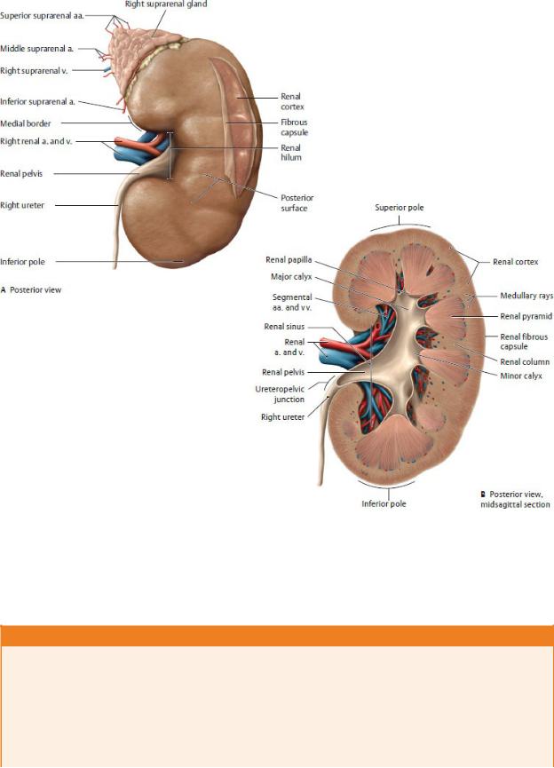
Fig. 12.31 Kidney: structure
Right kidney with suprarenal gland. (From Schuenke M, Schulte E, Schumacher U. THIEME Atlas of Anatomy, Vol 2. Illustrations by Voll M and
Wesker K. 3rd ed. New York: Thieme Publishers; 2020.)
BOX 12.17: DEVELOPMENTAL CORRELATION
COMMON RENAL ANOMALIES
Variations in renal vessels are common and usually asymptomatic. Kidneys develop in the pelvis and ascend to their location in the lum-bar region between the 6th and 9th fetal weeks. As they ascend, more superior renal arteries and veins replace the inferior renal ves-sels. In ~ 30% of the population, failure of these inferior vessels to degenerate results in multiple renal arteries and veins.
In some cases, one kidney may fail to ascend (a), resulting in a pelvic kidney. In other
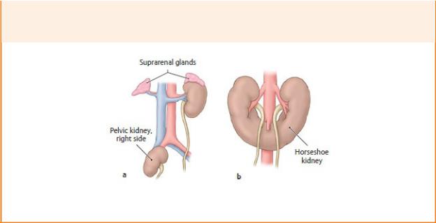
cases, the inferior poles of the kidneys may fuse to form a single U-shaped structure (b), although they remain functionally separate. These “horseshoe kidneys” get trapped in their ascent under the inferior mesenteric artery and remain at the L3 or L4 level.
(From Schuenke M, Schulte E, Schumacher U. THIEME Atlas of Anatomy, Vol 2. Illustrations by Voll M and Wesker K. 3rd ed. New York: Thieme Publishers; 2020.)
Ureters
The ureters are muscular tubes, 25 to 30 cm in length, that con-vey urine from the kidneys to the bladder through peristaltic (wavelike) action (Figs. 12.32 and 12.33). Both the abdominal and the pelvic parts of the ureters are retroperitoneal along their entire course. The pelvic ureter is discussed further in Chapter 11, Pelvic Viscera.
—Near the hilum of the kidney, the renal pelvis narrows to join with the origin of the ureter at the ureteropelvic junction (Fig. 12.31).

Fig. 12.32 Ureters in situ
Retroperitoneum of male abdomen, anterior view. Removed: Nonurinary organs and rectal stump. (From Schuenke M, Schulte E, Schumacher U. THIEME Atlas of Anatomy, Vol 2. Illustrations by Voll M and Wesker K. 3rd ed. New York: Thieme Publishers; 2020.)
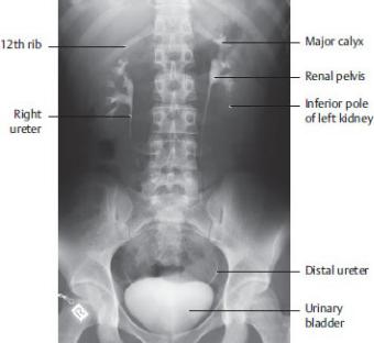
Fig. 12.33 Radiograph of intravenous pyelogram
Anterior view. (From Moeller TB, Reif E. Pocket Atlas of Sectional Anatomy, Vol 2, 3rd ed. New York: Thieme Publishers; 2007.)
—The abdominal part of the ureter descends along the anterior surface of the psoas muscle where it is crossed by the gonadal vessels.
—The ureter crosses over the pelvic brim to enter the pelvis at the bifurcation of the common iliac artery into internal and external iliac branches.
—The pelvic part of the ureter travels anteriorly along the lateral pelvic wall before it enters the urinary bladder at the ureterovesical junction.
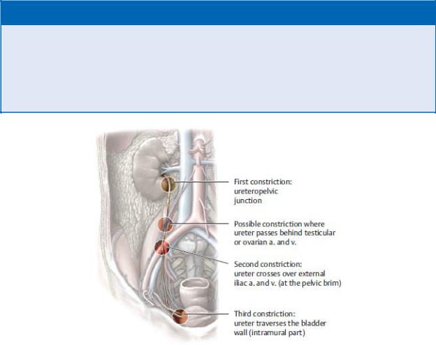
—Constrictions of the ureter, caused by ureteral narrowing or compression by adjacent structures, can occur near its origin and along its length (Fig. 12.34).
—Branches from several arteries form a delicate anastomosis along the length of the ureter (see Section 11.2).
•In the abdomen, this network of vessels usually includes contributions from the abdominal aorta and the renal, gonadal (testicular or ovarian), and common iliac arteries.
•Branches of the superior vesical, inferior vesical, and uterine arteries supply the pelvic ureter.
—The veins of the ureter accompany the arteries.
—Contributions from the renal, aortic, and superior hypogastric plexuses innervate the abdominal ureter. The inferior hypo-gastric plexus innervates the pelvic ureter (see Section 10.6).
—Pain from the ureter is relayed along sympathetic routes to spinal cord segments T11–L2 and is referred to the corre-sponding dermatomes of the lower abdominal wall, inguinal region, and medial aspects of the thigh.
BOX 12.18: CLINICAL CORRELATION
CALCULI OF THE KIDNEY AND URETER
Calculi (stones) formed in the urine can become lodged in the renal calyces, renal pelvis, or ureter. Calculi lodged in the ureters distend the ureteric walls and cause intense intermittent pain as peristaltic contractions move the calculi inferiorly. As the calculi move toward the pelvis, pain moves from the lumbar to inguinal regions (T11–L2 dermatomes) and may extend into the scrotum and anterior thigh with branches of the genitofemoral nerve.
Fig.12.34 Anatomic constrictions of the ureter
Right side, anterior view. (From Schuenke M, Schulte E, Schumacher U. THIEME Atlas of Anatomy, Vol 2. Illustrations by Voll M and Wesker K. 3rd ed. New York: Thieme Publishers; 2020.)
Suprarenal Glands
The paired suprarenal (adrenal) glands are located in the retro-peritoneum, where they cap the superior pole of each kidney and lie anterior to the crura of the diaphragm. The suprarenal glands are neuroendocrine glands that respond to stress.
—Perirenal fat and renal fascia surround both glands; a septum of the fascia separates them from the kidney.
—The right suprarenal gland is pyramidal, and the left is crescent shaped
(Figs. 12.35 and 12.36).
—The glands are composed of an outer cortex and an inner medulla. Both parts act as endocrine glands (i.e., secrete hormones) but differ from each other developmentally and functionally.
—The cortex
•is derived from mesoderm;
•is stimulated by hormones such as adrenocorticotropic hormone (ACTH); and
•secretes hormones (corticosteroids and androgens), which influence blood pressure and blood volume by regulating sodium and water retention in the kidneys.
—The medulla
•is primarily composed of nervous tissue that is derived from neural crest cells (embryonic cells that migrate from the developing neural tube and give rise to a variety of structures associated with the peripheral nervous system);
•is stimulated by preganglionic sympathetic fibers from the celiac plexus; and
•contains chromaffin cells that function like sympathetic ganglia and secrete hormones (catecholamines) that increase heart rate, blood pressure, blood flow, and respiration.
—Veins and lymphatic vessels exit at the hilum on the anterior surface, but arteries and nerves access the glands at multiple points.
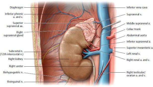
Fig. 12.35 Right kidney and suprarenal gland
Anterior view. Removed: Perirenal fat capsule. Retracted: Inferior vena cava. (From Schuenke M, Schulte E, Schumacher U. THIEME Atlas of Anatomy, Vol 2. Illustrations by Voll M and Wesker K. 3rd ed. New York: Thieme Publishers; 2020.)
—Each suprarenal gland has multiple superior, middle, and inferior suprarenal arteries, which branch from the inferior phrenic, abdominal aorta, and renal arteries, respectively.
—A single suprarenal vein drains each gland. The right vein drains to the inferior vena cava; the left vein may join with the inferior phrenic vein before draining to the left renal vein.
—Preganglionic sympathetic fibers of the greater splanchnic nerve combine with fibers of the celiac plexus to form the suprarenal plexus, but they do not synapse on the celiac ganglia. These preganglionic fibers, which may be considered homologous with postganglionic sympathetic neurons, terminate directly on the chromaffin cells of the medulla.
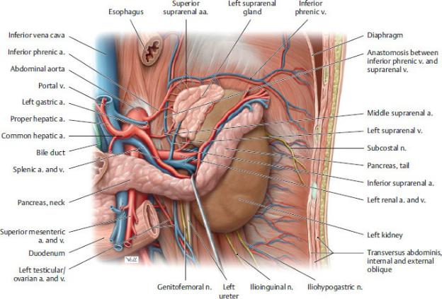
Fig. 12.36 Left kidney and suprarenal gland
Anterior view. Removed: Perirenal fat capsule. Retracted: Pancreas. (From Schuenke M, Schulte E, Schumacher U. THIEME Atlas of Anatomy, Vol 2. Illustrations by Voll M and Wesker K. 3rd ed. New York: Thieme Publishers; 2020.)