


20 Clinical Imaging Basics of the Upper Limb
Radiographs are always the first imaging choice in evaluation of bones and joints in the setting of trauma or pain ( Table 20.1). Radiographs, or x-rays, are highly sensitive for the detection of fractures and misalignment of joints.
Computed tomography (CT) provides significantly more detail of bone and is sometimes helpful for seeing subtle nondisplaced fractures, especially in older adults with decreased bone mass. The superior soft tissue contrast of magnetic resonance imaging (MRI) makes it the ideal imaging method for evaluation of the soft tissue components of joints.
Ultrasound has the benefit of real time imaging and is good for seeing superficial tissues, but its utility decreases substantially in larger patients. Ultrasound can also be very useful for imageguided procedures such as joint aspirations and joint injections. Additionally, in small children ultrasound can be useful for the anatomic evaluation of cartilaginous growth plates, especially in the growing elbow (see Table 20.1).
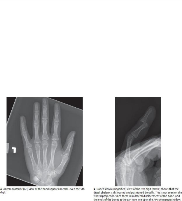
Since a radiograph is a summation shadow, all bones need to be evaluated in at least two projections (orthogonal) and all joints should be evaluated in at least three projections. The orthogonal view allows evaluation of the position of structures that may be overlapping in a single projection (Fig. 20.1). When viewing a bone x-ray, the cortical edge should be smooth along its entire length, and the trabecular pattern of the bone should be uniform throughout. Joints should be evaluated for even spacing, smoothness of the reticular surface, and alignment of the joint itself (Fig. 20.2).
Table 20.1 Suitability of Imaging Modalities for the Upper Limb
Modality |
Clinical Uses |
Radiographs (X-rays) |
Primarily used to evaluate the bones |
|
and alignment of joints |
|
|
CT (computed tomography) |
Usually reserved as a troubleshooting |
|
tool to evaluate for subtle |
|
nondisplaced fractures |
MRI (magnetic resonance |
One of the most important imaging |
imaging) |
modalities for the evaluation of joints, |
|
specifically the non-osseous |
|
components of the joint— cartilage, |
|
ligaments, tendons, and muscles |
|
|
Ultrasound |
Limited role in evaluation of superficial |
|
soft tissue abnormalities, and for |
|
guidance of interventional procedures |
|
involving the joints. In children, |
|
ultrasound plays a larger role in |
|
diagnosis of joint disorders and for |
|
evaluation of cartilaginous growth |
|
plates. |
|
|
Fig. 20.1 Radiograph of the hand demonstrating the importance of orthogonal views.
(Courtesy of Joseph Makris, MD, Baystate Medical Center.)
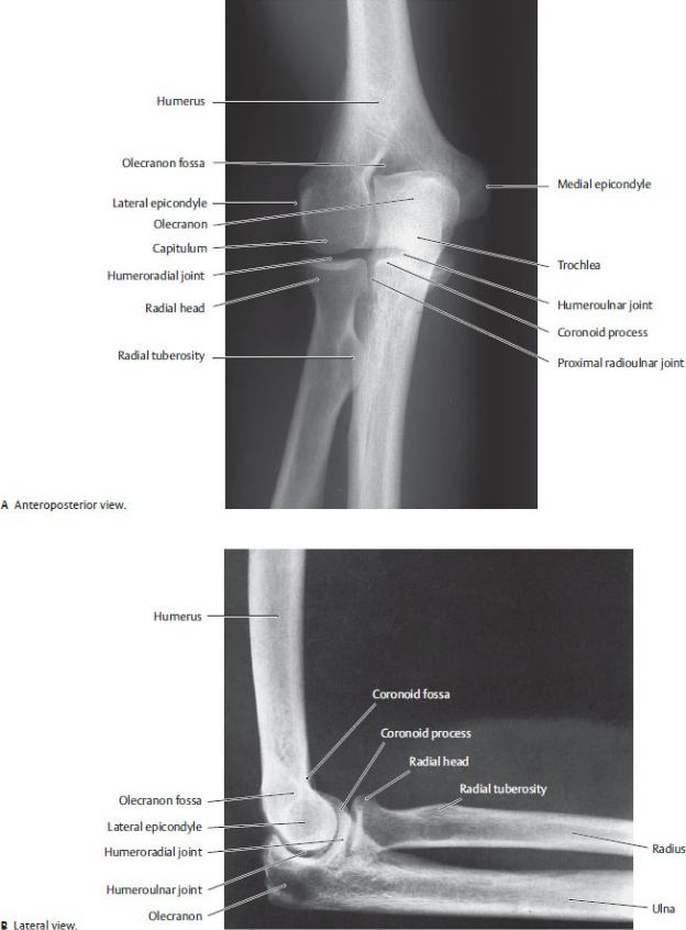
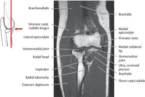
Fig. 20.2 Radiograph of the elbow
(From Moeller TB, Reif E. Pocket Atlas of Sectional Anatomy, Vol 3, 2nd ed. New York: Thieme Publishers; 2017.)
While x-rays are best for evaluating bones after trauma for fractures, MRI is the best choice for imaging soft tissues around the joints and for evaluation of the bone marrow (Figs. 20.3 and 20.4). Infiltration of the bone marrow from cancer or infection (osteomyelitis) can be invisible on x-rays early in the disease and still very subtle even as the disease progresses. MRI on the other hand is very sensitive for even the earliest changes in bone marrow and is essential for initial imaging and surveillance of these processes (Fig. 20.5). MRI is also helpful in assessing the extent of disease and adjacent soft tissue involvement, evaluation for metastases, surgical planning, disease staging, and monitoring effectiveness of therapy.
MRI arthrography involves injecting contrast material into a joint prior to MR imaging to better evaluate the key soft tissue components of the joint, such as the labrum, ligaments, and articular cartilage cartilage (Fig. 20.6). The joint injection procedure itself is usually performed with imaging guidance using ultrasound or fluoroscopy.
Fig. 20.3 MRI of the elbow
Coronal section.
In this sequence, fat is white (the fat in the bone marrow causes the bones to be a

very light gray/white as well). Muscles are dark gray and ligaments and cortical bone are black.
The utility of MRI is the ability to see the ligaments, tendons, muscles, and cartilage. A muscle tear, tendon rupture, or ligamentous tear is invisible on an X- ray, but well demonstrated on MRI. (From Moeller TB, Reif E. Pocket Atlas of Sectional Anatomy, Vol 3, 2nd ed. New York: Thieme Publishers; 2017.)
Fig. 20.4 MRI of the right wrist
Transverse (axial) section, distal view.
In this sequence, fat is white, muscles are dark gray, and nerves and tendons are black. Note the detail of the tendons and nerves crossing the wrist through the carpal tunnel. (From Moeller TB, Reif E. Pocket Atlas of Sectional Anatomy, Vol 3, 2nd ed. New York: Thieme Publishers; 2017.)
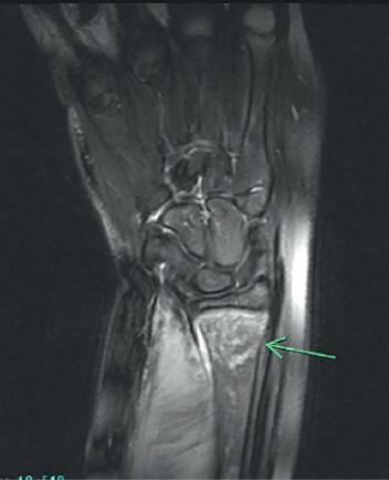
Fig. 20.5 Osteomyelitis
Coronal MRI of the right wrist in a teenager with pain, swelling, fever, and elevated white blood cell count. In this sequence, normal bone marrow should be uniformly dark. Note the patchy bright (whiter) areas in the distal radial metaphysis indicative of osteomyelitis in this patient (arrow). (Courtesy of Joseph Makris, MD, Baystate Medical Center.)
Fig. 20.6 MRI arthrogram of the shoulder
Axial image of a normal right shoulder at the level of the mid joint. The MRI was performed after contrast was injected directly into the joint space. The contrast fluid (white on this image) distends the joint capsule and outlines the articular cartilage, labrum, and ligaments, increasing sensitivity for evaluation of injuries to these structures. (Courtesy of Joseph Makris, MD, Baystate Medical Center.)

Unit VI Review Questions: Upper Limb
1.A patient complains of tingling and pain in the upper arm. Upon further
examination and additional tests, you determine that the axillary artery has been occluded by atherosclerosis. The patient, however, still has a radial pulse at the wrist. Which of the following arteries could provide a collateral circulation around the occlusion?
A.Suprascapular and circumflex scapular arteries
B.Suprascapular and lateral thoracic arteries
C.Posterior circumflex humeral and anterior circumflex humeral arteries
D.Superior thoracic and lateral thoracic arteries
E.Posterior intercostals and lateral thoracic arteries
2.The deep palmar arch of the hand is formed mainly by the
A.ulnar artery
B.radial artery
C.brachial artery
D.common palmar digital arteries
E.palmar metacarpal arteries
3.Each cord of the brachial plexus
A.contains nerve fibers from C5 to T1 levels of the spinal cord
B.is formed from the junction of one anterior and one posterior division
C.gives a branch to the median nerve
D.lies within the axilla
E.gives rise to a subscapular nerve (upper, middle, and lower)
4.Nerves often pair with arteries to travel as neurovascular bundles.
Which of the following is not an accurate pairing?
A. Axillary nerve and posterior circumflex humeral artery

B.Median nerve and brachial artery
C.Musculocutaneous nerve and circumflex scapular artery
D.Radial nerve and deep brachial artery
E.Long thoracic nerve and lateral thoracic artery
5.During a radical mastectomy on a 50-year-old woman, the surgeon does
a careful but thorough axillary node dissection. What nerve lies along the medial wall of the axilla and is particularly vulnerable during this procedure?
A.Lateral branch of posterior intercostal
B.Musculocutaneous
C.Lateral pectoral
D.Long thoracic
E.Dorsal scapular
6.One of your patients ruptured the tendon of the long head of the biceps
brachii. Although he is bothered by the unattractive bulge that formed in his anterior arm, he is surprised to find that he has lost little flexor strength. You confirm that the brachialis muscle has greater leverage at the joint and therefore is the more powerful flexor of the elbow. Where does the brachialis insert?
A.Radial tuberosity
B.Ulnar tuberosity
C.Medial epicondyle
D.Bicipital aponeurosis
E.Olecranon
7.Which part(s) of the triceps brachii crosses/cross the glenohumeral
joint?
A.Medial head
B.Lateral head
C.Long head
D.Lateral and medial heads
E.Lateral and long heads

8.Structures that pass through the quadrangular space of the upper limb
include the
A.radial nerve
B.suprascapular nerve
C.posterior humeral circumflex artery
D.anterior humeral circumflex artery
E.deep brachial artery
9.Which of the following bones articulates with the radius at the wrist?
A.Pisiform
B.Hamate
C.Capitate
D.Trapezium
E.Scaphoid
10.The flexor retinaculum
A.forms the floor of the ulnar tunnel
B.forms the roof of the carpal tunnel
C.is continuous with the palmar aponeurosis
D.is continuous with the palmaris longus
E.All of the above
11.As a second-year resident doing a pediatric surgery rotation, you set up
an arterial line on a 6-year-old girl prior to surgery. You choose the radial artery of her left (nondominant) hand. Because you know that the procedure can result in occlusion of the artery, you verify that the collateral circulation to the hand is patent. The radial artery
A.forms the superficial arch of the hand
B.passes superficial to the anatomic snuffbox
C.supplies the muscles of the posterior compartment through its posterior interosseous branch
D.supplies the princeps pollicis artery of the thumb

E.lies medial to the tendon of the flexor carpi radialis in the wrist
12.During your first training session as a phlebotomist, you are relieved to
find that your “patient” is a 24-year-old weight lifter whose superficial veins stand out dramatically against his overdeveloped muscles. Superficial veins of the upper limb
A.include a basilic vein that runs in the deltopectoral groove
B.include a cephalic vein that joins the brachial veins in the arm
C.drain into the veins of the deep venous system via perforating veins
D.course with the arteries as paired accompanying veins
E.have bidirectional valves that allow flow in either direction
13.An injury to the lower brachial plexus (Klumpke’s palsy) would affect
A.sensation in the nail bed of the 5th digit
B.abduction of the 2nd through 5th digits
C.adduction of the thumb
D.adduction of the wrist
E.All of the above
14.While moonlighting in the emergency department one night, you treat a
14-year-old gang member who was stabbed in the supraclavicular region of the neck 2 cm above the middle third of the clavicle. A chest X-ray confirms that he has a pneumothorax. What other structure could be injured in this area?
A.Axillary nerve
B.Pectoralis minor
C.Subscapular artery
D.Posterior cord of the brachial plexus
E.Cephalic vein
15.The serratus anterior muscle
A.forms a scapulothoracic joint with the external intercostal muscles

B.is innervated by a branch of the posterior cord
C.elevates the scapula off the thoracic wall
D.rotates the scapula laterally during abduction of the arm above the horizontal plane
E.originates from the subscapular fossa
16.A professional rodeo cowboy fell off his horse and fractured his
humerus at the anatomic neck and the lesser tubercle. Which of the following muscles inserts on this tubercle?
A.Supraspinatus
B.Infraspinatus
C.Subscapularis
D.Coracobrachialis
E.Teres major
17.Damage to which nerve would most affect elbow flexion?
A.Radial nerve
B.Ulnar nerve
C.Median nerve
D.Musculocutaneous nerve
E.Axillary nerve
18.At a neighborhood block party several children engage in a tug-of-war.
Suddenly, Jason, a 5-year-old boy, hugs his right elbow and cries out in pain. The inconsolable child is eventually taken to the local clinic, where the pediatrician recognizes that Jason has subluxed (partially dislocated) his radial head. By gently supinating the flexed arm, the doctor restores it to its normal position. Which of the following is true regarding the proximal radioulnar joint?
A.The radial head rotates within the annular ligament.
B.It includes a hinge-type joint between the head of the radius and capitulum of the humerus.
C.The biceps brachii pronates the joint.
D.An articular disk separates the radius and ulna.

E. The subluxation of the radial head results from a tear of the radial collateral ligament.
19.On your first day of shadowing in your preceptor’s office, you are asked
to get some baseline information on the patients. You start by taking their pulse. The radial artery is easiest to palpate in the wrist where it lies immediately lateral to the tendon of the
A.flexor carpi radialis
B.superficial flexor digitorum
C.flexor pollicis longus
D.palmaris longus
E.extensor carpi radialis
20.You examine a 14-year-old girl in the emergency department. She has a
puncture wound from a dog bite in the flesh over the middle phalanx of her 5th digit. The incident occurred 2 days ago, and the finger is inflamed and likely infected. What are your thoughts regarding possible spread of the infection via the synovial sheath?
A.It will spread into the superficial space on the dorsum of the hand.
B.It will spread to the common flexor sheath in the wrist.
C.It will spread to the adjacent finger.
D.It will remain confined to the sheath of the infected finger.
E.A and B are correct.
21.The axillary artery
A.begins at the lateral border of the 1st rib
B.lies anterior to the axillary vein
C.ends by dividing into brachial and deep brachial arteries
D.passes through the axilla between the pectoralis major and pectoralis minor muscles
E.branches include the thyrocervical trunk

22.Axillary lymph nodes that lie medial to the pectoralis minor include
A.pectoral nodes
B.humeral nodes
C.apical nodes
D.central nodes
E.subscapular nodes
23.The C4 anterior ramus is a component of
A.the pre-fixed plexus
B.the upper trunk
C.the posterior cord
D.the axillary nerve
E.All of the above
24.A young man suffered a crushing injury to his right arm, fracturing his
humerus at midshaft and damaging the nerve that runs in the posterior compartment. What functional loss would you expect from this injury?
A.Inability to extend the elbow
B.Inability to supinate the hand
C.Inability to abduct the thumb
D.Inability to extend the wrist
E.All of the above
25.One of your orthopedic colleagues introduces you, an anatomist, to his
patient, a famous baseball pitcher, who suffers from chronic rotator cuff pain. He asks you to demonstrate the rotator cuff on a cadaver specimen and explain the anatomy of this type of injury. You tell him the following:
A.Tendons of the rotator cuff muscles insert onto the capsule of the glenohumeral joint.
B.The supraspinatus tendon passes through the subacromial space between the shoulder joint and the coracoacromial arch.
C.An abnormal communication between the subacromial bursa and

glenohumeral joint cavity can result from rupture of the supraspinatus tendon.
D.Rupture of the supraspinatus tendon will impair the patient’s ability to initiate abduction of the arm.
E.All of the above
26.Which of the following muscles has no attachment on the humerus?
A.Deltoid
B.Coracobrachialis
C.Flexor digitorum superficialis
D.Pronator teres
E.Biceps brachii
27.Which muscle provides strong adduction of the glenohumeral joint?
A.Teres minor
B.Pectoralis major
C.Pectoralis minor
D.Short head of the biceps brachii
E.Subscapularis
28.A young woman riding a 10-speed bicycle accidentally engaged the
front brake, was thrown over the handlebars, and landed on her outstretched hands. In the emergency department, imaging of the elbow revealed a subtle fracture of the radial neck that allowed proximal movement of the radius relative to the ulna, which also suggested a tear in the interosseous membrane. Which movements were most impaired by this injury?
A.Flexion of the wrist
B.Extension of the elbow
C.Extension of the digits
D.Abduction of the thumb
E.Adduction of the thumb

29.An elderly man had injured his wrist in a fall and is now experiencing
tingling in his fingers and a weakened grip. A lateral X-ray of the wrist shows that his lunate has dislocated and is pressing on the structures within the carpal tunnel. Which of the following structures pass through the carpal tunnel?
A.Ulnar artery
B.Radial artery
C.Flexor carpi radialis
D.Flexor pollicis longus
E.Palmaris longus
30.In the anatomy lab you are fascinated by the deep dissection of the hand
because as an amateur violinist you appreciate that the intrinsic muscles of the palm are important for the fine movements of the hand. Which of the following is true for both the lumbricals and the interossei muscles?
A.All are innervated by the median nerve.
B.All are innervated by the ulnar nerve.
C.They flex the metacarpophalangeal joints.
D.They arise from the tendons of the flexor digitorum profundus.
E.They abduct or adduct the fingers.
31.Your 7-year-old nephew suffered a distal clavicular fracture after falling
from a tree. Which of the following structures would likely be disrupted with this type of injury?
A.Interclavicular ligaments
B.Coracohumeral ligament
C.Acromioclavicular joint
D.Scapulothoracic joint
E.Coracoacromial ligament
32.Your grandmother tripped on the stairs and landed on her outstretch arms. In the emergency department an initial physical exam strongly
indicated a fractured scaphoid. What part of the exam suggested this diagnosis?
A.Pain over palmar surface of the wrist
B.Pain over the ulnar styloid process
C.Displacement of the radial styloid process
D.Pain in the anatomic snuffbox
E.Inability to oppose the thumb
33.Deep fascia of the limbs forms discrete compartments of muscles that
usually share similar functions and innervations. The effects of trauma are often limited to the contents of a single compartment. Muscles effected by trauma to the anterior forearm compartment would include
A.brachioradialis
B.pronator teres
C.supinator
D.abductor pollicis longus
E.extensor carpi ulnaris
34.As a pediatric orthopedist, you’ve created some simple and fun
shortcuts for diagnosing nerve injuries to the hand in children. Which of the following activities would selectively test the indicated nerve?
A.“Thumbs up” sign (extending the thumb away from the fist): median nerve
B.“OK” sign (forming a circle with the 1st and 2nd digits): radial nerve
C.“Peace” sign (separating the 2nd and 3rd digits while extended): ulnar nerve
D.All the above
E.None of the above
35.A young woman is seen in the local clinic following a fall she suffered
while hiking. She has severe pain and swelling of her left elbow but a lateral x-ray confirms that there is no fracture. You suspect a soft tissue injury. What additional imaging modality would you choose to confirm this?

A.Frontal x-ray
B.MRI
C.CT
D.Ultrasound
36.You convinced your boyfriend to help you move some boxes of books
out of your basement. During the move he felt a sharp pain in his neck that remained throughout the following day. His physician recommended an MRI of his neck, which revealed a rupture of his middle scalene muscle caused by the strain of steadying his trunk as he lifted the heavy boxes. In addition, he demonstrated a “winged” scapula, indicating that there was significant damage to his long thoracic neve, which passes through the middle scalene before descending into the neck and axilla. Which activity would be affected by injury to the long thoracic nerve?
A.Adducting his arm to touch the elbow of the opposite side
B.Internally rotating the arm to touch the back of his waist
C.Externally rotating his arm to throw a baseball
D.Flexing his arm directly forward
E.Abducting his arm above his head
Answers and Explanations
1.A. The suprascapular and transverse cervical branches of the subclavian artery and the thoracodorsal and circumflex scapular branches of the distal segment of the axillary artery participate in a scapular arcade that provides a collateral circulation that would circumvent the occlusion (Section 18.4).
B.The lateral thoracic artery does not supply the scapula, nor does it anastomose with the suprascapular artery.
C.The anterior and posterior humeral circumflex arteries anastomose around the neck of the humerus but do not anastomose with arteries proximal to the occlusion.
D.Neither the superior thoracic artery nor the lateral thoracic artery supplies the scapular region.
E.Posterior intercostal arteries supply the medial scapular region, but they do not anastomose with the lateral thoracic artery.
2.B. The deep palmar arch is formed mainly by the radial artery (Section 18.4).
A. The ulnar artery forms the superficial palmar arch.
C.The brachial artery begins at the lateral border of the axilla and terminates in the cubital fossa.
D.The common palmar digital arteries are branches of the superficial palmar arch in the palm of the hand.
E.The palmar metacarpal arteries arise from the deep palmar arch in the palm of the hand.
3.D. Each cord of the brachial plexus lies within the axilla (Section 18.4).
A.The posterior cord contains fibers from C5 to T1; the medial cord contains fibers from C8 and T1; the lateral cord contains fibers from C5 to C7.
B.The posterior divisions form the posterior cord; only anterior divisions form the medial and the lateral cords.
C.The median nerve is formed from the medial and lateral cords.
E.The subscapular nerves arise from the posterior cord.
4.C. The musculocutaneous nerve passes from the axilla to pierce the coracobrachialis of the anterior arm. The circumflex scapular artery passes through the triangular space into the scapular region (Section 19.2).
A.The axillary artery and posterior circumflex humeral artery pass through the quadrangular space to the deltoid region.
B.The median nerve and brachial artery descend on the medial side of the biceps brachii and enter the cubital fossa.
D.The radial nerve and deep brachial artery wind around the posterior humerus through the triceps hiatus.
E.The long thoracic nerve and lateral thoracic artery descend on the medial wall of the axilla to supply the serratus anterior muscle.
5.D. The long thoracic nerve runs superficial to, and innervates, the serratus anterior, which forms the medial wall of the axilla. Injury to this nerve can result in a “winged” scapula (Section 19.1).
A.In the axilla the intercostal nerves run deep to the serratus anterior and external intercostal muscles and are not vulnerable to injury.
B.The musculocutaneous nerve leaves the axilla just inferior to the glenohumeral joint where it enters the coracobrachialis muscle.
C.The lateral pectoral nerve passes anteriorly to penetrate the pectoralis major muscle.
E.The dorsal scapular nerve descends posteriorly from the upper roots
of the plexus to innervate the levator scapulae and rhomboid muscles.
6.B. The brachialis muscle inserts on the ulnar tuberosity of the ulna (Section 19.3).
A. The biceps brachii inserts on the radial tuberosity.
C.The coronoid process forms the anterior lip of the trochlear notch of the ulna and is the origin of the ulnar head of the pronator teres.
D.The bicipital aponeurosis is a fascial extension of the biceps brachii.
E.The olecranon of the ulna forms the posterior prominence of the elbow and is the insertion for the triceps brachii and anconeus muscles.
7.C. The long head of the triceps brachii crosses the glenohumeral joint to attach to the infraglenoid tubercle, where it contributes to extension of the joint (Section 19.2).
A.The medial head arises from the medial aspect of the humeral shaft. It joins with the lateral and long heads to insert on the olecranon of the ulna.
B.The lateral head arises from the lateral aspect of the humeral shaft. It joins with the medial and long heads to insert on the olecranon of the ulna.
D.The medial and lateral heads arise from the humeral shaft and cross only the elbow joint.
E.The lateral head arises from the shaft of the humerus and crosses the elbow joint. The long head originates on the infraglenoid tubercle of the scapula and crosses the glenohumeral and elbow joints.
8.C. The posterior humeral circumflex artery and the axillary nerve pass through the quadrangular space (Section 19.2).
A.The radial nerve passes through the triceps hiatus.
B.The suprascapular nerve passes through the scapular notch.
D.The anterior humeral circumflex artery runs horizontally beneath the coracobrachialis and the short head of the biceps brachii to encircle the neck of the humerus.
E.The deep brachial artery passes through the triceps hiatus with the radial nerve.
9.E. The radius articulates distally with the scaphoid and lunate bones of the wrist (Section 19.5).
A.The pisiform on the medial side of the wrist articulates with the triquetrum.
B.The hamate articulates with the capitate, lunate, triquetrum, and 4th
and 5th metacarpal bones.
C.The capitate articulates with the hamate, trapezoid, scaphoid, lunate, and 3rd metacarpal.
D.The trapezium articulates with the trapezoid, scaphoid, and 1st metacarpal.
10.E. The flexor retinaculum forms the roof of the carpal tunnel and floor of the ulnar tunnel. As a thickening of the deep fascia of the palm, it is continuous with the palmar aponeurosis, palmaris longus, transverse metacarpal ligament, and fibrous digital sheaths (Sections 19.5 and 19.6).
A.The flexor retinaculum forms the floor of the ulnar tunnel; the palmar carpal ligament form its roof. B through D are also correct (E).
B.The carpal tunnel is a fascio-osseous tunnel. Carpal bones form the floor and sides; the flexor retinaculum forms the roof. A, C, and D are also correct (E).
C.The flexor retinaculum is continuous with the tough palmar aponeurosis of the hand. A, B, and D are also correct (E).
D.The palmaris longus passes over the carpal tunnel and inserts on the flexor retinaculum and palmar aponeurosis. A through C are also correct (E).
11.D. The princeps pollicis arises from the radial artery at the base of the 1st metacarpal and divides into two digital arteries of the thumb (Section 18.4).
A.The ulnar artery forms the superficial palmar arch. The radial artery forms the deep palmar arch.
B.The radial artery courses along the floor of the anatomic snuffbox.
C.The posterior interosseous artery, which supplies muscles of the posterior forearm compartment, arises from the common interosseous artery, a branch of the ulnar artery in the cubital fossa.
E.The radial artery lies lateral to the tendon of the flexor carpi radialis at the wrist.
12.C. The veins of the superficial venous system drain into the deep veins via perforating veins (Section 18.4).
A.The cephalic vein courses along the deltopectoral groove before terminating in the axillary vein.
B.The basilic vein joins with the paired brachial veins to form the axillary vein.
D.Veins of the deep venous system travel with the arteries as paired accompanying veins. There is no superficial arterial system that accompanies
the superficial veins.
E.Veins of the limbs have unidirectional valves that prevent pooling of blood and facilitate flow back toward the heart.
13.E. The ulnar nerve arises from the lower part of the plexus. Its dorsal cutaneous branch in the hand supplies the palmar and dorsal surfaces of the 4th (half) and 5th digits, and its deep branch supplies the dorsal interossei muscles that abduct the fingers and the adductor pollicis that adducts the thumb. It also innervates the flexor carpi ulnaris that assists in adduction of the wrist (Section 18.4).
A.The ulnar nerve’s (C8–T1) dorsal cutaneous branch in the hand supplies the palmar and dorsal surfaces of the 4th (half) and 5th digits. B through D are also correct (E).
B.The ulnar nerve’s (C8–T1) deep branch in the hand supplies the dorsal interossei muscles that abduct the fingers. A, C, and D are also correct
(E).
C.The ulnar nerve’s (C8–T1) deep branch in the hand supplies the adductor pollicis muscle that adducts the thumb. A, B, and D are also correct
(E).
D.The ulnar nerve (C8–T1) innervates the flexor carpi ulnaris that assists the extensor carpi ulnaris (radial nerve) in adduction of the wrist. A through C are also correct (E).
14.D. The posterior cord is part of the supraclavicular brachial plexus and could be injured in the area (Section 18.4).
A.The terminal nerves of the brachial plexus form below the clavicle. Although damage to the supraclavicular plexus could affect the axillary nerve, the stab wound would not directly damage the nerve itself.
B.The pectoralis minor attaches to the coracoid process, which lies below the middle part of the clavicle.
C.The subscapular artery is a branch of the distal part of the axillary artery, which is infraclavicular.
E.The cephalic vein drains into the axillary vein below the clavicle and most likely would not be affected by this injury.
15.D. The serratus anterior pulls the inferior angle of the scapula laterally when the glenohumeral joint is abducted above the horizontal plane (Section 19.1).
A.The scapulothoracic joint is a functional relationship between the
serratus anterior and subscapularis muscles.
B.The long thoracic nerve, which innervates the serratus anterior muscle, arises directly from the C5–C7 roots of the brachial plexus.
C.The serratus anterior supports the scapula against the thoracic wall. Denervation of the muscle allows the scapula to lift away from the thoracic wall, a condition known as a “winged” scapula.
E.The serratus anterior inserts on the entire medial border of the
scapula.
16.C. The subscapularis inserts on the lesser tubercle of the humerus and fibrous capsule of the glenohumeral joint (Section 19.2).
A.The supraspinatus inserts on the greater tubercle of the humerus.
B.The infraspinatus inserts on the greater tubercle of the humerus.
D.The coracobrachialis inserts on the shaft of the middle part of the humerus.
E.The teres major inserts on the crest that extends inferiorly from the lesser tuberosity.
17.D. The musculocutaneous nerve innervates the biceps brachii and brachialis muscles, the primary flexors of the elbow. There would also be loss of sensation to the skin of the lateral forearm (Sections 19.2 and 19.3).
A.Damage to the radial nerve results in wrist drop and loss of sensation to the dorsum of the hand and the proximal segments of the 1st through 3rd digits and half of the 4th digit.
B.Damage to the ulnar nerve may cause paresthesia (numbness and tingling) in the forearm, 4th and 5th fingers, or paralysis of most of the intrinsic muscles of the hand (so-called claw hand deformity).
C.Damage to the median nerve above the cubital fossa will cause the inability to flex the proximal interphalangeal joint of the 1st to 3rd digits and the inability to flex the distal interphalangeal joints of the 2nd and 3rd digits. This results in “benediction hand” (2nd and 3rd finger are partially extended) when trying to form a fist. It also causes the inability to flex the terminal phalanx of the thumb (due to damage of flexor pollicis longus) and loss of sensation over the lateral aspect of the hand.
E.Damage to the axillary nerve causes paralysis of the deltoid muscle, resulting in weakened flexion and extension of the shoulder and the inability to abduct the arm above the horizontal.
18.A. The annular ligament forms a circular cuff around the radial head that
allows the bone to rotate in the joint (Section 19.4).
B.The hinge-type joint between the radius and capitulum of the humerus is the humeroradial joint.
C.The biceps brachii inserts on the tuberosity of the radius and, therefore, supinates the joint when contracted.
D.In the proximal radioulnar joint the radius articulates at the radial notch of the ulna. An articular disk separates the radius and ulna in the distal radioulnar joint.
E.Subluxation of the radial head results from a laxity of the annular ligament.
19.A. The radial artery descends on the lateral side of the forearm and at the wrist lies immediately lateral to the tendon of the flexor carpi radialis (Section 19.5).
B.The tendons of the superficial flexor digitorum lie in the middle of the wrist, medial to the radial artery.
C.The tendon of the flexor pollicis longus lies lateral to the median nerve and medial to the tendon of the flexor carpi radialis and to the radial artery.
D.The palmaris longus runs superficial to the flexor retinaculum and medial to the flexor pollicis longus.
E.The radial artery lies medial (palmar) to the extensor carpi radialis
tendon.
20.B. The synovial sheath of the 5th digit normally communicates with the common synovial sheath (Section 19.6).
A.The synovial sheath of the 5th digit communicates with the common synovial sheath and with that of the thumb, but it does not normally communicate with the superficial space on the dorsum of the hand.
C.The synovial sheaths of the 2nd, 3rd, and 4th digits do not normally communicate with any other tendon sheaths. It is unlikely the infection will spread to these fingers.
D.Although the infection may remain confined to the infected finger, the synovial sheath of the 5th digit communicates with that of the thumb and with the common tendon sheath. Therefore, the concern is that it will spread via this route to the thumb, wrist, and forearm.
E.The synovial sheath of the 5th digit normally communicates with the common flexor sheath at the wrist but not with the superficial space on the dorsum of the hand.
21.A. The subclavian artery continues as the axillary artery at the lateral border of the 1st rib (Section 18.4).
B.The axillary artery lies posterior to the axillary vein.
C.The brachial artery is a continuation of the axillary artery in the axilla. The deep brachial artery is one of the branches of the brachial artery.
D.The axillary artery passes through the axilla posterior to the pectoralis minor muscle.
E.The thyrocervical trunk is a branch of the subclavian artery.
22.C. The apical nodes are the nodes of the upper infraclavicular group. These lie medial to the pectoralis minor muscle along the axillary vein and adjacent to the proximal part of the axillary artery (Section 18.4).
A.Pectoral nodes are part of the lower axillary group that lies lateral to the pectoralis minor.
B.Interpectoral nodes are part of the middle axillary group that lies between the pectoralis major and pectoralis minor muscles.
D.Central nodes are part of the lower axillary group that lies deep to the pectoralis minor.
E.The subscapular nodes are part of the lower axillary group that lies along the posterior axillary fold.
23.A. The brachial plexus contains the anterior rami of C5 to T1. A prefixed plexus also contains the C4 anterior ramus (Section 18.4).
B.The upper trunk contains C5 and C6 anterior rami.
C.The posterior cord contains the anterior rami of C5 to T1.
D.The axillary nerve contains the anterior rami of C5 and C6.
E.B, C, and D are incorrect.
24.D. Damage to the radial nerve from a midhumeral fracture results in loss of innervation to the extensor carpi radialis and extensor carpi ulnaris (Sections 18.4 and 19.4).
A.The branches of the radial nerve to the triceps brachii arise high in the arm, so a midhumeral lesion of the nerve would not affect the function of this muscle.
B.The radial nerve innervates only one of the muscles that supinate the hand, the supinator. The biceps brachii, also a supinator, is innervated by the musculocutaneous nerve.
C.Abduction of the thumb is weakened by damage to the radial nerve.
E.A, B, and C are incorrect.
25.E. All of the rotator cuff muscles insert on the fibrous capsule of the glenohumeral joint. The supraspinatus muscle, the most frequently involved in rotator cuff tears, passes through the narrow subacromial space and, with repetitive use, becomes frayed. Rupture of the tendon allows the overlying bursa to communicate with the joint cavity and impairs the initial phase of abduction. Later phases of abduction remain intact through the action of the deltoid muscle (Section 19.2).
A.The tendons of the rotator cuff muscles insert on, and reinforce, the fibrous capsule of the joint. B through D are also correct (E).
B.The supraspinatus tendon and subacromial bursa pass through the narrow subacromial space between the capsule of the shoulder joint and the coracoacromial arch. A, C, and D are also correct (E).
C.The supraspinatus tendon separates the subacromial and subdeltoid bursae from the joint cavity. With rupture of the tendon, a communication between the bursae and joint cavity may result. A, B, and D are also correct
(E).
D.The deltoid is the primary abductor of the glenohumeral joint, but the supraspinatus assists in the first 15 degrees of abduction. A through C are also correct (E).
26.E. The two heads of the biceps brachii originate on the supraglenoid tubercle and coracoid process of the scapula and insert on the radial tuberosity of the radius (Sections 19.2 and 19.3).
A.The deltoid originates along the scapular spine and clavicle and inserts on the deltoid tuberosity of the humerus.
B.The coracobrachialis originates on the coracoid process of the scapula and inserts on the shaft of the humerus.
C.The flexor digitorum superficialis originates on the medial epicondyle of the humerus and upper part of the radius and inserts on the middle phalanx of the 2nd through 4th digits.
D.The pronator teres originates on the medial epicondyle of the humerus and coronoid process of the ulna and inserts on the lateral radius.
27.B. The broad origin of the pectoralis major on the anterior trunk wall allows it to strongly adduct the arm (Section 19.2).
A.As part of the rotator cuff, the teres minor supports the head of the humerus in the glenohumeral joint. It also weakly adducts and laterally rotates the arm.
C.The pectoralis minor draws the scapula forward and down, causing
the scapula to rotate medially.
D.The short head of the biceps brachii provides some flexion, abduction, and internal rotation of the glenohumeral joint.
E.The subscapularis supports the head of the humerus in the glenohumeral joint and medially rotates the arm.
28.D. The function of the abductor pollicis muscle, the primary abductor of the thumb, would be greatly impaired because it originates on the interosseous membrane (Section 19.4).
A.The flexor carpi radialis and flexor carpi ulnaris originate from the medial epicondyle of the humerus and olecranon of the ulna and would be unaffected by this injury.
B.The triceps brachii, which extends the elbow, inserts on the olecranon. It would not be impaired by this injury.
C.The extensor digitorum originates from the humerus and ulna and would be unaffected by this injury.
E.The adductor of the thumb, adductor pollicis, attaches to the 2nd and 3rd metacarpals and the capitate. It would not be affected by this injury.
29.D. The flexor pollicis longus passes through the carpal tunnel with the flexor digitorum superficialis, flexor digitorum profundus, and median nerve (Section 19.5).
A.The ulnar artery and ulnar nerve pass through the ulnar canal at the
wrist.
B.At the wrist the radial artery turns dorsally to run through the floor of the anatomic snuffbox.
C.The flexor carpi radialis crosses the wrist lateral to the carpal tunnel and inserts on the base of the 2nd metacarpal.
E.The palmaris longus passes superficial to the flexor retinaculum to insert into the palmar aponeurosis.
30.C. The interossei and lumbricals flex the metacarpophalangeal joints and extend the interphalangeal joints (Section 19.6).
A.Only the lateral two lumbricals are innervated by the median nerve. The medial lumbricals and all of the interossei are innervated by the ulnar nerve.
B.The ulnar nerve innervates all of the interossei and the medial two lumbricals. The lateral two lumbricals are innervated by the median nerve.
D.Only the lumbricals arise from the flexor tendons. The interossei
arise from the shafts of the metacarpal bones.
E.The palmar interossei adduct the fingers, the dorsal interossei abduct the fingers. The lumbricals have no effect on these movements.
31.C. Distal clavicular fractures often disrupt the acromioclavicular joint and the coracoacromial ligaments (Sections 18.2 and 19.1).
A.Interclavicular ligaments connect the medial ends of the clavicles to the sternum and are less likely to be disrupted by a distal fracture.
B.The coracohumeral ligaments are part of the glenohumeral capsule and not at risk with this type of injury.
D.The scapulothoracic joint is a functional relationship between the serratus anterior and subscapularis muscles and not at risk in clavicular fractures.
E.The coracoacromial ligament is part of the coracoacromial arch, which protects the glenohumeral joint and is unlikely to be injured in this case.
32.D. The scaphoid is most easily palpated in the anatomic snuffbox, where it forms the floor of this space. Pain in this region is indicative of a fractured scaphoid (Section 19.5).
A.Pain over the palmar surface of the wrist would suggest a carpal fracture but not definitively indicate the scaphoid.
B.The ulna lies on the medial side of the wrist, the scaphoid is on the lateral side.
C.Displacement of the radius would suggest a fracture or rupture of some of the radial ligaments but would not be diagnostic of a scaphoid fracture.
E.Although movement of the thumb would likely be painful, the ability to oppose the thumb would remain intact since the opponens pollicis does not attach to the scaphoid.
33.B. The pronator teres is located in the superficial layer of the anterior forearm compartment.
A.The brachioradialis is a muscle of the posterior forearm compartment.
C.The supinator is a muscle of the posterior forearm compartment.
D.The abductor pollicis longus is a muscle of the posterior forearm
compartment.
E. The extensor carpi ulnaris is a muscle of the posterior forearm compartment.
34.C. The “peace” sign tests the ability of the dorsal interossei muscles, innervated by the ulnar nerve, to abduct the 2nd digit (Section 19.6).
A.A “thumbs up” sign requires extension of the thumb, performed by the extensor pollicis longus and brevis, which are innervated by the radial nerve.
B.The “OK” sign tests the ability of the opponens pollicis, which is innervated by the median nerve.
D.Only answer C is correct
E.Only A and B are incorrect
35.B. MRI is the modality of choice for visualizing soft tissues such as muscles, tendons, and cartilage.
A.A frontal X-ray would help confirm the presence or absence of a fracture, but soft tissue injuries are not visible on an X-ray.
C.CT is excellent for visualizing bone detail but less accurate than MRI for assessing soft tissue injuries.
D.Ultrasound is most useful for image-guided procedures and visualizing superficial tissues.
36.E. Injury to the long thoracic nerve inhibits the serratus anterior muscle, which rotates the inferior angle of the scapula laterally, tilting the glenoid superiorly. This rotation is necessary for abduction of the arm above 90 degrees.
A.Adducting the arm across the trunk is primarily the action of the pectoralis major muscle.
B.Touching the back of the waist requires both internal rotation and extension at the shoulder joint, which are actions of the latissimus dorsi in conjunction with the pectoralis major, deltoid, and teres major.
C.The deltoid and infraspinatus are the primary external rotators of the
arm.
D.The deltoid, pectoralis major, and biceps brachii are the primary flexors of the arm.

Unit VII Lower Limb
21Overview of the Lower Limb
21.1General Features
21.2Bones of the Lower Limb
21.1Clinical Correlation: Fractures of the Femur
21.2Developmental Correlation: Bipartite Patella
21.3Fascia and Compartments of the Lower Limb
21.4Neurovasculature of the Lower Limb
21.1Nerves of the Lower Limb
21.3Clinical Correlation: Femoral Head Necrosis
21.4Clinical Correlation: Popliteal Aneurysm
21.5Clinical Correlation: The Dorsal Pedal Pulse
21.6Clinical Correlation: Lower Limb Ischemia
21.7Clinical Correlation: Deep Vein Thromboses (DVT)
21.8Clinical Correlation: Varicose Veins
21.9Clinical Correlation: Femoral Nerve Injury
21.10Clinical Correlation: Obturator Nerve Injury
21.11Clinical Correlation: Superior Gluteal Nerve Injury
21.12Clinical Correlation: Sciatic Nerve Injury
21.13Clinical Correlation: Tibial Nerve Injury
21.14Clinical Correlation: Common Fibular Nerve Injury
21.15Clinical Correlation: Superficial and Deep Fibular Nerve Injury
22Functional Anatomy of the Lower Limb
22.1The Pelvic Girdle
22.2The Gluteal Region
22.1Gluteal Muscles
22.1Clinical Correlation: Piriformis Syndrome
22.3The Hip and Thigh
22.2Movements of the Hip Joint
22.3Iliopsoas Muscle
22.4Thigh Muscles, Anterior Compartment
22.5Thigh Muscles, Medial Compartment: Superficial and Deep Layers
22.6Thigh Muscles, Posterior Compartment
22.2Clinical Correlation: Congenital Hip Dislocation
22.3Clinical Correlation: Acquired Hip Dislocation
22.4Clinical Correlation: Hamstring Strains
22.5Clinical Correlation: Femoral Hernias
22.4The Knee and Popliteal Region
22.7Movements of the Knee Joint
22.6Clinical Correlation: Patellar Tendon Reflex
22.7Clinical Correlation: Ligamentous Injuries of the Knee
22.8Clinical Correlation: Popliteal (Baker’s) Cyst
22.9Clinical Correlation: Genu Varum and Genu Valgum
22.5The Leg
22.8Leg Muscles, Anterior Compartment
22.9Leg Muscles, Lateral Compartment
22.10Leg Muscles, Posterior Compartment: Superficial Flexors
22.11Leg Muscles, Posterior Compartment: Deep Flexors
22.10Clinical Correlation: Shin Splints
22.11Clinical Correlation: Rupture of the Calcaneal Tendon
22.12Clinical Correlation: Compartment Syndrome
22.13Clinical Correlation: Tarsal Tunnel Syndrome
22.6The Ankle (Talocrural) Joint
22.12Movements of the Ankle Joint
22.14Clinical Correlation: Ankle Sprains
22.7The Foot
22.13Movements of the Subtalor and Transverse Tarsal Joints
22.14Movements of the Metatarsophalangeal (MTP) and Interphalangeal (IP) Joints
22.15Intrinsic Muscles of the Dorsum of the Foot
22.16Intrinsic Muscles of the Sole of the Foot: Superficial layer
22.17Intrinsic Muscles of the Sole of the Foot: Deep Layer
22.15Clinical Correlation: Pes Planus
22.16Clinical Correlation: Plantar Fasciitis
22.17Clinical Correlation: The Plantar Reflex
22.8The Gait Cycle
22.18Muscle Action Sequence During the Gait Cycle
22.9Topographic Anatomy of Lower Limb Musculature
23 Clinical Imaging Basics of the Lower Limb
23.1Suitability of Imaging Modalities for the Lower Limb
Review Questions: Lower Limb