


23 Clinical Imaging Basics of the Lower Limb
As in the upper extremity, radiographs are always the first imaging choice in evaluation of bones and joints in the setting of trauma or pain, and magnetic resonance imaging (MRI) is the primary method for evaluating the soft tissue components of the joints. In infants, ultrasound is used to diagnose developmental hip dysplasia and to assess for abnormal joint fluid ( Table 23.1).
The principles and practices used in evaluating images of bones, joints, and soft tissues in the lower limb are similar to those used in the upper limb, except lower limb radiographs are often obtained with the patient standing or in a weight-bearing position. This distinction is important especially in the evaluation of weight-bearing joints (hips, knees, and ankles) because the joint is evaluated under the stress of everyday use (Figs. 23.1 and 23.2). MRIs cannot be taken with the patient in a weight-bearing position; nonetheless, they provide excellent detail of the internal soft tissue structures of the joint (Fig. 23.3).
Table 23.1 Suitability of Imaging Modalities for the Lower Limb
Modality |
Clinical Uses |
Radiographs (X-rays) |
First-line imaging evaluation tool for the |
|
extremities; used primarily to evaluate for |
|
fractures, bone lesions, and alignment of |
|
joints |
|
|
CT (computed |
Reserved as a troubleshooting tool to |
tomography) |
evaluate for subtle nondisplaced fractures |
|
|
MRI (magnetic |
One of the most important imaging modalities |
resonance imaging) |
for the evaluation of joints, specifically the |
|
non-osseous components of the joint, e.g., |
|
cartilage, ligaments, tendons, and muscles |
|
|
Ultrasound |
In children, due to their small size, it plays a |
|
larger role in diagnosis of hip disorders |
(developmental dysplasia, toxic synovitis, or joint effusion) and in evaluation of cartilaginous growth plates. Ultrasound has a limited role in evaluation of superficial soft tissue abnormalities and for guidance of interventional procedures involving the joints.
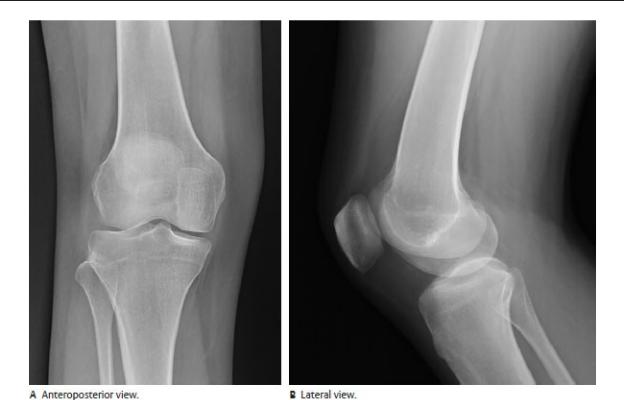
Fig. 23.1 Radiograph of the right knee
Knee radiographs are usually performed with the patient upright in order to assess the knee joint in a weight-bearing stance. The articular surfaces of the bones should be smooth and the medial and lateral joint compartments should be equally spaced. There should be no bone fragments in the joint spaces. The cortical edge of each bone should be traced around the entire bone and should be smooth. Note how the patella overlaps the femur in the frontal projection (A) and is difficult to see clearly. The lateral view (B) affords a non-overlapping view of the patella. (Courtesy of Joseph Makris, MD, Baystate Medical Center.)
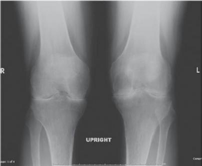
Fig. 23.2 Standing frontal radiographs of both knees in a 60-year-old man with chronic knee pain
Note the "bone on bone" appearance of the joint due to marked joint space narrowing. Compare this with the normal joint space appearance in Fig. 23.1. Although we don't see articular cartilage itself on an x-ray, we do see the space it creates between the bones, or, in this case, we see the results of chronic degenerative cartilage loss. (From Garcia G, ed. RadCases: Musculoskeletal Radiology 2nd ed. New York: Thieme Publishers; 2017.)
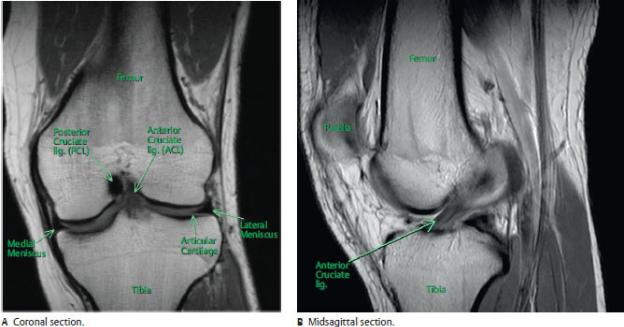
Fig. 23.3 MRI of the knee
In this sequence fat is bright (white), cortical bone is black, most normal ligaments and tendons are dark gray or black, and muscle is dark gray. The normal anterior cruciate ligament (ACL) has a unique striated appearance due to its linear bands of fibers. Note that the bone marrow is white because of its high fat content. (Courtesy of Joseph Makris, MD, Baystate Medical Center)

Unit VII Review Questions: Lower Limb
1.A young girl has recovered well from the severe injuries she sustained
in a motor vehicle accident 9 months ago, but because of some lingering nerve problems, she still has difficulty walking. She is balanced during the midstance phase but has difficulty bringing her limb forward to initiate movement. What nerve seems to be affected?
A.Superior gluteal
B.Inferior gluteal
C.Femoral
D.Obturator
E.Tibial
2.A young woman searching for driftwood walked several miles on the
soft sandy beach of a Caribbean island. While walking on this uneven surface, the balance between inversion and eversion at her subtalar joint was maintained by the opposing actions of the fibularis longus and the
A.fibularis brevis
B.fibularis tertius
C.soleus
D.tibialis anterior
E.extensor digitorum longus
3.Structures at the knee that are not attached to the joint capsule include
the
A.medial meniscus
B.medial collateral ligament
C.lateral collateral ligament
D.patellar retinacula
E.All of the above (A–D) are attached to the joint cavity.


each of the nerves that cross the ankle. Where would you anesthetize the sural nerve?
A.Anterior to the medial malleolus
B.Posterior to the medial malleolus
C.Posterior to the lateral malleolus
D.Anterior to the lateral malleolus
E.First web space
8.Sensation along the lateral side of the first toe is transmitted by
branches of the
A.saphenous nerve
B.medial plantar nerve
C.deep fibular nerve
D.superficial fibular nerve
E.lateral plantar nerve
9.Superficial veins of the lower limb
A.lie deep to the fascia lata
B.include the great saphenous vein that terminates at the popliteal vein
C.include the small saphenous vein whose course runs anterior to the lateral malleolus
D.drain to deep veins via perforating veins
E.originate from the plantar venous arch on the sole
10.The dorsal pedal artery is a continuation of the
A.posterior tibial artery
B.fibular artery
C.anterior tibial artery
D.popliteal artery
E.inferior medial genicular artery
11.The deep plantar arch of the foot is an anastomosis between the dorsal
pedal artery and which structure on the sole of the foot?
A.Fibular artery
B.Lateral plantar artery
C.Plantar metatarsal arteries
D.Plantar digital arteries
E.Arcuate artery
12.An obese woman suffers from peripheral edema (swelling of the legs)
and large varicose veins in her leg. Which of the following might be coexisting symptoms related to her condition?
A.Incompetent valves in the great saphenous vein
B.Reversed flow in the perforating veins
C.Deep vein thrombosis
D.Thrombophlebitis
E.All of the above
13.“Footdrop” occurs when the plantar flexor muscles are unopposed. This
condition may be caused by damage to which of the following nerves?
A.Superficial fibular
B.Deep fibular
C.Sural
D.Tibial
E.Medial plantar
14.As she was riding her bike home from her job as a camp counselor,
Kristin was sideswiped by a large pickup trunk. She suffered multiple bruises, a fractured tibia, and a fractured pelvis. Several months later she still had an area of numbness on the medial side of her thigh and walked with a lateral swing to her gait. Which muscle was affected by her injury?
A.Gluteus medius
B.Gluteus maximus
C.Semimembranosus
D.Adductor longus
E.Rectus femoris


posterior drawer sign. What structure appears to be injured?
A.Anterior cruciate ligament
B.Posterior cruciate ligament
C.Patellar ligament
D.Medial collateral ligament
E.Lateral collateral ligament
19.A single woman looking for adventure signed up for a walking tour of
the Scottish highlands. She was unprepared for the effort that it required but kept up with the more experienced walkers. After a few days she began to experience extreme tenderness in her anterior leg deep to the ridge of the tibia. The tour guide had seen this in previous clients and suggested that her pain was due to shin splints. Her pain originated from overuse of the
A.extensor digitorum longus
B.extensor digitorum brevis
C.extensor hallucis longus
D.fibularis tertius
E.tibialis anterior
20.Which fibrous structure on the sole of the foot originates at the
calcaneus and continues distally as digital fibrous sheaths?
A.Plantar aponeurosis
B.Plantar calcaneonavicular ligament
C.Long plantar ligament
D.Short plantar ligament
E.Deltoid ligament
21.A man was on a hunting trip with friends when he was accidentally shot
in the midthigh. He bled profusely from his femoral artery until his friends applied constant pressure over the wound and drove him to a local emergency clinic. Although examination of the wound site in surgery revealed a badly torn femoral artery, the man continued to have distal pulses in his leg. What vessel arises proximal to the injury site and can provide collateral blood supply to the distal limb?

A.Medial circumflex femoral artery
B.Lateral circumflex femoral artery
C.Obturator artery
D.Descending genicular artery
E.Popliteal artery
22.An elderly woman slipped on a patch of ice in her driveway when she
went to pick up the morning newspaper. Her neighbor witnessed the accident and called an ambulance. In the emergency department, X-rays revealed that she had fractured the neck of her femur. Her physician explained to her that avascular necrosis was a common complication of this injury in elderly women and convinced her to agree to a hip replacement. What vessel contributes most significantly to the blood supply of the hip joint?
A.Obturator
B.Superior gluteal
C.Inferior gluteal
D.Medial circumflex femoral
E.Lateral circumflex femoral
23.Which of the following spaces contain the femoral nerve?
A.Femoral sheath
B.Adductor hiatus
C.Popliteal fossa
D.Femoral canal
E.Retroinguinal space
24.The inferior gluteal nerve innervates the
A.gluteus maximus
B.gluteus medius
C.gluteus minimus
D.piriformis
E.All of the above

25.Which of the following muscles insert on the iliotibial tract?
A.Gluteus maximus
B.Gluteus medius
C.Gluteus minimus
D.Quadratus femoris
E.None of the above
26.Your 13-year-old son who studies martial arts is particularly flexible
and is praised for his high kicks. After several weeks of training in preparation for an upcoming tournament, he complains of tenderness in his lower buttocks during practice and even when he sits on the hard stadium benches. As his pediatrician, how do you explain his problem?
A.Inflammation of the ischial tuberosity
B.Rupture of the obturator internus tendon from its insertion
C.Compression of the sciatic nerve in the gluteal region
D.Irritation of the tibial nerve in the thigh
E.Irritation of the inferior gluteal nerve
27.Which of the following muscles are involved in knee flexion?
A.Flexor hallucis longus
B.Soleus
C.Tibialis posterior
D.Biceps femoris
E.Rectus femoris
28.The lateral border of the femoral triangle is formed by the
A.tensor fasciae latae
B.femoral nerve
C.sartorius
D.adductor longus
E.rectus femoris

29.An 18-year-old marathon runner finished fourth in the Detroit marathon
but limped away from the finish line. He had been suffering from excruciating pain on the sole of his foot and immediately sought advice from the medical volunteers. Following a thorough exam, the EMTs explained that the muscle tendons running through his tarsal tunnel had swollen and were compressing the nerve that accompanies them. What bones form the wall of this tunnel?
A.Navicular and talus
B.Cuboid and calcaneus
C.Talus and calcaneus
D.Medial malleolus of the tibia and calcaneus
E.Talus and base of the 1st metatarsal
30.An experienced 24-year-old backpacker has spent the last 4 months
hiking the entire Appalachian trail. Although he has sturdy hiking boots, he often changes to the lightweight sneakers he brought as a backup. For the last few miles of the trek, he has been experiencing shooting pain along the side of his foot at the apex of his medial arch. What structure is probably the cause of the pain?
A.Plantar calcaneonavicular (spring) ligament
B.Deltoid ligament
C.Posterior talofibular ligament
D.Superior extensor retinaculum
E.Tendon of fibularis longus
31.After retirement your uncle quickly grew bored with home activities
and began working at a local hardware store. He was a social person and enjoyed remaining active in the community but the long hours of standing on his feet was difficult. At his recent physical exam, he complained of foot pain and his physician explained that he was suffering from pes planus, or flatfoot. Which of the following are characteristics of this condition?
A.Increased laxity of the active stabilizers of the medial longitudinal arch
B.Increased laxity of the passive stabilizers of the medial longitudinal

arch
C.Eversion of the forefoot puts additional stress on the plantar calcaneonavicular ligament
D.Inferomedial displacement of the talus
E.All of the above
32.Crests, prominences, and depressions on bones are important for the
attachment sites they provide for muscles or the passageways used by nerves and vessels. Which of the following are correctly paired with the bone to which they belong?
A.Medial malleolus: tibia
B.Linea aspera: fibula
C.Intercondylar eminence: femur
D.Sustentaculum tali: talus
E.Adductor tubercle: tibia
33.A 19-year-old soccer player felt a sudden pain in his knee when, with
his right foot planted, he pivoted to the left to avoid an opposing player. Later at the emergency clinic, he learned that he would be out for the remainder of the season because he had ruptured his anterior cruciate ligament and one of the menisci. The orthopedist informed him that this was a fairly common type of knee injury. Which of the menisci is most likely injured and why?
A.The lateral meniscus because it’s tightly adherent to the fibrous capsule and lateral collateral ligament
B.The lateral meniscus because it’s more mobile during flexion of the knee
C.The medial meniscus because it’s relatively immobile and attached to the tibial collateral ligament
D.The medial meniscus because it’s more mobile during flexion and extension of the knee
34.Following an explosion at the local fertilizer factory, you volunteered to triage patients at the local hospital. A young female victim was brought in

with minor burns on her lower torso and a broken leg that appeared to be the cause of her intense pain. After a quick exam of the limb that revealed paresthesia and absence of a dorsal pedal pulse, you recognize that she was suffering from a compartment syndrome of her deep posterior compartment and called in the trauma surgeon. Without swift intervention, what would likely be the functional outcome of this case?
A.Weakening of the medial arch of the foot
B.Loss of dorsiflexion during the swing phase of gait
C.Weakened eversion of the foot
D.Loss of sensation on the dorsum of the foot
E.All of the above
35.Malia has been experiencing constant nagging pain in her left knee ever
since she landed the high jump in her track meet a week ago. An initial x- ray series did not reveal any obvious fractures or dislocations, but since the pain persists, her physician is concerned about the possibility of a meniscal tear. What further imaging studies would be useful?
A.CT
B.MRI
C.Ultrasound
D.Additional radiographic (x-ray) views
Answers and Explanations
1.C. Forward movement at the beginning of the swing phase depends on the action of her hip flexors, particularly the rectus femoris, which is innervated by the femoral nerve (Sections 22.2, 22.3, and 22.8).
A.The superior gluteal nerve innervates the abductors of the hip. Damage to this nerve would cause a sagging of the hip on the contralateral side just before the midstance.
B.The inferior gluteal nerve innervates the gluteus maximus. Injury to this nerve would affect the deceleration of the swing phase.
D.Injury to the obturator nerve, which innervates the adductors of the hip, would cause an outward swing of the limb but would not inhibit acceleration of the thigh.
E.The tibial nerve innervates the hamstring muscles of the posterior thigh, which are responsible for the deceleration at the end of the swing phase.
2.D. The tibialis anterior and tibialis posterior are the strong inverters of the foot that counter the action of the fibularis longus and fibularis brevis (Section 22.7).
A.The fibularis brevis, in the lateral crural compartment, inserts on the base of the 5th metatarsal, which allows it to evert the foot.
B.The fibularis tertius, a muscle of the anterior crural compartment, inserts on the base of the 5th metatarsal and everts the foot.
C.The soleus, in the posterior crural compartment, plantar flexes the
foot.
E.The extensor digitorum, in the anterior crural compartment, everts the
foot.
3.C. The lateral (fibular) collateral ligament is an extracapsular ligament that extends from the lateral epicondyle of the femur to the head of the fibula and remains separate from the joint capsule of the knee (Section 22.4).
A.The medial and lateral menisci attach along their outer rims to the joint capsule.
B.The medial (tibial) collateral ligament is a capsular ligament that extends from the medial epicondyle of the femur to the medial condyle and superior part of the medial tibia. It attaches to the joint capsule and to the medial meniscus.
D.The patellar retinacula are fibrous expansions of the tendons of the quadriceps femoris muscle. They form the joint capsule on either side of the patella.
E.Only the lateral (fibular) collateral ligament (C) is correct.
4.E. A vascular compartment of the retroinguinal space contains the femoral sheath and the femoral canal. The femoral ring defines the upper edge of the femoral canal. A femoral hernia protrudes into the femoral canal (Section 22.3).
A.The retroinguinal space contains the femoral canal, the site of the femoral hernia. B through D are also correct (E).
B.The femoral canal is a space within the femoral sheath that normally contains loose connective tissue, fat, and often a deep inguinal lymph node. A, C, and D are also correct (E).
C.The femoral sheath contains the femoral vessels and the femoral canal. A, B, and D are also correct (E).
D.The femoral ring defines the superior opening of the femoral canal. A, B, and C are also correct (E).
5.C. The pectineus in the medial thigh compartment adducts and laterally rotates the hip (Section 22.3).
A.The gluteus medius is an abductor of the hip.
B.The tensor fascia lata abducts, flexes, and internally rotates the hip.
D.The vastus lateralis extends the knee and has no influence over the
hip joint.
E.A, B, and D are incorrect.
6.E. The iliofemoral ligament attaches proximally to the anterior inferior iliac spine and rim of the acetabulum and distally to the intertrochanteric line of the femur. It supports the hip joint during standing (Section 22.3).
A.The transverse ligament completes the rim of the C-shaped acetabulum inferiorly.
B.The ligament of the head of the femur attaches to the acetabulum within the joint but provides little support. A small artery runs within the ligament to the femoral head.
C.The pubofemoral ligament runs laterally from the inferior aspect of the acetabular rim to merge with the iliofemoral ligament. It assists the iliofemoral ligament and limits abduction of the joint.
D.The ischiofemoral ligament is the weakest of the three ligaments of the capsule. It arises from the ischial part of the acetabular rim and spirals anteriorly to insert on the femoral neck.
7.C. The sural nerve innervates the lateral side of the foot and runs posterior to the lateral malleolus (Section 21.4).
A.The saphenous nerve runs anterior to the medial malleolus.
B.The tibial nerve courses through the tarsal tunnel posterior to the medial malleolus.
D.The superficial fibular nerve runs onto the dorsum of the foot anterior to the lateral malleolus.
E.The deep fibular nerve accompanies the dorsal pedal artery onto the dorsum of the foot. It innervates the skin of the first web space.
8.C. The deep fibular nerve, a branch of the common fibular nerve, supplies cutaneous innervation to the first web space, including the skin adjacent to the first and second digits. It also supplies motor innervation to the muscles of the anterior compartment of the leg (Section 21.4).
A.The saphenous nerve transmits sensation from the medial side of the
foot.
B.The medial plantar branch of the tibial nerve supplies cutaneous innervation to a large area of skin on the medial side of the foot and the medial three and a half digits.
D.The superficial fibular nerve supplies cutaneous innervation to the dorsum of the foot.
E.The lateral plantar branch supplies cutaneous innervation to the lateral foot and the lateral one and a half digits.
9.D. Similar to superficial veins of the upper limb, superficial veins of the lower limb drain to deep veins via perforating veins (Section 15.4).
A.Superficial veins lie in the subcutaneous tissue, superficial to the deep fascia (fascia lata).
B.The great saphenous vein pierces the fascia lata at the saphenous hiatus in the proximal thigh and terminates in the femoral vein.
C.The small saphenous vein runs posterior to the lateral malleolus and superiorly to the popliteal fossa.
E.The large superficial veins, the great and small saphenous veins, originate from the venous arch on the dorsum of the foot.
10.C. The anterior tibial artery descends within the anterior crural compartment and emerges onto the dorsum of the foot as the dorsal pedal artery (Section 21.4).
A.The posterior tibial artery descends within posterior crural compartment and branches into the medial plantar and lateral plantar arteries that supply the sole of the foot.
B.The fibular artery arises in the lateral part of the posterior leg and anastomoses with the anterior tibial artery to supply the ankle.
D.The popliteal artery lies posterior to the knee. It gives rise to the genicular and tibial arteries.
E.The inferior medial genicular artery is a branch of the popliteal artery and supplies blood to the patella and insertions of the sartorius, gracilis, and semitendinosus.
11.B. The lateral plantar artery, similar to the ulnar artery in the hand, is the largest branch of the posterior tibial artery. It supplies the lateral side of the foot and forms the deep plantar arch with the dorsal pedal artery (Section 21.4).
A.The fibular artery supplies muscles in the posterior and lateral compartments of the leg and forms an anastomosis with the anterior tibial
artery at the ankle, but it has no branches on the sole of the foot.
C.The plantar metatarsal arteries arise from the deep plantar arch.
D.Proper digital arteries arise from the plantar metatarsal arteries, which are branches of the deep plantar arch.
E.The arcuate artery, a branch of the dorsal pedal artery, forms a loop on the dorsum of the foot and supplies the 2nd, 3rd, and 4th dorsal metatarsal arteries.
12.E. Varicose, or dilated, veins can occur in conjunction with deep vein thromboses. When the deep veins are obstructed, the normal superficial-to- deep flow in the perforating veins reverses. With the increased volume, the superficial veins dilate, and their valves become incompetent. Thrombophlebitis, or venous inflammation, often occurs with thrombus formation (Section 21.4).
A.When venous valves are incompetent, upward flow is impeded, and blood pools in the veins. The resulting dilated veins are known as varicose veins. B through D are also correct (E).
B.Superficial veins normally drain into the deep venous system through the perforating veins. If the deep veins are obstructed by thrombus, blood flows outward through the perforating veins into the superficial system, dilating the superficial veins. A, C, and D are also correct (E).
C.When thrombus obstructs the deep veins of the leg, blood flows outward into the superficial veins, causing them to dilate. A, B, and D are also correct (E).
D.Inflammation of veins can occur with thrombus formation. A through C are also correct (E).
13.B. Footdrop is caused by an injury to the deep (or common) fibular nerve causing paralysis of the dorsiflexor muscles in the foot, leaving the plantar flexor muscles unopposed (Section 21.4).
A.Injury to the superficial fibular nerve results in an inability to evert the foot and therefore an overall loss of balance and loss of sensation on the dorsum of the foot.
C.Injury to the sural nerve causes loss of sensation over the skin on the lateral side of the foot.
D.Injury to the tibial nerve causes paralysis of all of the muscles of the posterior thigh (except the short head of the biceps femoris) and posterior leg.
E.Injury to the medial plantar nerve causes paralysis of the muscles on
the medial side of the foot and loss of sensation over a large area of skin on the medial side of the foot and the medial three and a half digits.
14.D. A lateral swinging gait is caused by unopposed hip abduction, which occurs when the adductors are paralyzed. The obturator nerve innervates the adductor muscles of the thigh and can be injured by pelvic fractures. The area of numbness on the medial thigh also suggests an obturator nerve injury (Section 21.4).
A.The gluteus medius is an abductor of the hip. Injury to this muscle results in a gluteal, or waddling, gait.
B.The injury to the inferior gluteal nerve, which innervates the gluteus maximus, impairs extension of the hip but does not cause any sensory loss.
C.Injury to the semimembranosus or its nerve, the tibial nerve, impairs hip extension, knee flexion, and sensation on the sole of the foot.
E.Injury to the femoral nerve, which innervates the rectus femoris, weakens flexion of the hip and extension of the knee. There is a sensory loss from the anterior thigh and the medial side of the leg and foot.
15.D. The common fibular nerve is the lateral component of the sciatic nerve. Compression will affect the dorsiflexors of the anterior leg compartment (through its deep fibular branch), resulting in footdrop (Sections 21.4 and 22.2).
A.Paresthesia on the sole of the foot would be a consequence of compression of the tibial nerve.
B.The tibial nerve, the medial component of the sciatic nerve, innervates the hamstring muscles, which flex the knee. This action would remain intact.
C.The knee extensors on the anterior thigh extend the knee and would not be affected by this anomaly.
E.Sagging of the unsupported hip is characteristic of an injury to the superior gluteal nerve.
16.A. The adductor magnus is the largest adductor of the hip but also works with the gluteus maximus to provide powerful extension of the hip joint (Section 22.3).
B.The adductor magnus has a dual innervation, the obturator and tibial
nerves.
C.The adductor magnus inserts entirely on the femur; the adductor tubercle is a feature on the distal femur.
D.The adductor canal passes between the anterior and medial thigh compartments. The vastus medialis forms the anterior wall, and the adductor muscles form the posterior wall.
E.The adductor magnus originates on the ischiopubic ramus, which includes the inferior pubic ramus and ischial ramus.
17.E. Semimembranosus (A) produces flexion at the knee joint and extension at the hip joint. Rectus femoris (B) produces flexion at the hip joint and extension at the knee joint. The lumbricals of the foot (C) produce flexion of the metatarsophalangeal (MTP) joints of the 2nd to 5th toes and extension of the interphalangeal (IP) joints of the 2nd to 5th toes. The long head of the biceps femoris (D) produces extension of the hip joint and flexion of the knee joint (Sections 22.3 and 22.7).
A.Semimembranosus produces flexion at the knee joint and extension at the hip joint. B through D are also correct (E).
B.Rectus femoris produces flexion at the hip joint and extension at the knee joint. A, C, and D are also correct (E).
C.The lumbricals of the foot produce flexion of the MTP joints of the 2nd to 5th toes and extension of the IP joints of the 2nd to 5th toes. A, B, and D are also correct (E).
D.The long head of the biceps femoris produces extension of the hip joint and flexion of the knee joint. A through C are also correct (E).
18.B. The posterior displacement of the tibia is a positive posterior drawer sign that indicates an injury to the posterior cruciate ligament (Section 22.4).
A.Injury to the anterior cruciate ligament is recognized by the positive anterior drawer sign in which the tibia can be pulled anteriorly from under the femur.
C.A positive drawer sign suggests anterior or posterior displacement of the tibia relative to the femur. Rupture of the patellar ligament destabilizes the knee joint but does not disrupt the alignment of the tibia and femur.
D.Although the medial collateral ligament may be damaged by a forceful blow, it is not diagnosed by a posterior drawer sign.
E.The lateral collateral ligament is unlikely to be damaged by a blow to the anterior knee and is not diagnosed by either anterior or posterior drawer signs.
19.E. Shin splints are a result of inflammation of the tibialis anterior and small tears of the periosteum where the muscle attaches to the bone (Section
22.5).
A.The extensor digitorum originates from the head of the fibula, lateral condyle of the tibia, and interosseous membrane.
B.The extensor digitorum brevis is an intrinsic muscle of the foot. It arises from the calcaneus.
C.The extensor hallucis longus arises from the middle of the fibular shaft and the interosseous membrane.
D.Fibularis tertius is a lateral muscle within the anterior compartment that arises from the distal fibula.
20.A. The plantar aponeurosis is a thick fibrous band on the sole that is continuous with the deep fascia of the leg (Section 22.7).
B.The plantar calcaneonavicular ligament, or spring ligament, supports the head of the talus and maintains the medial side of the longitudinal arch of the foot.
C.The long plantar ligament supports the lateral side of the longitudinal arch of the foot and extends from the calcaneus to the bases of the 1st, 2nd, and 3rd metatarsals.
D.The short plantar ligament, or plantar calcaneocuboid ligament, supports the lateral arch of the foot.
E.The deltoid ligament is a four-part ligament that supports the medial side of the ankle joint.
21.B. The lateral circumflex femoral artery a rises from the deep artery of the thigh in the proximal thigh. It supplies structures around the hip as well as a descending branch that anastomoses with the genicular arteries at the knee. Reverse flow in these branches could supply the popliteal artery and its branches in the leg (Section 21.4).
A.The medial circumflex femoral artery arises from the deep artery of the thigh and supplies the hip joint. It does not anastomose with the popliteal artery or other branches of the knee and leg.
C.The obturator artery supplies the medial compartment of the thigh and does not anastomose with vessels of the leg.
D.The descending genicular artery is a branch of the femoral artery, but it arises distal to the site of injury and therefore cannot provide collateral circulation.
E.The popliteal artery anastomoses with the proximal femoral artery only through the lateral circumflex femoral artery.
22.D. Although the medial and lateral circumflex femoral arteries and inferior gluteal artery anastomose around the hip, branches that supply the hip joint arise primarily from the medial circumflex femoral artery (Section 21.4).
A.The obturator artery supplies the medial thigh muscles and a small artery to the head of the femur. It is not a significant blood supply to the joint.
B.The superior gluteal artery supplies muscles of the gluteal region but does not supply the hip joint.
C.The inferior gluteal artery contributes to an anastomosis around the hip and primarily supplies muscles of the gluteal region.
E.The lateral circumflex femoral artery anastomoses with the medial circumflex artery around the femoral neck, but it contributes less to the hip joint and more to the muscles of the lateral thigh.
23.E. The femoral nerve enters the anterior thigh through the muscular compartment of the retroinguinal space. It branches immediately to innervate the muscles of the anterior thigh (Section 21.4).
A.The femoral sheath encloses only the femoral artery and vein.
B.The femoral artery and vein pass through the adductor hiatus into the popliteal fossa.
C.The popliteal fossa contains the tibial and common fibular nerves.
D.The femoral canal lies medially within the femoral sheath and contains only loose connective tissue, fat, and lymph nodes.
24.A. The inferior gluteal nerve innervates only the gluteus maximus muscle (Section 21.4).
B.The superior gluteal nerve innervates the gluteus medius and minimus and tensor of the fascia lata.
C.The superior gluteal nerve innervates the gluteus medius and minimus and tensor of the fascia lata.
D.The piriformis is innervated by the S1 and S2 branches of the sacral
plexus.
E.B, C, and D are incorrect
25.A. The upper fibers of the gluteus maximus insert onto the iliotibial tract (Section 22.2).
B.Gluteus medius inserts onto the lateral surface of the greater trochanter of the femur.
C.Gluteus minimus inserts onto the anterolateral surface of the greater
trochanter of the femur.
D.Quadratus femoris inserts onto the intertrochanteric crest of the
femur.
E.Not applicable.
26.A. The hamstring muscles originate at the ischial tuberosity and insert on the tibia and fibula. Repetitive stretching of these muscles over two joints (the flexed hip and extended knee) can irritate the site of origin (Section 22.3).
B.Pain from a tear at the insertion site of the obturator internus tendon would be focused over the greater trochanter of the femur.
C.The sciatic nerve emerges from behind the piriformis muscle and can be compressed at this location. However, pain would reflect the sensory areas of the tibial and common fibular nerves, which include the anterolateral and posterior leg and the dorsum and sole of the foot.
D.Irritation of the tibial nerve would manifest as pain in the sole of the
foot.
E.The inferior gluteal nerve does not have a sensory component. Injury to the nerve would affect the gluteus maximus muscle and manifest as weakened extension and lateral rotation.
27.D. The biceps femoris is one of the hamstring muscles (biceps femoris, semitendinosus, semimembranosus), which are the primary flexors of the knee (Section 22.3).
A.The flexor hallucis longus flexes the 1st digit and plantar flexes the
foot.
B.The soleus does not cross the knee joint and only plantar flexes the foot at the ankle.
C.The tibialis posterior plantar flexes and inverts the foot.
E.The rectus femoris extends the leg at the knee.
28.C. The borders of the femoral triangle are the sartorius, adductor longus, and inguinal ligament (Section 22.3).
A.The tensor fasciae latae is the lateral boundary of the anterior thigh. The femoral triangle lies between the anterior and medial thigh compartments.
B.The femoral nerve is one of the contents (lateral) of the femoral triangle but not a boundary.
D.The adductor longus is the medial border of the triangle.
E.The rectus femoris is lateral to the femoral triangle and lies inside the anterior compartment of the thigh.
29.D. The tarsal tunnel is formed by the flexor retinaculum and its attachments to the calcaneus and medial malleolus of the tibia (Section 22.5).
A.The navicular and talus lie distal to the tarsal tunnel, and neither provide attachment for the flexor retinaculum, which forms the roof of the tunnel.
B.The cuboid lies on the lateral side of the foot; the tarsal tunnel lies on the medial side of the ankle.
C.The talus is not part of the tarsal tunnel.
E.Neither the talus nor the 1st metatarsal is part of the tarsal tunnel.
30.A. The plantar calcaneonavicular ligament supports the head of the talus, which forms the apex of the medial longitudinal arch (Section 22.7).
B.The deltoid ligament is the collateral ligament on the medial side of the ankle. It is not associated with the medial arch.
C.The posterior talofibular ligament is part of the lateral ligament of the ankle and is not associated with the medial arch.
D.The superior extensor retinaculum restrains the tendons on the dorsum of the foot (dorsiflexors).
E.The tendon of the fibularis longus supports the transverse arch on the lateral side of the sole of the foot.
31.E.
A.B. Pes planus is a condition that occurs most commonly in older adults who stand for long periods. It’s characterized by laxity in both active and passive stabilizers of the medial arch.
C.As the forefoot everts and abducts, additional pressure is put on the plantar calcaneonavicular ligament.
D.The lack of support for the head of the talus causes it to displace inferiorly and medially.
32.A. The medial malleolus of the distal tibia forms part of the talocrural joint (Section 21.2 and 22.6).
B.The linea aspera are paired ridges on the posterior surface of the femur that serve as attachments sites for thigh muscles.
C.The intercondylar eminence, part of the tibial plateau, separates the medial and lateral condyles.
D.The sustentaculum tali is a part of the calcaneus, which forms part of the medial arch of the foot.
E.The adductor tubercle is an attachment site for the adductor magnus on the distal femur.
33.C. The medial meniscus is relatively immobile during flexion and extension because of its attachment to the tibial collateral ligament. This makes it more vulnerable to injury and is frequently injured in conjunction with the anterior cruciate and tibial collateral ligaments (Section 22.4).
A.The lateral meniscus is attached to the joint capsule but not to the collateral ligament.
B.The lateral meniscus is more mobile during movements of the knee joint and therefore less vulnerable to injury.
D.The medial meniscus is relatively immobile during movements at the
knee.
34.A. The tibial nerve, which passes through the deep posterior compartment, innervates many of the active stabilizers of the medial longitudinal arch including the posterior tibialis and intrinsic muscles of the foot (Section
22.5and 22.7).
B.Dorsiflexion is a function of muscles of the anterior compartment, which would remain intact in this patient.
C.Eversion of the foot is a function primarily of muscles of the lateral compartment.
D.Sensation on the dorsum of the foot is mediated by the branches of the superficial fibular neve, which travel in the anterior and lateral compartments.
35.B. The soft tissue contrast of MRI makes it the most effective imaging modality for assessing non-osseous structures, such as ligaments, tendons and cartilaginous structures like menisci (Chapter 23).
A.CT would be most useful for evaluating subtle fractures but MRI is the modality of choice for assessing the integrity of cartilaginous structures.
C.Ultrasound is useful for evaluating superficial soft tissue abnormalities but is limited by the depth of structures and doesn’t provide the soft tissue contrast of MRIs.
D.Cartilaginous structures are not visible on x-rays.

Unit VIII Head and Neck
24Overview of the Head and Neck
24.1Bones of the Head: The Skull
24.1Bones of the Neurocranium and Viscerocranium
24.2Landmarks of the Skull
24.1Clinical Correlation: Fractures of the Face
24.2Developmental Correlation: Craniosynostosis
24.3Clinical Correlation: Skull Fractures at the Pterion
24.2Bones of the Neck
24.3Arteries of the Head and Neck
24.3Anterior, Medial, and Posterior Branches of the External Carotid Artery
24.4Terminal Branches of the External Carotid Artery
24.4Clinical Correlation: Subclavian Steal Syndrome
24.5Clinical Correlation: Anastomoses between External Carotid and Internal Carotid Arteries
24.4Veins of the Head and Neck
24.6Clinical Correlation: Venous Anastomoses as Portals of Infection
24.5Lymphatic Drainage of the Head and Neck
24.5Superficial Cervical Lymph Nodes
24.6Deep Cervical Lymph Nodes
24.6Nerves of the Head and Neck
25The Neck
25.1Regions of the Neck
25.1Regions of the Neck
25.2Deep Fascia of the Neck
25.2Deep Cervical Fascia
25.3Muscles of the Neck
25.3Superficial Muscles of the Neck
25.4Infrahyoid Muscles
25.5Deep Muscles of the Neck
25.1Clinical Correlation: Congenital Torticollis
25.4Nerves of the Neck
25.6Branches of the Spinal Nerves in the Neck
25.5Esophagus
25.6Larynx and Trachea
25.7Actions of the Laryngeal Muscles
25.2Clinical Correlation: Tracheostomy and Cricothyroidotomy
25.3Clinical Correlation: Recurrent Laryngeal Nerve Paralysis with Thyroidectomy
25.7Thyroid and Parathyroid Glands
25.4Developmental Correlation: Thyroglossal Duct Cyst
25.8Topography of the Neck
26Meninges, Brain, and Cranial Nerves
26.1The Meninges
26.1Principal Dural Sinuses
26.1Clinical Correlation: Tentorial Herniation
26.2Clinical Correlation: Cavernous Sinus Thrombophlebitis
26.3Clinical Correlation: Extracerebral Hemorrhage
26.2The Brain
26.2Distribution of the Cerebral Arteries
26.4Clinical Correlation: Hydrocephalus
26.5Clinical Correlation: Stroke
26.3Cranial Nerves
26.3Classification of Cranial Nerve Fibers
26.4Cranial Nerves: Function Overview
26.5Glossopharyngeal Nerve Branches
26.6Vagus Nerve Branches in the Neck
26.6Clinical Correlation: Trigeminal Neuralgia
26.7Clinical Correlation: Bell’s Palsy
26.8Clinical Correlation: Branching Pattern of the Facial Nerve: Diagnostic Significance in Temporal Bone Fractures
26.9Anatomic Notes: Petrosal Nerves of the Head
26.10Clinical Correlation: Injury to the Hypoglossal Nerve
26.4Autonomic Nerves of the Head
26.7Sympathetic Fibers in the Head
26.8Parasympathetic Ganglia in the Head
27Anterior, Lateral, and Deep Regions of the Head
27.1The Scalp and Face
27.1Muscles of Facial Expression: Forehead, Nose, and Ear
27.2Muscles of Facial Expression: Mouth and Neck
27.1Clinical Correlation: Scalp Infections
27.2The Temporomandibular Joint and Muscles of Mastication
27.3Muscles of Mastication
27.2Clinical Correlation: Dislocation of the Temporomandibular Joint
27.3Parotid Region
27.4Temporal Fossa
27.5Infratemporal Fossa
27.4Branches of the Maxillary Artery
27.5Nerves of the Infratemporal Fossa
27.6Pterygopalatine Fossa
27.6Communications of the Pterygopalatine Fossa
27.7Nerves of the Pterygopalatine Fossa
27.7Nasal Cavity
27.8Nasal Passages into which Sinuses Empty
27.3Clinical Correlation: Infection of the Maxillary Sinuses
27.4Clinical Correlation: Epistaxis
27.8Oral Region
27.9Suprahyoid Muscles
27.10Muscles of the Soft Palate
27.11Muscles of the Tongue
27.12Lymphatic Drainage of the Tongue
27.13Innervation of the Tongue
27.5Developmental Correlation: Cleft Lip
27.6Developmental Correlation: Cleft Palate
27.9Pharynx and Tonsils
27.14Muscles of the Pharynx: Pharyngeal Constrictors
27.15Muscles of the Pharynx: Pharyngeal Elevators
27.7Clinical Correlation: Piriform Recesses
27.8Clinical Correlation: Tonsillectomy
27.9Clinical Correlation: Gag Reflex
27.10Clinical Correlation: Fasciae and Potential Tissue Spaces in the Head
28The Eye and Ear
28.1The Eye
28.1Openings in the Orbit for Neurovascular Structures
28.2Actions of the Extraocular Muscles
28.3Nerves of the Orbit
28.1Clinical Correlation: Presbyopia and Cataracts
28.2Clinical Correlation: Glaucoma
28.3Clinical Correlation: Corneal Reflex
28.4Clinical Correlation: Pupillary Light Reflex
28.5Clinical Correlation: Oculomotor Nerve Injury
28.6Clinical Correlation: Horner’s Syndrome
28.2The Ear
28.7Clinical Correlation: Otitis Media
28.8Clinical Correlation: Hyperacusis
28.9Clinical Correlation: Meniere’s Disease
28.10Clinical Correlation: Vertigo, Tinnitus, and Hearing Loss
29Clinical Imaging Basics of the Head and Neck
29.1Suitability of Imaging Modalities for the Head and Neck
Review Questions: Head and Neck