


26 Meninges, Brain, and Cranial Nerves
The cranial meninges continuous with the meninges of the spinal cord, as well as the 12 cranial nerves that arise from the brain, are essential parts of the gross anatomy of the head and neck region and are discussed in detail in this unit. The study of the brain, however, is generally confined to the neuroanatomy curriculum, and only a brief overview is provided here.
26.1 The Meninges
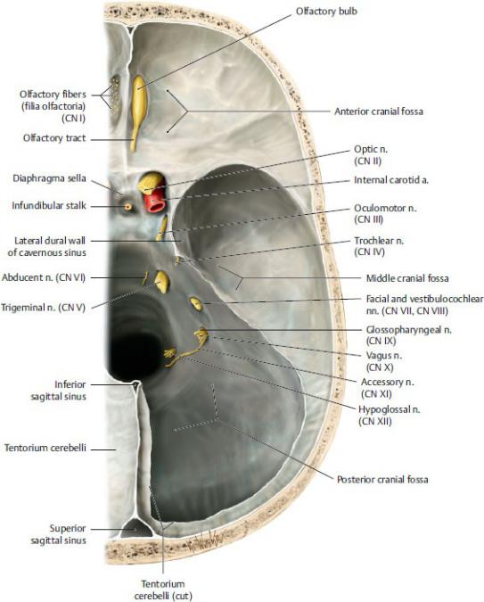
The cranial meninges, coverings that protect the brain, consist of the external fibrous dura mater, the thin intermediate arachnoid mater, and the delicate inner pia mater (Fig. 26.1).
composed of a periosteal (endosteal) layer and a meningeal layer. The two layers are inseparable except where they enclose the venous sinuses that drain the brain (e.g., the superior sagittal sinus; see Fig. 26.4).
•The outer periosteal layer, formed by the periosteum of the skull, adheres tightly to the inner surface of the skull, particularly at the sutures. This layer ends at the foramen magnum and is not continuous with the dura around the spinal cord.
•The inner meningeal layer, a strong membranous sheet that adheres to the inner surface of the periosteal layer, provides sheaths for the cranial nerves as they pass through the skull foramina. It is closely applied, although not attached, to the underlying arachnoid mater. It continues into the vertebral canal as dura of the spinal cord (Fig. 26.2).
—The middle meningeal arteries, branches of the maxillary arteries, supply most of the dura, with contributions from the ophthalmic, occipital, and vertebral arteries. Veins accompany the arteries and drain into the pterygoid venous plexus.
—Branches of the trigeminal nerve (cranial nerve [CN] V) transmit sensation from the dura of the anterior and middle cranial fossa. Spinal nerves C1, C2, and C3 and small branches of the vagus nerve (CN X) innervate the dura of the posterior cranial fossa.
Dural Partitions
Infoldings of the meningeal layer of the dura form incomplete membranous partitions that separate and support parts of the brain (Fig. 26.3).
—The falx cerebri, a vertical sickle-shaped partition separating the right and left cerebral hemispheres, is attached anteriorly to the crista galli and the inner crest of the frontal bone and is continuous posteriorly with the tentorium cerebelli. The inferior, free edge of the falx cerebri is unattached.
—The tentorium cerebelli, a horizontal continuation of the falx cerebri, separates the occipital lobes of the cerebrum from the cerebellar hemispheres in the posterior cranial fossae.
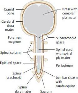
Fig. 26.2 Meninges in the cranial cavity and spinal cord
The two layers of the dura mater (periosteal and meningeal) form a single structural unit in the cranial cavity that adheres to the inner surface of the skull. In the spinal canal, however, the dura is separated from the periosteum of the vertebrae by the epidural space. (From Schuenke M, Schulte E, Schumacher U. THIEME Atlas of Anatomy, Vol 3. Illustrations by Voll M and Wesker K. 3rd ed. New York: Thieme Publishers; 2020.)
•It is attached to the posterior clinoid processes and the petrous part of the temporal bones anteriorly and to the parietal and occipital bones posterolaterally.
•A U-shaped tentorial notch separates the attachments to the petrous ridge on each side and connects the middle and posterior cranial fossae.
—The falx cerebelli, a vertical partition separating the cerebellar hemispheres, is continuous superiorly with the tentorium cerebelli and is attached posteriorly to the occipital crest.
—The diaphragma sellae, a small dural fold attached to the anterior and posterior clinoid processes, forms a roof over the sella turcica, which encloses the hypophysis (pituitary gland).
Fig. 26.3 Dural septa (folds)
Left anterior oblique view. (From Schuenke M, Schulte E, Schumacher U. THIEME Atlas of Anatomy, Vol 3. Illustrations by Voll M and Wesker K. 3rd ed. New York: Thieme Publishers; 2020.)
BOX 26.1: CLINICAL CORRELATION
TENTORIAL HERNIATION
Increased pressure within the middle cranial fossa created by edema or a space-occupying lesion such as a tumor can squeeze the brain tissue and force part of the temporal lobe to herniate through the tentorial notch. Pressure on the adjacent brainstem can be fatal in this situation. The oculomotor nerve (CN III) can also be stretched or damaged, leading to fixed pupil dilation (loss of parasympathetic function) and a “down and out” gaze due to paralysis of most of the extraocular muscles.
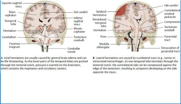
Potential sites of herniation beneath the free edges of the meninges
Coronal section, anterior view. (From Schuenke M, Schulte E, Schumacher U. THIEME Atlas of Anatomy, Vol 3. Illustrations by Voll M and Wesker K. 3rd ed. New York: Thieme Publishers; 2020.)
Dural Venous Sinuses
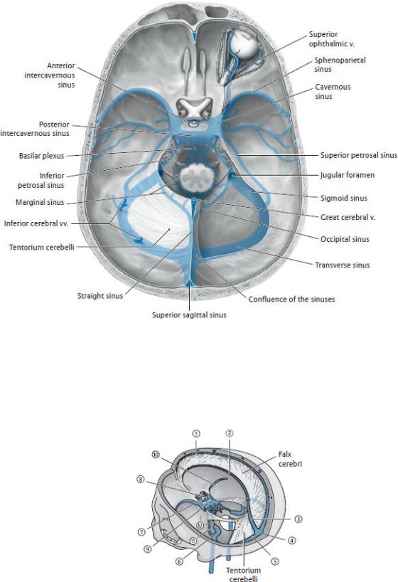
Dural venous sinuses are valveless venous spaces that form as a result of the separation of the periosteal and meningeal layers of the dura. Most of the large veins of the brain, skull, orbit, and inner ear drain through the dural sinuses and into the internal jugular veins in the neck (Figs. 26.4 and 26.5; Table 26.1).
—The confluence of sinuses at the posterior edge of the tentorium cerebelli is a junction of the superior sagittal, straight, occipital, and transverse sinuses.
—The superior sagittal sinus runs in the attached superior border of the falx cerebri and ends in the confluence of sinuses.
—The inferior sagittal sinus runs in the free inferior edge of the falx cerebri and ends in the straight sinus.
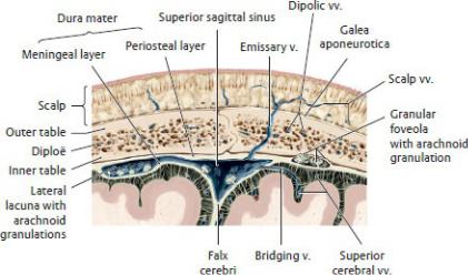
Fig. 26.4 Structure of a dural sinus
Superior sagittal sinus, coronal section, anterior view. (From Schuenke M, Schulte E, Schumacher U. THIEME Atlas of Anatomy, Vol 3. Illustrations by Voll M and Wesker K. 3rd ed. New York: Thieme Publishers; 2020.)
—The straight sinus runs in the space formed by the union of the falx cerebri and tentorium cerebelli. It receives the inferior sagittal sinus and great cerebral vein and drains into the confluence of sinuses.
—The paired transverse sinuses run along the attached posterolateral margins of the tentorium cerebelli. Posteriorly, they join at the confluence of sinuses, and anteriorly they drain into the sigmoid sinuses, forming grooves in the occipital and parietal bones along their course.
—The paired sigmoid sinuses run in deep grooves of the occipital and temporal bones and drain into the internal jugular veins at the jugular foramen.
—The occipital sinus runs in the free edge of the falx cerebelli and ends in the confluence of sinuses.

Publishers; 2020.)
Table 26.1 Principal Dural Sinuses
Upper Group |
Lower Group |
Superior sagittal sinus |
Cavernous sinus |
Inferior sagittal sinus |
Anterior intercavernous sinus |
Straight sinus |
Posterior intercavernous sinus |
Confluence of the sinuses |
Sphenoparietal sinus |
Transverse sinus |
Superior petrosal sinus |
Sigmoid sinus |
Inferior petrosal sinus |
Note: The occipital sinus is also included in the upper group.
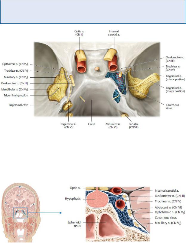
—The paired cavernous sinuses, located on either side of the sella turcica, have characteristics that distinguish them from other dural sinuses (Figs. 26.6 and 26.7).
•Each cavernous sinus contains a large plexus of thinwalled veins.
•Several important structures are associated with each cavernous sinus:
◦Internal carotid artery, which is surrounded by the sympathetic internal carotid nerve plexus
◦Oculomotor nerve (CN III)
◦Trochlear nerve (CN IV)
◦Ophthalmic and maxillary divisions (CN V1, V2) of the trigeminal nerve
◦Abducent nerve (CN VI)
BOX 26.2: CLINICAL CORRELATION
CAVERNOUS SINUS THROMBOPHLEBITIS
Cavernous sinus thrombophlebitis can occur secondary to thrombophlebitis of the facial vein. Although blood from the angle of the eye, lips, nose, and face usually drains inferiorly, it can also drain through the veins of the orbit to the cavernous sinus. Infections from the face, particularly from the danger zone of the face (which extends from the bridge of the nose to the angles of the mouth) can spread
MacPherson BR, Wikenheiser JC. Atlas of Anatomy. Illustrations by Voll M and Wesker K. 4th ed. New York: Thieme Publishers; 2020.)
•The cavernous sinuses receive the superior and inferior ophthalmic veins, the sphenoparietal sinuses, the superficial middle cerebral veins, and the central veins of the retina.
•The cavernous sinuses drain into the superior and inferior petrosal sinuses posteriorly and the pterygoid venous plexus inferiorly.
•Anterior and posterior intercavernous sinuses (see Fig. 26.5) connect the right and left cavernous sinuses.
—Paired superior petrosal sinuses, which drain the cavernous sinuses, travel within the attached margins of the tentorium cerebelli along the top of the petrous part of the temporal bones and empty into the sigmoid sinuses.
—Paired inferior petrosal sinuses drain the cavernous sinuses, passing through a groove between the petrous part of the temporal bones and the basilar part of the occipital bone and emptying into the sigmoid sinuses at the origin of the internal jugular veins. The inferior petrosal sinuses communicate, through a basilar plexus, with the vertebral venous plexus.
Arachnoid and Pia Mater (see Figs. 26.1, 26.4, 26.8)
—Arachnoid is a thin, avascular, fibrous layer underlying the meningeal layer of the dura.
•Cerebrospinal fluid in the subarachnoid space presses the arachnoid against the overlying dura, but the two layers are not attached. Weblike arachnoid trabeculae attach the arachnoid to the underlying pia mater.
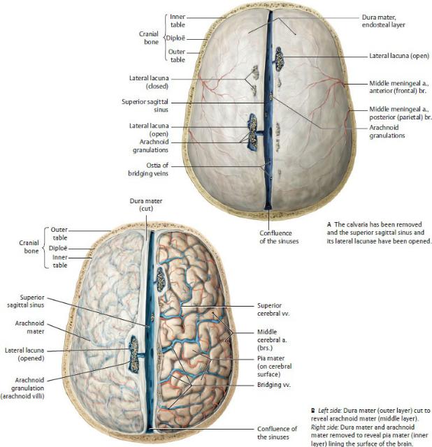
•Delicate fingers of the arachnoid layer, the arachnoid villi, pierce the dura to allow the reabsorption of cerebrospinal fluid into the venous circulation and are especially numerous in the superior sagittal sinus. They form aggregations called arachnoid granulations that protrude into the largest dural venous sinuses and can push the dura ahead of them into the parietal bone, forming “pits.”
•Congregations of arachnoid granulations also occur in lateral lacunae, lateral expansions of the superior sagittal sinus.
—Pia mater, or pia, is a thin, highly vascular layer that adheres to the surface of the brain and closely follows its contours.
Meningeal Spaces (Fig. 26.8)
—The epidural space between the cranium and dura is not a natural space because the dura adheres to the skull. Meningeal vessels that supply the
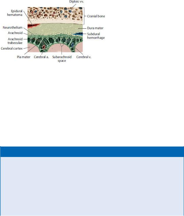
skull and dura travel in this space.
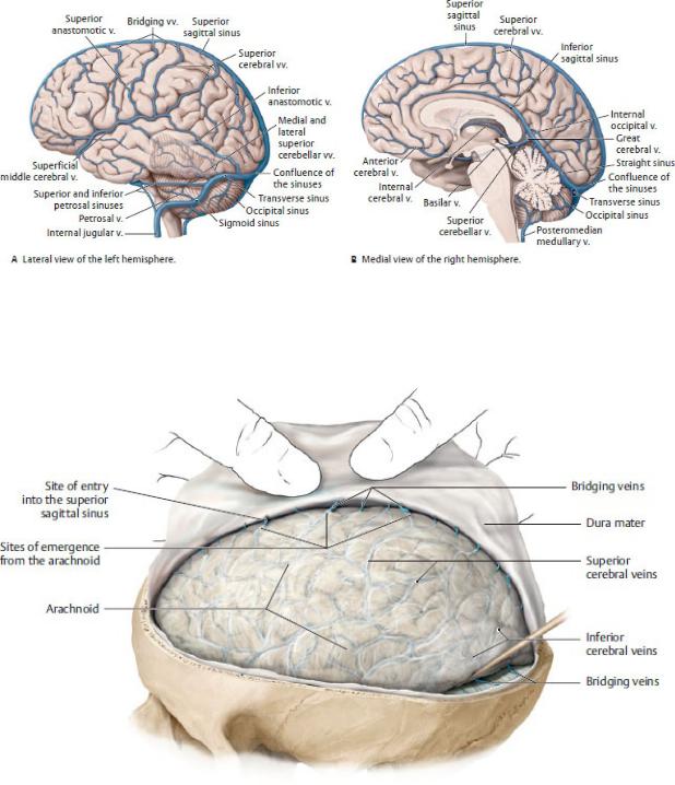
—The subdural space between the dura and arachnoid is a potential space, open only in pathologic conditions such as a subdural hematoma. Superficial cerebral veins (“bridging veins”) cross this space, connecting the venous circulation of the brain with the dural venous sinuses.
Fig. 26.8 Meningeal spaces
Meninges, coronal section, anterior view. (From Schuenke M, Schulte E, Schumacher U. THIEME Atlas of Anatomy, Vol 3. Illustrations by Voll M and Wesker K. 3rd ed. New York: Thieme Publishers; 2020.)
—The subarachnoid space, between the arachnoid and pia layers, contains cerebrospinal fluid, arteries, and veins.
•Subarachnoid cisterns are spaces that form where the subarachnoid space enlarges around large infoldings of the brain. The largest of these include the cerebellomedullary, pontomedullary, interpeduncular, chiasmatic, quadrigeminal, and ambient cisterns (see Section 26.2; see Fig. 26.11).
BOX 26.3: CLINICAL CORRELATION
EXTRACEREBRAL HEMORRHAGE
Bleeding from vessels between the bony skull and the brain (extracerebral hemorrhage) increases intracranial pressure and can damage brain tissue. Three types of cerebral hemorrhages are distinguished based on their relationship to the meningeal layers.
Epidural hemorrhages commonly originate from a torn middle meningeal artery following a skull fracture at the pterion and result in bleeding into the epidural space. The hemorrhagic spread is usually

—The cerebrum is the largest part of the brain and the center for integration within the central nervous system.
•The falx cerebri lies in a longitudinal fissure between the right and left cerebral hemispheres.
•Each cerebral hemisphere is further divided into frontal, parietal, occipital, and temporal lobes that occupy the anterior and middle cranial fossae.
•Posteriorly, the cerebrum rests on the tentorium cerebelli.
•The surface layer of the cerebrum (cortex) forms gyri (folds) separated by sulci (grooves).
—The diencephalon forms the central core of the brain and consists of the thalamus, hypophysis, and hypothalamus.
—The mesencephalon (midbrain), the most anterior part of the brainstem, passes through the tentorial notch between the middle and posterior cranial fossae.
•It is associated with the oculomotor (CN III) and trochlear (CN IV) nerves.
—The pons, the middle part of the brainstem, lies in the anterior part of the posterior cranial fossa below the mesencephalon.
Fig. 26.9 Adult brain
(From Gilroy AM, MacPherson BR, Wikenheiser JC. Atlas of Anatomy. Illustrations by Voll M and Wesker K. 4th ed. New York: Thieme Publishers; 2020.)
•Several ascending and descending fiber tracts connect the pons to the cerebellum.
•The pons is associated with the trigeminal (CN V), abducent (CN VI), and facial (CN VII) nerves.
—The medulla oblongata, the most posterior part of the brainstem, connects the brain and spinal cord.
•It contains nuclei for the vestibulocochlear (CN VIII), glossopharyngeal

(CN IX), vagus (CN X), and hypoglossal (XII) nerves.
—The cerebellum, which occupies most of the posterior cranial fossa, lies inferior to the cerebrum and is separated from it by the tentorium cerebelli.
• It consists of paired hemispheres and a small middle section, the vermis.
Ventricular System and Cerebrospinal Fluid
The brain and spinal cord are suspended in cerebrospinal fluid (CSF). The buoyant environment created by the CSF reduces the pressure of the brain on the nerves and vessels on its inferior surface.
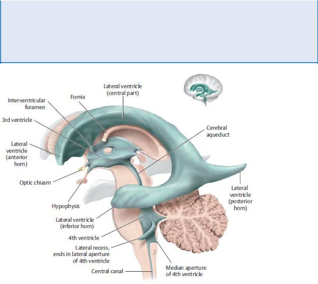
—CSF is produced in the choroid plexuses, vascular networks within four ventricles (spaces) of the brain. The first two of these ventricles are large and paired; the third and fourth are smaller and lie in the midline (Fig. 26.10).
•The 1st and 2nd (lateral) ventricles, paired cavities that occupy a large portion of each cerebral hemisphere, communicate with the 3rd ventricle through the interventricular foramina.
•The 3rd ventricle, a slitlike space between the two halves of the diencephalon, communicates posteriorly with the 4th ventricle through a narrow passage, the cerebral aqueduct, which passes through the mesencephalon.
•The 4th ventricle, a pyramidally shaped space that extends from the pons to the medulla oblongata, is continuous with the spinal canal inferiorly and with the subarachnoid space through the median and lateral apertures in its roof.
—CSF circulates through the ventricles and passes into the subarachnoid space and subarachnoid cisterns through the median and lateral apertures of the 4th ventricle. It flows superiorly through the fissures and sulci of the cerebrum and is reabsorbed into the venous circulation through the arachnoid granulations that protrude into the superior sagittal sinus (Fig. 26.11).
BOX 26.4: CLINICAL CORRELATION
HYDROCEPHALUS
Hydrocephalus, an excessive accumulation of cerebrospinal fluid (CSF) in the ventricles of the brain, can occur as a result of partial obstruction of the flow of CSF within the ventricular system, interference of CSF reabsorption into the venous circulation, or, in rare cases, overproduction of CSF. Excess CSF in the ventricles causes
them to dilate and exert pressure on the surrounding cortex, causing the bones of the calvaria to separate, thus creating the characteristic increase in head size. Treatment involves the placement of a shunt between the ventricles and the abdomen, which allows CSF to drain to the peritoneal cavity, where it can be easily absorbed.
Fig. 26.10 Ventricular system in situ
Ventricular system with neighboring structures, left lateral view. (From Schuenke M, Schulte E, Schumacher U. THIEME Atlas of Anatomy, Vol 3. Illustrations by Voll M and Wesker K. 3rd ed. New York: Thieme Publishers; 2020.)
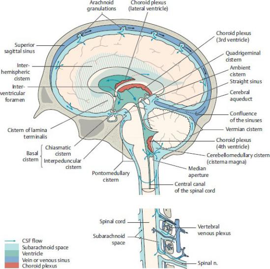
Fig. 26.11 Circulation of cerebrospinal fluid (CSF)
(From Schuenke M, Schulte E, Schumacher U. THIEME Atlas of Anatomy, Vol 3. Illustrations by Voll M and Wesker K. 3rd ed. New York: Thieme Publishers; 2020.)
Arteries of the Brain
As a result of its high metabolic demand, the brain receives one sixth of the cardiac output and one fifth of the oxygen consumed by the body at rest. This blood supply, derived from the internal carotid and vertebral arteries, is divided into anterior and posterior cerebral circulations (Fig. 26.12), which unite on the ventral surface of the brain to form a cerebral arterial circle (of Willis).
—The internal carotid artery supplies the anterior cerebral circulation (see Fig. 24.25).
• Its petrous part has a tortuous course as it enters the skull and follows

the carotid canal horizontally and medially within the temporal bone. Small branches pass into the middle ear and pterygoid canal.
•The cavernous part crosses over the foramen lacerum and runs anteriorly within the cavernous sinus (Fig. 26.13). Small branches supply the meninges, hypophysis, and cranial nerves within the cavernous sinus.
•The cerebral part in the middle cranial fossa gives off the ophthalmic artery (see Fig. 28.12) and immediately makes a U-turn to run posteriorly, where it divides into the anterior cerebral and middle cerebral arteries.
—The vertebral and basilar arteries supply the posterior cerebral circulation.
•The vertebral artery enters the skull through the foramen magnum and supplies branches to the spinal cord and cerebellum before merging with the opposite vertebral artery to form a single basilar artery.
◦Intracranial branches of the vertebral artery include the posterior inferior cerebellar artery and the anterior and posterior spinal arteries.
BOX 26.5: CLINICAL CORRELATION
STROKE
A stroke is the manifestation of a neurologic deficiency resulting from a cerebral vascular impairment. Ischemic strokes are usually caused by an embolus obstructing one of the major cerebral arteries. Although the vessels of the circle of Willis can provide collateral circulation to circumvent the obstruction, anastomoses between the vessels are often incomplete or of insufficient size to provide adequate flow. Hemorrhagic strokes are usually due to rupture of an aneurysm, most often a saccular, or berry, aneurysm that bleeds into the subarachnoid space. Symptoms occur shortly after the cerebral event and relate to the area of brain affected. They may include difficulty speaking, understanding language, or walking; vision problems; contralateral paralysis or numbness; and headache.
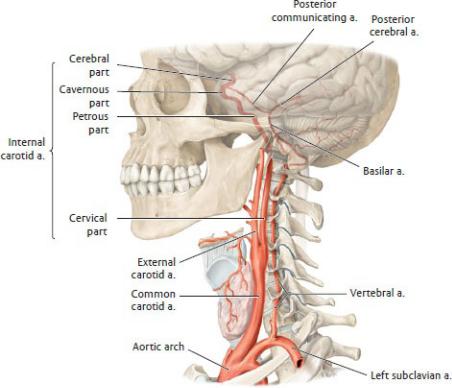
Fig. 26.12 Internal carotid artery
Left lateral view. (From Schuenke M, Schulte E, Schumacher U. THIEME Atlas of Anatomy, Vol 3. Illustrations by Voll M and Wesker K. 3rd ed. New York: Thieme Publishers; 2020.)
•The basilar artery ascends on the ventral surface of the brainstem, distributing branches to the brainstem, cerebellum, and cerebrum. It terminates as the right and left posterior cerebral arteries.
◦Major branches of the basilar artery are the anterior inferior cerebellar artery and the superior cerebellar artery.
—The cerebral arterial circle (of Willis), an important arterial anastomosis on the ventral surface of the brain, supplies the brain and connects the circulations of the internal carotid and vertebral arteries (Figs. 26.14 and
26.15).
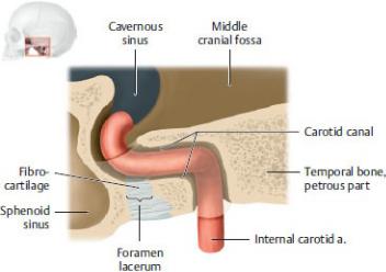
Fig. 26.13 The foramen lacerum and the internal carotid artery in the carotid canal
Left lateral view. The foramen lacerum is not a true aperture, being occluded in life by a layer of fibrocartilage; it appears as an opening only in the dried skull. It is closely related to the internal carotid artery that traverses the canal. (From Schuenke M, Schulte E, Schumacher U. THIEME Atlas of Anatomy, Vol 3. Illustrations by Voll M and Wesker K. 3rd ed. New York: Thieme Publishers; 2020.)
•A small anterior communicating artery connects the two anterior cerebral arteries, linking the right and left anterior cerebral circulations.
•A pair of posterior communicating arteries connects the internal carotid and posterior cerebral arteries on each side, completing the communication between the anterior and posterior cerebral circulations.
•The vessels that form the circle are
◦the anterior communicating arteries,
◦the anterior cerebral arteries,
◦the internal carotid arteries,
◦the posterior communicating arteries, and
◦the posterior cerebral arteries.
—The cerebral arteries that arise from the cerebral arterial circle provide the blood supply to the cerebral hemispheres (Table 26.2).
Table 26.2 Distribution of the Cerebral Arteries
Artery |
Origin |
Distribution |
Anterior cerebral |
Internal carotid artery |
Frontal pole and medial |
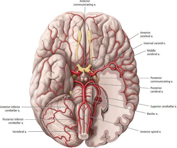
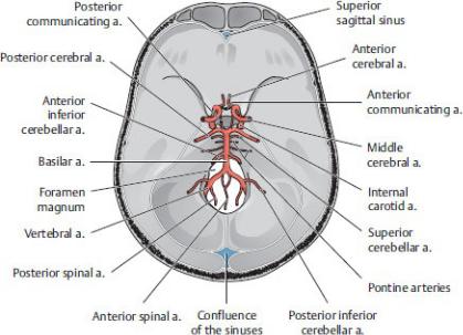
Fig. 26.15 Projection of the circle of Willis onto the base of the skull
Superior view. (From Schuenke M, Schulte E, Schumacher U. THIEME Atlas of Anatomy, Vol 3. Illustrations by Voll M and Wesker K. 3rd ed. New York: Thieme Publishers; 2020.)
Veins of the Brain
Veins that drain the brain are thin-walled and valveless and usually drain into one of the dural venous sinuses (Fig. 26.16).
—Superficial (external) veins that drain the cerebral hemispheres include
•the superior cerebral veins, which drain the supralateral and medial aspects. These “bridging veins” traverse the subdural space and drain into the superior sagittal sinus (Fig. 26.17);
•the middle cerebral veins, which drain the lateral hemispheres and empty into the cavernous sinus and from there into the petrosal and transverse sinuses; and
•the inferior cerebral veins, which drain inferior aspects of the brain and join either the superior cerebral or the basilar veins.
—Basilar veins drain the small anterior cerebral veins and deep middle cerebral veins.
—Internal cerebral veins drain the 3rd and 4th ventricles and deep parts of the cerebrum. These unite to form the great cerebral vein.
—The great cerebral vein receives the basilar veins and merges with the inferior sagittal sinus to form the straight sinus.
—Superior and inferior cerebellar veins drain the cerebellum into adjacent dural sinuses or superficially into the great cerebral vein.
hemorrhage. (From Schuenke M, Schulte E, Schumacher U. THIEME Atlas of Anatomy, Vol 3. Illustrations by Voll M and Wesker K. 3rd ed. New York: Thieme Publishers; 2020.)
26.3 Cranial Nerves
The 12 cranial nerves arise from the base of the brain (Figs. 26.18 and 26.19; Tables. 26.3 and 26.4). Like spinal nerves, cranial nerves can stimulate muscles or transmit sensation from a peripheral structure to the central nervous system. Some cranial nerves also carry fibers from the cranial portion of the parasympathetic nervous system. Seven types of nerve fibers are found (alone or in combination) in cranial nerves.
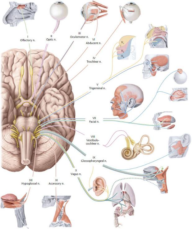
Fig. 26.18 Cranial nerves
Inferior (basal) view. The 12 pairs of cranial nerves (CN) are numbered according to their emergence from the brainstem. (See Table 26.3 for explanation of color coding.) (From Gilroy AM, MacPherson BR, Wikenheiser JC. Atlas of Anatomy. Illustrations by Voll M and Wesker K. 4th ed. New York:
2020.)
Table 26.3 Classification of Cranial Nerve Fibers
Fiber Type |
Function |
General somatomotor |
○ Innervate voluntary muscle |
|
|
General visceromotor |
○ Constitute the cranial component of the |
(parasympathetic) |
parasympathetic system, innervate |
|
involuntary muscles and glands |
|
|
Special visceromotor |
○ Innervate muscles that developed from |
(branchiomotor) |
the primitive pharynx (pharyngeal arches) |
|
|
General somatosensory |
○ Carry sensations such as touch, |
|
temperature, pain, and pressure |
|
|
Special somatosensory |
○ Carry impulses from the eye for sight |
|
and from the ear for hearing and balance |
|
|
General viscerosensory |
○ Transmit information from viscera such |
|
as carotid bodies, the heart, esophagus, |
|
trachea, and gastrointestinal tract |
|
|
Special viscerosensory |
○ Transmit information regarding smell |
|
and taste |
|
|
See Fig. 26.18 for explanation of color coding.
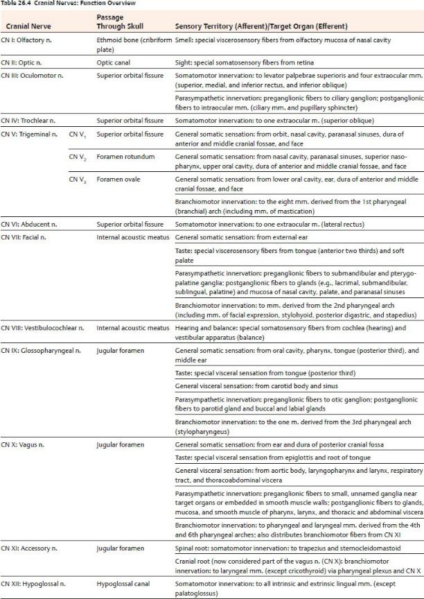
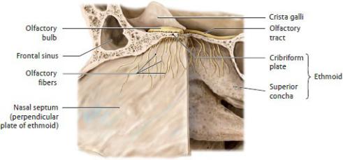
Fig. 26.20 Olfactory nerve (CN I)
Olfactory fibers, bulb, and tract. Portion of left nasal septum and lateral wall of right nasal cavity, left lateral view. (From Schuenke M, Schulte E, Schumacher U. THIEME Atlas of Anatomy, Vol 3. Illustrations by Voll M and Wesker K. 3rd ed. New York: Thieme Publishers; 2020.)
CN I, the olfactory nerve, carries special sensory fibers that transmit sensation of smell from the superior aspect of the lateral and septal walls of the nasal cavity (Fig. 26.20).
—Olfactory neurons pass through the cribriform plate of the ethmoid bone and synapse with secondary neurons in the olfactory bulbs.
•The axons of these secondary neurons form the olfactory tracts.
•The olfactory bulbs and tracts are extensions of the cerebral cortex.
CN II, the optic nerve, is a collection of special sensory nerve fibers that originate on the retina of the eye and converge at the optic disk at the back of the eyeball (Fig. 26.21; see also Section 28.1).
—The nerve exits the orbit through the optic canal and joins the contralateral optic nerve to form the optic chiasm.
—The optic chiasm is a redistribution center where nerve fibers from the medial half of each optic nerve cross to the opposite side.
—Two optic tracts diverge from the chiasm. Each tract contains nerve fibers from the medial half of one eye and the lateral half of the other eye.
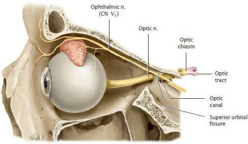
Fig. 26.21 Optic nerve (CN II)
Optic nerve in the left orbit, left lateral view. (From Schuenke M, Schulte E, Schumacher U. THIEME Atlas of Anatomy, Vol 3. Illustrations by Voll M and Wesker K. 3rd ed. New York: Thieme Publishers; 2020.)
CN III, the oculomotor nerve; CN IV, the trochlear nerve; and CN VI, the abducent (abducens) nerve innervate structures of the orbit (Fig. 26.22; see also Section 28.1). They pass through the cavernous sinus before entering the orbit through the superior orbital fissure.
—The oculomotor nerve has somatic and visceral components.
•General somatic motor fibers innervate four of the extraocular muscles (superior rectus, medial rectus, inferior rectus, and inferior oblique), which move the eyeball, and the levator palpebrae superioris muscle, which elevates the eyelid.
•General visceral motor fibers carry preganglionic parasympathetic fibers that synapse in the ciliary ganglion and innervate the pupillary sphincter muscle (which constricts the pupil) and ciliary body (which changes the curvature of the lens of the eye) (see Fig. 28.7).
—The trochlear nerve carries general somatic motor fibers and innervates the superior oblique muscle, which depresses and medially rotates the eye.
—The abducent nerve carries general somatic motor fibers and innervates the lateral rectus muscle, which abducts the eye.
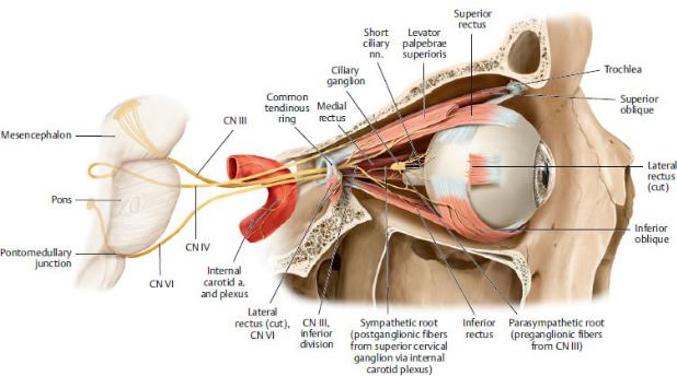
Fig. 26.22 Oculomotor (CN III), trochlear (IV), and abducent (VI) nerves
Course of the nerves innervating the extraocular muscles, right orbit, lateral view. (From Gilroy AM, MacPherson BR, Wikenheiser JC. Atlas of Anatomy. Illustrations by Voll M and Wesker K. 4th ed. New York: Thieme Publishers; 2020.)
CN V, the trigeminal nerve, is the primary sensory nerve of the face (Figs. 26.23 and 26.24). Its small motor component innervates the muscles of mastication.
—The general somatic sensory neurons, which form the sensory root, synapse in sensory nuclei that are distributed along the brainstem and down into the cervical spinal cord.
—A small motor root in the mandibular division (CN V3) contains branchial motor fibers.
—Branches of the trigeminal nerve are spatially associated with the parasympathetic ganglia of the head and distribute postganglionic parasympathetic fibers to their target organs.
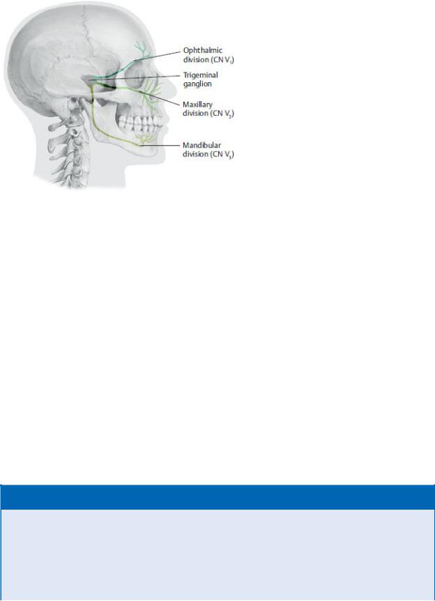
Fig. 26.23 Divisions of the trigeminal nerve
Right lateral view. (From Schuenke M, Schulte E, Schumacher U. THIEME Atlas of Anatomy, Vol 3. Illustrations by Voll M and Wesker K. 3rd ed. New York: Thieme Publishers; 2020.)
—The trigeminal nerve has three divisions:
1.The ophthalmic division (CN V1) (see Section 28.1)
◦contains only somatic sensory fibers;
◦passes through the wall of the cavernous sinus and superior orbital fissure into the orbit;
◦is associated with the ciliary ganglion (see Section 24.4);
◦distributes visceral motor fibers from the facial nerve (CN VII) to the lacrimal gland via the lacrimal nerve;
◦innervates the orbit, the cornea, and the skin on the top of the nose, forehead, and scalp;
◦functions as the sensory component of the corneal reflex via the nasociliary branch; and
◦has lacrimal, frontal, and nasociliary branches.
BOX 26.6: CLINICAL CORRELATION
TRIGEMINAL NEURALGIA
Trigeminal neuralgia, a pathology of the sensory root of the trigeminal nerve (CN V), most commonly affects the maxillary division (CN V2) and least frequently affects the ophthalmic division (CN V1). The disorder is characterized by unilateral electric shock-like pain in the

area supplied by the nerve. It usually lasts several seconds to several minutes. As the condition progresses, the pain may last longer, and there may be a shorter period between attacks. The pain may be initiated by touching a trigger point in the face by eating, talking, brushing teeth, or shaving. It is believed that trigeminal neuralgia is caused by the loss of myelin on the sensory root due to the pressure from an abnormal blood vessel. Surgery to destroy the nerve root or ganglion may be effective but may lead to permanent facial numbness.
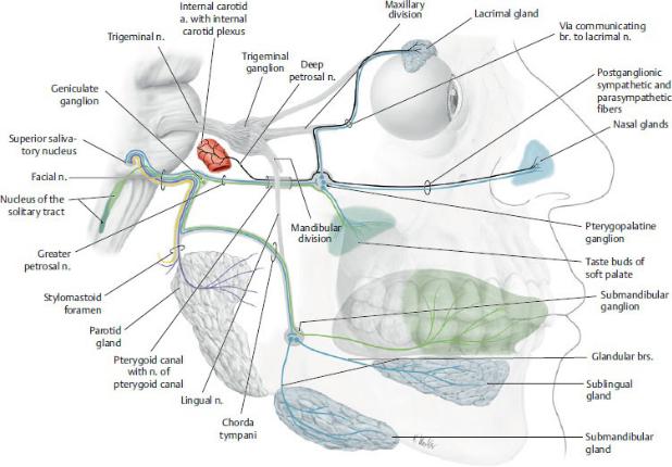
sensory and visceral components (Figs. 26.25, 26.26, 26.27). It contains a motor root that innervates the muscles of facial expression, and an intermediate nerve that carries special sensory (for taste) and visceral motor fibers (parasympathetic) and somatic sensory fibers. Both the motor root and the intermediate nerve pass through the internal acoustic meatus into the facial canal of the temporal bone.
Fig. 26.25 Course of the facial nerve
Visceral motor (parasympathetic) and special visceral sensory (taste) fibers shown in blue and green respectively. Postganglionic sympathetic fibers are shown in black, right lateral view. (From Gilroy AM, MacPherson BR, Wikenheiser JC. Atlas of Anatomy. Illustrations by Voll M and Wesker K. 4th ed. New York: Thieme Publishers; 2020.)
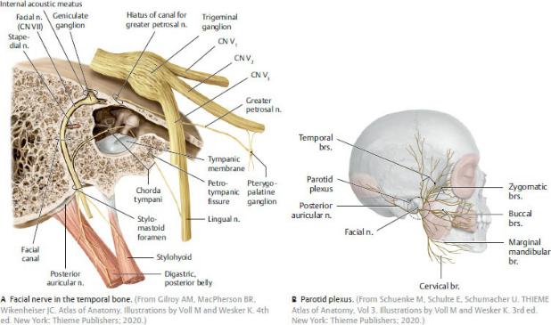
Fig. 26.26 Facial nerve (CN VII)
Branches of the facial nerve, right lateral view.
—The motor root
•exits the skull through the stylomastoid foramen
•contains branchiomotor fibers, which
◦innervate the stylohyoid, stapedius, and digastric (posterior belly) muscles (see Figs. 27.28 and 27.29);
◦form most of the posterior auricular nerve, which innervates the posterior auricular muscles and the posterior belly of the occipitofrontalis; and
◦form the nerves of the intraparotid plexus within the parotid gland, which innervates the muscles of facial expression (see Section 27.1). Branches of the parotid plexus include the temporal, zygomatic, buccal, marginal mandibular, and cervical branches.
—The three branches of the intermediate nerve (nervus intermedius) arise within the facial canal, including
•the greater petrosal nerve (parasympathetic), which passes through the middle cranial fossa and combines with the deep petrosal nerve (sympathetic) to form the nerve of the pterygoid canal (see Section 27.6). The visceral motor (parasympathetic) fibers synapse in the pterygopalatine ganglion and are distributed to glands of the nasal

mucosa and palate and to the lacrimal gland.
•the chorda tympani, which passes through the middle ear cavity, exits through the petrotympanic fissure to the infratemporal fossa and travels with the lingual nerve of CN V3. It carries
◦visceral motor fibers that synapse in the submandibular ganglion and supply the submandibular and sublingual salivary glands, and
◦special visceral sensory fibers for taste from the anterior part of the tongue and palate.
•general somatic sensory fibers carried by the posterior auricular nerve, which transmit sensations from the external ear to the geniculate ganglion, the sensory ganglion of the facial nerve, located in the temporal bone.
BOX 26.7: CLINICAL CORRELATION
BELL’S PALSY
Bell’s palsy is paralysis of the facial muscles due to a lesion of the facial nerve (CN VII). Symptoms usually begin suddenly and affect one side of the face only. They include drooping of the corner of the mouth, eyebrow, and lower eyelid, and the inability to smile, whistle, blow out cheeks, wrinkle the forehead, blink, or close the eyes forcefully. Taste is impaired on the anterior two thirds of the tongue (due to involvement of the chorda tympani), decreased tear production leads to dry eyes (due to involvement of the greater petrosal nerve), sensitivity to sounds is increased (due to paralysis of the stapedius), and the lower jaw and tongue deviate to the opposite side (due to paralysis of the posterior belly of the digastric muscle).
BOX 26.8: CLINICAL CORRELATION
(From Schuenke M, Schulte E, Schumacher U. THIEME Atlas of Anatomy, Vol 3. Illustrations by Voll M and Wesker K. 3rd ed. New York: Thieme Publishers; 2020.
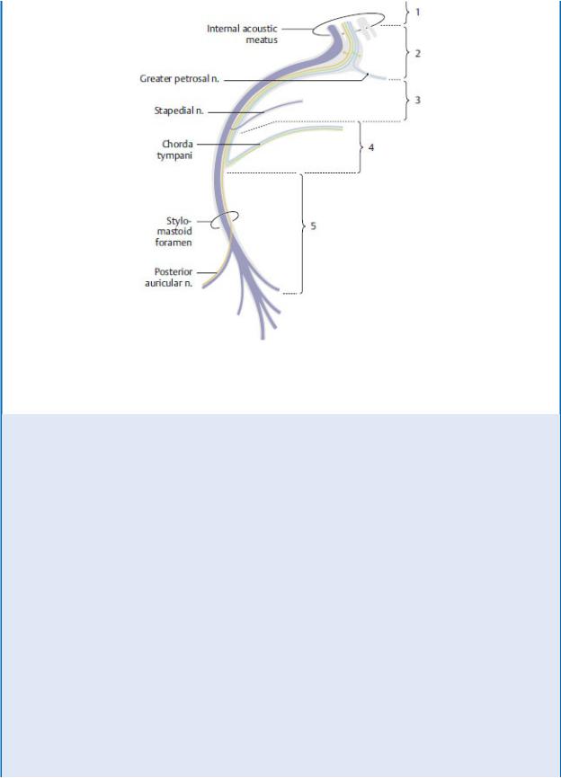
BRANCHING PATTERN OF THE FACIAL NERVE: DIAGNOSTIC SIGNIFICANCE IN TEMPORAL BONE FRACTURES
Blue: visceral motor (parasympathetic); purple: branchial motor; yellow: general somatic sensory; green: special visceral sensory. The principal signs and symptoms are different depending upon the exact site of the lesion in the course of the facial nerve through the petrous bone.
Note: Only the principal signs and symptoms associated with a particular lesion site are described here. The more peripheral the site of the nerve injury, the less diverse the signs and symptoms become.
1 A lesion at this level affects the facial nerve in addition to the vestibulochochlear nerve. As a result, peripheral motor facial paralysis is accompanied by hearing loss (deafness) and vestibular dysfunction (dizziness).
2 Peripheral motor facial paralysis is accompanied by disturbances of taste sensation (chorda tympani), lacrimation, and salivation.
3 Motor paralysis is accompanied by disturbances of salivation and
taste. Hyperacusis results from paralysis of the stapedius muscle.
4 Peripheral motor paralysis is accompanied by disturbances of taste and salivation.
5 Peripheral motor (facial) paralysis is the only manifestation of a lesion at this level.
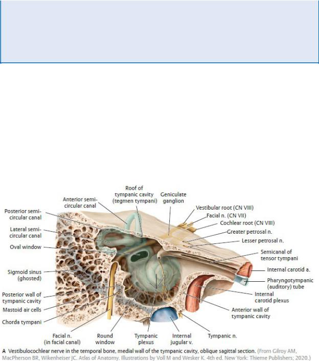
CN VIII, the vestibulocochlear nerve, is the sensory nerve of hearing and balance. The nerve enters the temporal bone with the facial nerve through the internal acoustic meatus.
—The two branches of the vestibulocochlear nerve carry special sensory fibers (Fig. 26.27; see Section 28.2):
•The cochlear root supplies the cochlea and its spiral organ, the organ of hearing.
•The vestibular root, which contains the vestibular ganglia, supplies the utricle, saccule, and semicircular ducts, the organs of balance.
Fig. 26.27 Vestibulocochlear nerve (CN VIII)
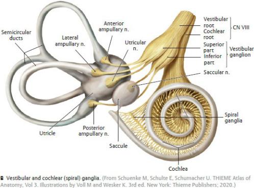
Fig. 26.27 (continued) Vestibulocochlear nerve (CN VIII)
CN IX, the glossopharyngeal nerve, leaves the skull through the jugular foramen and contains special sensory (taste), visceral sensory, somatic motor, and visceral motor components (Figs. 26.28 and 26.29; Table 26.5).
—Somatic motor fibers innervate the stylopharyngeus muscle.
—Visceral motor fibers arise with the tympanic nerve, a branch of the glossopharyngeal nerve. Carrying sensory and visceral motor fibers, it runs through the tympanic cavity of the middle ear (see Section 28.2), where it contributes to the tympanic plexus. It gives rise to the lesser petrosal nerve.
◦The lesser petrosal nerve passes through the middle cranial fossa and foramen ovale carrying visceral motor (preganglionic parasympathetic) fibers that synapse in the otic ganglion. Postganglionic fibers travel with the auriculotemporal nerve (CN V3) to innervate the parotid gland.
◦Sensory fibers of the tympanic plexus supply the tympanic cavity and pharyngotympanic (auditory) tube.
—Special sensory fibers transmit taste from the posterior third of the tongue.
—Visceral sensory fibers transmit information from the tonsils, soft palate, posterior third of the tongue, pharynx, and, via the branch to the carotid sinus, from receptors in the carotid body and carotid sinus at the bifurcation of the common carotid artery.

BOX 26.9: ANATOMIC NOTES
PETROSAL NERVES OF THE HEAD
Three petrosal nerves are associated with autonomic innervation of the head. Two nerves carry preganglionic parasympathetic nerves:
—The greater petrosal nerve, a branch of CN VII, forms the parasympathetic part of the nerve of the pterygoid canal, which synapses in the pterygopalatine ganglion. Postganglionic fibers travel via the zygomatic n. (CN V2) to the lacrimal n. (CN V1) in the orbit where they innervate the lacrimal gland. They also innervate glands in the nasal cavity via branches of the maxillary nerve (CN V2).
—The lesser petrosal nerve, a branch of CN IX, arises from the tympanic plexus in the middle ear and synapses in the otic ganglion. Postganglionic fibers travel briefly with the auriculotemporal n. (CN V3) before innervating the parotid gland.
One nerve carries postganglionic sympathetic fibers:
—The deep petrosal nerve arises from the internal carotid plexus and forms the sympathetic component of the nerve of the pterygoid canal. These fibers pass through the pterygopalatine fossa without synapsing in the ganglion, and are distributed to the lacrimal gland and nasal glands along the same routes as the greater petrosal nerve.
Table 26.5 Glossopharyngeal Nerve Branches
|
Tympanic n. |
|
Br. to carotid sinus |
|
Br. to stylopharyngeus muscle |
|
Tonsillar brs. |
|
Lingual brs. |
|
Pharyngeal brs. |
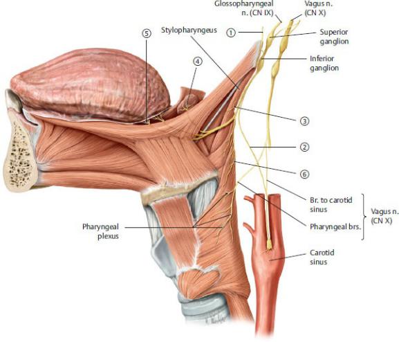
Fig. 26.28 Glossopharyngeal nerve (CN IX)
Course of the glossopharyngeal nerve, left lateral view. The numbers are explained in Table 26.5. (From Schuenke M, Schulte E, Schumacher U. THIEME Atlas of Anatomy, Vol 3. Illustrations by Voll M and Wesker K. 3rd ed. New York: Thieme Publishers; 2020.)
Fig. 26.29 Branches of the glossopharyngeal nerve
(From Schuenke M, Schulte E, Schumacher U. THIEME Atlas of Anatomy, Vol 3. Illustrations by Voll M and Wesker K. 3rd ed. New York: Thieme Publishers; 2020.)
CN X, the vagus nerve, has the most extensive distribution of the cranial nerves (Fig. 26.30; Table 26.6).
—Branchial motor fibers innervate the muscles of the soft palate (except tensor veli palatini), pharynx (except stylopharyngeus), and larynx, and the palatoglossus muscle of the tongue.
—Visceral motor fibers innervate smooth muscle and glands of the pharynx, larynx, thoracic organs, and abdominal foregut and midgut.

—General somatic sensory fibers transmit sensation from the dura in the posterior cranial fossa, the skin of the external ear, and the external auditory canal.
—Visceral sensory fibers transmit sensation from mucosa of the lower pharynx, the larynx, lungs and airway, the heart, the abdominal foregut and midgut, and the chemoreceptors of the aortic body and baroreceptors of the aortic arch.
—Special sensory fibers carry taste from the epiglottis.
—The vagus nerve has cervical, thoracic, and abdominal segments.
•In the neck
◦each vagus nerve leaves the skull through the jugular foramen and descends within the carotid sheath of the neck, and
◦its branches are pharyngeal branches, the superior laryngeal nerve, cervical cardiac (parasympathetic) branches, and the right recurrent laryngeal nerve (which arises from the right vagus and recurs around the right subclavian artery).
Table 26.6 Vagus Nerve Branches in the Neck
|
Pharyngeal brs. |
|
Superior laryngeal n. |
3R |
Right recurrent laryngeal n. |
|
|
3L |
Left recurrent laryngeal n. |
|
|
|
Cervical cardiac brs. |
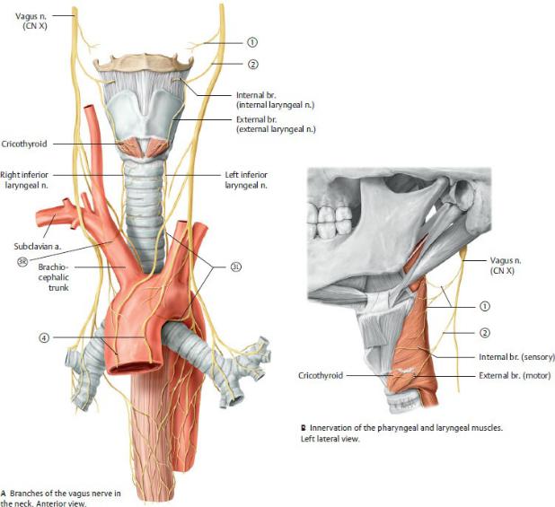
Fig. 26.30 Vagus nerve (CN X)
The numbers are explained in Table 26.6. (From Schuenke M, Schulte E, Schumacher U. THIEME Atlas of Anatomy, Vol 3. Illustrations by Voll M and Wesker K. 3rd ed. New York: Thieme Publishers; 2020.)
•In the thorax
◦the right and left vagus nerves enter the thorax posterior to the sternoclavicular joints and merge on the surface of the esophagus as the esophageal plexus (see Section 5.2), and
◦their branches are the left recurrent laryngeal nerve (which arises from the left vagus and recurs around the arch of the aorta, where it becomes known as the inferior laryngeal nerve) and the thoracic cardiac and pulmonary branches (parasympathetic).

•In the abdomen
◦right and left vagal trunks arise from the esophageal plexus and pass through the esophageal hiatus of the diaphragm as the anterior and posterior vagal trunks; and
◦their parasympathetic branches are distributed to organs of the foregut, midgut, and retroperitoneum.
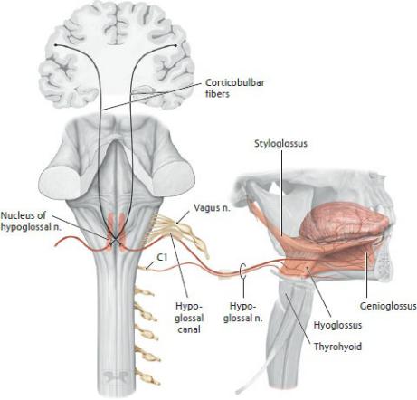
CN XI, the accessory nerve, contains general somatic motor fibers, which originate in a nucleus of the upper segments of the spinal cord (Fig. 26.31).
—The nerve emerges with the upper five or six cervical spinal nerves and ascends within the vertebral canal. It enters the skull through the foramen magnum and exits through the jugular foramen with the vagus (CN X) and glossopharyngeal (CN IX) nerves.
—It innervates the sternocleidomastoid muscle and then crosses the lateral region of the neck to innervate the trapezius muscle.
—Traditionally this nerve was believed to have a spinal root, as described above, and a cranial root, arising from the nucleus ambiguous in the
medulla. The two roots travel together through the jugular foramen before the cranial root splits off to join the vagus nerve. Current thinking distinguishes the cranial root as part of the vagus nerve; the spinal root is now considered to be the accessory nerve (CN XI).
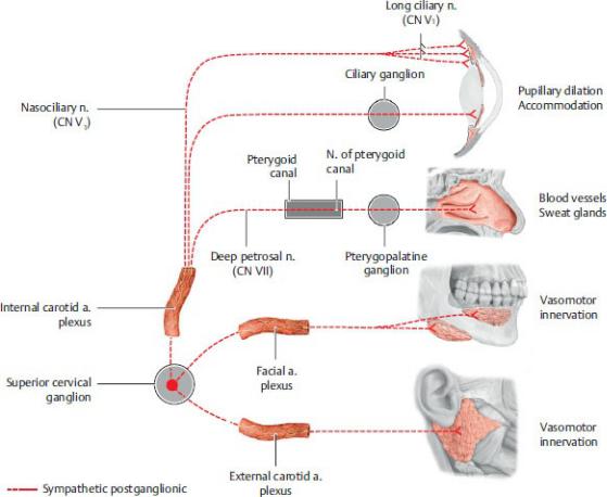
CN XII, the hypoglossal nerve, contains only general somatic motor fibers (Fig. 26.32).
—The hypoglossal nerve leaves the skull through the hypoglossal canal and runs forward, medial to the angle of the mandible, to enter the oral cavity.
—It innervates all of the muscles of the tongue except the palatoglossus.
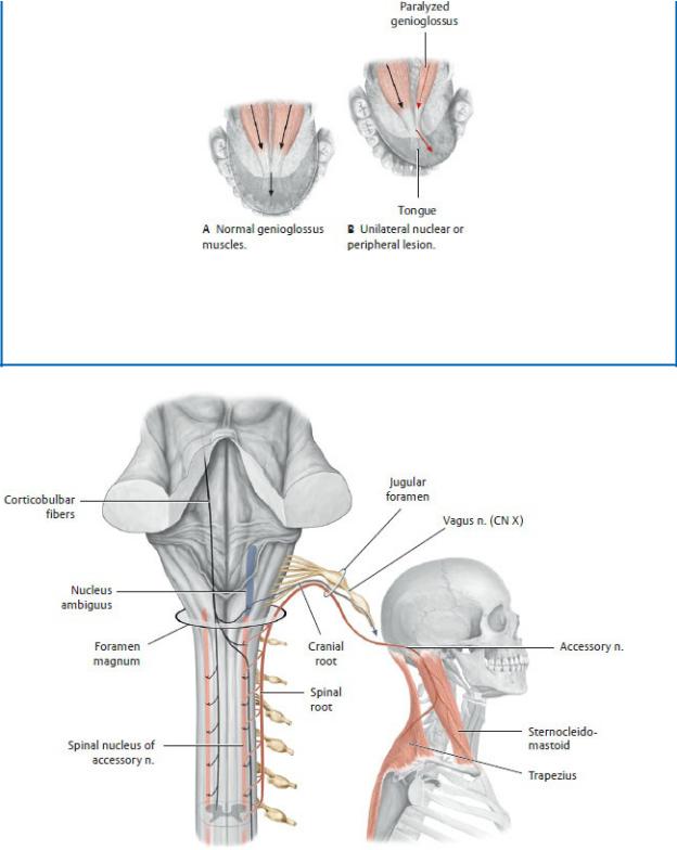
BOX 26.10: CLINICAL CORRELATION
INJURY TO THE HYPOGLOSSAL NERVE
Injury to the hypoglossal nerve causes ipsilateral paralysis of half of the tongue. When the tongue is protruded, the tip deviates toward the paralyzed side because the action of the genioglossus muscle on the unaffected side is unopposed. Symptoms mainly manifest as slurring of speech. Over time, the tongue becomes weak and atrophies.
Voll M and Wesker K. 3rd ed. New York: Thieme Publishers; 2020.)
Fig. 26.32 Hypoglossal nerve (CN XII)
Brainstem with the cerebellum removed, posterior view. Note: C1, which innervates the thyrohyoid and geniohyoid, runs briefly with the hypoglossal nerve. (From Schuenke M, Schulte E, Schumacher U. THIEME Atlas of Anatomy, Vol 3. Illustrations by Voll M and Wesker K. 3rd ed. New York: Thieme Publishers; 2020.)
26.4 Autonomic Nerves of the Head
—Sympathetic nerves of the head arise as postganglionic fibers from the superior cervical ganglia (Fig. 26.33; Table 26.7; see Section 25.4).
•The internal carotid plexus of sympathetic fibers surrounds the internal carotid artery and its branches within the skull. A similar external carotid plexus follows the branches of the external carotid artery on the face.
•Sympathetic fibers often travel with the parasympathetic nerves, but
they do not synapse in the parasympathetic ganglia.
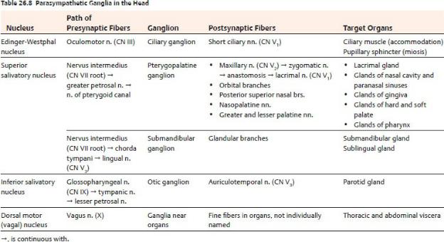
—The cranial portion of the parasympathetic (visceral motor) system is associated with the oculomotor (CN III), facial (CN VII), glossopharyngeal (CN IX), and vagus (CN X) nerves (Fig. 26.34; Table 26.8).
•Preganglionic parasympathetic fibers traveling with the oculomotor (CN III), facial (CN VII), and glossopharyngeal (CN IX) nerves synapse in the four parasympathetic ganglia of the head: the ciliary, pterygopalatine, submandibular, and otic ganglia.
•Parasympathetic nerves traveling with the vagus nerve (CN X) extend into the thorax and abdomen and synapse in ganglia of nerve plexuses in those regions.
•Parasympathetic ganglia of the head are usually attached to, or in close association with, a branch of the trigeminal nerve (CN V). The postganglionic parasympathetic fibers travel to their target organ by “piggybacking” on these trigeminal branches.
Fig. 26.33 Sympathetic innervation of the head
Sympathetic preganglionic fibers of the head originate in the lateral horn of the
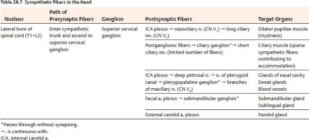
T1–T3 spinal cord. They exit into the sympathetic trunk and ascend to synapse in the superior cervical ganglion. Postganglionic fibers then travel with arterial plexuses (internal carotid a., facial a., and external carotid a.). Although these fibers often travel with parasympathetic fibers through the parasympathetic ganglia, they do not synapse in these ganglia. Similar to parasympathetic fibers, sympathetic nerves may “piggyback” on branches of the trigeminal nerve (CN V) to reach their target organ. (From Schuenke M, Schulte E, Schumacher U. THIEME Atlas of Anatomy, Vol 3. Illustrations by Voll M and Wesker K. 3rd ed. New York: Thieme Publishers; 2020.)
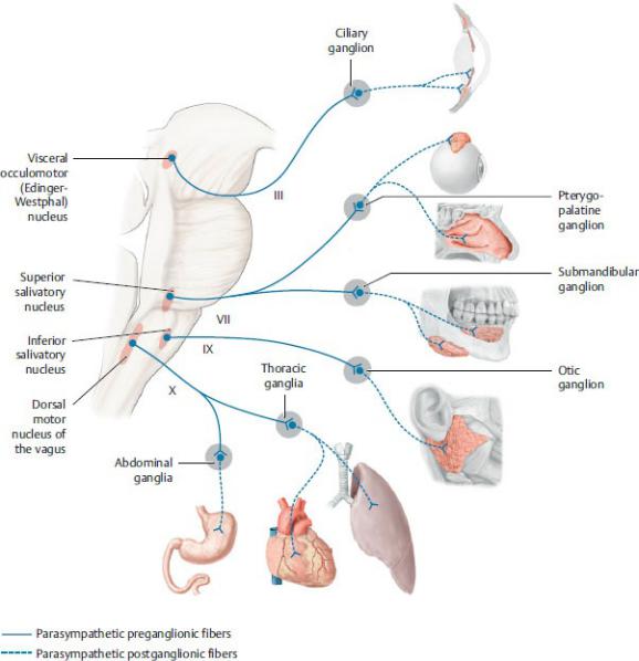
Fig. 26.34 Parasympathetic nervous system (cranial part): Overview
There are four parasympathetic nuclei in the brainstem. The visceral motor fibers of these nuclei travel along specific cranial nerves as shown. The postganglionic fibers often travel with branches of the trigeminal nerve (CN V) to reach their target organs. (From Gilroy AM, MacPherson BR, Wikenheiser JC. Atlas of Anatomy. Illustrations by Voll M and Wesker K. 4th ed. New York: Thieme Publishers; 2020.)
27 Anterior, Lateral, and Deep Regions of the
Head
The anatomy of the head can be divided into smaller regions that lie anterior and lateral to the neurocranium and form the superficial and deep structures of the face. They include the scalp; the parotid region; the temporal, infratemporal, and pterygopalatine fossae; and the nasal and oral cavities.
27.1 The Scalp and Face
The scalp covers the neurocranium and extends from the superior nuchal lines of the occipital bone, which mark the superior limit of the neck, to the supraorbital ridge of the frontal bone. The face extends from the forehead to the chin and to the ears on either side.
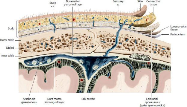
—The scalp is composed of five layers (Fig. 27.1):
•Skin
•Connective tissue containing the vessels of the scalp
•Aponeurosis of the occipitofrontalis, temporoparietalis, and superior auricular muscles (gala aponeurotica)
•Loose areolar tissue
•Periosteum of the skull (pericranium)
Note that the first letter of each layer spells “SCALP”—a handy mnemonic.
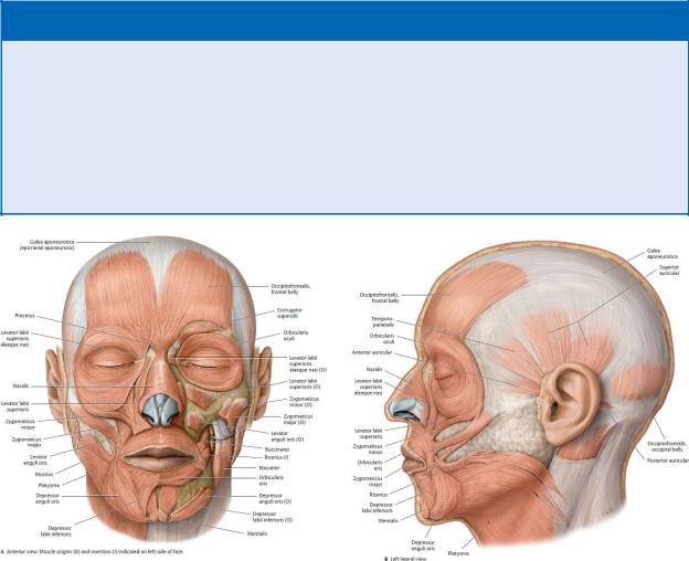
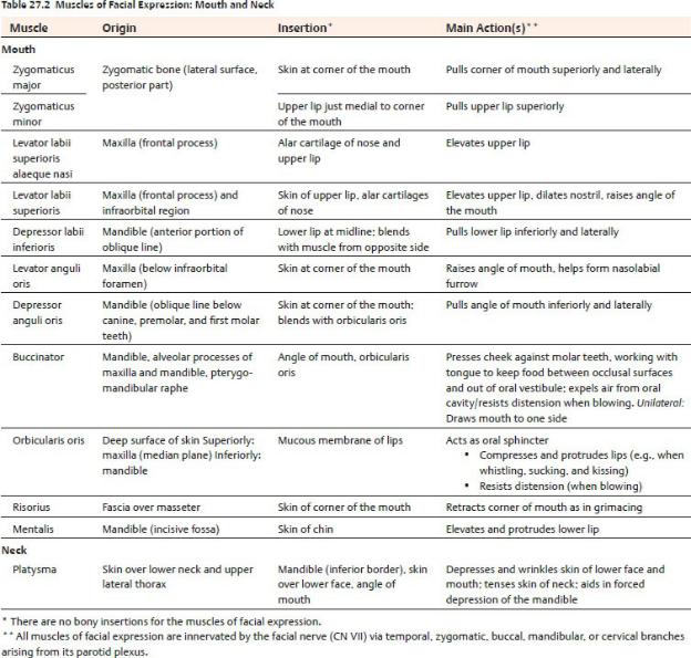
—Muscles of facial expression lie in the loose connective tissue layer of the face and scalp. Their origin on bones of the face and insertion into the overlying skin allows them to create facial movements (Fig. 27.2; Tables 27.1 and 27.2).
—Most arteries that supply the face and scalp are branches of the external carotid artery (see Fig. 24.21) and include the following:
• The superior and inferior labial arteries, the lateral nasal artery, and the angular branches of the facial artery, which supply the face between the eye and lower lip
• The mental branch of the inferior alveolar artery, which supplies the chin
•The superficial temporal, posterior auricular, and occipital arteries, which supply the lateral and posterior parts of the scalp
•The supratrochlear and supraorbital branches of the ophthalmic artery (a branch of the internal carotid artery), which supply the anterior scalp. These arteries anastomose with the angular artery on the face and form a connection between internal carotid and external carotid circulations (see Box 24.5).
Fig. 27.1 The scalp
(From Schuenke M, Schulte E, Schumacher U. THIEME Atlas of Anatomy, Vol 3. Illustrations by Voll M and Wesker K. 3rd ed. New York: Thieme Publishers; 2020.)
—Superficial veins of the head drain the face and scalp. Most of these veins follow the arteries of similar name and territory but drain to the facial and retromandibular veins, which terminate in the internal and external jugular veins, respectively.
—Veins of the scalp have deep connections to
•diploic veins, which run within the diploë layer of the skull, and
•emissary veins, which drain through the skull from the dural venous sinuses.
BOX 27.1: CLINICAL CORRELATION
SCALP INFECTIONS
Infections of the scalp spread easily over the calvaria through the loose connective tissue layer. Spread into the posterior neck is inhibited by the attachment of the occipitofrontalis muscle to the occipital and temporal bones. Laterally, spread is inhibited beyond the zygomatic arches by the attachment of the epicranial aponeu-rosis to the zygomatic arches via the temporal fascia. Anteriorly, however, infections can spread to the eyelids and nose under the frontalis muscle. Additionally, emissary veins may carry infections intracranially to the dural sinuses and can result in meningitis.
Fig. 27.2 Muscles of facial expression (continued on page 504)
(From Schuenke M, Schulte E, Schumacher U. THIEME Atlas of Anatomy, Vol 3. Illustrations by Voll M and Wesker K. 3rd ed. New York: Thieme Publishers; 2020.)

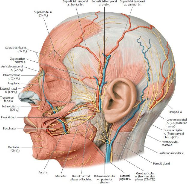
—Primary sensory nerves of the face and scalp (Fig. 27.3) include
•the supraorbital and supratrochlear nerves (cranial nerve [CN] V1);
•the infraorbital nerve and zygomaticotemporal and zygomaticofacial nerves (V2);
•the auriculotemporal, buccal, and mental (a branch of the inferior alveolar nerve) nerves (V3);
•the great auricular and lesser occipital nerve, which are anterior rami of C2 and C3 via the cervical plexus; and
•the greater occipital and third occipital nerves, which are posterior rami of C2 and C3, respectively.
—Motor nerves of the face and scalp (Fig. 27.4) include

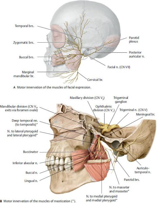
Fig. 27.4 Motor innervation of the face
Left lateral view. (A) Five branches of the facial nerve (CN VII) provide motor innervation to the muscles of facial expression. (B) The mandibular division of the trigeminal nerve (CN V3) supplies motor innervation to the muscles of mastication. (From Gilroy AM, MacPherson BR, Wikenheiser JC. Atlas of Anatomy. Illustrations by Voll M and Wesker K. 4th ed. New
Left lateral view. (From Schuenke M, Schulte E, Schumacher U. THIEME Atlas of Anatomy, Vol 3. Illustrations by Voll M and Wesker K. 3rd ed. New York:
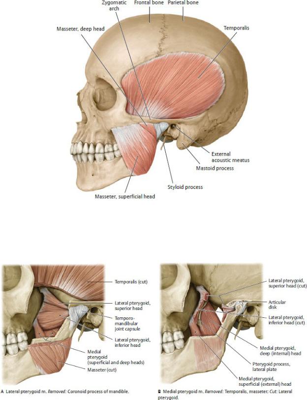
Fig. 27.9 Masticatory muscular sling
Oblique posterior view. Revealed: Muscular sling formed by the masseter and medial pterygoid muscles that embed the mandible. (From Schuenke M, Schulte E, Schumacher U. THIEME Atlas of Anatomy, Vol 3. Illustrations by Voll M and Wesker K. 3rd ed. New York: Thieme Publishers; 2020.)
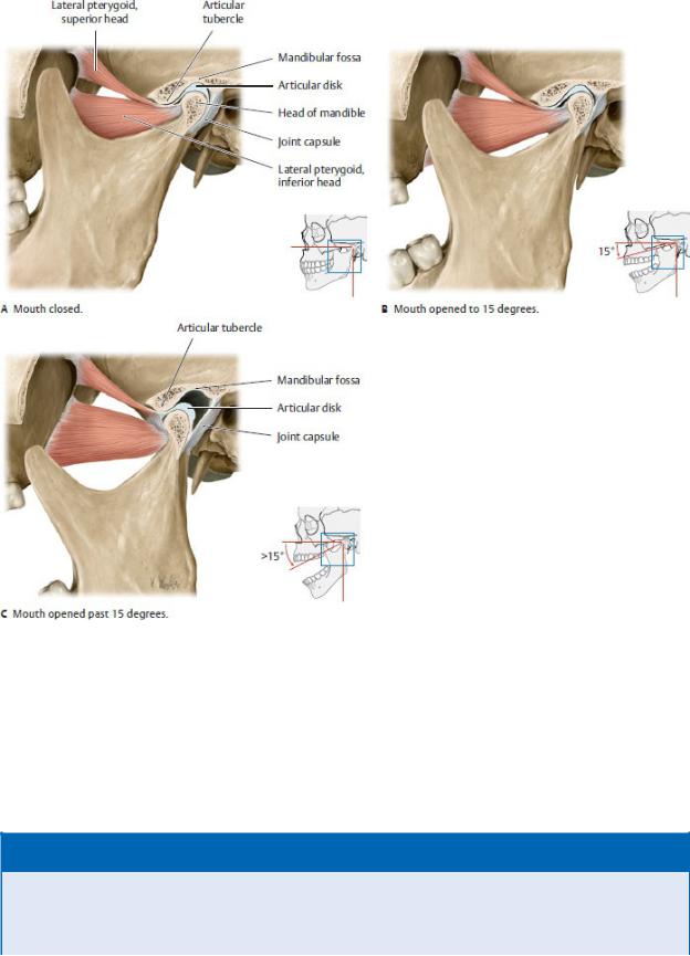
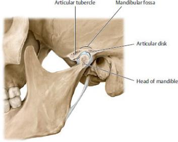
Fig. 27.10 Movement of the temporomandibular joint
Left lateral view. During the first 15 degrees of mandibular depression (opening of the mouth), the head of the mandible remains in the mandibular fossa. Past 15 degrees, the head of the mandible glides forward onto the articular tubercle. (From Gilroy AM, MacPherson BR, Wikenheiser JC. Atlas of Anatomy. Illustrations by Voll M and Wesker K. 4th ed. New York: Thieme Publishers; 2020.)
BOX 27.2: CLINICAL CORRELATION
DISLOCATION OF THE TEMPOROMANDIBULAR JOINT
During yawning (or other activities in which the mouth opens very widely), the head of the mandible moves forward from the mandibular fossa onto the articular tubercle. In some

individuals this may cause the head of the mandible to slide in front of the anterior tubercle, where it locks the mandible in the protruded position. The ligaments supporting the joint become stretched, which causes severe spasm (trismus) of the masseter, medial pterygoid, and temporalis muscles.
•The muscles of mastication act primarily to close the jaw and move the upper teeth against the lower teeth in a grinding motion. The mouth is opened primarily by the suprahyoid muscles with the assistance of the lateral pterygoid.
•The temporalis is the most powerful of these muscles and does approximately half of the work of mastication.
•The masseter, which has superficial and deep parts, raises the mandible and closes the mouth.
•The lateral pterygoid initiates opening of the mouth, which is then continued by the suprahyoid muscles. Because it’s attached to the articular disk, it guides the movement of the joint.
•The medial pterygoid muscle runs almost perpendicular to the lateral pterygoid and contributes to the masticatory muscular sling.
•The masseter and medial pterygoid form a muscular sling that suspends the mandible. By combining the actions of both muscles, the sling enables powerful closure of the jaws.
27.3 Parotid Region
The parotid region lies superficial to the ramus of the mandible and includes the parotid gland and its surrounding structures (Figs. 27.11 and 27.12).
—The parotid gland, the largest of the three salivary glands, lies anterior to the ear on the lateral side of the face.
•A superficial part of the gland lies on the masseter muscle.
•A deep part of the gland curves around the posterior edge of the ramus of the mandible.
•A tough parotid fascia derived from the deep cervical fascia encloses the gland.
•Secretomotor (parasympathetic) fibers to the parotid gland arise with the glossopharyngeal nerve (CN IX) and synapse in the otic ganglion. Postganglionic fibers hitchhike on the auriculotemporal nerve of CN V3.
—The parotid (Stensen’s) duct crosses the masseter muscle superficially to pierce the buccinator muscle and enter the mouth, where it opens into the oral vestibule opposite the second upper molar tooth.
—The parotid plexus, formed by the facial nerve (CN VII), is embedded within the parotid gland and gives rise to five branches that supply muscles of the face: the temporal, zygomatic, buccal, marginal mandibular, and cervical branches (see Fig. 27.24).
Fig. 27.11 Parotid gland
Left lateral view. Note: The parotid duct penetrates the buccinator muscle to open opposite the second upper molar. (From Gilroy AM, MacPherson BR, Wikenheiser JC. Atlas of Anatomy. Illustrations by Voll M and Wesker K. 4th ed. New York: Thieme Publishers; 2020.)
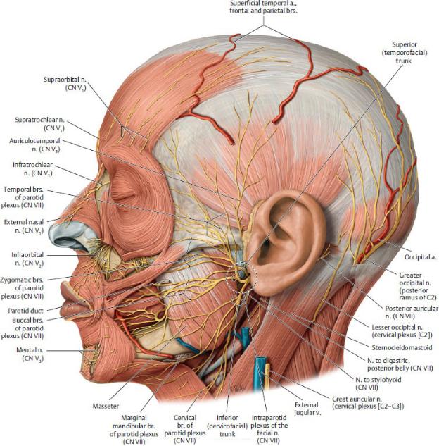
Fig. 27.12 Parotid region
Left lateral view. Removed: Parotid gland, sternocleidomastoid, and veins of the head. (From Gilroy AM, MacPherson BR, Wikenheiser JC. Atlas of Anatomy. Illustrations by Voll M and Wesker K. 4th ed. New York: Thieme Publishers; 2020.)
—Structures traversing, or embedded within, the parotid gland include
•the parotid plexus of the facial nerve (CN VII);
•the retromandibular vein, formed by the superficial temporal and
M and Wesker K. 4th ed. New York: Thieme Publishers; 2020.)
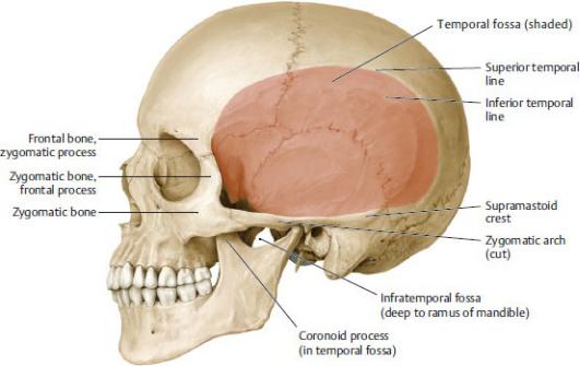
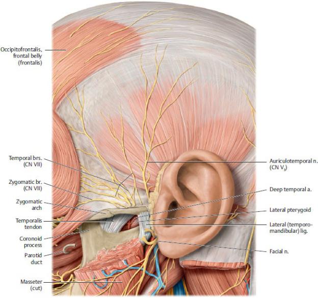
Fig. 27.14 Topography of the temporal fossa
Left lateral view. Cut: Masseter. Revealed: Temporal fossa and temporomandibular joint. (From Baker EW. Anatomy for Dental Medicine, 2nd ed. New York: Thieme; 2015)
—The temporal fossa contains
•the temporalis muscle and temporal fascia,
•the deep temporal branches of the maxillary artery, and
•the deep temporal nerves and auriculotemporal nerve of the
mandibular division of the trigeminal nerve (CN V3).
27.5 Infratemporal Fossa
The infratemporal fossa lies deep to the ramus of the mandible and is continuous superiorly with the temporal fossa (Fig. 27.15).
—The bony boundaries of the infratemporal region are
•anteriorly, the posterior wall of the maxilla;
•posteriorly, the mandibular fossa of the temporal bone;
•medially, the lateral pterygoid plate of the sphenoid bone;
•laterally the ramus of the mandible; and
•superiorly, the temporal bone and greater wing of the sphenoid bone.
—The infratemporal fossa communicates with the orbit anteriorly, the pterygopalatine fossa medially, and the middle cranial fossa superiorly.
—The contents of the infratemporal fossa (Figs. 27.16 and 27.17) include
•the temporomandibular joint,
•the medial and lateral pterygoid muscles and the inferior part of the temporalis muscle,
•the maxillary artery and its branches (Table 27.4),
•the pterygoid venous plexus,
•the mandibular division of the trigeminal nerve (CN V3) and its branches,
•the otic ganglion, and
•the chorda tympani of the facial nerve (CN VII).
—The mandibular nerve (CN V3), the nerve of the infratemporal fossa and the only division of the trigeminal nerve that carries general sensory and somatic motor fibers, distributes postganglionic parasympathetic (visceral motor) fibers that arise from the otic and submandibular ganglia (Fig. 27.18, Table 27.5; see Fig. 27.4).
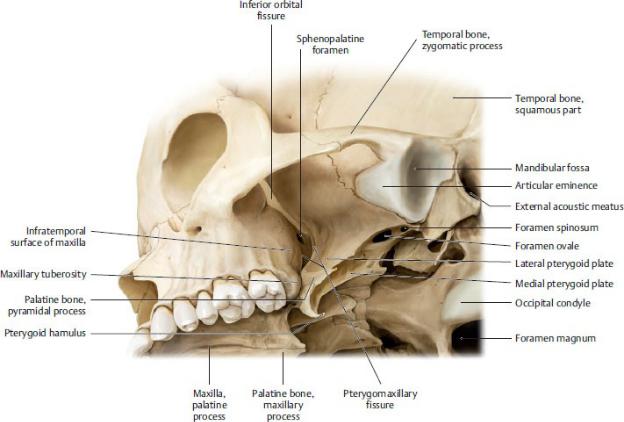
Fig. 27.15 Bony boundaries of the infratemporal fossa
Oblique external view of the base of the skull. (From Gilroy AM, MacPherson BR, Wikenheiser JC. Atlas of Anatomy. Illustrations by Voll M and Wesker K. 4th ed. New York: Thieme Publishers; 2020.)
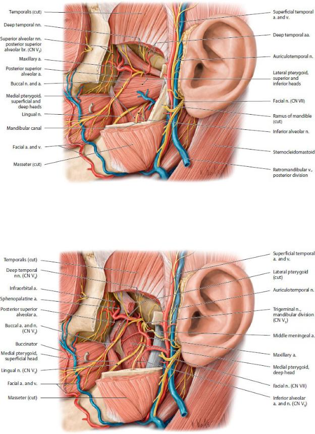
Fig. 27.16 Infratemporal fossa: Superficial layer
Left lateral view. Removed: Ramus of the mandible. (From Gilroy AM, MacPherson BR, Wikenheiser JC. Atlas of Anatomy. Illustrations by Voll M and Wesker K. 4th ed. New York: Thieme Publishers; 2020.)
Fig. 27.17 Infratemporal fossa: Deep dissection
Left lateral view. Removed: Lateral pterygoid muscle (both heads). Revealed: Deep infratemporal fossa and mandibular nerve as it enters the mandibular canal via the foramen ovale in the roof of the fossa.

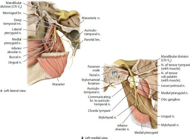
Fig. 27.18 Mandibular nerve (CN V3) in the infratemporal fossa
(From Gilroy AM, MacPherson BR, Wikenheiser JC. Atlas of Anatomy. Illustrations by Voll M and Wesker K. 4th ed. New York: Thieme Publishers; 2020.)
Table 27.5 Nerves of the Infratemporal Fossa
Nerve |
Nerve Fibers |
Distribution |
Muscular |
Branchial motor |
Muscles of mastication; |
branches (CN |
|
mylohyoid; tensor tympani; tensor |
V3) |
|
veli palatini; anterior belly of |
|
|
digastric |
|
|
|
Auriculotemporal |
General sensory |
Auricle, temporal region, and |
(CN V3) |
|
temporomandibular joint |
|
Visceral motor |
Parotid gland |

from
glossopharyngeal n. (CN IX)
Inferior alveolar |
General sensory |
Mandibular teeth; mental branch |
(CN V3) |
|
supplies skin of lower lip and chin |
Lingual (CN V3) |
General sensory |
Anterior two thirds of tongue, floor |
|
|
of mouth, and lingual mandibular |
|
|
gingiva |
|
|
|
Buccal (CN V3) |
General sensory |
Skin and mucous membrane of |
|
|
cheek |
|
|
|
Meningeal (CN |
General sensory |
Dura of middle cranial fossa |
V3) |
|
|
Chorda tympani |
Special sensory |
Anterior two thirds of tongue |
(CN VII) |
taste |
|
|
|
|
|
Visceral motor |
Submandibular and sublingual |
|
|
glands via submandibular |
|
|
ganglion and lingual n. (CN V3). |
27.6 Pterygopalatine Fossa
The pterygopalatine fossa, a narrow space located medial to the infratemporal fossa, is an important distribution center for branches of the maxillary division of the trigeminal nerve (CN V2) and the accompanying branches of the maxillary artery.
—The bony boundaries of the pterygopalatine fossa (Fig. 27.19) are
•superiorly, the apex of the orbit;
•anteriorly, the maxillary sinus;
•posteriorly, the lateral pterygoid plate of the sphenoid bone;
•laterally, the pterygomaxillary fissure; and
•medially, the vertical plate of the palatine bone.
—Contents of the pterygopalatine fossa include
•the pterygopalatine part of the maxillary artery, its branches, and its accompanying veins;
•the nerve of the pterygoid canal;
•the pterygopalatine ganglion; and
•the maxillary division of the trigeminal nerve (CN V2) and its branches.
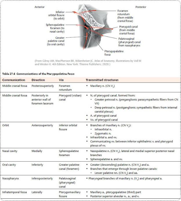
—The pterygopalatine fossa communicates anteriorly with the orbit, medially with the nasal cavity and palate, and posteriorly with the middle cranial fossa and base of the skull (Table 27.6).
—The maxillary artery passes from the infratemporal fossa to the pterygopalatine fossa through the pterygomaxillary fissure (see Table 27.4). Its branches accompany the branches of the maxillary nerve (CN V2) to supply the nose, palate, and pharynx.
—The nerve of the pterygoid canal, which enters the pterygo palatine fossa from the middle cranial fossa, is an autonomic nerve that carries
•preganglionic parasympathetic fibers from the greater petrosal nerve, a branch of the facial nerve (CN VII); and
•postganglionic sympathetic fibers from the deep petrosal nerve, which arises from the internal carotid plexus.
—The pterygopalatine ganglion receives general sensory fibers from the maxillary nerve (CN V2) and parasympathetic and sympathetic fibers from the nerve of the pterygoid canal. Only the parasympathetic fibers synapse in the ganglion; general sensory and sympathetic fibers pass through the ganglion but do not synapse there (Table 27.7).
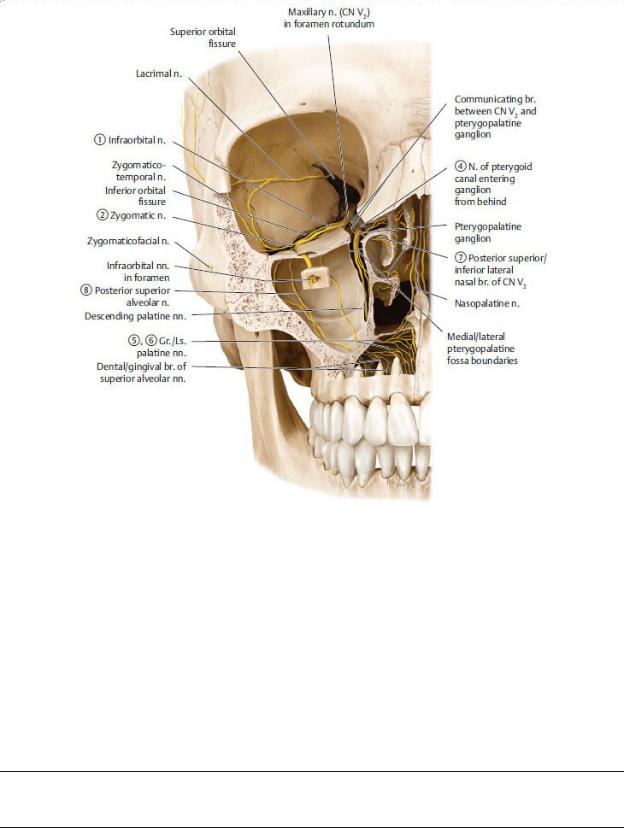
—The maxillary division of the trigeminal nerve (CN V2)
•passes from the middle cranial fossa to the pterygopalatine fossa through the foramen rotundum;
•suspends the pterygopalatine ganglion by two small ganglionic nerves, which transmit general sensory fibers of the maxillary nerve;
•distributes postganglionic parasympathetic (secretomotor) and sympathetic (vasoconstrictive) fibers to the lacrimal, nasal, palatal, and pharyngeal glands; and
•distributes general sensory fibers to the midface, maxillary sinus, maxillary teeth, nasal cavity, palate, and superior pharynx.
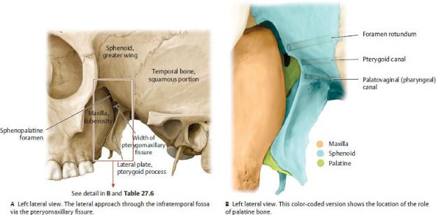
Fig. 27.19 Pterygopalatine fossa
(From Gilroy AM, MacPherson BR, Wikenheiser JC. Atlas of Anatomy. Illustrations by Voll M and Wesker K. 4th Edition. New York: Thieme Publishers; 2020.)

sphenoid sinuses
N. of the pterygoid |
Visceral motor |
canal (CN VII) |
(preganglionic |
|
parasympathetic |
|
and postganglionic |
|
sympathetic) to |
|
glands of the nasal |
|
mucosa, and palate; |
|
lacrimal gland |
Greater palatine n. (CN V2) |
Hard palate and palatal |
|
gingiva |
|
|
Lesser palatine n. (CN V2) |
Soft palate, palatine |
|
tonsil |
|
|
Medial and lateral posterior superior and |
Nasal septum, upper |
posterior inferior nasal nn. (CN V2) |
lateral nasal wall, |
|
ethmoid sinuses |
|
|
Posterior superior alveolar n. (CN V2) |
Maxillary sinus, cheeks, |
|
buccal gingiva, |
|
maxillary molars |
|
|
27.7 Nasal Cavity
The nasal cavity is located in the middle of the face between the orbits and maxillary sinuses and above the oral cavity.
Structure of the Nasal Cavity
The nose has an external portion and paired internal nasal cavities that are separated by a nasal septum (Figs. 27.20 and 27.21).
—The external nose consists of
•anteriorly, the alar and lateral nasal cartilages, which form the nasal ala and crura surrounding the nostrils and the apex, or tip of the nose; and
•posteriorly, the frontal, maxillary, and nasal bones, which form the root, or bridge, of the nose.
—The nasal cavities are pyramidally shaped spaces that communicate anteriorly with the outside through the nares (anterior nasal apertures) and
posteriorly with the nasopharynx through the choanae.
—The lateral walls of the nasal cavities are formed by the superior and middle nasal conchae of the ethmoid bone; the inferior nasal conchae; and the maxillary, palatine, lacrimal, and nasal bones.
•The superior, middle, and inferior nasal conchae are scrolllike bony processes that project into the nasal cavity.
•The superior, middle, and inferior nasal meatuses are recesses below the respective nasal conchae.
—The nasal septum forms the medial wall of each nasal cavity and consists of the vomer, the perpendicular plate of the ethmoid bone, and the septal cartilage.
—The hard palate, consisting of the maxilla and palatine bones, forms the floor of the nasal cavities and separates them from the oral cavity (see Section 27.8).
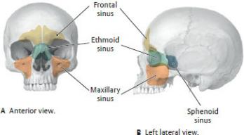
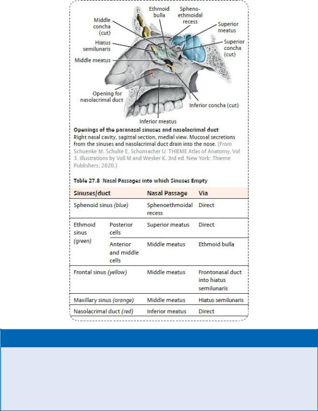
—Paranasal sinuses are air-filled cavities within bones of the skull that communicate with the nasal cavities (Fig. 27.22; Table 27.8).
•Paired frontal sinuses, which are usually asymmetrical, lie above the root of the nose and drain into the middle meatus through a frontonasal duct into the hiatus semilunaris.
•Sphenoid sinuses, which form within the body of the sphenoid bone, lie between the right and left cavernous sinuses and drain into the sphenoethmoidal recess in the posterosuperior part of the nasal cavities above the superior concha.
•Ethmoid sinuses compose the medial wall of the orbit and are formed by numerous thin-walled ethmoid air cells. They lie between the orbits above the nasal cavities and drain into the superior and middle meatuses.
•Paired maxillary sinuses, the largest of the paranasal sinuses, lie on either side of the nasal cavities, inferior to the orbits, and drain into the middle meatuses.
—A nasolacrimal duct drains tears from the medial corner of each eye and empties into the inferior meatus on each side.
Fig. 27.20 Skeleton of the nose
The skeleton of the nose is composed of an upper bony portion and a lower cartilaginous portion. The proximal portions of the nostrils (alae) are composed of connective tissue with small embedded pieces of cartilage. (From Schuenke M, Schulte E, Schumacher U. THIEME Atlas of Anatomy, Vol 3. Illustrations by Voll M and Wesker K. 3rd ed. New York: Thieme Publishers; 2020.)
BOX 27.3: CLINICAL CORRELATION
INFECTION OF THE MAXILLARY SINUSES
Infections arising in the nasal cavity may spread to any of the paranasal sinuses, but the maxillary sinuses are the most com-monly affected. Mucus builds up within the maxillary sinuses and is unable to drain because their ostia are located high on the superomedial walls. The ostia are also commonly obstructed by inflammation of the mucous membranes of the
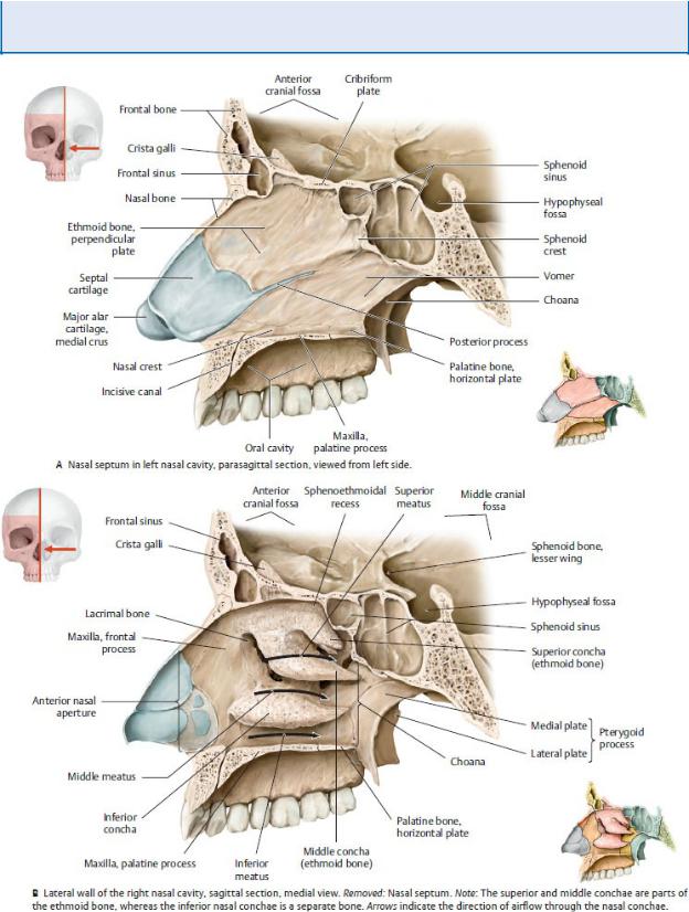
The left and right nasal cavities are flanked by lateral walls and separated by the nasal septum. Air enters the nasal cavity through the anterior nasal aperture and travels through three passages: the superior, middle, and inferior meatuses (B, arrows). These passages are separated by the superior, middle, and inferior conchae. Air leaves the nose through the choanae, entering the nasopharynx. (From Schuenke M, Schulte E, Schumacher U. THIEME Atlas of Anatomy, Vol 3. Illustrations by Voll M and Wesker K. 3rd ed. New York: Thieme Publishers; 2020.)
Fig. 27.22 Location of the paranasal sinuses
The paranasal sinuses (frontal, ethmoid, maxillary, and sphenoid) are airfilled cavities that reduce the weight of the skull. (From Schuenke M, Schulte E, Schumacher U. THIEME Atlas of Anatomy, Vol 3. Illustrations by Voll M and Wesker K. 3rd ed. New York: Thieme Publishers; 2020.)
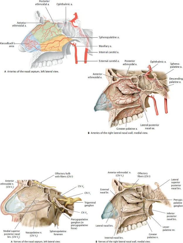
Neurovasculature of the Nasal Cavity
—The external carotid artery, via its maxillary and facial branches, and the internal carotid artery, via its ophthalmic branch, supply the nasal cavity. The area of overlap between the external and internal circulations is referred to as Kiessel-bach’s area (Fig. 27.23).
•Nasal branches of the maxillary artery include
◦the lateral posterior nasal and posterior septal arteries, branches of the sphenopalatine artery; and
◦the greater palatine artery, a branch of the descending palatine artery.
•Branches of the facial artery include the lateral nasal artery and a septal artery, a branch of the superior labial artery.
•Branches of the ophthalmic artery include anterior and posterior ethmoidal arteries.
—Veins that drain the nasal cavity form a submucosal venous plexus that drains into the ophthalmic, facial, and spheno palatine veins.
—The olfactory nerve (CN I) and the ophthalmic (CN V1) and maxillary (CN V2) divisions of the trigeminal nerve innervate the nose (Fig. 27.24).
•Olfactory nerves, which are responsible for smell, arise from the olfactory epithelium in the roof of the nasal cavity. They pass through the cribriform plate and end in the olfactory bulbs.
•Infratrochlear and anterior ethmoid branches of CN V1 and the infraorbital branch of CN V2 innervate the external nose.
•The anterior and posterior ethmoidal branches of CN V1 innervate the external nose and the mucosa of the anterosuperior nasal cavity through internal, external, medial, and lateral nasal branches.
•The posterior nasal branches of the nasopalatine nerve on the septum and nasal branches of the greater pala-tine nerve on the lateral walls (both branches of CN V2) innervate the mucosa of the posteroinferior nasal cavity.

(From Schuenke M, Schulte E, Schumacher U. THIEME Atlas of Anatomy, Vol 3. Illustrations by Voll M and Wesker K. 3rd ed. New York: Thieme Publishers; 2020.)
27.8 Oral Region
The oral cavity, located below the nasal cavity and anterior to the pharynx, is bounded superiorly by the palate, inferiorly by the tongue and muscular floor, anteriorly by the lips, posteriorly by the uvula, and laterally by the cheeks (Fig. 27.25).
Lips, Cheeks, Gingiva, Teeth, and Oral Cavity
—The lips frame the mouth and surround the oral fissure, the opening into the oral cavity.
•The lips contain the sphincter-like obicularis oris muscle and superior and inferior labial muscles. Externally, they are covered by skin; internally, they are covered by the mucous membrane of the oral cavity.
•The philtrum, an external midline depression on the upper lip, extends superiorly to the nasal septum.
•Labial frenula are midline folds of mucous membrane that attach the inner surfaces of the upper and lower lips to the gums.
BOX 27.5: DEVELOPMENTAL CORRELATION
CLEFT LIP
Cleft lip is a congenital defect that occurs early in embryonic life when the maxillary prominence and the median nasal promi-nence fail to fuse. It is seen in ~ 1:1,000 live births and is more common in males than females. The cleft may be unilateral or bilateral and is described as complete when it extends into the nose and incomplete when it appears as a notch in the lip. Corrective surgery is usually performed when the infant is around 10 weeks old.
—The cheeks, continuous with the lips, form the walls of the mouth and the buccal region of the face.
•The buccinator muscle, innervated by the buccal branch of the facial nerve (CN VII), forms the movable wall of the cheek.
•Buccal fat pads, encapsulated cushions of fat that lie superficial to the buccinator muscles, are proportionately large in infants and reduced in adults.
•The zygomatic bone and zygomatic arch form the “cheek bone.”
—Teeth are anchored in alveoli (sockets) of the maxillary and mandibular dental arches (Figs. 27.26).
•Children have 20 deciduous teeth, which are replaced at predictable intervals between ages 7 and 25 years.
•Adult have 32 teeth, consisting of incisors, canines, premolars, and molars. Teeth are numbered 1 to 16 from right to left along the maxillary arch, and 17 to 32 from left to right along the mandibular arch.
—The gingivae, or gums, made of fibrous tissue covered by the mucous membrane of the oral cavity, are firmly attached to the maxilla and mandible.
—The oral cavity, or mouth, is divided into two regions: the oral vestibule and the oral cavity proper (Fig. 27.27).
•The oral vestibule is the narrow space between the lips and cheeks and the dental arches of the maxilla and mandible.
•The oral cavity proper is the space bounded anteriorly and laterally by the upper and lower dental arches. Superiorly, the palate forms the roof, and inferiorly, the tongue rests on a muscular floor.
—The oral cavity connects posteriorly to the pharynx through a narrow space, the faucial isthmus.
—Muscles that form the floor of the mouth, the suprahyoid muscles, are attached to the hyoid bone in the neck (Fig. 27.28; Table 27.9). They have a complex innervation with contributions from the trigeminal and facial nerves and the C1 spinal nerve via the hypoglossal nerve (Fig. 27.29; see also Fig. 25.7).
—The lingual, facial, and maxillary branches of the external carotid artery supply the lips, cheeks, floor of the mouth, and upper and lower teeth.
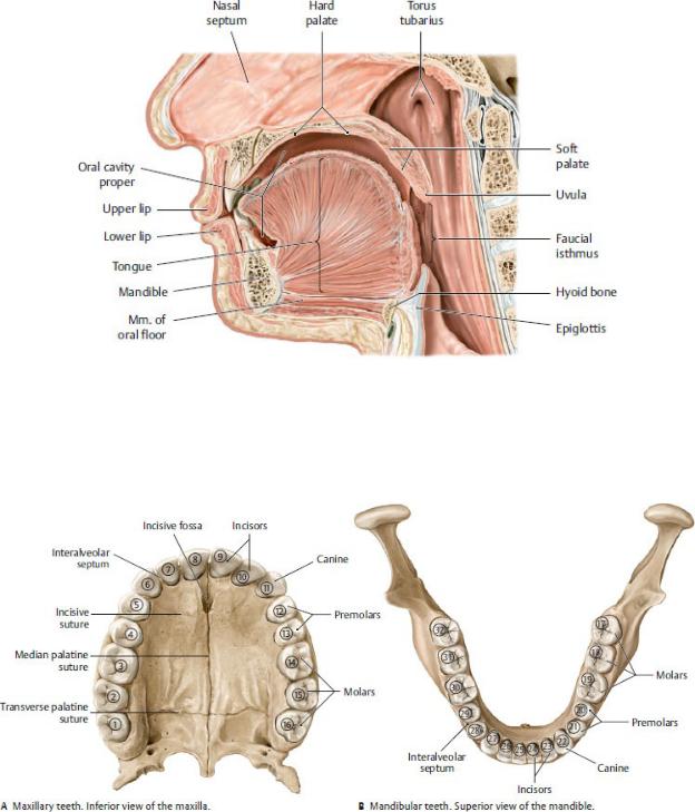
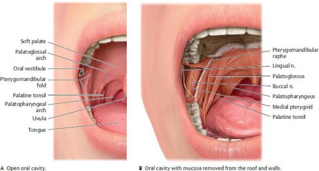
Fig. 27.27 Oral cavity topography
Right side, anterior view. (From Gilroy AM, MacPherson BR, Wikenheiser JC. Atlas of Anatomy. Illustrations by Voll M and Wesker K. 4th ed. New York: Thieme Publishers; 2020.)
—The trigeminal nerve (CN V) transmits sensation from the mouth.
•The superior alveolar branch of the maxillary division (CN V2) innervates the upper teeth.
•Inferior alveolar, lingual, and buccal branches of the mandibular division (CN V3) innervate the cheek, lower teeth, and floor of the mouth.
—Visceral motor fibers carried by the chorda tympani (CN VII) synapse in the submandibular ganglion in the floor of the mouth. Postganglionic fibers travel via the lingual nerve to innervate the submandibular and sublingual glands (Fig. 27.29).
The Palate
The palate forms the roof of the oral cavity and the floor of the nasal cavity and separates the oral cavity from the pharynx posteriorly.
—Nasal mucosa covers the superior surface, and oral mucosa, densely packed with mucus-secreting palatine glands, covers the inferior surface.
—The palate has anterior and posterior regions.
• The hard palate, formed by the palatine processes of the maxillary
bones and the horizontal processes of the palatine bones, makes up the anterior two thirds of the palate (Fig. 27.30).
Fig. 27.28 Muscles of the oral floor: suprahyoid muscles
(From Gilroy AM, MacPherson BR, Wikenheiser JC. Atlas of Anatomy. Illustrations by Voll M and Wesker K. 4th ed. New York: Thieme Publishers; 2020.)
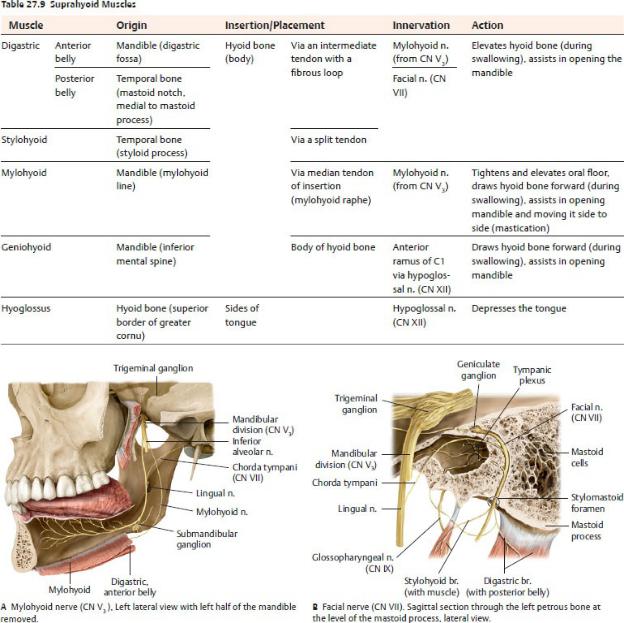
Fig. 27.29 Nerves in the floor of the mouth
(From Schuenke M, Schulte E, Schumacher U. THIEME Atlas of Anatomy, Vol 3. Illustrations by Voll M and Wesker K. 3rd ed. New York: Thieme Publishers; 2020.)
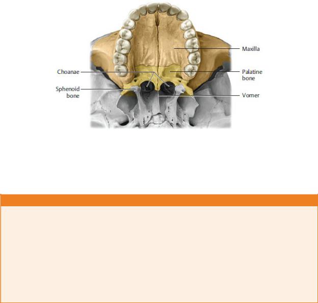
Fig. 27.30 Hard palate
Inferior view. (From Schuenke M, Schulte E, Schumacher U. THIEME Atlas of Anatomy, Vol 3. Illustrations by Voll M and Wesker K. 3rd ed. New York: Thieme Publishers; 2020.)
BOX 27.6: DEVELOPMENTAL CORRELATION
CLEFT PALATE
Cleft palate is a congenital defect that occurs early in embryonic life when the lateral palatine processes fail to fuse with each other, with the nasal septum, and/or with the median palatine pro-cesses. It is seen in ~ 1:2,500 live births and is more common in females than males. The cleft (a fissure or opening) is described as complete when it involves the soft and hard palate and incom-plete when it appears as a “hole” in the roof of the mouth (usually in the soft palate). In each case the uvula is usually also split. The cleft connects the oral cavity directly to the nasal cavity. Initial treatment involves the use of a prosthetic device called a palatal obturator to seal the cleft until corrective surgery is performed when the infant is between 6 and 12 months old.
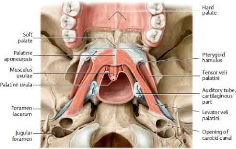
•The soft palate, the posterior third of the palate, has an anterior aponeurotic part that is attached to the hard palate and a posterior muscular part with an unattached posterior margin that ends in a conical projection, the uvula (see Fig. 27.25).
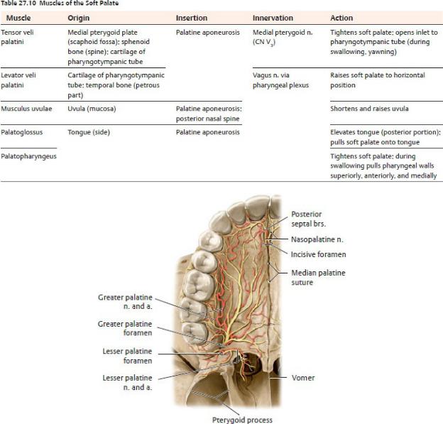
—During swallowing, muscles of the soft palate can tense and elevate it against the posterior pharyngeal wall to prevent food from passing into the nasal cavity. They can also draw the palate downward against the tongue to prevent food from entering the pharynx. Muscles of the soft palate include the tensor veli palatini, levator veli palatini, musculus uvulae, palatoglossus, and palatopharyngeus (Fig. 27.31; Table 27.10).
—The paired palatoglossal and palatopharyngeal arches, formed by the
palatoglossus and palatopharyngeus muscles, respectively, anchor the soft palate to the tongue and pharynx (see Fig. 27.27).
—Greater palatine, lesser palatine, and sphenopalatine branches of the maxillary artery supply the palate (Fig. 27.32; see also Fig. 27.23B).
—The greater palatine, lesser palatine, and nasopalatine branches of the maxillary division (CN V2) carry sensory innervation from the palate (see
Fig. 27.24B).
Fig. 27.31 Muscles of the soft palate
Inferior view. The soft palate forms the posterior boundary of the oral cavity, separating it from the oropharynx. (From Gilroy AM, MacPherson BR, Wikenheiser JC. Atlas of Anatomy. Illustrations by Voll M and Wesker K. 4th ed. New York: Thieme Publishers; 2020.)

Extrinsic muscles |
Extrinsic muscles |
|
||
• |
Genioglossus |
• |
Superior |
longitudinal |
• |
Hyoglossus |
|
muscle |
|
• |
Styloglossus |
• |
Inferior longitudinal muscle |
|
|
|
• |
Transverse muscle |
|
|
|
• |
Vertical muscle |
|
* All extrinsic and intrinsic muscles of the tongue are innervated by the hypoglossal n. (CN XII).
Fig. 27.33 Structure of the tongue
Superior view. The V-shaped sulcus terminalis divides the tongue into an anterior (oral, presulcal) and a posterior (pharyngeal, postsulcal) part. (From Schuenke M, Schulte E, Schumacher U. THIEME Atlas of Anatomy, Vol 3. Illustrations by Voll M and Wesker K. 3rd ed. New York: Thieme Publishers; 2020.)
—All extrinsic and intrinsic muscles of the tongue are innervated by the hypoglossal nerve (CN XII).
—The lingual arteries, branches of the external carotid arteries, supply the tongue. The lingual veins accompany the arteries and drain into the internal jugular vein.
—Lymph from the tongue ultimately drains to deep cervical and jugular nodes in the neck, although the drainage from the various parts of the tongue follows four different pathways (Fig. 27.35; Table 27.12). These drainage
routes can be clinically important in the metastasis of lingual tumors.
—The five cranial nerves (trigeminal, facial, glossopharyngeal, vagus, and hypoglossal) that innervate the tongue carry motor, general sensory, and special sensory fibers (Fig. 27.36; Table 27.13).
Table 27.12 Lymphatic Drainage of the Tongue
Area of the Tongue |
Drainage Pattern |
Primary Nodes |
Root |
Bilaterally |
Superior deep |
|
|
cervical |
|
|
|
Medial part of the body |
Bilaterally |
Inferior deep |
|
|
cervical |
|
|
|
Lateral parts of the body |
Ipsilaterally |
Submandibular |
|
|
|
Apex and frenulum |
Midline—bilaterally |
Submental |
|
Sides—ipsilaterally |
|
|
|
|
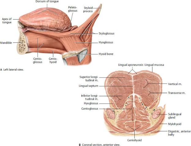
Fig. 27.34 Extrinsic muscles of the tongue
(From Schuenke M, Schulte E, Schumacher U. THIEME Atlas of Anatomy, Vol 3. Illustrations by Voll M and Wesker K. 3rd ed. New York: Thieme Publishers; 2020.)
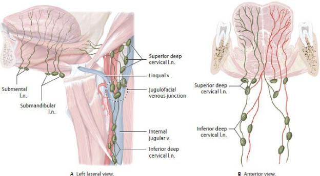
Fig. 27.35 Lymphatic drainage of the tongue and oral floor
Lymph flows to the submental and submandibular lymph nodes of the tongue and oral floor, which ultimately drain into the jugular lymph nodes along the internal jugular vein. Because the lymph nodes receive drainage from both the ipsilateral and contralateral sides (B), tumor cells may become widely disseminated in this region (e.g., metastatic squamous cell carcinoma, especially on the lateral border of the tongue, frequently metastasizes to the opposite side). (From Schuenke M, Schulte E, Schumacher U. THIEME Atlas of Anatomy, Vol 3. Illustrations by Voll M and Wesker K. 3rd ed. New York: Thieme Publishers; 2020.)
Fig. 27.36 Neurovasculature of the tongue
Left lateral view. (From Schuenke M, Schulte E, Schumacher U. THIEME Atlas of Anatomy, Vol 3. Illustrations by Voll M and Wesker K. 3rd ed. New York: Thieme Publishers; 2020.)
Somatosensory and taste innervation of the tongue
Superior view. (From Gilroy AM, MacPherson BR, Wikenheiser JC. Atlas of Anatomy. Illustrations by Voll M and Wesker K. 4th ed. New York: Thieme Publishers; 2020.)
Table 27.13 Innervation of the Tongue
Nerve |
Nerve Fibers |
Distribution |
Lingual nerve (CN |
General sensory |
Anterior two thirds of the |
V3) |
|
tongue |
Chorda tympani (CN |
Special sensory |
Anterior two thirds of the |
VII) |
|
tongue |
|
|
|
Glossopharyngeal |
General sensory |
Posterior third of the tongue |
nerve (CN IX) |
and special sensory |
|
|
|
|
Vagus nerve (CN X) |
General sensory |
Root of the tongue |
|
and special sensory |
|
|
|
|
Hypoglossal nerve |
Somatic motor |
Muscles of the tongue, |
(CN XII) |
|
except the palatoglossus, |
|
|
which is innervated by the |
|
|
vagus nerve (CN X) |
|
|
|
Salivary Glands
Three pairs of salivary glands, the parotid, sublingual, and sub mandibular, produce and secrete saliva into the mouth (Fig. 27.37).
—The sublingual gland, the smallest of the three glands, lies deep to the mucosa of the floor of the mouth, forming the sublingual folds, onto which it secretes saliva through numerous small ducts.
—The submandibular gland has a superficial part located in the neck and a deep part located in the floor of the mouth, which connect around the posterior border of the mylohyoid muscle. The submandibular (Wharton’s) duct opens via the sublingual papilla at the base of the lingual frenulum.
—The parotid gland, the largest of the three glands, is located in the parotid region on the lateral side of the head, anterior to the ear. The parotid duct opens into the vestibule of the mouth at the parotid papilla located opposite to the second maxillary molar (see Section 27.3).
—The facial, lingual, maxillary, and superficial temporal arteries supply the salivary glands. Veins of similar names accompany the arteries and drain into the retromandibular vein.
—The sublingual and submandibular glands receive parasympa thetic secretomotor fibers via the chorda tympani (a branch of CN VII) and submandibular ganglion.
—The parotid gland receives secretomotor (parasympathetic) fibers of the glossopharyngeal nerve (CN IX) that synapse in the otic ganglion.
—Sympathetic innervation to the glands arises as postganglionic fibers from the superior cervical ganglion and follows the branches of the external carotid artery.
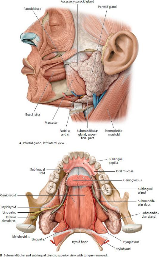
Fig. 27.37 Salivary glands
(From Schuenke M, Schulte E, Schumacher U. THIEME Atlas of Anatomy, Vol 3. Illustrations by Voll M and Wesker K. 3rd ed. New York: Thieme Publishers;
27.9 Pharynx and Tonsils
The pharynx, a fibromuscular tube that forms part of the upper airway and the upper digestive tract, transmits air from the nasal cavity to the trachea and food from the oral cavity to the esophagus.
Regions of the Pharynx
The pharynx, which extends from the base of the skull to the inferior aspect of the larynx (cricoid cartilage), is divided into three regions (Figs. 27.38 and 27.39): the nasopharynx, the orophar ynx, and the laryngopharynx.
—The nasopharynx, the uppermost part of the pharynx, lies posterior to the nasal cavity and above the soft palate. The body of the sphenoid forms its roof. Anteriorly, the nasopharynx communicates with the nasal cavity through the paired choanae.
•The orifices (openings) of the pharyngotympanic tubes are located on the lateral pharyngeal wall. Above the orifice, the cartilaginous part of the tube protrudes into the pharynx to form a bulge, the torus tubarius.
•A salpingopharyngeal fold, formed by the salpingopha-ryngeus muscle deep to the mucosa, extends inferiorly from the torus tubarius.
—The oropharynx, which lies posterior to the oral cavity, extends superiorly to the soft palate and inferiorly to the top of the larynx. The posterior tongue forms the anterior boundary.
•Two arches, the anterior palatoglossal arch (fold) and the posterior palatopharyngeal arch (fold), formed by the palatoglossus and palatopharyngeal muscles, respectively, separate the oropharynx from the oral cavity. Both muscles are innervated by the vagus nerve (CN X).
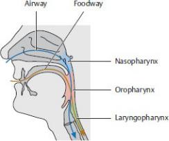
Fig. 27.38 Regions of the pharynx
Midsagittal section, left lateral view. The blue arrow indicates flow of air and the red arrow, passage of food. (From Schuenke M, Schulte E, Schumacher U. THIEME Atlas of Anatomy, Vol 3. Illustrations by Voll M and Wesker K. 3rd ed. New York: Thieme Publishers; 2020.)
•The palatine tonsil lies between these arches in the tonsillar fossa.
•A median fold of mucosa divides the space between the base of the tongue and the epiglottis. The spaces on either side of the fold are called valleculae.
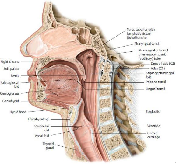
Fig. 27.39 Pharynx Midsagittal section, left lateral
Midsagittal section, left lateral view. (From Gilroy AM, MacPherson BR, Wikenheiser JC. Atlas of Anatomy. Illustrations by Voll M and Wesker K. 4th ed. New York: Thieme Publishers; 2020.)
—The laryngopharynx, which lies posterior to the larynx, extends from the epiglottis to the inferior border of the cricoid cartilage, where it narrows and is continuous with the esophagus.
•The laryngopharynx communicates with the larynx through the laryngeal inlet.
•Aryepiglottic folds separate the laryngeal inlet from mucosal-lined fossae on the lateral walls of the pharynx called piriform recesses.

BOX 27.7: CLINICAL CORRELATION
PIRIFORM RECESSES
The piriform recesses are small fossae that lie on either side of the laryngeal inlet. Occasionally small foreign objects that are swal-lowed or inhaled or bits of food (such as a peanut) can become lodged in these spaces. In such cases, the internal laryngeal and inferior laryngeal nerves, which lie deep to the mucosa in this area, can be vulnerablcongenital defect that occurs early ine to injury.
Muscles of the Pharynx
An outer circular layer and inner longitudinal layer of skeletal muscles form the walls of the pharynx (Figs. 27.40 and 27.41; Tables 27.14 and 27.15). Pharyngeal muscles coordinate with muscles of the floor of the mouth and soft palate during swallowing (Fig. 27.42).
—Circular superior, middle, and inferior pharyngeal constric-tor muscles propel a bolus of food downward through the oropharynx and laryngopharynx; all are innervated by the vagus nerve (CN X).
—Longitudinal muscles, the salpingopharyngeus and palatopharyngeus innervated by the vagus nerve (CN X) and pharyngeal plexus, and the stylopharyngeus innervated by the glossopharyngeal nerve (CN IX), elevate the pharynx to prevent food from entering the nasopharynx.
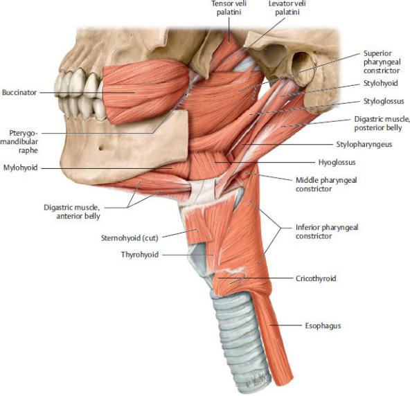
Fig. 27.40 Pharyngeal muscles
Left lateral view. (From Gilroy AM, MacPherson BR, Wikenheiser JC. Atlas of Anatomy. Illustrations by Voll M and Wesker K. 4th ed. New York: Thieme Publishers; 2020.)
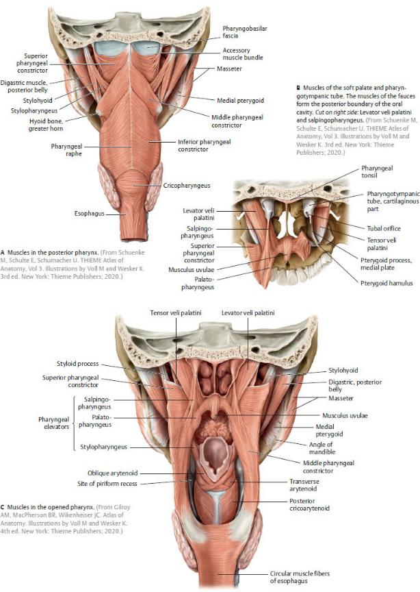
Fig. 27.41 Pharyngeal muscles
Posterior view.
Fig. 27.42 Anatomy of swallowing
As part of the airway, the larynx in the adult is located at the inlet to the digestive tract (A). During swallowing, therefore, the airway must be briefly occluded to keep food from entering the trachea. During the reflexive phase of swallowing, muscles on the floor of the mouth and the thyrohyoid elevate the larynx while the epiglottis covers the laryngeal inlet, sealing off the lower airway. In addition, the soft palate tightens,
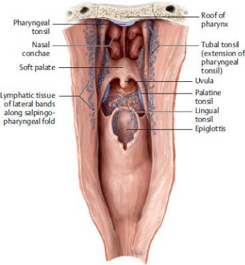
elevates, and opposes the posterior pharyngeal wall, sealing off the upper airway (B). (From Schuenke M, Schulte E, Schumacher U. THIEME Atlas of Anatomy, Vol 3. Illustra-tions by Voll M and Wesker K. 3rd ed. New York: Thieme Publishers; 2020.)
Tonsils
—Tonsils, masses of lymphoid tissue found in the mucosal lining of the pharynx, form an incomplete circular ring called the pharyngeal lymphatic ring (Waldeyer’s ring) around the superior part of the pharynx (Fig. 27.43). They include
•one pharyngeal tonsil (also known as adenoid) in the mucous membrane of the roof and posterior wall of the pharynx;
•paired tubal tonsils, extensions of the pharyngeal tonsil, near the orifices of the pharyngotympanic tube;
•paired palatine tonsils in the fossae between the palatoglossal and palatopharyngeal arches;
•lingual tonsils on the posterior one third of the dorsum of the tongue; and
•pairs of lateral bands that lie along the salpingopharyn geal folds.
—Tonsils are supplied by branches of the facial, ascending palatine, lingual, descending palatine, and ascending pharyngeal arteries.

Fig. 27.43 Tonsils: Waldeyer’s ring
Posterior view of the opened pharynx. (From Schuenke M, Schulte E, Schumacher U. THIEME Atlas of Anatomy, Vol 3. Illustrations by Voll M and Wesker K. 3rd ed. New York: Thieme Publishers; 2020.)
—Tonsillar lymphatic vessels drain to the jugulodigastric node near the angle of the mandible before draining to the deep cervical lymph nodes.
—A tonsillar nerve plexus is formed by the glossopharyngeal (CN IX) and vagus (CN X) nerves.
BOX 27.8: CLINICAL CORRELATION
TONSILLECTOMY
During a tonsillectomy, the palatine tonsils are removed from the tonsillar bed along with their accompanying fascia. The glosso-pharyngeal nerve, which lies on the lateral wall of the pharynx, is vulnerable to injury during the procedure and may result in loss of sensation and taste from the posterior one third of the tongue. Bleeding may occur from the large external palatine vein or from the tonsillar branches of the facial, ascending pharyngeal, maxil-lary, and lingual arteries. The internal carotid artery may also be vulnerable because it runs just lateral to the tonsil.
Neurovasculature of the Pharynx
—Direct and indirect branches of the external carotid artery supply the pharynx. These include the facial, lingual, ascending palatine, descending palatine, and ascending pharyngeal arteries (see Fig. 24.21).
—Venous drainage of the pharynx passes through the pharyngeal venous plexus to the internal jugular vein (see Fig. 24.26).
—Sensory innervation of the pharyngeal mucosa varies by region (Fig. 27.44).
•The maxillary nerve (CN V2) innervates the upper part of the nasopharynx.
•The glossopharyngeal nerve (CN IX) primarily innervates the oropharynx, although its territory extends into both the nasopharynx and laryngopharynx.
•The vagus nerve (CN X) via its internal laryngeal branch innervates the laryngopharynx.
BOX 27.9: CLINICAL CORRELATION
GAG REFLEX
The gag reflex is a reflexive contraction of the pharyngeal muscles that protects the airway by preventing accidental ingestion and aspiration. It is elicited by touching the soft palate or back
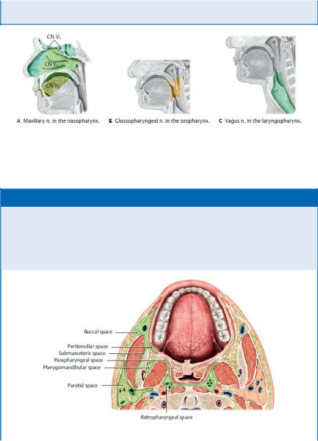

MacPherson BR, Wikenheiser JC. Atlas of Anatomy. Illustrations by Voll M and Wesker K. 4th Edition. New York: Thieme Publishers; 2020.)

28 The Eye and Ear
The eye, the organ of vision, and the ear, which contains the organs for hearing and balance, are anatomically the most complex of the sense organs. The eyes, enclosed within the bony orbits, are prominent features of the face. The ears have superficial and deep components that are related to the temporal bones on the sides of the head.
28.1 The Eye
The anatomy of the eye includes the bony orbit, the eyelids and lacrimal apparatus, the eyeball, and six extraocular muscles.
The Bony Orbit
The paired orbits lie on either side of the superior part of the nasal cavity, above the maxillary sinuses and below the anterior cranial fossae (see Fig. 24.15). These cavities are shaped like quadrangular pyramids with the apex directed posteriorly and the base opening onto the face (Fig. 28.1).
—Seven bones of the skull form the bony orbit:
1.the frontal bone, which forms the roof;
2.the maxilla, which forms the floor;
3.the ethmoid,
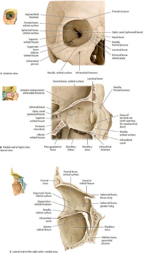

Fig. 28.1 Bones of the orbit
(From Schuenke M, Schulte E, Schumacher U. THIEME Atlas of Anatomy, Vol 3. Illustrations by Voll M and Wesker K. 3rd ed. New York: Thieme Publishers; 2020.)
4.lacrimal,
5.palatine, and
6.sphenoid bones, which form the medial wall; and
7.the zygomatic bone, which, along with the sphenoid bone, forms the lateral wall.
—The eyeball occupies the anterior part of the orbit, accompanied by six extraocular muscles, ophthalmic vessels, and six cranial nerves (optic [CN II], oculomotor [CN III], trochlear [CN IV], trigeminal [CN V], abducent [CN VI], and facial [CN VII]) (Fig. 28.2). Periorbital fat supports and surrounds these structures.
—Three openings at the apex of the orbit allow passage of nerves and vessels between the middle cranial fossa and the orbit: the optic canal, superior orbital fissure, and inferior orbital fissure (Table 28.1).
—Several other openings transmit nerves and vessels onto the face
—supraorbital and infraorbital foramina, frontal incisure, and zygomatico-orbital foramen—and into the nasal cavity—anterior and posterior ethmoidal foramina. The nasolacrimal canal, a passageway between the orbit and nasal cavity, transmits the nasolacrimal duct.
Table 28.1 Openings in the Orbit for Neurovascular Structures
Opening* |
Nerves |
Vessels |
Optic canal |
Optic n. (CN II) |
Ophthalmic a. |
|
|
|
Superior |
Oculomotor n. (CN III) |
|
orbital |
Trochlear n. (CN IV) |
|
fissure |
Abducent n. (CN VI) |
|
|
Trigeminal n., ophthalmic |
|
|
division (CN V1) |
|
• Lacrimal n.
• Frontal n.
• Nasociliary n.
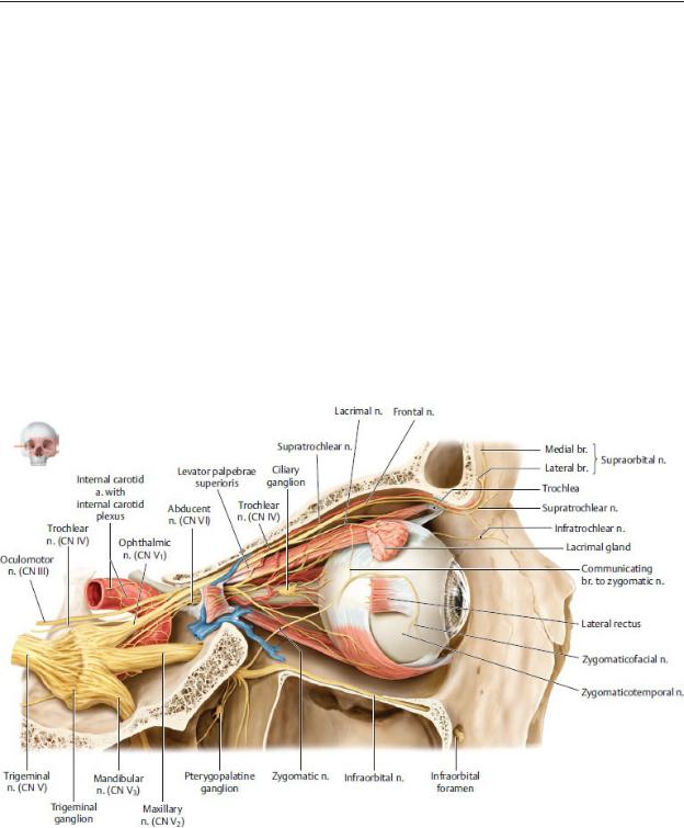
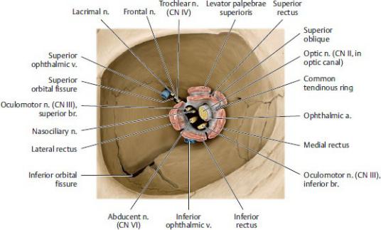
Fig. 28.2 Topography of the orbit
Right orbit, lateral view. (From Schuenke M, Schulte E, Schumacher U. THIEME Atlas of Anatomy, Vol 3. Illustrations by Voll M and Wesker K. 3rd ed. New York: Thieme Publishers; 2020.)
Fig. 28.3 Passage of neurovascular structures through the apex of the orbit
Anterior view. (From Gilroy AM, MacPherson BR, Wikenheiser JC. Atlas of Anatomy. Illustrations by Voll M and Wesker K. 4th ed. New York: Thieme Publishers; 2020.)
—A common tendinous ring at the apex of the orbit surrounds the optic canal and part of the superior orbital fissure and serves as the origin for four of the extraocular muscles (Fig. 28.3). The optic nerve (CN II) and ophthalmic artery enter the orbit through the optic canal and thus pass through the tendinous ring. Other structures entering through part of the superior orbital fissure enclosed by the ring include the superior and inferior branches of the oculomotor nerve (CN III), the nasociliary branch of the ophthalmic nerve (CN V1), and the abducens nerve (CN VI).
Orbital Region, Eyelids, and Lacrimal Apparatus
The upper and lower eyelids are movable folds of skin that protect the eyeball from injury, irritation, and light. They are separated from each other by the palpebral fissure (Fig. 28.4).
— The eyelids are covered externally by skin and internally by the palpebral
conjunctiva. This thin inner membrane reflects at the superior and inferior fornices onto the anterior eyeball as the bulbar (ocular) conjunctiva. When the eyes are closed, the palpebral and bulbar conjunctivae form the conjunctival sac.
—Tarsal plates or tarsus, bands of dense connective tissue that provide support for both the upper and lower eyelids, are attached to medial and lateral palpebral ligaments, which connect to the medial and lateral margins of the orbit, respectively. Tarsal glands within the tarsal plates lubricate the edges of the eyelids to prevent them from sticking together.
—The orbicularis oculi muscle (see Fig. 27.2), innervated by the facial nerve (cranial nerve [CN] VII), closes the eye in a sphincter-like fashion. The levator palpebrae superioris muscle, innervated by the oculomotor nerve (CN III) and attached to the superior tarsal plate, opens the eye by lifting the upper eyelid.
—The orbital septum, a thin membranous sheet, extends from the orbital rim, where it’s continuous with the periosteum, into the tarsal plates of the
eyelids. On the upper eyelid it also blends with the tendon of the levator palpebrae superioris. The septum holds the orbital fat within the orbit and helps limit the spread of infection to and from the orbit.
The lacrimal apparatus produces and drains tears that cleanse and lubricate the outer surface of the eye (Fig. 28.5).
—The lacrimal gland, which produces and secretes tears, resides in the lacrimal fossa in the supralateral aspect of the orbit. Secretomotor parasympathetic fibers from the facial nerve (CN VII) stimulate the gland (see Fig. 26.25).
—Blinking of the eye sweeps the tears across the eye toward the medial angle, where they drain via superior and inferior puncta (openings) into lacrimal canaliculi and the lacrimal sac, the dilated superior part of the nasolacrimal duct.
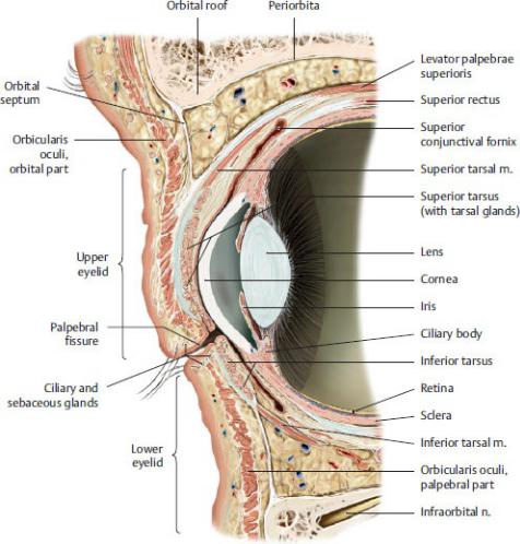
Fig. 28.4 Eyelids and conjunctiva
Sagittal section through the anterior orbital cavity.
(From Gilroy AM, MacPherson BR, Wikenheiser JC. Atlas of Anatomy. Illustrations by Voll M and Wesker K. 4th ed. New York: Thieme Publishers; 2020.)
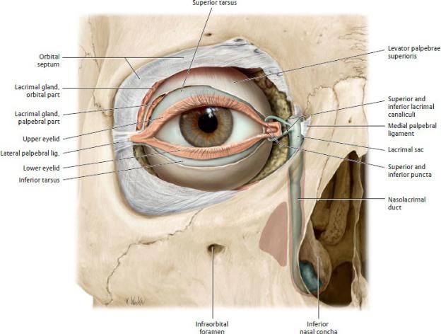
Fig. 28.5 Lacrimal apparatus
Right eye, anterior view.
Removed: Orbital septum (partial). Divided: Levator palpebrae superioris (tendon of insertion). (From Schuenke M, Schulte E, Schumacher U. THIEME Atlas of Anatomy, Vol 3. Illustrations by Voll M and Wesker K. 3rd ed. New York: Thieme Publishers; 2020.)
—The nasolacrimal duct is a membranous structure that begins in the medial angle of the eye and terminates in the inferior meatus of the nasal cavity. Tears drain into the nasal cavity via this duct.
The Eyeball
The eyeball, the organ of vision, has three concentric layers that form its outer walls: the sclera, the choroid, and the retina (Fig. 28.6).
—The sclera, the white part of the eye, forms the posterior five sixths of the outer fibrous layer of the eye; the cornea, the transparent part of the sclera, forms the anterior sixth. This outer layer is largely avascular but provides
structure to the eyeball.
—The choroid, the middle vascular layer, provides oxygen and nutrients to the underlying retina (Fig. 28.7).
•The ciliary body connects the choroid with the circum ference of the iris. Short, smooth muscle fibers, zonular fibers, which attach the ciliary body to the lens, control the thickness and refractive power of the lens and therefore the focus of the eye.
•The iris, an adjustable muscular diaphragm, surrounds a central aperture, the pupil (Fig. 28.8).
◦The pupillary sphincter muscle of the iris responds to parasympathetic stimulation to constrict the pupil.
◦The pupillary dilator muscle of the iris responds to sympathetic stimulation to dilate the pupil.
—The retina, the inner sensory layer, has a posterior optic part that is sensitive to light and a nonvisual part that continues anteriorly over the ciliary body and iris.
•The optic disk, a point on the retina where the optic nerve exits the eyeball, lacks photoreceptors and therefore is insensitive to light and is known as the blind spot.
•The macula of the retina, a spot lateral to the optic disk, is an area of intense visual acuity.
•The fovea centralis, a depression in the macula, is the area of greatest visual acuity.
—Light passes through four refractive media before focusing on the retina of the eye:
1. The cornea, the primary refractive medium for light entering the eye

Fig. 28.6 Structure of the eyeball
Transverse section through the right eyeball, superior view. (From Gilroy AM, MacPherson BR, Wikenheiser JC. Atlas of Anatomy. Illustrations by Voll M and Wesker K. 4th ed. New York: Thieme Publishers; 2020.)
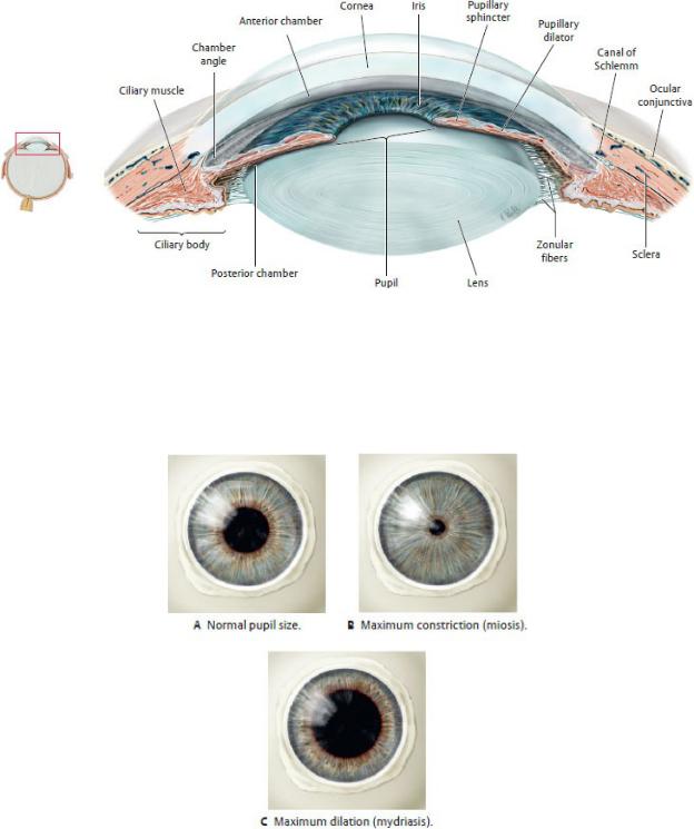

Schuenke M, Schulte E, Schumacher U. THIEME Atlas of Anatomy, Vol 3. Illustrations by Voll M and Wesker K. 3rd ed. New York: Thieme Publishers; 2020.)
2.The aqueous humor, a watery solution that fills the anterior and posterior chambers of the eye that lie anterior to the lens and ciliary body. The balance between its production and drainage determines the intraocular pressure.
3.The lens, a transparent biconcave disk that focuses objects on the retina by changing its thickness. In the process of accommodation, which is mediated by parasympathetic stimulation, the ciliary muscle contracts, causing the lens to thicken and bring near objects into focus. When the ciiary muscle relaxes, the lens flattens, allowing the eye to focus on far objects (Fig. 28.9).
4.The vitreous body, a jelly-like substance that fills the chamber of the eye posterior to the lens.
BOX 28.1: CLINICAL CORRELATION
PRESBYOPIA AND CATARACTS
The lens of the eye undergoes age-related changes that affect vision in the older patient. The loss of elasticity of the lens, and the subsequent loss of accommodation, diminishes a patient’s ability to focus on near objects, a condition known as presbyopia. Opacities of the lens or its capsule, known as cataracts, reduce the amount of light that reaches the retina, resulting in blurred, cloudy vision. Treatment involves surgical removal of the affected lens and replacement with a plastic lens implant.
BOX 28.2: CLINICAL CORRELATION
GLAUCOMA
Glaucoma refers to a group of eye diseases that involve increased intraocular pressure and atrophy of the optic nerve. In primary open-angle glaucoma, the most common form, venous channels in the angle between the cornea and the iris that allow drainage of the aqueous humor from the anterior and posterior cham-bers are blocked. The subsequent buildup of aqueous humor results in raised intraocular pressure and eventual damage to the optic nerve. This leads to a gradual loss of peripheral vision that progresses to tunnel vision. Pressure on the retina can lead to blindness.
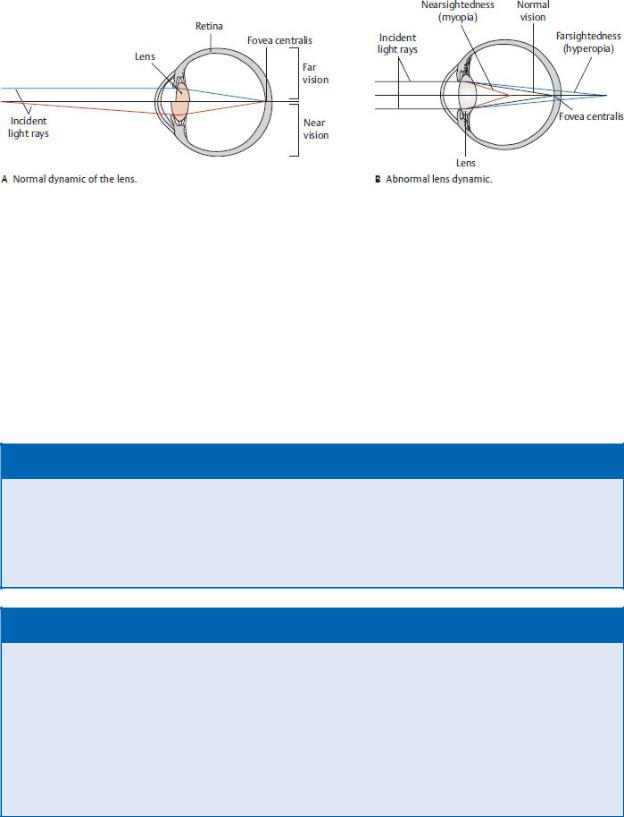
—The six extraocular muscles that control the movement of the eyeball (Fig. 28.10; Table 28.2) include
•four rectus muscles, the superior rectus, medial rectus, inferior rectus, and lateral rectus, that originate from a common tendinous ring at the apex of the orbit; and
•two oblique muscles, the superior oblique that arises near the apex and reflects back via the trochlea to attach to the eyeball, and the inferior oblique that arises from the medial aspect of the orbital floor.
—The muscles allow six cardinal directions of gaze, which are the normal movements of the eyeball tested during clinical evaluation of ocular mobility.
Neurovasculature of the Orbit
The orbital region is an area of arterial and venous anastomoses (Fig. 28.11; see Sections 24.3 and 24.4).
•Branches of the external carotid artery, the infraorbital (a branch of the maxillary artery), and facial arteries, anastomose with the supraorbital branch of the internal carotid artery. This potential anastomosis can serve an important function if the maxillary artery is ligated (such as for severe nosebleeds).
•The anastomosis between the extracranial angular vein and intracranial superior ophthalmic veins can serve as a conduit for bacterial infections of the face passing into intracranial venous pathways.
—The ophthalmic artery supplies most structures of the orbit (Fig. 28.12). One of its branches, the central retinal artery, runs within the optic nerve and is the sole arterial supply to the retina through its terminal branches.
•The ophthalmic artery anastomoses with the facial artery through its supratrochlear branch and with the maxillary artery through its anterior and posterior ethmoidal and middle meningeal branches.
—The superior and inferior ophthalmic veins, which drain structures in the orbit, primarily drain into the cavernous sinus but also communicate with the facial vein and pterygoid venous plexus (Fig. 28.13).
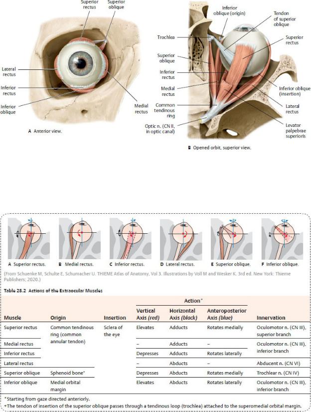
Fig. 28.10 Extraocular muscles
Right eye. (From Schuenke M, Schulte E, Schumacher U. THIEME Atlas of Anatomy, Vol 3. Illustrations by Voll M and Wesker K. 3rd ed. New York: Thieme Publishers; 2020.)
BOX 28.5: CLINICAL CORRELATION
OCULOMOTOR NERVE INJURY
The oculomotor nerve innervates most of the extraocular mus-cles. With paralysis of these muscles, the eye is directed down-ward and outward because of the unopposed action of the lateral rectus (innervated by the abducent nerve) and superior oblique (innervated by the trochlear nerve) (A). The dilator pupillae is also unopposed, so the pupil remains fully dilated. Paralysis of the levator palpebrae superior causes the superior eyelid to droop (B).
(From Schuenke M, Schulte E, Schumacher U. THIEME Atlas of Anatomy, Vol 3. Illustrations by Voll M and Wesker K. 3rd ed. New York: Thieme Publishers; 2020.)
—Six cranial nerves (optic, oculomotor, trochlear, trigeminal, abducent, and facial) innervate structures in the orbit. All of these nerves pass through the cavernous sinus before entering the orbit at its apex (Table 28.3; Figs. 28.14 and 28.15).
•The optic nerve (CN I) transmits images from the retina.
•The oculomotor nerve (CN III), trochlear nerve (CN IV), and abducent nerve (CN VI) innervate the extraocular muscles.
•The ophthalmic division of the trigeminal nerve (CN V1) carries general sensory fibers from structures in the orbit and distributes postganglionic autonomic fibers to target organs of the orbit and face.
•The facial nerve (CN VII) provides secretomotor (parasympathetic) innervation to the lacrimal gland.
—Autonomic innervation of structures in the orbit includes the following:
•Sympathetic fibers from the carotid plexus innervate the ciliary body and pupillary dilator muscle (responsible for pupil dilation).
•Parasympathetic fibers from the oculomotor nerve (CN III) synapse in the ciliary ganglion and travel via the short ciliary nerves to innervate the ciliary body and pupillary sphincter muscles (responsible for pupil constriction).
•Parasympathetic fibers from the facial nerve (CN VII) synapse in the
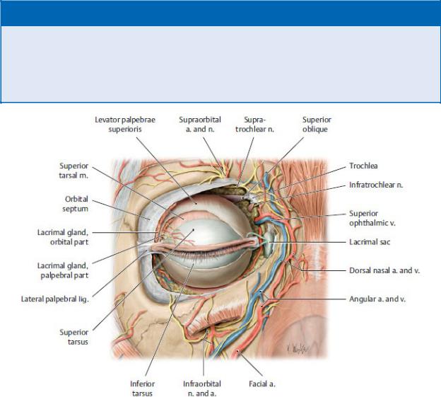
pterygopalatine ganglion and travel via the zygomatic nerve (CN V2) to innervate the lacrimal gland (responsible for tear secretion).
BOX 28.6: CLINICAL CORRELATION
HORNER’S SYNDROME
Horner’s syndrome is a kaleidoscope of symptoms resulting from disruption of the cervical sympathetic trunk in the neck. The absence of sympathetic innervation is manifested on the affected side of the face as pupillary constriction (miosis), a sunken eye (enophthalmos), drooping of the upper eyelid (ptosis), loss of sweating (anhydrosis), and vasodilation.
Fig. 28.11 Neurovascular structures of the orbital region
Anterior orbital structures have been exposed by partial removal of the orbital septum. (From Schuenke M, Schulte E, Schumacher U. THIEME Atlas of Anatomy, Vol 3. Illustrations by Voll M and Wesker K. 3rd ed. New York: Thieme Publishers; 2020.)
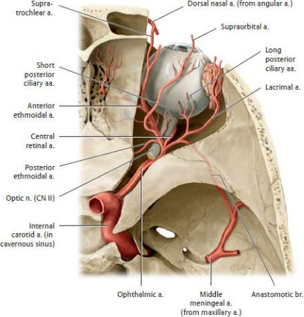
Fig. 28.12 Arteries of the orbit
Right orbit, superior view. Opened: optic canal and orbital roof. (From Schuenke M, Schulte E, Schumacher U. THIEME Atlas of Anatomy, Vol 3. Illustra-tions by Voll M and Wesker K. 3rd ed. New York: Thieme Publishers; 2020.)
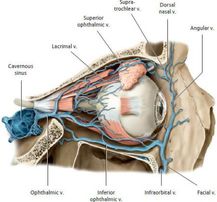
Fig. 28.13 Veins of the orbit
Right orbit, lateral view. Removed:lateral orbital wall. Opened: maxillary sinus. (From Schuenke M, Schulte E, Schumacher U. THIEME Atlas of Anatomy, Vol 3. Illustrations by Voll M and Wesker K. 3rd ed. New York: Thieme Publishers; 2020.)
Table 28.3 Nerves of the Orbit
Nerve |
Nerve Fibers |
Distribution |
Optic (CN II) |
Special sensory for |
Retina |
|
vision |
|
|
|
|
Oculomotor (CN III) |
Somatic motor |
Muscles of the orbit, |
|
|
except the lateral |
|
|
rectus and superior |
|
|
oblique muscles |
|
Parasympathetic: |
Pupilary sphincter |
|
synapse in ciliary |
muscle and ciliary |
|
ganglion; postsynaptic |
body |

fibers travel with short ciliary nn. (CN V1)
Trochlear (CN IV) |
Somatic motor |
Superior oblique |
|
|
|
|
muscle |
|
|
|
|
Ophthalmic (CN V1) |
|
|
|
Lacrimal |
General sensory |
Lacrimal gland, upper |
|
|
|
|
lateral eyeball |
Frontal |
|
|
|
- |
Supratrochlear |
General sensory |
Anterior scalp |
- |
Supraorbital |
General sensory |
Anterior scalp |
Short ciliary |
General sensory |
Ciliary body and iris |
|
|
|
Parasympathetic (CN |
|
|
|
III) and sympathetic |
|
Nasociliary
- |
Anterior and |
General sensory |
Nasal cavity, ethmoid |
|
posterior |
|
and sphenoid sinuses |
|
ethmoidal |
|
|
- |
Infratrochlear |
General sensory |
External nose, |
|
|
|
conjunctiva, lacrimal |
|
|
|
sac |
- |
Long ciliary |
General sensory |
Iris and cornea |
|
|
Sympathetic (carotid |
Pupil dilator |
|
|
plexus) |
|
Abducent (CN VI) |
Somatic motor |
Lateral rectus muscle |
|
|
|
|
|
Facial (CN VII) |
Parasympathetic: |
Lacrimal gland |
|
|
|
synapse in |
|
|
|
pterygopalatine |
|
|
|
ganglion; postsynaptic |
|
|
|
fibers travel with |
|
|
|
zygomatic n. (CN V2) |
|
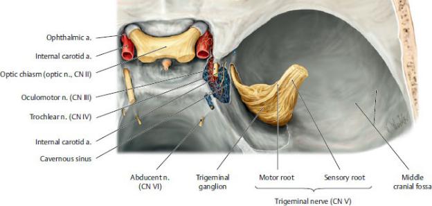
Fig. 28.14 Intracavernous course of the cranial nerves that enter the orbit
Superior view of the sella turcica and middle cranial fossae, right side. The lateral and superior walls of the cavernous sinus have been opened and the trigeminal ganglion has been retracted laterally. The three cranial nerves that supply the ocular muscles (CN III, CN IV, and CN VI) pass through the cavernous sinus with CN V1 and CN V2 and the internal carotid artery. All of the nerves course along the lateral wall of the sinus except CN VI, which runs directly through the sinus in close proximity to the artery. Because of this relationship, the CN VI may be damaged as a result of a sinus thrombosis or an intracavernous aneurysm of the internal carotid artery. (From Gilroy AM, MacPherson BR, Wikenheiser JC. Atlas of Anatomy. Illustrations by Voll M and Wesker K. 4th ed. New York: Thieme Publishers; 2020.)
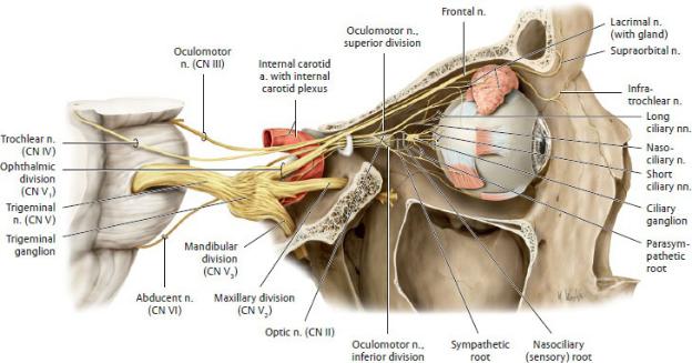
Fig. 28.15 Innervation of the orbit
Right orbit, lateral view. Removed: Temporal bony wall. (From Schuenke M, Schulte E, Schumacher U. THIEME Atlas of Anatomy, Vol 3. Illustrations by Voll M and Wesker K. 3rd ed. New York: Thieme Publishers; 2020.)
28.2 The Ear
The ear, which contains the organs for hearing and equilibrium, is divided into external, middle, and internal parts (Fig. 28.16).
The External Ear
The external ear collects and conducts sound.
—The auricle, the visible external part of the ear, is composed mostly of a skeleton of elastic cartilage that is covered by skin (Fig. 28.17).
•Inferiorly is the soft (non-cartilaginous) lobule.
•Anteriorly, the small part projecting posteriorly over the opening of the auditory canal is the tragus, which is separated from the antitragus by a small notch.
•The posterior margin of the auricle is defined by the helix, which curves around the scaphoid fossa and ends at the concha, a depression at the entrance to the auditory canal.
•The rim of the antihelix begins at the antitragus and curves superiorly,
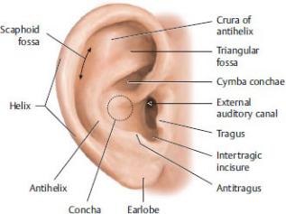
forming the cymba concha. Superiorly its crura diverge to form the triangular fossa.
—The external acoustic meatus (external auditory canal), a canal that extends 2 to 3 cm from the auricle to the tympanic membrane, conducts sound waves toward the middle ear. The outer third of the canal is cartilaginous, and the inner two thirds are formed by the temporal bone. Ceruminous and sebaceous glands in the subcutaneous tissue lining the cartilaginous part secrete earwax.
Fig. 28.17 Structure of the auricle
Right auricle, lateral view. (From Gilroy AM, MacPherson BR, Wikenheiser JC. Atlas of Anatomy. Illustrations by Voll M and Wesker K. 4th ed. New York: Thieme Publishers; 2020.)
—The tympanic membrane, a thin, transparent membrane, separates the external and middle ear.
•Skin covers the tympanic membrane externally, and a mucous membrane lines it internally.
•The concave outer surface of the membrane has a central conelike depression, the umbo.
•A thin, superior portion of the membrane, the flaccid part (pars flaccida), is distinct from the rest of the membrane, the tense part (pars tensa).
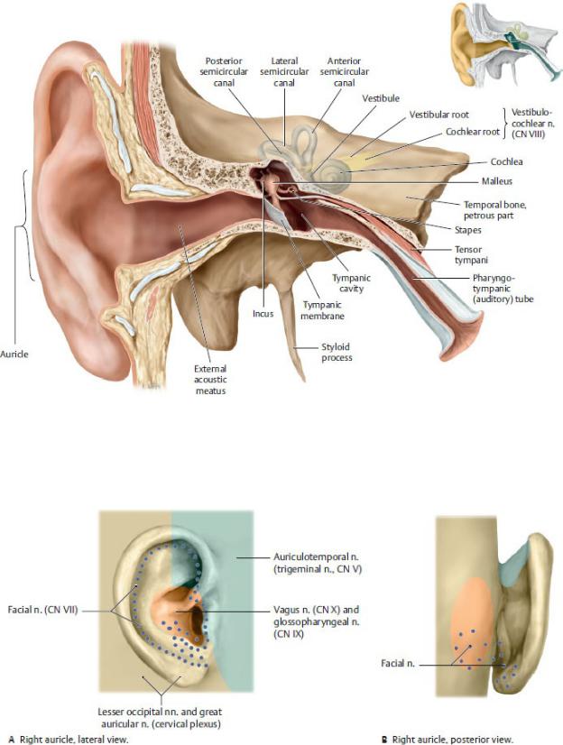
(From Schuenke M, Schulte E, Schumacher U. THIEME Atlas of Anatomy, Vol 3. Illustrations by Voll M and Wesker K. 3rd ed. New York: Thieme Publishers; 2020.)
—Posterior auricular and anterior auricular arteries, branches of the superficial temporal artery, supply the external ear.
—Sensation from the external ear is transmitted by the cervical plexus and three cranial nerves (Fig. 28.18):
•the great auricular nerve (cervical plexus) from the auricle;
•the auriculotemporal nerve, a branch of CN V3, from the auricle and external surface of the tympanic membrane;
•the auricular branch of the vagus nerve (CN X) from the external surface of the tympanic membrane; and
•the glossopharyngeal nerve (CN IX) from the internal surface of the tympanic membrane.
The Middle Ear
—The middle ear, also known as the tympanic cavity, is an air-filled chamber housed in the petrous portion of the temporal bone (Figs. 28.19, 28.20, 28.21).
•Anteriorly, a pharyngotympanic tube, which connects the tympanic cavity to the nasopharynx, helps equalize pressure in the middle ear.
•Posteriorly, the aditus (inlet) to the mastoid antrum (a cavity in the mastoid process of the temporal bone) connects the tympanic cavity to the bony meshwork of mastoid air cells.
Fig. 28.19 Tympanic cavity and pharyngotympanic tube
Medial view of opened tympanic cavity. (From Gilroy AM, MacPherson BR, Wikenheiser JC. Atlas of Anatomy. Illustrations by Voll M and Wesker K. 4th ed. New York: Thieme Publishers; 2020.)
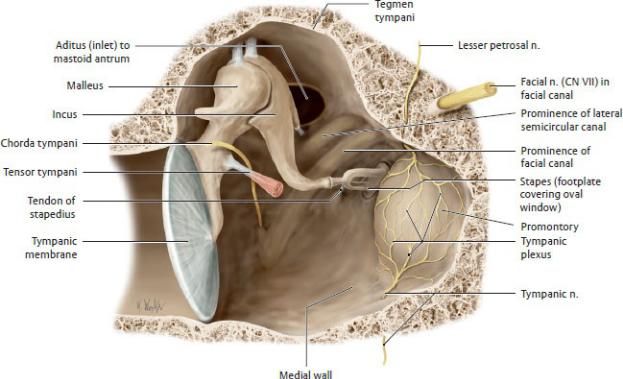
Fig. 28.20 Tympanic cavity
Right tympanic cavity, anterior view. Removed: Anterior wall. (From Gilroy AM, MacPherson BR, Wikenheiser JC. Atlas of Anatomy. Illustrations by Voll M and Wesker K. 4th ed. New York: Thieme Publishers; 2020.)
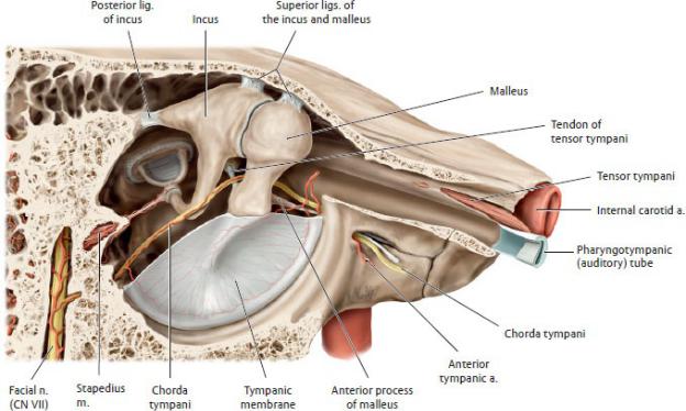
Fig. 28.21 Muscles and neurovascular relations in the tympanic cavity
Right middle ear, lateral view. (From Schuenke M, Schulte E, Schumacher U. THIEME Atlas of Anatomy, Vol 3. Illustrations by Voll M and Wesker K. 3rd ed. New York: Thieme Publishers; 2020.)
•A thin bony plate, the tegmen tympani, forms the roof of the tympanic cavity and separates it from the middle cranial fossa.
•The medial wall, which separates the tympanic cavity from the internal ear, has a promontory, covered by the tympanic nerve plexus, and two openings, the oval and round windows (see Fig. 28.20).
—The auditory ossicles, the malleus, incus, and stapes bones of the middle ear, articulate with each other through synovial joints and form a bony chain between the tympanic mem-brane and the oval window of the internal ear.
•The handle of the malleus is embedded in the tym-panic membrane, and its head articulates with the incus.
•The incus articulates with the malleus and stapes.
•The head of the stapes articulates with the incus, and its base fits into the oval window of the bony labyrinth of the internal ear.
—Muscles of the middle ear dampen the movements of the auditory ossicles, thereby lessening the sound transmitted from the external ear.
•The tensor tympani, which is innervated by a branch of the mandibular

nerve (CN V3), lessens the damage from loud sounds by tensing the tympanic membrane.
•The stapedius, which is innervated by a branch of the facial nerve (CN VII), dampens the vibrations of the stapes on the oval window.
—The ascending pharyngeal, maxillary, and posterior auricular branches of the external carotid artery and a small branch of the internal carotid artery supply the middle ear.
—The chorda tympani, a branch of the facial nerve (CN VII), has no branches in the middle ear but passes between the malleus and incus to exit the cavity through a small opening in the temporal bone.
—The glossopharyngeal nerve (CN IX) transmits sensation from the tympanic cavity and pharyngotympanic tube. Pregangli-onic parasympathetic fibers carried in the tympanic nerve (a branch of the glossopharyngeal nerve) synapse in the otic ganglion. The postganglionic fibers join with sympathetic fibers of the internal carotid plexus to form the tympanic plexus (see Fig. 26.29).
BOX 28.7: CLINICAL CORRELATION
OTITIS MEDIA
Otitis media is an infection of the middle ear that occurs com-monly in children often following an upper respiratory tract infec-tion. Fluid that accumulates in the middle ear can temporarily diminish hearing, and inflammation of the lining of the tympanic cavity can block the pharyngotympanic tube.
BOX 28.8: CLINICAL CORRELATION
HYPERACUSIS
The stapedius muscle protects the delicate inner ear by modi-fying the vibrations of very loud sounds as they are transmitted through the middle ear to the stapes. Paralysis of the muscle resulting from a lesion of the facial nerve causes an extreme sen-sitivity to sound, a condition known as hyperacusis.
The Internal Ear
—The internal ear, which contains the organ for hearing, the auditory apparatus, and the organ for balance, the vestibular apparatus, is encased within the petrous part of the temporal bone (Figs. 28.22 and 28.23) and consists of the following:
• A bony otic capsule, which forms the walls of the bony labyrinth
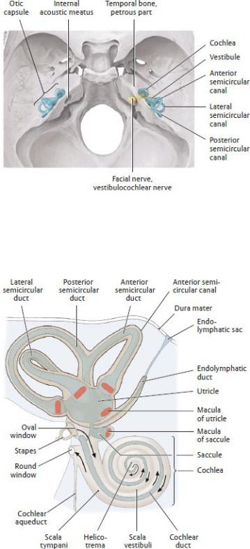
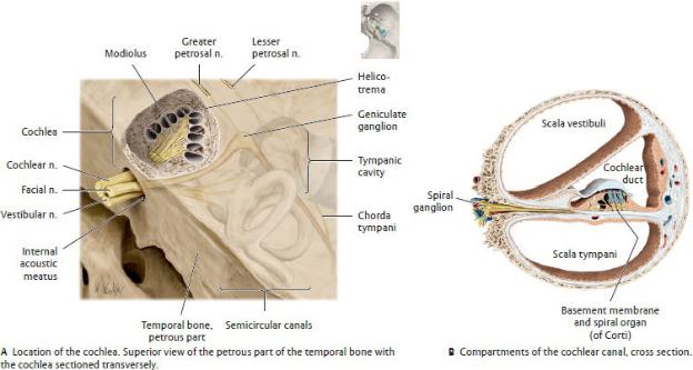
the temporal bone. It is composed of a membranous labyrinth filled with endolymph floating within a similarly shaped bony labyrinth filled with perilymph. (From Schuenke M, Schulte E, Schumacher U. THIEME Atlas of Anatomy, Vol 3. Illustrations by Voll M and Wesker K. 3rd ed. New York: Thieme Publishers; 2020.)
Fig. 28.24 Auditory apparatus
(From Schuenke M, Schulte E, Schumacher U. THIEME Atlas of Anatomy, Vol 3. Illustrations by Voll M and Wesker K. 3rd ed. New York: Thieme Publishers; 2020.)
•A bony labyrinth, a series of chambers and canals within the otic capsule containing the fluid perilymph. It includes the cochlea, the vestibule, and the semi-circular canals.
•A membranous labyrinth, a series of sacs and ducts sus-pended within the bony labyrinth, and filled with a fluid, endolymph. The membranous labyrinth is made up of
◦the cochlear duct contained within the cochlea,
◦the utricle and saccule contained within the vestibule, and
◦the semicircular ducts contained within the semi circular canals.
—The auditory apparatus consists of the following:
•The cochlea, a space within the bony labyrinth that includes the bony cochlear (spiral) canal, which makes 2.5 turns around its axis, the
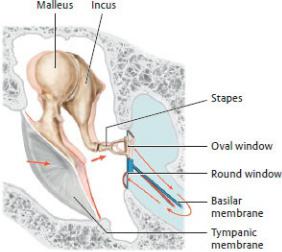
modiolus (Fig. 28.24). The basal turn of the cochlea forms the promontory on the medial wall of the middle ear and contains the round window, which is closed by a membrane.
•The cochlear duct, part of the membranous labyrinth, which is a blindended duct filled with endolymph and suspended within the cochlear canal (Fig. 28.24):
◦The cochlear duct divides the cochlear canal into two channels, the scala vestibuli and scala tympani, which are continuous with each other at the helicotrema, a space at the apex of the canal.
◦At the base of the cochlea, the scala vestibuli lies against the oval window, and the scala tympani lies against the round window.
•The spiral organ (of Corti), which contains the sensory receptors for hearing, and is embedded in the basement membrane on the floor of the cochlear duct
Fig. 28.25 Propagation of sound waves by the ossicular chain
(From Schuenke M, Schulte E, Schumacher U. THIEME Atlas of Anatomy, Vol 3. Illustrations by Voll M and Wesker K. 3rd ed. New York: Thieme Publishers; 2020.)
—The sequence of sound transmission through the ear involves (Fig. 28.25) the following:
1.Transmission of sound waves from the external ear and external acoustic meatus to the tympanic membrane of the middle ear. The waves vibrate the auditory ossicles and, in turn, the oval window that is attached to the base of the stapes.
2.Transmission of vibrations of the oval window to the perilymph of the
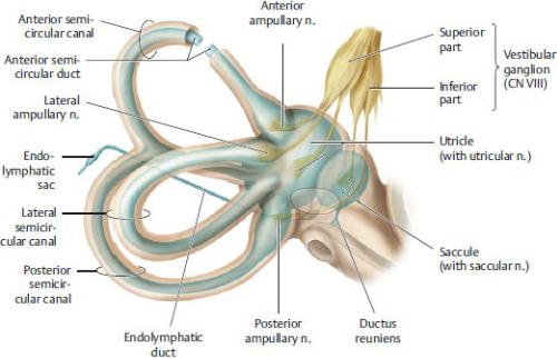
scala vestibuli, which creates pressure waves that displace the basement membrane and spiral organ of the cochlear duct. Nerve endings in the spiral organ transmit impulses to the brain along the cochlear nerve.
Fig. 28.26 Structure of the vestibular apparatus
(From Gilroy AM, MacPherson BR, Wikenheiser JC. Atlas of Anatomy. Illustrations by Voll M and Wesker K. 4th ed. New York: Thieme Publishers; 2020.)
3.Transmission of the pressure waves of the perilymph from the scala vestibuli along the scala tympani to the round window, with dissipation into the tympanic cavity.
—The vestibular apparatus consists of the following (Fig. 28.26):
• The vestibule of the bony labyrinth that communicates with the cochlea and semicircular canals
◦A small extension, the vestibular aqueduct, communi-cates with the posterior cranial fossa and contains the endolymphatic sac, a membranous storage space for excess endolymph.
•The utricle and saccule, part of the membranous labyrinth, which lie within the vestibule
◦The utricle communicates with the semicircular ducts; the saccule communicates with the cochlear duct.
◦Both the uticle and saccule contain specialized sensory fields called

maculae, which occupy different positions in space and are sensitive to movement of the endo-lymph in the horizontal and vertical planes.
•Three semicircular canals of the bony labyrinth, which are arranged perpendicular to one another and communicate with the vestibule. Each canal has a swelling at one end, the bony ampulla.
•Three semicircular ducts, parts of the membranous labyrinth contained within the semicircular canals, that communicate with the utricle
◦An ampulla at one end of each semicircular duct contains the ampullary crest, an area of sensory epithelium. The ampullary crests respond to motion of the endolymph within the ducts caused by rotation of the head.
—The membranous labyrinth receives its blood supply from the internal auditory artery, a branch of the basilar artery via its anterior inferior cerebellar artery.
—The vestibular and cochlear nerves within the internal acoustic meatus form the vestibulocochlear nerve (CN VIII) (see Fig. 26.27)
•The vestibular nerve innervates organs of the vestibular apparatus: the maculae of the utricle and saccule and the ampullary crests of the semicircular canals. Neuron cell bodies lie within the vestibular ganglion in the internal acoustic meatus.
•The cochlear nerve innervates the spiral organ (of Corti) in the cochlea. Neuron cell bodies lie in the spiral ganglion of the cochlea at the modiolus.
BOX 28.9: CLINICAL CORRELATION
MENIERE’S DISEASE
Meniere’s disease is an inner ear disorder resulting from block-age of the cochlear duct. Recurring episodes are characterized by tinnitus (ringing or buzzing in the ear), vertigo (the illusion of movement), and hearing loss. The hearing loss may fluctuate in intensity and from ear to ear but eventually becomes permanent.
BOX 28.10: CLINICAL CORRELATION
VERTIGO, TINNITUS, AND HEARING LOSS
Trauma to the ear can cause three types of symptoms: vertigo, tinnitus, and hearing loss. Vertigo refers to the illusion of move-ment, or dizziness, and results from injury to the semicircular canals. Tinnitus refers to a ringing in the ears and is a disorder involving the cochlear duct. Causes of hearing loss can be either peripheral or centrally located. Conductive hearing loss occurs when there is impaired transmission of sound waves through the auditory

canal to the auditory ossicles. Sensorineural hearing loss occurs when there is damage to the pathway between the cochlea and the brain.