


16 The Perineum
The perineum is the space inferior to the pelvic floor, which is divided into a urogenital triangle and an anal triangle. The urogenital triangle contains the male and female external genital structures, and the anal triangle contains the anal canal and anus.
16.1 Perineal Spaces
—The boundaries of the diamond-shaped perineum (Figs. 16.1 and 16.2; see also Fig. 14.4) are
•the pelvic outlet (pubic symphysis, ischiopubic rami, sacrotuberous ligaments, and coccyx), which forms the perimeter;
•the lower parts of the obturator internus muscles and their obturator fasciae, which line the lateral walls;
•the inferior surface of the pelvic diaphragm, which forms the roof; and
•the skin of the perineum, which forms the floor.
—A line connecting the ischial tuberosities separates the perineum into an anterior urogenital triangle and a posterior anal triangle
—The perineal membrane, a tough, fibrous sheet, stretches between the ischiopubic rami, extending anteriorly almost to the pubic symphysis and posteriorly to the ischial tuberosities.
•It separates the urogenital triangle into the deep perineal and superficial perineal spaces (see Fig. 14.3).
•It forms a platform for the attachment of the cavernous bodies (which become engorged during arousal) of the external genitalia.
—The superficial perineal space is a potential space bounded above by the perineal membrane and below by the superficial perineal fascia (Colles’ fascia), the extension of the membranous layer of superficial fascia (Scarpa’s fascia) on the abdominal wall.
•In both sexes it contains
◦the bulbospongiosus, ischiocavernosus, and superficial transverse perineal muscles (see Section 16.2) and
◦the deep perineal branches of the internal pudendal vessels and pudendal nerve.
•In males (see Section 16.3) it also contains
◦the root of the penis and
◦the proximal portion of the spongy (penile) urethra.
•In females (see Section 16.4) it also contains
◦the clitoris and associated muscles,
◦the bulbs of the vestibule and
◦the greater vestibular glands.
—The deep perineal space is bounded inferiorly by the perineal membrane and superiorly by the inferior fascia of the pelvic diaphragm.
•In both sexes it contains
◦part of the urethra and inferior part of the external urethral sphincter,
◦the anterior recesses of the ischioanal fat pads, and
◦neurovascular structures to the penis or clitoris.
•In males it also contains
◦the membranous part of the urethra,
◦the bulbourethral glands, and
◦the deep transverse perineal muscles. (In females this is often replaced by smooth muscle.)
•In females it also contains
◦compressor urethrae, urethrovaginal sphincter, and parts of the external urethral sphincter; and
◦the proximal part of the urethra.
Fig. 16.1 Muscles and fascia of the female perineum
Lithotomy position, caudal (inferior) view. The green arrow is pointing forward to the anterior recess of the ischioanal fossa. (From Schuenke M, Schulte E, Schumacher U. THIEME Atlas of Anatomy, Vol 1. Illustrations by Voll M and Wesker K. 3rd ed. New York: Thieme Publishers; 2020.)
Fig. 16.2 Muscles and fascia of the male perineum
Lithotomy position, caudal (inferior) view. The green arrow is pointing forward

to the anterior recess of the ischioanal fossa. (From Schuenke M, Schulte E, Schumacher U. THIEME Atlas of Anatomy, Vol 1. Illustrations by Voll M and Wesker K. 3rd ed. New York: Thieme Publishers; 2020.)
16.2 Muscles of the Perineum
—Muscles of the perineum support the pelvic floor, surround the orifices of the urethra and anus, and assist in the erection of genital structures ( Table
16.1; Fig. 16.3).
—The perineal body is an irregular subcutaneous mass of fibro-muscular tissue formed by converging fibers of the levator ani, the superficial transverse perineal and deep transverse perineal muscles, bulbospongiosus muscles, and the external anal sphincter.
•It lies between the rectum and bulb of the penis in males and between the rectum and vagina in females.
•It supports the pelvic diaphragm and pelvic viscera.
—Perineal branches of the internal pudendal artery supply muscles of the perineum. Venous blood drains to the internal pudendal vein and the internal iliac vein.
—The pudendal nerve (S2–S4) innervates the muscles of the perineum.
BOX 16.1: CLINICAL CORRELATION
EPISIOTOMY
During vaginal delivery, pressure on the perineum risks tearing the perineal muscles. A clean incision through the posterior vaginal orifice into the perineal body, known as an episiotomy, is often performed to enlarge the opening and prevent damage to perineal muscles. A median, or midline, episiotomy extends only into the perineal body, but if further traumatic tearing occurs to extend the cut, it can damage the external anal sphincter (resulting in fecal incontinence) or create an anovaginal fistula. Mediolateral episiotomies often replace the midline incisions. These extend laterally from the vaginal orifice to the superficial perineal muscles, thus avoiding the perineal body and the possible sequelae from extensive tearing. However, while the more lateral incisions provide greater access, they are more difficult to repair.
BOX 16.2: CLINICAL CORRELATION
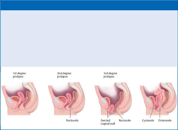
PROLAPSE OF PELVIC ORGANS
The pelvic diaphragm, pelvic ligaments, and perineal body provide important structural support for the pelvic viscera. Stretching or disruption of these tissues often occurs during childbirth and results in prolapse of the uterus into the vagina. A widening of the genital hiatus from an atrophic pelvic floor or a weakened perineal body can allow the bladder (cystocele), rectum (rectocele), or rectovesical pouch (enterocele) to bulge into the vaginal wall.
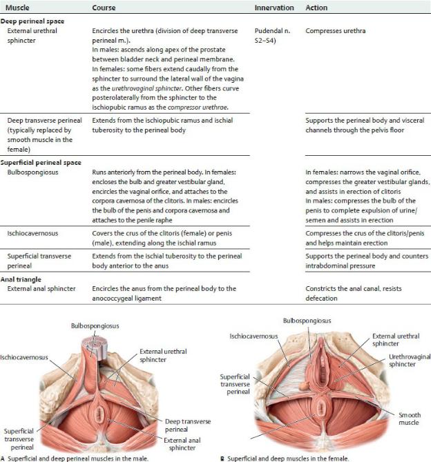
Fig. 16.3 Muscles of the perineum
Inferior view. (From Gilroy AM, MacPherson BR, Wikenheiser JC. Atlas of Anatomy. Illustrations by Voll M and Wesker K. 4th Edition. New York: Thieme Publishers; 2020.)
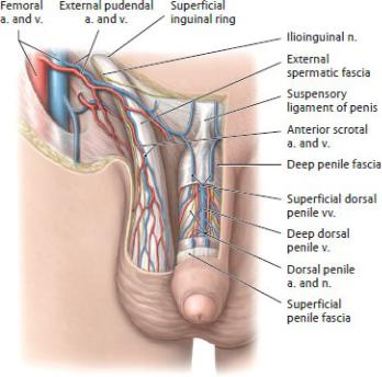
Fig. 16.4 Neurovasculature of the penis and scrotum
Anterior view. Partially removed: Skin and fascia. (From Schuenke M, Schulte E, Schumacher U. THIEME Atlas of Anatomy, Vol 1. Illustrations by Voll M and Wesker K. 3rd ed. New York: Thieme Publishers; 2020.)
16.3 Male Urogenital Triangle
The male urogenital triangle contains the scrotum, penis, bulbourethral glands, perineal muscles, and associated neurovasculature.
The Scrotum
—The scrotum is a saccular extension of the anterior abdominal wall that encloses the testes and spermatic cord (see Section 10.4).
—The subcutaneous layer of the skin over the scrotum is devoid of fat, but the dartos fascia that underlies the skin is continuous with the deep membranous layer (Scarpa’s fascia) of the abdomen and with the superficial perineal fascia (Colles’ fascia) of the perineum.
—An extension of the dartos fascia divides the scrotum into right and left compartments and is visible externally as the scrotal raphe.
—Scrotal branches of the internal pudendal and external pudendal arteries, and cremasteric branches of the inferior epigastric artery supply the scrotum. Veins of similar names accompany the arteries (Figs. 16.4 and 16.5).
—Lymph from the scrotum drains to superficial inguinal nodes. Recall that lymph from the contents within the scrotum (i.e., the testis and epididymis) drains directly to the para-aortic nodes.
—Innervation of the scrotum (see Fig. 14.21) is supplied by
•the ilioinguinal and genitofemoral (genital branch) nerves (lumbar plexus), which innervate the anterior scrotum, and
•the pudendal and posterior femoral cutaneous nerves of the thigh (sacral plexus), which innervate the posterior scrotum.
Fig. 16.5 Blood vessels of the male genitalia
Left lateral view. (From Schuenke M, Schulte E, Schumacher U. THIEME Atlas
of Anatomy, Vol 1. Illustrations by Voll M and Wesker K. 3rd ed. New York: Thieme Publishers; 2020.)
The Penis
The penis serves both copulatory and urinary functions. It contains three cylindrical bodies of erectile tissue, one of which surrounds the spongy urethra that transmits urine from the bladder as well as semen during sexual intercourse
(Figs. 16.6 and 16.7).
—The penis has three parts:
1.The root, the most proximal part, is attached to the perineal membrane and is covered by muscles. It is composed of
◦right and left crura that are attached to the ischiopubic rami on either side and are covered by the ischiocavernosus muscles, and
◦the bulb of the penis, which is attached to the perineal membrane and covered by the bulbospongiosus muscle. The spongy (penile) urethra enters on its dorsal surface.
2.The body, pendulous and not covered by muscle, is made up of three cylindrical bodies of erectile tissue. A tunica albuginea, a dense fibrous coat, surrounds each erectile body, and distally a deep penile fascia (Buck’s fascia) binds the three bodies together. The three erectile bodies include
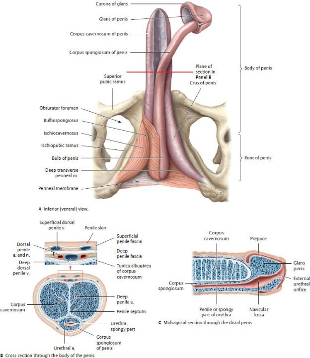
Fig. 16.6 Penis
(From Schuenke M, Schulte E, Schumacher U. THIEME Atlas of Anatomy, Vol 1. Illustrations by Voll M and Wesker K. 3rd ed. New York: Thieme Publishers; 2020.)
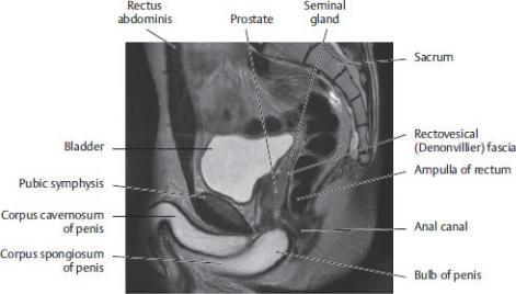
Fig. 16.7 MRI of the male pelvis
Sagittal section, left lateral view. (From Hamm B, et al. MRI Imaging of the Abdomen and Pelvis, 2nd ed. New York: Thieme Publishers; 2009.)
◦two corpora cavernosa, continuations of the crura, which lie side by side on the dorsum of the penis, and
◦one corpus spongiosum, a continuation of the bulb of the penis, which lies ventral to the two corpora cavernosa and is traversed by the spongy (penile) urethra.
3.The glans penis (also known as the glans), an expansion of the distal end of the corpus spongiosum, is characterized by
◦the corona, which overhangs the distal ends of the corpora cavernosa, and
◦a fusiform dilation of the spongy urethra, the navicular fossa, which terminates at the external urethral orifice at its tip.
—External pudendal arteries supply the skin and subcutaneous tissue of the penis. Venous drainage of this tissue passes through the superficial dorsal veins, which drain to the external pudendal veins (see Fig. 16.4).
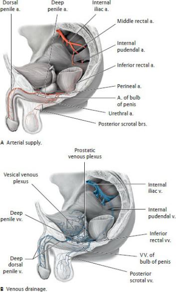
—Internal pudendal arteries supply the deep penile structures. Their branches
(Fig. 16.8; see also Fig. 16.6B) include
• the artery of the bulb of the penis, which supplies the bulb of the penis, the urethra within the bulb, and the bulbourethral glands;
• the dorsal penile artery, which runs between the deep penile fascia and tunica albuginea to supply the penile fascia and skin and the glans; and
• the deep penile artery, which runs within the corpus cavernosum and gives off helicine arteries that supply the erectile tissue and are
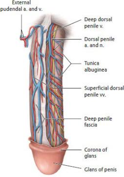
responsible for engorgement of the corpora during erection.
—The erectile bodies are drained by venous plexuses that empty into the single deep dorsal vein, which passes under the pubic symphysis to join the prostatic venous plexus in the pelvis.
—Lymphatic drainage areas of the penis include
•the erectile bodies of the penis, which drain to internal iliac nodes;
•the glans penis, which drains to deep inguinal nodes; and
•the urethra, which drains to internal iliac and deep inguinal nodes.
Fig. 16.8 Neurovasculature on the dorsum of the penis
Superior (dorsal) view. Removed: Skin. (From Schuenke M, Schulte E, Schumacher U. THIEME Atlas of Anatomy, Vol 1. Illustrations by Voll M and Wesker K. 3rd ed. New York: Thieme Publishers; 2020.)
—The glans of the penis is richly innervated by sensory fibers via the dorsal penile nerve, a branch of the pudendal nerve. Sympathetic fibers from the hypogastric nerve plexus are also carried along this route.
—Parasympathetic fibers carried by the cavernous nerves derived from the prostatic plexus innervate the helicine arteries within the erectile tissue and are responsible for penile erection.
Bulbourethral Glands
The bulbourethral glands are paired mucus-secreting glands (see Figs. 15.2 and
15.3).
—They lie on either side of the urethra below the prostate, surrounded by the urethral sphincter.
—Their ducts open into the proximal part of the spongy urethra.
—They are active during sexual arousal.
Erection, Emission, and Ejaculation
The sexual responses of erection, emission, and ejaculation involve sympathetic, parasympathetic, and somatic (via the pudendal nerve) pathways.
—During erection
•constriction of the helicine arteries, normally maintained by sympathetic innervation, is inhibited by parasympathetic stimulation. As the arteries relax, the cavernous spaces within the erectile bodies dilate and become engorged;
•contractions of the bulbocavernosus and ischiocavernosus muscles, innervated by the pudendal nerve, impede venous outflow and maintain the erection.
—During emission
•parasympathetic stimulation mediates the secretion of seminal fluid from the seminal glands, bulbourethral glands, and prostate gland, and
•sympathetic stimulation mediates emission (the movement of seminal fluid through the ducts) by initiating peristalsis of the ductus deferens and seminal glands. This propels the seminal fluid into the prostatic urethra, where additional fluid is added as the prostate contracts.
—During ejaculation
•sympathetic stimulation constricts the internal urethral sphincter, which prevents the seminal fluid from entering the bladder (retrograde ejaculation);
•parasympathetic stimulation contracts the urethral muscles; and
•the pudendal nerve contracts the bulbospongiosus muscle.
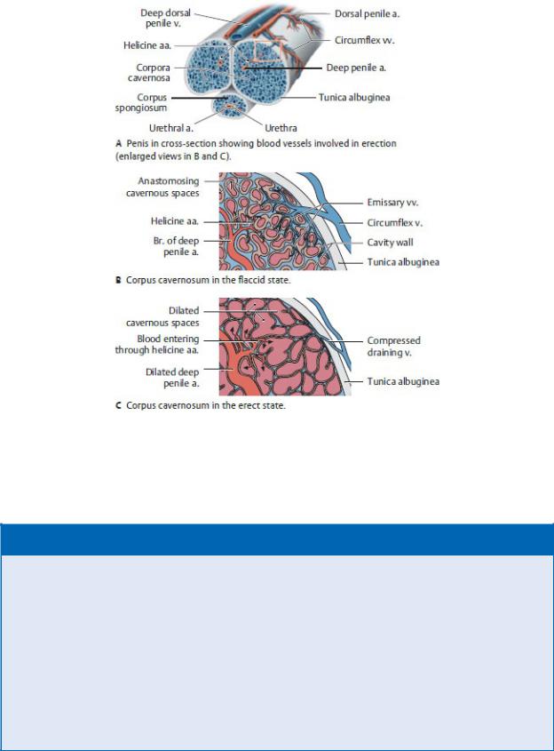
Fig. 16.9 Mechanism of penile erection (after Lehnert)
(From Schuenke M, Schulte E, Schumacher U. THIEME Atlas of Anatomy, Vol 1. Illustrations by Voll M and Wesker K. 3rd ed. New York: Thieme Publishers; 2020.)
BOX 16.3: CLINICAL CORRELATION
RETROGRADE EJACULATION
Relaxation of the detrusor muscle and contraction of the internal urethral sphincter in males are controlled by sympathetic fibers from the superior hypogastric plexus. Closure of the sphincter occurs during ejaculation, preventing the retrograde flow of semen into the bladder. Disruption of the sympathetic nerves can result in retrograde ejaculation. This can occur during the repair of an abdominal aortic aneurysm, for example, if the aneurysm involves the bifurcation of the aorta.
16.4 Female Urogenital Triangle
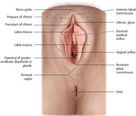
As in the male perineum, the female perineum contains erectile bodies, secretory glands, and their associated neurovasculature. In addition, it contains paired folds of skin that surround the urethral and vaginal orifices. These external genitalia are known collectively as the vulva (Figs. 16.10, 16.11, 16.12, 16.13).
—The mons pubis, a superficial mound of fatty subcutaneous tissue that is continuous with the superficial fatty layer of the abdominal wall, lies anterior to the pubic symphysis and is continuous with the labia majora.
—Labia majora, bilateral folds of fatty subcutaneous tissue, flank the pudendal cleft, the opening between the labia. The labia join anteriorly at the anterior commissure and posteriorly at the posterior commissure.
Pigmented skin and coarse pubic hair cover the labia’s outer surface; the inner surface is smooth and hairless.
—Labia minora, bilateral folds of hairless skin within the pudendal cleft, flank the vestibule of the vagina.
—The vestibule of the vagina is a space surrounded by the two labia minora. It contains the urethral and vaginal orifices and the openings of the ducts of the greater vestibular and lesser vestibular glands.
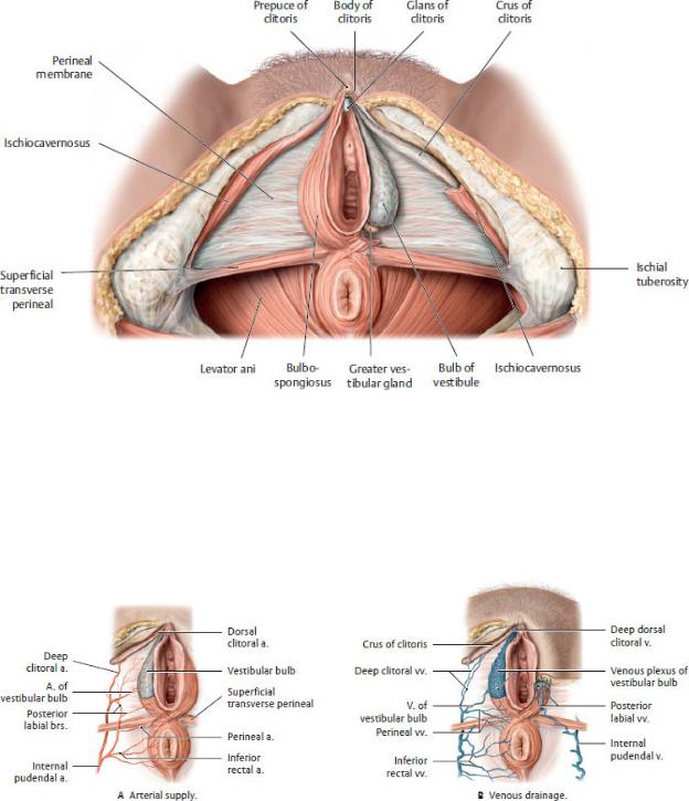
—Bulbs of the vestibule are paired masses of erectile tissue that lie deep to the labia minora and are covered by the bulbospongiosus muscles.
—Greater vestibular (Bartholin’s) glands, small glands that lie under the posterior end of the vestibular bulbs, help to lubricate the vestibule during sexual arousal.
—Small lesser vestibular glands lie on each side of the vestibule and secrete mucus to moisten the labia and vestibule.
—The clitoris, a highly sensitive erectile organ, is located at the anterior junction of the paired labia minora (Fig 16.12).
•Paired erectile bodies, the corpora cavernosa, make up the crura, which join to form the body of the clitoris. The prepuce (hood) covers the body.
•The glans at the tip of the clitoris is its most sensitive part.
—The external pudendal arteries supply the skin over the mons pubis and labia majora. Similar to those in the male, these superficial structures drain to the external pudendal veins.
—The internal pudendal arteries supply most of the external genitalia through branches that are similar to those in the male perineum (Fig. 16.14A).
•The perineal arteries supply perineal muscles and labia minora.
•The arteries of the vestibular bulb supply the bulbs of the vestibule
and greater vestibular glands.
Fig. 16.10 Female external genitalia
Lithotomy position with labia minora separated. (From Schuenke M, Schulte E, Schumacher U. THIEME Atlas of Anatomy, Vol 1. Illustrations by Voll M and Wesker K. 3rd ed. New York: Thieme Publishers; 2020.)
Fig. 16.11 Vestibule and vestibular glands
Lithotomy position with labia separated. (From Schuenke M, Schulte E, Schumacher U. THIEME Atlas of Anatomy, Vol 1. Illustrations by Voll M and Wesker K. 3rd ed. New York: Thieme Publishers; 2020.)
Fig. 16.12 Erectile tissue in the female perineum
(From Gilroy AM, MacPherson BR, Wikenheiser JC. Atlas of Anatomy.
Illustrations by Voll M and Wesker K. 4th ed. New York: Thieme Publishers; 2020.)
Fig. 16.13 Erectile muscles and tissue of the female genitalia
Lithotomy position. Removed: Labia, skin, and perineal membrane. Removed from leftside: Ischiocavernosus and bulbospongiosus muscles; greater vestibular (Bartholin’s) gland. (From Gilroy AM, MacPherson BR, Wikenheiser JC. Atlas of Anatomy. Illustrations by Voll M and Wesker K. 4th ed. New York: Thieme Publishers; 2020.)
Fig. 16.14 Blood vessels of the female external genitalia
Inferior view. (From Schuenke M, Schulte E, Schumacher U. THIEME Atlas of
Anatomy, Vol 1. Illustrations by Voll M and Wesker K. 3rd ed. New York: Thieme Publishers; 2020.)
Fig. 16.15 Anal canal
Coronal section, anterior view. (From Schuenke M, Schulte E, Schumacher U. THIEME Atlas of Anatomy, Vol 2. Illustrations by Voll M and Wesker K. 3rd ed. New York: Thieme Publishers; 2020.)
•The dorsal clitoral arteries supply the glans of the clitoris.
•The deep clitoral arteries supply the corpora cavernosa and are responsible for engorgement during arousal.
—Tributaries of the internal pudendal veins drain most perineal structures and accompany the arteries. A single deep dorsal clitoral vein drains the venous plexuses of the erectile tissue and passes under the pubic symphysis to join the venous plexuses in the pelvis (Fig. 16.14B).
—Most lymph from the female perineum drains to superficial inguinal lymph nodes. Exceptions include
•the clitoris, bulbs of the vestibule, and anterior labia, which drain to deep inguinal or internal iliac nodes; and
•the urethra, which drains to sacral or internal iliac nodes.
—The right and left pudendal nerves are the primary nerves of the perineum

(see Fig. 14.20). They supply
•perineal nerves to the vaginal orifice and superficial perineal muscles;
•dorsal clitoral nerves to deep perineal muscles and sensation from the clitoris, especially the glans; and
•posterior labial nerves to the posterior vulva.
—Similar to the innervation of the male scrotum, the anterior vulva receives sensory innervation (see Fig. 14.20) from
•the ilioinguinal nerves and genital branch of the genitofemoral nerves, which supply branches to the mons pubis and anterior labia; and
•posterior femoral cutaneous nerves, which supply the posterolateral vulva.
—Sympathetic fibers to the perineum travel with the hypogastric nerve plexes; parasympathetic fibers travel with cavernous nerves of the uterovaginal plexus. Both innervate the erectile tissue of the clitoris and bulbs of the vestibule (Fig.14.23).
16.5 Anal Triangle
The anal triangle contains the anal canal and ischioanal fossae.
The Anal Canal
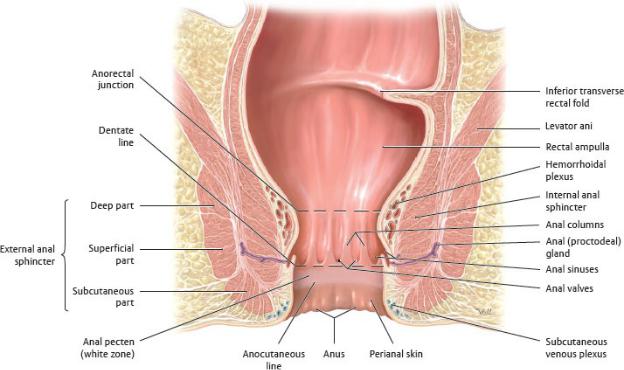
The anal canal, the terminal part of the gastrointestinal tract, controls fecal continence and the defecation response. It extends from the anorectal junction at the pelvic diaphragm to the anus (Fig. 16.15).
—The puborectalis muscle forms a sling around the anorectal junction, pulling it anteriorly and creating the perineal flexure (Fig. 16.16). From this angle the anal canal descends inferiorly and posteriorly between the anococcygeal ligament (levator plate) and the perineal body (see Fig. 16.1).
Fig. 16.16 Closure of the rectum Left lateral view.
The puborectalis acts as a muscular sling that kinks the anorectal junction. It functions in the maintenance of fecal continence. (From Gilroy AM, MacPherson BR, Wikenheiser JC. Atlas of Anatomy. Illustrations by Voll M and Wesker K. 4th ed. New York: Thieme Publishers; 2020.)
—Two sphincters surround the anal canal:
1.The internal anal sphincter is a thickening of the circular muscular layer that surrounds the upper part of the anal canal.
◦It is an involuntary sphincter.
◦It remains contracted via sympathetic innervation except in response to distension of the rectal ampulla. Parasympathetic innervation relaxes the sphincter.
2.The external anal sphincter is a broad band of muscle that extends anteriorly to merge with the perineal body, posteriorly to attach to the coccyx (through the anococcygeal ligament), and superiorly to merge with the puborectalis muscle of the pelvic floor (see also Figs. 15.1, 16.1, 16.2). Although it is described as having deep, superficial, and subcutaneous parts, they are functionally, and often anatomically, indistinct.
◦It is a voluntary sphincter.
◦The inferior rectal nerve, a branch of the pudendal nerve, innervates this sphincter.
—The inner surface of the anal canal is characterized by
• anal columns, vertical ridges formed by underlying branches of the superior rectal vessels;
• anal valves that connect the inferior edges of the anal columns; and
• anal sinuses, recesses at the base of the anal columns that secrete mucus to facilitate defecation.
—The dentate (pectinate) line is an irregular ridge at the base of the anal columns.
• It divides the anal canal into a superior part derived from the endoderm of the embryonic hindgut and an inferior part derived from the embryonic ectoderm.
• It divides the anal canal in its blood supply, lymphatic drainage, and innervation.
—Below the dentate line, a smooth lining devoid of glands and hair, the anal pecten, extends down to the anocutaneous line, or intersphincteric groove.
—Below the anocutaneous line, the anal canal is lined by hair-bearing skin that is continuous with perianal skin that surrounds the anus.

—Above the dentate line the neurovasculature of the anal canal is similar to that of the distal gastrointestinal tract (see Section 14.6).
•It receives its blood supply from the superior rectal artery, a branch of the inferior mesenteric artery.
•The rectal venous plexus drains through the superior rectal vein into the portal venous system.
•Lymph drains to the internal iliac nodes.
•Visceral innervation is transmitted via rectal plexuses to the inferior hypogastric plexus.
◦Sympathetic stimulation maintains the tone of the sphincter.
◦Parasympathetic innervation relaxes the sphincter and stimulates peristalsis of the rectum.
◦Visceral sensory fibers travel with pelvic splanchnic (parasympathetic) nerves and convey only sensation of stretching (no sensitivity to pain).
—Below the dentate line the neurovasculature of the anal canal is similar to that of the perineum (see Section 14.6).
•It receives its blood supply from the right and left inferior rectal arteries, branches of the internal pudendal arteries.
•The rectal venous plexus drains into inferior rectal veins, which in turn drain to internal iliac veins of the venae caval system.
•Lymph drains to superficial inguinal nodes.
•Somatic innervation is transmitted via the inferior rectal nerve, a branch of the pudendal nerve.
◦Somatic motor fibers stimulate contraction of the external anal sphincter.
◦Somatic sensory fibers transmit pain, touch, and temperature.
BOX 16.4: CLINICAL CORRELATION
HEMORRHOIDS
External hemorrhoids are thrombosed veins of the external rectal plexus, commonly associated with pregnancy or chronic constipation. They lie below the dentate line and are covered by skin. Because they are somatically innervated, they are more painful than internal hemorrhoids.
Internal hemorrhoids contain dilated veins of the internal rectal plexus. With a breakdown of the muscularis layer, these vascular cushions prolapse into the anal canal and may become ulcerated.
Because they lie above the dentate line and are viscerally innervated, these hemorrhoids are painless. They are generally not associated with portal hypertension, but bleeding is characteristically bright red due to the arteriovenous anastomoses between the venous plexus and branches of the rectal arteries.
BOX 16.5: CLINICAL CORRELATION
ANAL FISSURES
Anal fissures are tears in the mucosa around the anus (usually in the posterior midline) that are caused by the passing of hard or large stools. Because they are below the pectinate line and innervated by the inferior rectal nerves, these lesions are painful. Most anal fissures heal spontaneously within a few weeks if care is taken to prevent constipation. Perianal abscesses that develop from the fissures can spread into the adjacent ischioanal fossae.
Defecation and Continence
The opening (defecation) and closing (continence) of the anal canal is controlled by a complex apparatus that involves muscular, vascular, and neural components.
—The internal anal sphincter, under visceromotor control, is responsible for 70% of fecal continence. Sympathetic stimulation allows it to maintain a constant constriction of the anal canal until it relaxes in response to a rise in intra-rectal pressure.
—The external anal sphincter and puborectalis of the levator ani, under somatomotor control, constrict the anal canal and maintain the anorectal angle, respectively. Voluntary relaxation during defecation results in widening of the anal canal and straightening of the puborectalis sling.
—The vascular part of the continence apparatus involves the hemorrhoidal plexus, a permanently distended cavernous body within the submucosa, which forms circular cushions above the anal columns (Fig. 16.17). When filled with blood (supplied by the superior rectal artery) these cushions serve as an effective continence mechanism that ensures liquidand gas-tight closure. The sustained contraction of the muscular sphincter apparatus inhibits venous drainage, but when the sphincter relaxes during defecation, blood is allowed to drain via arteriovenous anastomoses to the inferior mesenteric vein (to the portal system) and middle and inferior rectal veins
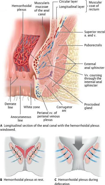
(to the systemic system).
Fig. 16.17 Structure of the vascular continence mechanism
(From Schuenke M, Schulte E, Schumacher U. THIEME Atlas of Anatomy, Vol 2. Illustrations by Voll M and Wesker K. 3rd ed. New York: Thieme Publishers; 2020.)
—The neural component of this mechanism involves (see Fig. 14.22)
•somatic motor nerves primarily via the pudendal nerve (S2–S4) to the external anal sphincter and puborectalis muscle, and somatic sensory
nerves via inferior rectal nerves from the anus and perianal skin.
•visceral motor (parasympathetic) nerves via pelvis splanchnic nerves to the internal anal sphincter in the rectal plexus and visceral sensory nerves from the wall of the rectum.
Ischioanal Fossae and Pudendal Canal
—The ischioanal fossae are paired wedge-shaped spaces on either side of the anal canal bounded by the pelvic diaphragm superiorly and the skin of the anal region inferiorly (see Fig. 15.24).
•Fat and loose connective tissue, strengthened by strong fibrous bands, fill the fossae. These tissues support the anal canal but can be displaced readily when the anal canal distends with feces.
•The inferior rectal vessels and nerve, branches of the internal pudendal vessels and pudendal nerve, traverse the fossae.
•The ischioanal fossae extend anteriorly into the urogenital triangle superior to the perineal membrane.
—The pudendal canal is a passageway formed by the splitting of the fascia of the obturator internus muscle on the lateral wall of the ischioanal fossa.
•The internal pudendal artery and vein and the pudendal nerve enter the canal after exiting the lesser sciatic foramen and giving off inferior rectal branches.

17 Clinical Imaging Basics of the Pelvis and
Perineum
Radiography of the pelvis is used in the evaluation of trauma patients and for a quick first-line assessment of the hip joints. If greater detail of the soft tissues is required, magnetic resonance imaging (MRI) is the next step. For evaluation of pelvic contents in female patients, ultrasound provides superb images quickly and inexpensively and without exposing the gonads to radiation. Therefore, ultrasound is very useful in emergent situations (e.g., acute pelvic pain). MRI also shows excellent detail of the pelvis, but because it takes longer to acquire the images, it is less useful in emergencies ( Table 17.1).
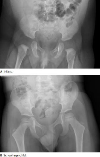
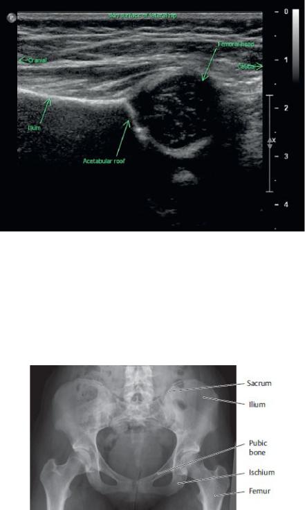
In children, ultrasound can also be valuable in evaluation of the hips. The developing skeleton is only partially ossified, thus making portions of it invisible on x-rays (Fig. 17.1). To assess the hip joint, such as for developmental dysplasia of the hip (DDH), ultrasound is used to assess the position of the cartilaginous femoral head (Fig 17.2).
Standard radiographic views of the bony pelvis include an anteroposterior (AP) projection showing both hip joints (Fig. 17.3). In patients with pelvic fractures, different obliquities are often used to assess alignment. There are several approaches to imaging the contents of the pelvis. As in the abdomen, computed tomography (CT) is often used with oral or intravenous contrast to enhance the pelvic organs (Fig. 17.4), but in non-emergent conditions, MRI provides the best anatomic detail (Fig. 17.5). In comparison, ultrasound offers less detail, but because it is safer and faster, it is usually the first choice for imaging female pelvic organs (Figs. 17.6 and 17.7).
Table 17.1 Suitability of Imaging Modalities for the Pelvis
Modality |
Clinical Uses |
Radiographs (x-rays) |
Used primarily to evaluate the pelvic |
|
bones |
|
|
CT (computed tomography) |
Provides excellent anatomic detail, but at |
|
the expense of radiation |
MRI (magnetic resonance |
Ideal for evaluation of the pelvic bones |
imaging) |
and surrounding muscles and soft tissues; |
|
also useful as an adjunct to ultrasound for |
|
the female pelvis |
|
|
Ultrasound |
The primary imaging modality for |
|
evaluation of the female pelvis, specifically |
|
the uterus and ovaries; in men, the |
|
mainstay of imaging of the scrotum and |
|
testicles |
|
|
Fig. 17.1 Frontal radiographs of the pelvis illustrating the developmental ossification of
the skeleton
Note how the femoral head and pelvic growth plates progressively ossifiy with age and thus become visible as bone by radiography. Compare to the fully grown adult pelvis in 17.3. (Courtesy of Joseph Makris, MD, Baystate Medical Center.)
Fig. 17.2 Infant hip ultrasound
The probe is positioned at the lateral aspect of the infant’s hip sagittally. The cartilaginous (has not ossified yet) femoral head is well seen by ultrasound due to the water component of the growing epiphyseal cartilage. The shape of the developing acetabulum and the position of the femoral head relative to the acetabulum are evaluated for signs of developmental dysplasia. (Courtesy of Joseph Makris, MD, Baystate Medical Center.)
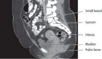
Fig. 17.3 Radiograph of the pelvis in an adult
Anterior view.
Note that the rounded femoral heads sit uniformly in each acetabulum, and the sacroiliac joints and pubic symphysis are sharply seen. The lower part of the sacrum is partially obscured by gas and stool in the rectum (irregular dark and light gray shapes in the mid-pelvis). The iliac crests can be traced along their edges to the acetabulum and into the ischium and pubic symphysis. The edges of the bones should curve smoothly and be sharply defined. (Courtesy of Joseph Makris, MD, Baystate Medical Center.)
Fig. 17.4 CT of the female pelvis
Left lateral view.
This CT reconstruction, shown in the “soft tissue window,” highlights the relationships of the pelvic organs. Oral contrast causes the bowel to appear white; intravenous contrast enhances the vascularized soft tissues (light gray). The urine filled bladder has a water density of dark gray. The uterus is enhanced by the intravenous contrast and is easily seen against the bladder anteriorly and fat superiorly. The endometrium is barely seen within the uterus. It is difficult to precisely define the plane between the uterus and the adjacent rectum. (Courtesy of Joseph Makris, MD, Baystste Medical Center.)
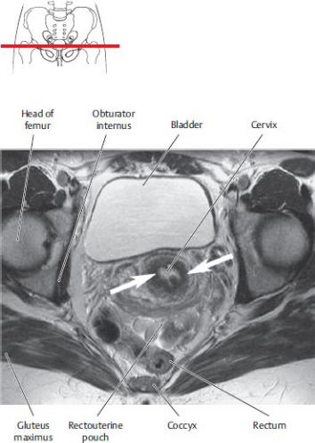
Fig. 17.5 MRI of the female pelvis
Transverse section through the level of the cervix. In this MRI sequence, fluid is white, muscle is black, and other soft tissues are gray. Soft tissue detail seen on MRI is superior to that seen on CT and ultrasound. Note the dark (black) cervical stroma (arrows) that surrounds the white fluid in the cervical canal. Because MRI is very sensitive in differentiating tissue types, it is easy to distinguish the pelvic organs from each other and from the adjacent bowel. The bright fat planes between structures provide good contrast. (From Hamm B, et al. MRI Imaging of the Abdomen and Pelvis, 2nd ed. New York: Thieme Publishers; 2009.)
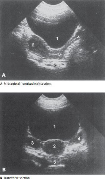
Fig. 17.6 Transabdominal ultrasound of the uterus and ovaries
With transabdominal imaging of the pelvis, the ultrasound probe is placed on the lower abdominal wall anterior to the bladder. A full bladder provides an acoustic “window” to better visualize the uterus and related structures. (Patients are required to drink plenty of fluid prior to the exam.) The urine in the bladder is anechoic (black). The muscular and vascular structure of the uterus and follicular and vascular structure of the ovaries make these structures hypoechoic (dark gray). The ovaries should appear relatively symmetric in size and shape. (From Gunderman R. Essential Radiology, 3rd ed. New York: Thieme Publishers; 2014.) 1, urine filled bladder; 2, uterus; 3, cervix; 4, vagina; 5, rectum.
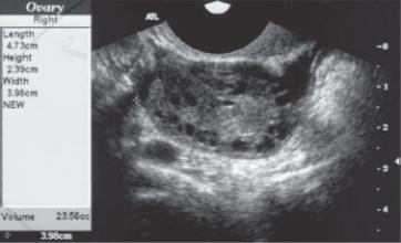
Fig. 17.7 Transvaginal ultrasound of the right ovary
An empty bladder (patient is asked to urinate immediately before the exam) positions the uterus and associated structures for optimal visualization. The tip of the ultrasound probe is positioned directly against the cervix and pointed toward the side of interest until the ovary is found. The close proximity of the tip of the ultrasound probe to the pelvic structures provides significantly improved image detail over transabdominal images. This patient has enlarged ovaries due to polycystic ovarian syndrome. Note the detail of the numerous small ovarian follicles (black, since they are fluid filled). (From Gunderman R. Essential Radiology, 3rd ed. New York: Thieme Publishers; 2014)
Unit V Review Questions: Pelvis and Perineum
1.After a prolonged labor, Janice delivered a healthy 9 lb 8 oz baby boy
through a normal vaginal birth, although the delivery required an episiotomy to prevent tearing of the perineum. During the procedure, her external anal sphincter and some fibers of the puborectalis muscle were cut. As a medical student who understands the role of these muscles, Janice is concerned about her ability to maintain fecal continence. Relaxation of these muscles
A.straightens the perineal flexure of the anorectal junction
B.allows venous drainage from the vascular cushions of the hemorrhoid plexus
C.widens the anal canal
D.would result from damage to the inferior rectal branches of the pudendal nerve
E.all of the above
2.Which one of the following statements best describes the muscles of the
pelvis?
A.The obturator internus muscle leaves the pelvis through the greater sciatic foramen.
B.The levator ani muscle is composed of the pubococcygeus, puborectalis, and iliococcygeus.
C.The pelvic diaphragm is a sling of muscle that separates the true pelvis from the false pelvis.
D.The piriformis muscle forms the lateral wall of the pelvis.
E.The coccygeus muscle attaches to the sacrotuberous ligament.
3.Typically, the posterior division of the internal iliac artery supplies
A.structures of the perineum
B.muscles of the medial thigh
C.meninges of the sacral spinal roots

D. uterus and uterine tubes
E.prostate
4.The components of the pelvic splanchnic nerves are most similar to the
components of the
A.lumbar splanchnic nerves
B.sacral splanchnic nerves
C.pudendal nerve
D.vagus nerve
E.hypogastric nerves
5.Carcinoma of the prostate can metastasize to bone and the brain through
its connections with the vertebral venous plexus. Which other structures communicate with this venous plexus?
A.Breast
B.Spinal cord
C.Intercostal muscles
D.Esophagus
E.All of the above
6.Although branches of the uterine artery anastomose extensively
throughout the pelvis, the main stem of the vessel travels within the
A.proper ovarian ligament
B.cardinal ligament
C.uterosacral ligament
D.ovarian suspensory ligament
E.round ligament
7.Which of the following statements accurately describes the relations of
the ureter?
A. It is crossed anteriorly by the gonadal vessels on the posterior abdominal wall.

B.It crosses the brim of the pelvis at the bifurcation of the common iliac artery.
C.In females it passes under the uterine artery ~ 2 cm lateral to the cervix.
D.It enters the bladder at the posterolateral aspect of the trigone.
E.All of the above
8.During a routine physical exam on a male patient, you test the integrity
of the external anal sphincter. What spinal cord segments are involved in this?
A.T12–L1
B.L2–L4
C.L4–L5
D.S1–S2
E.S2–S4
9.The superficial perineal fascia is continuous with the
A.perineal membrane
B.dartos fascia
C.deep penile fascia
D.tunica albuginea of the penis
E.endopelvic fascia
10.The boundary of the pelvic inlet A. provides the attachment site for the
pelvic diaphragm
B.includes the iliac crests
C.includes the ramus of the ischium
D.is crossed by the ovarian and testicular arteries
E.separates the cavity of the true pelvis from the abdominal cavity
11.Several years after delivering twin boys, a woman experienced mild uterine prolapse and urinary incontinence. Her gynecologist was able to

confirm that the angle of her anococcygeal ligament had changed, suggesting a laxity in her pelvic floor muscles. Which muscles insert on the anococcygeal ligament?
A.Coccygeus
B.Iliococcygeus
C.Piriformis
D.Deep transverse perineal
E.Obturator internus
12.Tumors that metastasize via the bloodstream often form metastases in
the first capillary bed that the cells reach after they enter the bloodstream. Based on their venous drainage, tumors in which of the following locations are likely to reach the lung before they reach the liver?
A.Ascending colon
B.Sigmoid colon
C.Pancreas
D.Superior (proximal) rectum
E.Inferior (distal) rectum
13.Which one of the following structures passes through the genital hiatus?
A.Ductus deferens
B.Cavernous nerves
C.Round ligament
D.Obturator nerve
E.External iliac artery
14.A 44-year-old woman undergoes a total hysterectomy for painful
fibroids. The ovaries will not be removed during the procedure. Which of the following ligaments must be preserved?
A.Suspensory ligament
B.Ovarian ligament
C.Uterosacral ligament
D.Transverse cervical ligament
E.Round ligament

15.Which of the following statements best describes the uterine cervix?
A.In a normally anteflexed uterus, the cervix is tilted posteriorly.
B.Vaginal fornices surround its supravaginal part.
C.It is the attachment site for the round ligament.
D.It makes up the inferior third of the uterus.
E.It communicates with the uterine cavity through the external os.
16.A 53-year-old man had an aortic aneurysm that extended through his
aortic bifurcation into his common iliac arteries. During the open repair, the surgeon opened the vessels longitudinally and fixed a synthetic graft to the walls of the vessels above and below the aneurysm. Following surgery, the man experienced retrograde ejaculation because of damage to nerves that innervated his internal urethral sphincter. Which nerves were damaged?
A.Sympathetic nerves of the superior hypogastric plexus
B.Parasympathetic nerves of the superior hypogastric plexus
C.Somatic nerves of the sacral plexus
D.Pelvic splanchnic nerves
E.Sympathetic trunk
17.You are treating a 34-year-old woman for hemorrhoids. Although she
does not complain of pain, the hemorrhoids protrude into her anal canal and are becoming ulcerated. There is no evidence of portal hypertension, but blood from the ulcers is bright red. Based on her brief history, what can you surmise about her condition?
A.The prolapsed tissue contains the dilated veins of the external rectal plexus.
B.The hemorrhoids originate from below the dentate line.
C.The dilated veins in the hemorrhoids drain to the inferior rectal veins.
D.The dilated veins communicate with the rectal arteries to form an arteriovenous hemorrhoidal plexus.
E.All of the above

18.During a robotic prostatectomy on a 41-year-old man, the cavernous
nerves were inadvertently damaged. What symptoms would you expect in this patient?
A.Loss of tone in his external anal sphincter
B.Urinary incontinence
C.Inability to ejaculate
D.Inability to form an erection
E.Loss of sensation at the tip of the penis
19.Structures that pass through the greater sciatic foramen include the
A.obturator internus
B.coccygeus
C.iliopsoas
D.piriformis
E.obturator nerve
20.The tendinous arch of the pelvic fascia
A.is a condensation of endopelvic fascia
B.includes the lateral ligaments of the rectum
C.supports the pelvic viscera
D.provides an attachment site for the pelvic diaphragm
E.All of the above
21.Structures that drain (directly or indirectly) into the deep inguinal lymph
nodes include the
A.glans of the penis
B.perianal skin
C.supralateral part of the uterus via the round ligament
D.scrotum
E.All of the above

22.During the national championships an Olympic gymnast fell backward
off the balance beam, fracturing the tip of her coccyx and subluxing (dislocating) her sacroiliac joint. The team physician was concerned about damage to her sacral plexus and its branches that exit the pelvis. Nerves of the sacral plexus pass through the
A.posterior sacral foramina
B.obturator canal
C.greater sciatic foramen
D.superficial inguinal ring
E.deep inguinal ring
23.A young, pregnant woman in her third trimester was alarmed when she
experienced a sharp pain in the anterior part of her labia majora when she stood up. Her obstetrician assured her that this was a common problem in late pregnancy and was most likely caused by
A.stretching of the round ligament
B.tightening of the inguinal ligament
C.pressure on the obturator nerve
D.irritation of the perineal branch of the pudendal nerve
E.stretching of the iliohypogastric nerve
24.A vascular surgeon is repairing an aneurysm of the aortic bifurcation
that extends along the right common iliac artery to its division into internal iliac and external iliac branches. What structure does the surgeon encounter that is at risk as it crosses over the pelvic brim at this distal end of the aneurysm?
A.Ureter
B.Testicular artery
C.Lumbosacral trunk
D.Sciatic nerve
E.Ductus deferens

25.Similar to other sections of the large intestine, the rectum is
characterized by
A.teniae coli
B.haustra
C.a mesentery
D.epiploic appendices
E.None of the above
26.Which of the following are found within the superficial perineal space?
A.Bulbourethral glands
B.External urethral sphincter
C.Bulbospongiosus muscle
D.Anterior extension of ischioanal fat pad
E.Inferior rectal nerve
27.The spongy part of the male urethra
A.passes through the corpus spongiosum of the penis
B.is surrounded by the internal urethral sphincter
C.is surrounded by the external urethral sphincter
D.is characterized by a vertical ridge, the urethral crest, on its posterior wall
E.contains openings for the ejaculatory ducts
28.Similar to the penis in the male, the clitoris is formed by highly
sensitive erectile tissue. Which of the following structures is composed of erectile tissue but does not form part of the clitoris?
A.Corpora cavernosa
B.Bulbs of the vestibule
C.Greater vestibular glands
D.Prepuce
E.Glans

29.During a routine physical, which included a digital rectal exam, your
physician discovered a firm nodule on your prostate, suggesting prostate carcinoma. This was confirmed in subsequent blood tests that revealed increased levels of prostate specific antigen (PSA). Prostate carcinoma is
A.a common and non-malignant disease of older men
B.usually found in the peripheral zone
C.usually found in the largely glandular anterior lobe
D.a disease of the periurethral zone, causing urethral obstruction in the early stages
E.also known as benign prostatic hypertrophy
30.What imaging modality is best for routine evaluation of a developing
fetus?
A.CT
B.Ultrasound
C.X-rays
D.MRI
31.A 24-year-old male has acute onset of severe scrotal pain. You suspect
that he may have testicular torsion (twisting of the spermatic cord and testicle, restricting blood to the testicle) and send the patient for an emergent ultrasound of his scrotum to confirm your suspicion. What feature of ultrasound imaging is useful for assessing blood flow to the testicles?
A.Low cost
B.Lack of ionizing radiation
C.Real-time color and spectral Doppler imaging
D.Ultrasound waves travel best through water
Answers and Explanations
1.E. All are correct (Section 16.5).
A.Contraction of the muscles maintains the flexure, relaxation of the muscles straightens it.
B.Contraction of the muscular sphincter apparatus inhibits the venous drainage of the vascular cushions. They drain when the sphincters relax
during defecation.
C.The function of the external anal canal is to widen the anal canal during defecation.
D.The inferior rectal nerves maintain the tone of the external anal sphincter and puborectalis muscles. Denervaton of these muscles would result in their relaxation.
2.B. The levator ani muscle is composed of the pubococcygeus, puborectalis, and iliococcygeus muscles (Section 14.3).
A.The obturator internus muscle covers the sidewall of the pelvis and perineum. Its tendon passes from the perineum to the gluteal region through the lesser sciatic foramen.
C.The pelvic diaphragm separates the true pelvis from the perineum, which lies below it.
D.The piriformis muscle forms the posterior muscular wall of the
pelvis.
E.The coccygeus muscle attaches to the sacrospinous ligament along its entire length.
3.C. The posterior division supplies parietal branches to the posterior abdominal wall, some gluteal muscles, and the meninges of the sacral spinal roots (Section 14.6).
A.The internal pudendal artery, a branch of the anterior division of the internal iliac artery, supplies most structures of the perineum.
B.The obturator artery, a branch of the anterior division of the internal iliac artery, supplies muscles of the medial thigh.
D.The uterine artery, a branch of the anterior division of the internal iliac artery, supplies the uterus and uterine tubes.
E.The prostatic arteries, usually branches of the inferior vesical arteries, are derived from the anterior division of the internal iliac artery.
4.D. The pelvic splanchnic nerves represent the sacral component of the parasympathetic system; the vagus nerve represents the cranial component of the parasympathetic system (Section 14.6).
A.Lumbar splanchnic nerves arise from the lumbar sympathetic trunk and carry postganglionic sympathetic fibers.
B.Sacral splanchnic nerves arise from the sacral sympathetic trunk and carry postganglionic sympathetic fibers.
C.The pudendal nerve arises from the sacral plexus and carries somatic
sensory and motor fibers.
E.Hypogastric nerves derive from the superior hypogastric plexus carrying postganglionic sympathetic fibers to the pelvic plexus.
5.E. The vertebral venous system is a tributary of the azygos system. The breast, intercostal muscles, and esophagus drain via intercostal veins to the azygos system, and the spinal cord drains directly into the vertebral venous plexus (Section 15.1 and Unit III, Thorax).
A.Blood from the breast drains to intercostal veins and the azygos system, which communicates with the vertebral venous plexus. B through D are also correct (E).
B.The veins of the spinal cord drain to the vertebral venous plexus. A, C, and D are also correct (E).
C.The intercostal veins that drain intercostal muscles terminate in the azygos system, which also receives the vertebral venous plexus. A, B, and D are also correct (E).
D.Veins of the lower esophagus drain to the portal system, but in the thorax esophageal veins drain to the azygos system, which also receives the vertebral venous plexus. A through C are also correct (E).
6.B. The uterine artery and vein travel within the cardinal ligament at the base of the broad ligament (Section 15.2).
A.The proper ovarian ligament connects the ovary to the uterus and does not contain any major vessels.
C.The uterosacral ligament is a thickening of endopelvic fascia that connects the cervix of the uterus to the sacrum. It does not contain any major vessels.
D.The ovarian suspensory ligament is a fold of peritoneum that contains the ovarian vessels as they cross over the pelvic brim.
E.The round ligament extends from the uterus through the inguinal canal to the labia majora. Although it is accompanied by small vessels, it does not contain any major arteries.
7.E. All are correct (Section 15.3).
A.The gonadal vessels cross anterior to the ureter as it descends along the posterior abdominal wall. B through D are also correct (E).
B.The bifurcation of the common iliac artery is a useful landmark for locating the ureter as it crosses the pelvic brim. A, C, and D are also correct
(E).
C.The ureter passes under the uterine artery lateral to the uterine cervix and therefore is at risk of injury during a hysterectomy. A, B, and D are also correct (E).
D.The ureteral openings posterolaterally and the urethral opening in the anterior midline define the apexes of the trigone. A through C are also correct (E).
8.E. The pudendal nerve (S2–S4) provides motor innervation to the external sphincter (Sections 14.6 and 15.4).
A.T12–L1 nerve roots contribute to the subcostal, iliohypogastric, and ilioinguinal nerves, which innervate muscles of the anterior abdominal wall.
B.The L2–L4 nerve roots form the femoral and obturator nerves, which innervate muscles of the anterior and medial thigh.
C.Nerves from L4–L5 spinal cord segments form the lumbosacral trunk, which joins the sacral plexus to innervate muscles of the lower limb.
D.S1–S2 nerve roots contribute to the sacral plexus, which innervates muscles of the lower limb.
9.B. The superficial perineal fascia is continuous with the dartos fascia that lines the scrotum and with the membranous layer (Scarpa’s fascia) of the superficial fascia of the abdominal wall (Section 16.1).
A.The perineal membrane is a fibrous sheet that forms the roof of the superficial perineal space.
C.Deep penile fascia is a fibrous layer that binds the three erectile bodies of the penis.
D.Each of the three erectile bodies of the penis is surrounded by the dense, fibrous tunica albuginea.
E.Endopelvic fascia is the loose connective tissue matrix that fills the subperitoneal spaces of the pelvis.
10.E. The pelvic inlet separates the true pelvis from the false pelvis. The cavity of the false pelvis is the lower part of the abdominal cavity and contains abdominal viscera (Section 14.1).
A.The pelvic diaphragm attaches to the superior pubic ramus, tendinous arch of the levator ani, and sacrospinous ligament.
B.The iliac crests form the upper boundary of the false pelvis. The pelvic inlet forms its lower boundary.
C.The ramus of the ischium forms part of the pelvic outlet but not the pelvic inlet.
D.The ovarian arteries cross the pelvic inlet, but the testicular arteries pass along the brim of the pelvis (pelvic inlet) to the deep inguinal ring, where they enter the inguinal canal as part of the spermatic cord.
11.B. The anococcygeal ligament (also known as the levator plate) is a midline raphe between the anus and coccyx that serves as the insertion for the pubococcygeus and iliococcygeus muscles (Section 14.3).
A.The coccygeus inserts on the ischial spine.
C.The piriformis inserts on the greater trochanter of the femur.
D.Deep transverse perineal muscles insert on the vagina (or prostate) and perineal body.
E.The obturator internus inserts on the greater trochanter of the femur.
12.E. The inferior rectal veins drain through the caval system (via the internal iliac vein and inferior vena cava) back to the heart and pulmonary circulation. All other choices are tributaries of the portal system, which drains through the liver before returning to the heart (Sections 11.2 and 15.4).
A.Veins of the ascending colon are tributaries of the portal system. Therefore, blood passes through the liver before returning to the heart and pulmonary circulation.
B.Veins of the sigmoid colon, and all parts of the gastrointestinal tract, are tributaries of the portal system. Therefore, blood passes through the liver before returning to the heart and pulmonary circulation.
C.Veins of the pancreas are tributaries of the portal system. Therefore, blood from the pancreas drains through the liver before returning to the heart and pulmonary circulation.
D.The superior rectal vein that drains the superior rectum is a tributary of the portal system. Therefore, this blood passes through the liver before returning to the heart and pulmonary circulation.
13.B. The cavernous nerves carry parasympathetic innervation from the pelvic plexus to the perineum by passing through the genital hiatus (Section 14.6).
A.The ductus deferens passes through the deep inguinal ring and inguinal canal of the male.
C.The round ligament traverses the female inguinal canal and terminates in the labia majora.
D.The obturator nerve passes through the obturator canal into the medial thigh.
E.The external iliac artery passes below the inguinal ligament into the anterior thigh.
14.A. The suspensory ligament contains the ovarian vessels and is removed only during the removal of the ovary (Section 15.2).
B.The ovarian ligament attaches the ovary to the uterus and must be ligated to extract the uterus.
C.The uterosacral ligament connects the cervix to the sacrum.
D.The transverse cervical ligament contains the uterine arteries, which are ligated during a hysterectomy.
E.The round ligament passes through the inguinal canal and connects the uterus to the labia majora.
15.D. The uterus comprises a body that forms the superior two thirds and a cervix that forms the inferior one third (Section 15.2).
A.In an anteflexed uterus, the cervix is tilted anteriorly.
B.The vaginal part of the cervix is surrounded by the vaginal fornices.
C.The round ligaments arise from the upper part of the uterus.
E.The cervix communicates with the body of the uterus through the internal os.
16.A. The superior hypogastric plexus, a sympathetic nerve plexus, drapes over the aortic bifurcation as it enters the pelvis. It is frequently cut during surgery for the repair of an aneurysm at the location. The sympathetic nerves stimulate closure of the internal urethral sphincter during ejaculation, preventing the retrograde flow of seminal fluid into the bladder. Damage to these nerves can result in retrograde ejaculation (Sections 14.6 and 16.3).
B.The superior hypogastric plexus is a sympathetic nerve plexus derived from the intermesenteric plexus and lumbar splanchnic nerves.
C.Somatic nerves do not stimulate visceral responses such as closure of the sphincter.
D.Pelvic splanchnic nerves do not innervate the internal urethral sphincter.
E.The paired sympathetic trunks run along the vertebral bodies on either side and would not be damaged in this procedure.
17.D. The submucosal venous plexus of the anorectal region communicates with branches of the rectal arteries in an arteriovenous anastomosis,
creating a thickened vascular tissue known as the hemorrhoidal plexus. As a result of these anastomoses, bleeding from the internal rectal plexus appears bright red (Section 15.4).
A.Prolapsed internal hemorrhoids contain the dilated veins of the internal rectal plexus and are painless. blood passes through the liver before returning to the heart and pulmonary circulation.
13.B. The cavernous nerves carry parasympathetic innervation from the pelvic plexus to the perineum by passing through the genital hiatus (Section 14.6).
A.The ductus deferens passes through the deep inguinal ring and inguinal canal of the male.
C.The round ligament traverses the female inguinal canal and terminates in the labia majora.
D.The obturator nerve passes through the obturator canal into the medial thigh.
E.The external iliac artery passes below the inguinal ligament into the anterior thigh.
14.A. The suspensory ligament contains the ovarian vessels and is removed only during the removal of the ovary (Section 15.2).
B.The ovarian ligament attaches the ovary to the uterus and must be ligated to extract the uterus.
C.The uterosacral ligament connects the cervix to the sacrum.
D.The transverse cervical ligament contains the uterine arteries, which are ligated during a hysterectomy.
E.The round ligament passes through the inguinal canal and connects the uterus to the labia majora.
15.D. The uterus comprises a body that forms the superior two thirds and a cervix that forms the inferior one third (Section 15.2).
A.In an anteflexed uterus, the cervix is tilted anteriorly.
B.The vaginal part of the cervix is surrounded by the vaginal fornices.
C.The round ligaments arise from the upper part of the uterus.
E. The cervix communicates with the body of the uterus through the internal os.
16.A. The superior hypogastric plexus, a sympathetic nerve plexus, drapes over the aortic bifurcation as it enters the pelvis. It is frequently cut during surgery for the repair of an aneurysm at the location. The sympathetic
nerves stimulate closure of the internal urethral sphincter during ejaculation, preventing the retrograde flow of seminal fluid into the bladder. Damage to these nerves can result in retrograde ejaculation (Sections 14.6 and 16.3).
B.The superior hypogastric plexus is a sympathetic nerve plexus derived from the intermesenteric plexus and lumbar splanchnic nerves.
C.Somatic nerves do not stimulate visceral responses such as closure of the sphincter.
D.Pelvic splanchnic nerves do not innervate the internal urethral sphincter.
E.The paired sympathetic trunks run along the vertebral bodies on either side and would not be damaged in this procedure.
17.D. The submucosal venous plexus of the anorectal region communicates with branches of the rectal arteries in an arteriovenous anastomosis, creating a thickened vascular tissue known as the hemorrhoidal plexus. As a result of these anastomoses, bleeding from the internal rectal plexus appears bright red (Section 15.4).
A.Prolapsed internal hemorrhoids contain the dilated veins of the internal rectal plexus and are painless.
A.Lymph from the glans of the penis drains to deep inguinal nodes. B through D are also correct (E).
B.Lymph from perianal skin, like all skin of the perineum, drains to superficial inguinal nodes, which drain to deep inguinal nodes. A, C, and D are also correct (E).
C.Lymph from the supralateral parts of the uterus drain along the round ligaments (which terminate in the labia majora) to superficial inguinal nodes. These nodes drain to the deep inguinal nodes. A, B, and D are also correct
(E).
D.Lymph from the scrotum drains to superficial inguinal nodes, which drain to deep inguinal nodes. A through C are also correct (E).
22.C. Most nerves of the sacral plexus pass through the greater sciatic foramen to innervate muscles of the gluteal region and lower limb (Section 14.6).
A.Only posterior rami of sacral spinal nerves pass through the posterior sacral foramina. The sacral plexus is formed by anterior rami of sacral spinal nerves.
B.The obturator nerve, which passes through the obturator canal, is a branch of the lumbar plexus.
D.The ilioinguinal and genitofemoral nerves, which pass through the superficial inguinal ring, are branches of the lumbar plexus.
E.The genitofemoral nerve, a branch of the lumbar plexus, passes through the deep inguinal ring.
23.A. The round ligament originates on the superior aspect of the anterolateral uterus and inserts in the labia majora. As the uterus enlarges and stretches the ligament, pain is felt in the labia (Section 15.2).
B.During late pregnancy pelvic ligaments are more relaxed in preparation for the passage of the child through the birth canal.
C.Pressure on the obturator nerve would be felt on the medial thigh.
D.The anterior part of the labia majora is innervated by the labial branches of the genitofemoral nerve, not the pudendal nerve.
E.The iliohypogastric nerve innervates the skin above the pubic crest and does not extend inferiorly to the labia.
24.A. The ureter crosses the pelvic brim at the bifurcation of the common iliac artery into its internal iliac and external iliac branches (Section 15.3).
B.The testicular artery does not cross the pelvic brim but enters the deep inguinal ring.
C.The lumbosacral trunk crosses the pelvic brim posterior to the common iliac vessels and anterior to the sacroiliac joint. It enters the pelvis to join the sacral plexus.
D.The sciatic nerve passes through the greater sciatic foramen and does not cross the pelvic brim.
E.The ductus deferens courses around the inferior epigastric vessels and over the distal end of the external iliac vessels to descend into the pelvis.
25.E. The rectum is devoid of teniae coli, haustra, a mesentery, and epiploic appendices (Section 15.4).
A.The teniae coli of the large intestine converge to form a uniform layer surrounding the rectum.
B.The haustra of the large intestine disappear at the rectosigmoid junction.
C.The rectum has no mesentery. The upper rectum is covered anteriorly by peritoneum, but the lower rectum lies in the subperitoneal space.
D.Epiploic appendices are not present in the rectum.
26.C. The bulbospongiosus muscle is found within the superficial perineal
space (Section 16.1).
A.The bulbourethral glands are found in the deep perineal space.
B.The external urethral sphincter is located in the deep perineal space.
D.The anterior extensions of the ischioanal fat pads are found in the
deep perineal space.
E.The inferior rectal nerve is found in the anal triangle, not the deep perineal or the superficial perineal spaces of the urogenital triangle.
27.A. The spongy urethra passes through the bulb and corpus spongiosum of the penis (Section 15.3).
B.The internal urethral sphincter surrounds the preprostatic urethra at the neck of the bladder.
C.Part of the external urethral sphincter surrounds the membranous
urethra.
D.The urethral crest is a feature of the prostatic urethra.
E.Ejaculatory ducts open into the prostatic urethra on the urethral crest.
28.B. The bulbs of the vestibule are masses of erectile tissue that lie deep to the labia minora. Unlike in the male, where the bulb and corpus spongiosum forms part of the penis, the bulbs of the vestibule do not form part of the clitoris (Section 16.4).
A.The paired corpora cavernosa converge to form the body of the
clitoris.
C.Greater vestibular glands, which lie deep to the bulbs of the vestibule, are not part of the clitoris and are not composed of erectile tissue.
D.The prepuce is a part of the clitoris that forms a hood over the body.
E.The glans is the highly sensitive tip of the clitoris.
29.B. Prostate tumors most often grow deep to the capsule in the peripheral zone.
A.Prostatic carcinoma is a common disease of older men but can metastasize via the vertebral venous plexus to the bony pelvis, vertebral column, skull, and brain, and via other venous routes, to the heart and lungs.
C.The anterior lobe, or isthmus, is fibromuscular and not a common site of carcinoma.
D.Although prostate carcinoma may eventually invade the periurethral zone, it generally does not cause urethral obstruction in the early stages.
E.Benign prostatic hypertrophy results from proliferation of the epithelial and stromal tissues, particularly of the periurethral zone, and is
unrelated to carcinoma.
30.B. Ultrasound is the optimal imaging tool for fetal evaluation due to its lack of ionizing radiation and relative low cost. Additionally, the fluid-filled amniotic sac provides an excellent acoustic window.
A.A developing fetus is particularly vulnerable to the potential deleterious effects of radiation and thus CT is contraindicated in fetal evaluation.
C.X-rays would also expose the fetus to radiation, although at a lower dose than CT. Additionally, a radiograph would show only limited detail of the developing fetal skeleton, and would not be sufficient in this setting.
D.MRI is safe for fetal imaging and provides excellent anatomic detail. However, it is expensive, less available, and requires very long scan times, making it impractical for use in routine or screening fetal evaluation. MRI is used as a supplement to ultrasound for further evaluation of fetal and maternal abnormalities detected at screening ultrasound.
31.C. Doppler ultrasound can detect motion and is excellent for assessment of blood flow. The use of Doppler ultrasound is an important step in the evaluation of acute scrotal pain as it can be obtained very quickly and provides excellent imaging detail of the testes and scrotal contents. Testicular torsion is a surgical emergency, and rapid diagnosis and treatment are key to preventing infarction and subsequent loss of the testicle.
A.The relative low cost of ultrasound contributes to its high availability, but is not related to blood flow assessment.
B.Lack of radiation makes ultrasound particularly well suited for imaging the gonads but is not related to blood flow assessment.
D.Ultrasound waves do travel well through water/fluid but since the testicles are located in the scrotum, this is not a critical factor. Transabdominal imaging of the uterus on the other hand, requires a full bladder to be used as an acoustic “window”.

Unit VI Upper Limb
18Overview of the Upper Limb
18.1General Features
18.2Bones of the Upper Limb
18.1Clinical Correlation: Clavicular Fractures
18.2Clinical Correlation: Humeral Fractures
18.3Clinical Correlation: Colles’ Fractures
18.4Clinical Correlation: Lunate Dislocation
18.5Clinical Correlation: Scaphoid Fractures
18.3Fascia and Compartments of the Upper Limb
18.4Neurovasculature of the Upper Limb
18.1Nerves of the Brachial Plexus
18.6Clinical Correlation: Rootand Trunk-Level Injuries
18.7Clinical Correlation: Musculocutaneous Nerve Injury
18.8Clinical Correlation: Median Nerve Injury
18.9Clinical Correlation: Ulnar Nerve Injury
18.10Clinical Correlation: Axillary Nerve Injury
18.11Clinical Correlation: Radial Nerve Injury
19Functional Anatomy of the Upper Limb
19.1The Pectoral Girdle
19.1Pectoral Girdle Muscles
19.2Movements of the Pectoral Girdle
19.1Clinical Correlation: Long Thoracic Nerve Injury
19.2The Shoulder Region
19.3Shoulder Muscles
19.4Deltoid and Rotator Cuff Muscles
19.5Movements at the Glenohumeral Joint
19.2Clinical Correlation: Glenohumeral Dislocation
19.3Clinical Correlation: Rotator Cuff Tears
19.3The Arm and Cubital Region
19.6Arm Muscles: Anterior and Posterior Compartments
19.7Movements at the Humeroulnar and Humeroradial Joints
19.4The Forearm
19.8Movements at the Radioulnar Joints
19.9Forearm Muscles: Anterior Compartment
19.10Forearm Muscles: Posterior Compartment
19.4Clinical Correlation: Subluxation of the Radial Head (Nursemaid’s Elbow)
19.5Clinical Correlation: Lateral Epicondylitis (“Tennis Elbow”)
19.5The Wrist
19.11Movements at the Wrist Joints
19.12Dorsal Compartments for Extensor Tendons
19.6Clinical Correlation: Carpal Tunnel Syndrome
19.7Clinical Correlation: Ulnar Nerve Compression
19.6The Hand
19.13Movements at the Joints of the Fingers (Digits 2 through 5)
19.14Movements at Joints of the Thumb
19.15Thenar Muscles
19.16Hypothenar Muscles
19.17Metacarpal Muscles
19.8Clinical Correlation: Dupuytren’s Disease
19.9Clinical Correlation: Tendon Sheath Communication
19.7Topographic Views of Upper Limb Musculature
20Clinical Imaging Basics of the Upper Limb
20.1Suitability of Imaging Modalities for the Upper Limb
Review Questions: Upper Limb