

29 Clinical Imaging Basics of the Head and Neck
Ultrasound provides excellent high-resolution detail of superficial structures of the neck (such as the thyroid (Fig. 29.1) and neck blood vessels) quickly, inexpensively, and safely—that is, without radiation exposure. Evaluation of deeper and intracranial struc-tures requires computed tomography (CT) or magnetic resonance imaging (MRI). CT is fast, and best for emergent situations, such as trauma or other acute clinical scenarios (Fig. 29.2). CT is also superior in evaluation of the bones of the skull and skull base. MRI, however, is the workhorse for nonemergent imaging of the head and neck. MRI’s superior soft tissue contrast makes it highly suitable for the brain, and for evaluating tumors of the head and neck (Table 29.1).
Vascular structures are an important focus in the radiology of the head and neck anatomy. Vessels can be imaged by ultrasound (Fig. 29.3), CT angiogram, MRI angiogram, or fluoroscopic cathe-ter-based angiogram (Fig. 29.4). For evaluating the bone and air-filled spaces of the skull, CT provides the clearest detail (Fig. 29.5) but the soft tissues of the brain are seen best with MRI (Figs. 29.6 and 29.7).
X-rays of the skull have a limited clinical role but can be useful as a screening evaluation for developmental or acquired abnor-malities of the cranium (such as abnormal skull shape or size) in children (Fig. 29.8).
Table 29.1 Suitability of Imaging Modalities for the Head and Neck
Modality |
Clinical Uses |
Radiographs (X-rays) |
Used primarily for evaluation of the skull, and |
|
the soft tissues of the neck in children. Also |
|
used with angiographic studies (fluoroscopy) |
|
of the neck and intracranial vessels |
|
|
CT (computed |
Excellent for high detail evaluation of the skull |
tomography) |
and skull base, sinuses, cervical spine, and |
|
for evaluation of the deeper spaces of the |
|
neck |
MRI (magnetic |
Excellent for evaluation of the soft tissues of |
resonance imaging) |
the neck, orbits, cranial nerves and brain |
|
|
Ultrasound |
Used primarily for the thyroid and neck |
|
vessels. Also used for evaluation of other |
|
abnormalities in the more peripheral soft |
|
tissue structures of the neck especially in |
|
children (e.g., lymph nodes, branchial cleft |
|
cysts, thyroglossal duct cysts). |
|
|
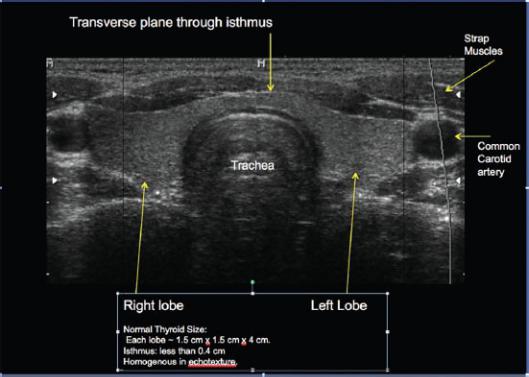
Fig. 29.1 Ultrasound of the thyroid gland
Transverse (axial) plane.
The thyroid gland is perfectly suited for ultrasound because of its position deep to the skin and subcutaneous tissues of the neck. This allows the use of a high-frequency linear transducer probe, which gives the highest resolution possible with ultrasound. The thyroid gland is homogeneously echoic, slightly more echogenic (whiter) than muscle. Note how the echogenic fascial planes delineate the strap muscles. The common carotid artery appears black because of the fluid (blood) it contains.
Ultrasound waves do not travel well through air, so the trachea is seen as a curved line anteriorly where the sound waves bounce off the air-filled structure. (Courtesy of Joseph Makris, MD, Baystate Medical Center.)
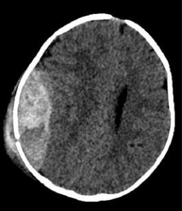
Fig. 29.2 CT of the head showing large epidural hematoma
This is a single axial image from a CT scan of the head in an unconscious patient after a severe motor vehicle accident. There is a large right-sided epidural hematoma (white = acute blood in the brain) compressing and displacing the brain. This patient requires immediate surgical evacuation of the hematoma. (Courtesy of Joseph Makris, MD, Baystate Medical Center.)
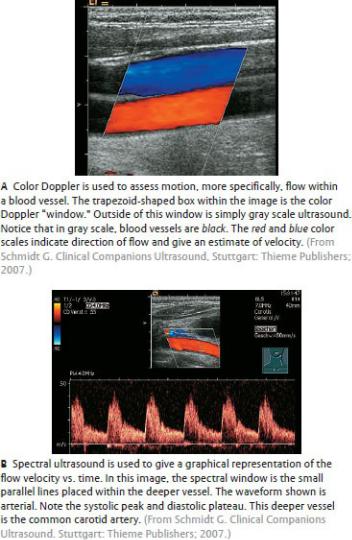
Fig. 29.3 Ultrasound of the common carotid artery and jugular vein
The skin surface is at the top of the image.
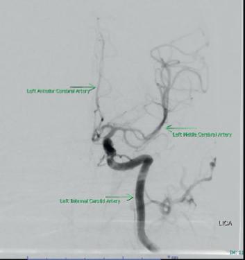
Fig. 29.4 Angiogram of the left internal carotid artery
Anteroposterior view.
In this fluoroscopic study, bones are digitally subtracted in order to isolate the vessels. The image is a photo negative of the x-ray in which contrast in the vessels appears black. The catheter, inserted into the patient’s groin and threaded through the aorta, enters the left internal carotid artery. Contrast material is then injected directly into the artery, and fluoroscopic x-rays are taken of the area during the injection. Note that in the normal condition, blood vessels are smooth and without focal or diffuse dilation and the caliber of the vessels is uniform, tapering slightly as they progress distally, without narrowing or ending abruptly. (Courtesy of Joseph Makris, MD, Baystate Medical Center.)

Fig. 29.5 CT of the head through the nasal cavity and maxillary sinuses
Transverse (axial) plane, inferior view.
CT is superior for imaging the paranasal sinuses because the high contrast readily differentiates air and bone from other structures. The airfilled sinuses are sharply contrasted against the soft tissues and even more so against the white bones. This feature also makes CT optimal for skull base imaging. Note in this patient there is some fluid in the right maxillary sinus producing an air-fluid level (patient is supine, so the fluid settles in the posterior aspect of the sinus) and the mastoid air cells on the right are filled with fluid instead of air. Compare the normal left side with the abnormal right side. This patient has right maxillary sinusitis and mastoiditis. (Courtesy of Joseph Makris, MD, Baystate Medical Center.)
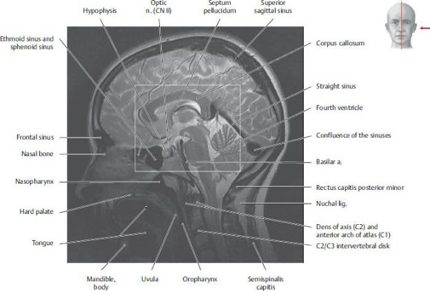
Fig. 29.6 MRI of the neck
Transverse (axial) plane, inferior view.
In this MRI sequence, fat is bright and muscles are gray. The fat planes between muscles allow differentiation of the adjacent muscles and identification of the spaces of the neck. (From Moeller TB, Reif E. Pocket Atlas of Sectional Anatomy, Vol 1, 4th ed. New York: Thieme Publishers; 2013.)
Fig. 29.7 MRI of the head
Midsagittal section.
In this MRI sequence, fluid is bright (note the bright CSF in the 4th ventricle) and soft tissues are shades of gray. However, note the subtle differences in gray scale that allow differentiation of the various soft tissues. This feature makes MRI superior to CT for use in imaging the brain. The brainstem structures and corpus callosum are sharply defined. The small amount of fluid interdigitating between the sulci of the cerebrum highlights the gross architecture of the brain. The layers of the scalp and skull are also well differentiated. (From Moeller TB, Reif E. Pocket Atlas of Sectional Anatomy, Vol 1, 4th ed. New York: Thieme
Publishers; 2013.)
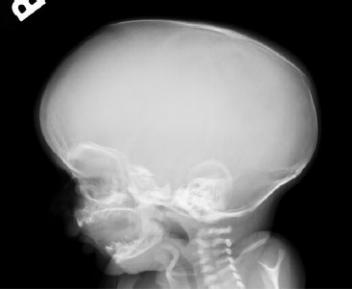
Fig. 29.8 Skull x-ray in infant
This infant was imaged for “abnormal head shape.” Note the elongated appearance of the skull (scaphocephaly) in this child with sagittal suture craniosynostosis—premature fusion of the sagittal suture. The sutures of the skull normally remain open during childhood to allow for brain and head growth. Premature fusion of the sagittal suture limits transverse growth of the skull. To compensate, the skull grows abnormally long. These infants require surgical correction by re-opening the affected fused suture. (Courtesy of Joseph Makris, MD, Baystate Medical Center.)

Unit VIII Review Questions: Head and Neck
1.Which bone of the skull contains the optic foramen, foramen ovale, and
foramen spinosum?
A.Frontal
B.Temporal
C.Sphenoid
D.Ethmoid
E.Occipital
2.The internal jugular vein
A.is located within the carotid sheath
B.receives venous drainage from the brain
C.is a tributary of the brachiocephalic vein
D.receives blood from the facial vein
E.All of the above
3.The following structure runs within the falx cerebri.
A.Inferior sagittal sinus
B.Transverse sinus
C.Sigmoid sinus
D.Cavernous sinus
E.3rd ventricle
4.The vertebral artery
A.ascends through the transverse foramina of all seven cervical vertebra
B.arises from the first part of the axillary artery
C.terminates by joining with the posterior cerebellar artery
D.supplies branches to the thyroid gland
E.supplies the posterior cerebral circulation

septum is removed. Which bone forms this part of the nasal septum?
A.Vomer
B.Ethmoid
C.Palatine
D.Temporal
E.Sphenoid
8.The lacrimal gland is innervated by
A.the facial nerve (VII)
B.the postganglionic neurons with the cell bodies in the pterygopalatine ganglion
C.the parasympathetic neurons
D.the lacrimal nerve
E.All of the above
10.A 2-year-old boy seen by his pediatrician was noticeably irritable and
tugging at his ear. When his mother asked him in a normal tone to sit quietly, he covered his ears screaming in pain and asked her not to shout at him. On exam he was found to have a fever, and his tympanic membrane was red and bulging. He was diagnosed with otitis media and started on antibiotics. The child’s extreme sensitivity to sound (hyperacusis) suggests that the infection affected the stapedius muscle. The nerve that innervates this muscle is a branch of what nerve?
A.Maxillary division of trigeminal nerve
B.Mandibular division of trigeminal nerve
C.Facial nerve
D.Glossopharyngeal nerve
E.Vagus nerve
11.The innervation for the sternothyroid muscle is the
A.ansa cervicalis
B.facial nerve
C.hypoglossal nerve
D.accessory nerve

E.recurrent laryngeal nerve
12.A man was having an animated discussion with his friends when he
started coughing after swallowing a piece of chicken at a fast-food restaurant. He soon became unable to speak or cough. Two paramedics who were taking a break at a nearby table quickly came to his help, and one stood behind the man and performed forceful abdominal thrusts (Heimlich maneuver). After several unsuccessful attempts to dislodge the aspirated food, the paramedics decide to perform an emergency airway procedure. What is the most superior site to perform this procedure so that the airway is secured below the vocal folds?
A.Superior to the hyoid bone
B.Between the hyoid bone and the thyroid cartilage
C.Between the thyroid cartilage and the cricoid cartilage
D.Between the thyroid cartilage and the arytenoid cartilage
E.Superior to the manubrium in the jugular fossa
13.A man is found to have a nodule in his neck. His calcitonin level is
elevated, and a biopsy reveals medullary carcinoma of the thyroid. A thyroidectomy is performed. After surgery the patient’s voice is found to be hoarse. What nerve was most likely injured during surgery?
A.The glossopharyngeal
B.The hypoglossal
C.The ansa cervicalis
D.The recurrent laryngeal
E.The phrenic
14.A boy who has been vomiting is diagnosed with an obstruction in his
gastrointestinal tract. He becomes severely dehydrated, and a sunken anterior fontanelle is felt. What is the youngest age at which this sign no longer would appear in a dehydrated child?
A.1 month
B.3 months
C.6 months


hypertension (pseudotumor cerebri). One complication of this disorder is downward displacement of the brainstem, which leads to stretching of the abducent nerve. How would you diagnose if this complication occurred in this patient?
A.She would be unable to move her eyes laterally.
B.She would be unable to move her eyes medially.
C.She would be unable to move her eyes superiorly.
D.She would be unable to move her eyes inferiorly
E.She would be unable to rotate her eyes internally.
19.A 61-year-old firefighter was trapped for 12 hours in a collapsed
building following an earthquake. Although he suffered a fractured tibia, the emergency department physician was also concerned about a deep scalp laceration that extended across the top of the patient’s head. Infection from this laceration could spread
A.to dural venous sinuses through emissary veins
B.into the neck in the loose areolar tissue
C.laterally beyond the zygomatic arches
D.to the epidural space
E.All of the above
20.A dentist who specializes in relief of facial pain is experimenting with a
procedure to inject anesthetic into the pterygopalatine fossa through a lateral approach. The needle passes through the mandibular notch of the mandible, traverses the infratemporal fossa, and enters the pterygomaxillary fissure. Which of the following structures would be at risk during the procedure?
A.Maxillary artery
B.Mandibular division of the trigeminal nerve (CN III)
C.Pterygoid venous plexus
D.Otic ganglion
E.All of the above
21.Which of the following drains into the inferior meatus of the nasal

cavity?
A.Nasolacrimal duct
B.Frontal sinus
C.Ethmoid sinus
D.Sphenoid sinus
E.Maxillary sinus
22.Which of the following vessels accompanies the optic nerve through the
optic canal?
A.Supratrochlear artery
B.Supraorbital artery
C.Ophthalmic artery
D.Ophthalmic vein
E.Ophthalmic nerve
23.The nerve that innervates the middle ear muscle that changes the shape
of the tympanic membrane is the
A.optic nerve
B.oculomotor nerve
C.trochlear nerve
D.trigeminal nerve
E.facial nerve
24.The branches of the cervical plexus
A.originate from the anterior rami of C1 through C4
B.innervate the sternocleidomastoid
C.carry preganglionic fibers that synapse in parasympathetic ganglia of the head
D.include only cutaneous nerves
E.include the greater occipital nerve
25.Intubation is necessary during surgery because, due to muscle relaxants

and other drugs administered during the procedure, patients are unable to fully relax the vocal folds. Paralysis of which muscle prevents the relaxation of the vocal folds?
A.Thyroarytenoid muscles
B.Posterior cricoarytenoid muscles
C.Cricothyroid muscles
D.Transverse arytenoid muscles
E.Lateral arytenoids muscles
26.All of the innervation of the larynx, both sensory and motor, is supplied
by
A.the vagus nerve
B.the glossopharyngeal nerve
C.the recurrent laryngeal nerve
D.the superior laryngeal nerve
E.None of the above
27.Which is the only laryngeal cartilage to completely encircle the airway?
A.Thyroid
B.Cricoid
C.Epiglottic
D.Arytenoid
E.Corniculate
28.A man is in a knife fight and receives a stab wound between the
sternocleidomastoid muscle and the superior belly of the omohyoid muscle. Profuse bleeding ensues due to penetration of the common carotid artery. Which structure within the same fascial sheath may also have been injured?
A.External jugular vein
B.Phrenic nerve
C.Internal jugular vein
D.Superior thyroid artery
E.Sympathetic trunk

29.A man with a long history of tobacco use goes to the dentist for a
cleaning. The dentist notices a lesion on the lateral part of the body of the tongue and refers the patient to an otolaryngologist (ENT), who biopsies the lesion and determines that the patient has squamous cell carcinoma of the tongue. A CT of the patient’s head and neck demon-strates metastasis to the lymph nodes. The primary group of lymph nodes for the drainage of this patient’s cancer is
A.superior deep cervical
B.inferior deep cervical
C.anterior superficial cervical
D.submandibular
E.submental
30.All of the following bones form a part of the nasal walls or floor except
A.Frontal
B.Ethmoid
C.Maxilla
D.Vomer
E.Palatine
31.The action of the medial pterygoid muscle is to
A.elevate the mandible
B.tense the soft palate
C.elevate the soft palate
D.elevate the hyoid bone
E.retract the mandible
32.A 63-year-old man complains of difficulty speaking, chewing, and
swallowing. On physical exam, you note that the patient’s tongue is atrophied, and when asked to stick out his tongue and say “ahh,” the patient’s tongue deviates to the left. In obtaining a medical history, you learn that the patient underwent recent surgery on his common carotid

artery. Damage to which nerve has most likely caused the observed deficits?
A.Right glossopharyngeal nerve
B.Right hypoglossal nerve
C.Left hypoglossal nerve
D.Right lingual nerve
E.Left lingual nerve
33.The mandibular division of the trigeminal nerve (V3) passes through the
foramen ovale into the
A.infratemporal fossa
B.pterygopalatine fossa
C.orbit
D.nasal cavity
E.oral cavity
34.A man with hypertension and a long history of smoking suffers a series
of transient ischemic attacks (TIAs). In the evaluation, a carotid bruit is heard. Further testing reveals severe stenosis of his internal carotid artery due to atherosclerosis. The TIAs were due to embolism of the atherosclerotic plaque. Which of the following is the first branch of the internal carotid artery likely to be affected by an embolism?
A.Superior thyroid
B.Lingual
C.Facial
D.Maxillary
E.Ophthalmic
35.A patient has thrombophlebitis of the right cavernous sinus due to a
facial skin infection. The infection traveled via the angular vein to the superior ophthalmic vein and into the cavernous sinus. Which of the following is an additional symptom that this patient might exhibit?
A.Jugular venous distension on the right
B.Inability to smile


cranial fossa between the frontal and temporal bones. Foramina of the sphenoid include the optic foramen, foramen ovale, foramen rotundum, foramen spinosum, and superior orbital fissure (Section 24.1).
A.The frontal bone forms the forehead, the roof and superior rim of the orbit, and the floor of the anterior cranial fossa.
B.The temporal bone forms part of the middle and posterior cranial fossa. Its foramina include the internal and external acoustic meatuses, carotid canal, and stylomastoid foramen.
D.The ethmoid bone forms part of the anterior cranial fossa, medial walls of the orbit, and parts of the nasal septum and lateral nasal walls,
E.The occipital bone forms most of the posterior cranial fossa. Its foramina include the foramen magnum, condylar canals, and jugular foramina.
2.E. The internal jugular vein is located within the carotid sheath (A),
contains venous drainage from the brain (B), is a tributary of the brachiocephalic vein (C), and receives blood from the facial vein (D) (Section 24.4).
A.The internal jugular vein runs within the carotid sheath with the common carotid artery and vagus nerve. B, C, and D are also correct (E).
B.Cerebral venous drainage flow primarily to the internal jugular veins. A, C, and D are also correct (E).
C.The internal jugular veins join the subclavian veins to form the brachiocephalic veins. A, B, and D are also correct (E).
D.The facial vein is a tributary of the internal jugular vein. A through C are also correct (E).
3.A. The inferior sagittal sinus runs in the inferior edge of the falx cerebri
and ends as the straight sinus (Section 26.1).
B.The transverse sinus runs along the posterolateral margins of the tentorium cerebelli.
C.The sigmoid sinuses run in grooves of the occipital and temporal
bones.
D.The cavernous sinuses lie lateral to the sella turcica between the meningeal and periosteal dura.
E. The 3rd ventricle lies between the right and left thalami of the diencephalon.
4.E. The right and left vertebral arteries join to form the basilar artery.
Together these arteries supply the posterior circulation of the brain (Section 26.2).
A.The vertebral artery ascends through the transverse foramina of C1– C6 vertebrae.
B.The vertebral artery is a branch of the subclavian artery.
C.The vertebral artery terminates by joining with the contralateral vertebral artery to form the basilar artery.
D.The vertebral artery does not supply branches to the thyroid gland.
5.B. CN VII, the facial nerve, exits the stylomastoid foramen and is
responsible for the muscles of facial expression, including the occipitofrontalis muscle, which elevates the eyebrows (Section 26.3).
A. he levator palpebrae superioris muscle elevates the eyelid and is innervated by CN III, the oculomotor nerve. In addition to the levator palpebrae superioris, the occipitofrontalis muscle assists in raising the eyebrow, but because CN III is not affected the patient is able to open his eye. He is not, however, able to close his eye completely due to the paralysis of the orbicularis oculi muscle.
C.CN XII, the hypoglossal nerve, innervates most of the tongue musculature. A left-sided hypoglossal injury would lead to the inability to protrude his tongue to the right.
D.The muscles of mastication are innervated by the mandibular division of CN V, the trigeminal nerve. Although his ability to chew is unaffected, the patient does have some trouble eating because of paralysis to the buccinator muscle, which assists in positioning food in the oral cavity.
E.Sensation to the cheek is conveyed through the maxillary division of the trigeminal nerve.
6.D. The inferior alveolar nerve courses within the mandibular canal of the mandible and would be injured in this patient (Section 26.3).

A.The lingual nerve courses through the infratemporal fossa and into the floor of the mouth.
B.The hypoglossal nerve courses anteriorly below the angle of the mandible before entering the mouth at the posterior border of the mylohyoid muscle.
C.Zygomatic branches of the facial nerve pass lateral to the masseter muscle as they cross the cheek.
E.The chorda tympani travels with the lingual nerve in the infratemporal fossa and floor of the mouth.
7.B. The lateral pterygoid muscle inserts into the condylar process of the
mandible and its articular disk and the capsule of the temporomandibular joint (TMJ) (Section 27.2).
A. The medial pterygoid inserts into the pterygoid tuberosity on the medial surface of the angle of the mandible.
C.The temporalis inserts into the apex and medial surface of the coronoid process of the mandible.
D.The masseter inserts into the masseteric tuberosity at the angle of the mandible.
E.The buccinator inserts into the angle of the mouth and orbicularis
oris.
8.A. The vomer makes up the inferior and posterior parts of the nasal
septum (Section 27.7).
B.The ethmoid bone, through its perpendicular plate, makes up the superior and posterior parts of the nasal septum.
C.The palatine forms the posterior palate and does not contribute to the nasal septum.
D.The temporal forms the base and lateral side of the skull and does not contribute to the nasal septum.
E.Although some of the sphenoid bone will be removed as a part of the procedure, it does not contribute to the nasal septum.
9.E. The lacrimal gland is innervated by visceral motor fibers

(preganglionic parasympathetic [C]) of the greater petrosal branch of the facial nerve (A). The postganglionic neuron is the zygomatic branch of the maxillary nerve (V2), which has its cell body in the pterygopalatine ganglion (B) and is then distributed to the lacrimal gland. The lacrimal nerve (D) provides sensory innervation to the lacrimal gland, conjunctiva, and upper eyelids (Section 26.3).
A.The lacrimal gland is innervated by visceral motor fibers (preganglionic parasympathetic of the greater petrosal branch of the facial nerve). B through D are also correct (E).
B.The zygomatic branch of the facial nerve carries postganglionic parasympathetic fibers from the pterygo-palatine ganglion to the lacrimal gland. A, C, and D are also correct (E).
C.Parasympathetic fibers of the zygomatic nerve inner-vate the lacrimal gland. A, B, and D are also correct (E).
D.The lacrimal nerve provides sensory innervation to the lacrimal gland, conjunctiva, and upper eyelids. A through C are also correct (E).
10.C. The stapedius nerve arises from the facial nerve in the facial canal to
innervate the stapedius muscle, which dampens sound waves transmitted through the middle ear (Section 28.2).
A.The maxillary nerve is distributed to the orbit, nasal cavity, and oral cavity, but it has no branches in the middle ear.
B.The mandibular nerve innervates the tensor tympani, which tenses the tympanic membrane.
D.The glossopharyngeal nerve transmits sensation from the tympanic cavity and pharyngotympanic tube and joins with sympathetic fibers of the internal carotid plexus to form the tympanic plexus.
E.The vagus nerve transmits sensation from the external surface of the tympanic membrane.
11.A. The ansa cervicalis innervates all of the infrahyoid muscles except
the thyrohyoid (Section 25.3 and Table 21.4).
B.The cervical branch of the facial nerve innervates the platysma.
C.The hypoglossal nerve innervates only muscles of the tongue, including the genioglossus, hyoglossus, and intrinsic tongue muscles.


frontal and parietal bones. It closes at 18 to 24 months of age (Section 24.1).
A., B., C., D. The anterior fontanelle remains open until 18 to 24 months of age.
15.B. The superior deep cervical nodes lie between the jugular and facial
veins and the anterior belly of the digastric (Section 24.5).
A. The parotid nodes are superficial nodes that overlie the parotid gland on the side of the face.
C.The inferior deep cervical nodes lie in the neck near the inferior part of the internal jugular vein.
D.The retroauricular nodes are superficial nodes that lie along the posterior margin of the auricle.
E.The lateral cervical nodes are superficial nodes that lie along the external jugular vein in the neck.
16.E. The CSF is reabsorbed into the venous system by the arachnoid
granulations, which protrude into the superior sagittal sinus (Section 26.2).
A.The choroid plexus is the site of CSF production in each of the four ventricles (the first and second [or lateral], the third, and the fourth).
B.The interventricular foramen is the communication between the lateral ventricles.
C.The cerebral aqueduct is the communication between the third and fourth ventricles.
D.The lateral apertures are the communication between the fourth ventricle and the subarachnoid space.
17.E. None of the answers above describes the vagus nerve correctly
(Section 26.3).
A.The vagus nerve leaves the skull through the jugular foramen with the glossopharyngeal and spinal accessory nerves.
B.The carotid sinus is innervated by the glossopharyn-geal nerve.
C.The oculomotor nerve (III), trochlear nerve (IV), abducent nerve (VI), ophthalmic nerve (V1), and ophthal-mic vein are transmitted through

the superior orbital fissure.
D. The vagus nerve innervates all of the muscles of the soft palate except the tensor veli palatini, which is supplied by the mandibular division of the trigeminal nerve.
18.A. The abducent nerves innervate the lateral rectus muscles, which
move the eyes laterally (Section 26.3).
B.Medial movement of the eyes is controlled by the oculomotor nerves via the medial rectus muscles.
C.Superior movement of the eyes is controlled by the oculomotor and trochlear nerves via the superior rectus and inferior oblique muscles.
D.Inferior movement of the eyes is controlled by the oculomotor nerves via the inferior rectus and the superior oblique muscles.
E.Internal rotation of the eyes, also called intorsion or rotation along the long axis of the eye, is controlled by the trochlear nerves via the superior oblique muscles. This movement is normally prevented by the action of the inferior oblique muscles.
19.A. Emissary veins communicate with veins of the scalp and can carry
infections intracranially to the dural venous sinuses (Section 27.1).
B.The attachment of the occipitofrontalis muscle to the skull prevents infections of the scalp from spreading into the neck.
C.The attachment of the epicranial aponeurosis to the zygomatic arches prevents further lateral spread of infection.
D.Spread of infection intracranially occurs through the emissary veins, which communicate with the dural sinuses, not with the epidural space.
E.Not applicable.
20.E. The infratemporal fossa contains the maxillary artery and many of its
branches, the mandibular nerve, the pterygoid plexus, and the otic ganglion, as well as the medial and lateral pterygoid muscles (Section 27.5).
A.The infratemporal fossa contains the maxillary artery, but choices B, C, and D are also correct (E).
B.The infratemporal fossa contains the mandibular nerve, but choices

A, C, and D are also correct (E).
C.The infratemporal fossa contains the pterygoid plexus, but choices A, B, and D are also correct (E).
D.The infratemporal fossa contains the otic ganglion, but choices A, B, and C are also correct (E).
21.A. The nasolacrimal duct drains tears from the medial corner of each
eye into the inferior meatus (Sections 27.7 and 28.1).
B.The frontal sinus drains into the middle meatus through a frontonasal
duct.
C.The ethmoid sinus drains into the superior and middle meatuses.
D.The sphenoid sinus drains into the sphenoethmoidal recess in the posterosuperior part of the nasal cavity.
E.The maxillary sinus drains into the middle meatus.
22.C. Only the ophthalmic artery and optic nerve enter the orbit through
the optic canal (Sections 24.1 and 28.1).
A.The supratrochlear artery is a branch of the ophthalmic artery in the orbit that supplies the scalp.
B.The supraorbital artery is a branch of the ophthalmic artery in the orbit that supplies the scalp.
D.The ophthalmic vein enters the orbit through the superior orbital
fissure.
E.The ophthalmic nerve (CN V1) enters the orbit through the superior
orbital fissure.
23.D. The mandibular division of the trigeminal nerve innervates the tensor
tympani muscle, which lessens damage from loud sounds by tensing the tympanic membrane (Section 28.2).
A.The optic nerve carries sensory innervation from the neural retina to the lateral geniculate nucleus.
B.The oculomotor nerve innervates most of the extraocular muscles of the eye and the intrinsic muscles of the eye.
C.The trochlear nerve innervates the superior oblique extraocular

muscle of the eye.
E. The facial nerve innervates the stapedius muscle, which dampens the vibrations of the stapes on the oval window.
24.A. The cervical plexus originates from the anterior rami of C1–C4
(Section 25.4).
B.The accessory nerve (CN XI) innervates the sternocleidomastoid.
C.Only the oculomotor, facial, and glossopharyngeal nerves carry preganglionic parasympathetic nerves that synapse in the head.
D.The cervical plexus has both sensory and motor components. The sensory nerves of the plexus, the lesser occipital, great auricular, transverse cervical, and supracla-vicular nerves innervate the skin of the anterior and lateral neck and lateral scalp. The ansa cervicalis, the motor part of the plexus, innervates most of the infrahyoid muscles.
E.The greater occipital nerve is supplied by the posterior rami of spinal nerves C1–C3 and is therefore not a branch of the cervical plexus.
25.B. The only muscles that relax the vocal folds are the posterior
cricoarytenoid muscles. Note that there are other results of anesthesia that also necessitate intubation (Section 25.6).
A. The thyroarytenoid closes the vocal folds.
C.The cricothyroid muscles tighten the vocal folds.
D.The transverse arytenoid muscles close the vocal folds.
E.The lateral arytenoid muscles close the vocal folds.
26.A. All of the innervation of the larynx, both sensory and motor, is
supplied by the superior and inferior (recurrent) laryngeal branches of the vagus nerve (CN X) (Section 25.6).
B.The glossopharyngeal nerve (IX) innervates the tympanic cavity and pharyngotympanic (auditory) tube; the pharynx (sensory and motor), the tonsils, palate, posterior one third of the tongue (sensory and taste), and the stylopharyngeus muscle. It also innervates the carotid body and carotid sinus.
C.The recurrent laryngeal nerve is a branch of the vagus nerve that innervates all of the muscles of the larynx except the cricothyroid and carries

sensation from the lower half of the larynx (from the vocal cords downward).
D.The superior laryngeal nerve innervates the cricothyroid muscle, which helps to tense the vocal folds, and provides sensation to the upper half of the larynx (above the vocal cords).
E.Not applicable.
27.B. The only laryngeal cartilage to completely encircle the airway is the
cricoid cartilage (Section 25.6).
A. The U-shaped thyroid cartilage has two laminae that join in the anterior midline to form a laryngeal prominence.
C.The epiglottic cartilage, a single leaf-shaped cartilage, forms the anterior wall of the laryngeal inlet at the root of the tongue.
D.Paired arytenoid cartilages articulate with the superior borders of the cricoid laminae. Their vocal processes attach to the thyroid cartilage through the vocal ligaments.
E.Corniculate cartilages appear as small tubercles within the aryepiglottic fold.
28.C. The common carotid artery, internal jugular vein, and vagus nerve
are contained within the carotid sheath (Section 25.2).
A.The external jugular vein is not within the carotid sheath.
B.The phrenic nerve lies on the anterior scalene muscle behind the carotid sheath.
D.The superior thyroid artery is a branch of the external carotid artery and does not travel in the carotid sheath.
E.The sympathetic trunk lies behind the carotid sheath.
29.D. The submandibular lymph nodes are the primary nodes of the lateral
part of the body of the tongue (Section 27.8).
A.The superior deep cervical nodes are the primary nodes of the root of the tongue.
B.The inferior deep cervical are the primary nodes of the medial parts of the body of the tongue.
C.The anterior superficial cervical nodes are the primary nodes of the

skin anterior muscles of the neck and are not the primary lymph nodes of any part of the tongue.
E. The submental nodes are the primary nodes of the apex and frenulum of the tongue.
30.A. The ethmoid, maxilla, vomer, palatine, lacrimal, nasal, and inferior
nasal concha form the bony skeleton of the nasal cavity (Section 27.7).
B.The ethmoid forms the cribriform plate at the roof of the nasal cavity, the superior and middle nasal concha of the lateral walls, and part of the nasal septum.
C.The maxilla forms the anterior part of the palate on the floor of the nasal cavity.
D.The vomer forms part of the nasal septum.
E.The palatine bone forms the posterior part of the palate on the floor of the nasal cavity.
31.A. The medial pterygoid muscle forms a sling with the masseter muscle
to elevate the mandible (Section 27.2 and Table 27.3).
B.The tensor veli palatini tenses the soft palate.
C.The levator veli palatini elevates the soft palate.
D.The suprahyoid muscles, including digastric, geniohyoid, stylohyoid, and mylohyoid, elevate the hyoid. The medial pterygoid can only elevate the hyoid second-arily by first elevating the mandible.
E.The posterior part of the temporalis muscle retracts the mandible.
32.C. The hypoglossal nerve (CN XII) innervates all muscles of the tongue
except for the palatoglossus. When damaged, muscles on the affected side atrophy and fail to contribute to protrusion of the tongue. This causes the tongue to deviate to the side of the lesion (left side in this patient). The nerve is at risk during surgery on the common carotid artery because of its proximity to the carotid bifurcation (Section 26.3).
A.The stylopharyngeus is the only muscle innervated by the glossopharyngeal nerve.
B.Damage to the right hypoglossal nerve would cause atrophy of the

(Section 26.1).
A.Jugular venous distension is caused by a decrease of venous return to the heart.
B.The inability to smile is caused by a lesion to the facial nerve.
C.The inability to chew is caused by a lesion to the mandibular division of the trigeminal nerve.
D.Loss of vision is caused by a lesion to the optic nerve, which does not travel through the cavernous sinus. Note that there are additional causes of loss of vision, none of which are related to the cavernous sinus.
36.D. The ophthalmic artery is the first major branch of the internal carotid
artery in the anterior cranial fossa (Section 24.3).
A.The posterior cerebral is the terminal branch of the basilar artery.
B.The occipital artery is a branch of the external carotid artery.
C.The labyrinthine artery is a branch of the basilar artery.
E. The middle meningeal artery is a branch of the maxillary artery, which is a branch of the external carotid artery.
37.B. Ultrasound is a dynamic modality in which the structures are seen in
real time as the sonographer manipulates the probe. Veins and arteries can be quickly and easily distinguished from each other by the application of gentle pressure on the probe. This will cause the veins with lower internal pressure to collapse temporarily, but the higher pressure arteries remain distended (Chapter 21).
A. The blue and red colors only indicate direction of blood flow relative to the transducer (red toward the transducer).
C.Although a normal vein is black (anechoic), a normal artery is black as well. Fluid, including un-clotted normal blood is black on grayscale ultrasound.
D.A normal vein is not echogenic. This would indicate a blood clot or other abnormality.
38.E. CT has excellent spatial resolution and is optimal for evaluating the bones of the skull base and assessing the anatomy/integrity of the tiny
middle ear ossicles (Chapter 21).
A.Ultrasound would be a poor choice for bony evalua-tion and for deep structures.
B.Although MRI would be sensitive to the edema seen with skull base injuries, it has lower specificity for fractures and decreased spatial resolution compared with CT.
C.X-rays would not show the skull base or ossicles well, due to the overlapping cranium.
D.Angiography may be used to assess vascular integrity after trauma, but would not be helpful in this case.