

3.3. Structure of the eukaryotic cell
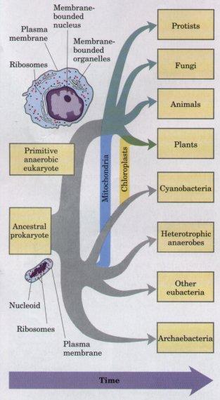
Eukaryotic cells (eu - 'true', karyo - 'nucleus') probably arose a little over 1000 million years ago, nearly 2500 million years after their prokaryotic ancestors. The development of eukaryotic cells from prokaryotic ones involved considerable changes, as can be seen from the Figure.
Three major changes must have occurred as prokaryotes gave rise to eukaryotes. First, as cells acquired more DNA, mechanisms evolved to fold it compactly into discrete complexes with specific proteins and to divide it equally between daughter cells at cell division. Second, as cells became larger, a system of intracellular membranes developed, including a double membrane surrounding the DNA. Finally, primitive eukaryotic cells, which were incapable of photosynthesis or
of aerobic metabolism formed permanent symbiotic associations with aerobic and photosynthetic bacteria. Some aerobic bacteria evolved into the mitochondria of modern eukaryotes, and some photosynthetic cyanobacteria became the chloroplasts of modern plant cells.
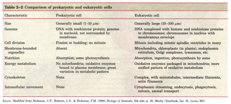
Prokaryotic and eukaryotic cells are compared in the Table2.2.
Typical eukaryotic cells (Fig.3) are much larger than prokaryotic cells-commonly 10 to 30 μm in diameter, with cell volumes 1,000 to 10,000 times larger than those of bacteria.
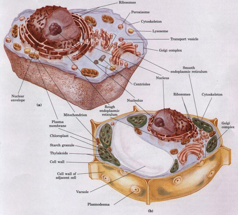
Figure 3. Schematic illustration of the two types of eukaryotic cell: a representative animal cell (a) and a representative plant cell (b).
The distinguishing characteristic of eukaryotes is the nucleus with a complex internal structure, surrounded by a double membrane. The other striking difference between eukaryotes and prokaryotes is that eukaryotes contain a number of other membrane-bounded organelles.
3.4 The nucleus
When viewed under a microscope, the most prominent feature of a cell is the nucleus. While its shape, size, position and chemical composition vary from cell to cell, its functions are always the same, namely, to control the cell’s activity and to retain the organism’s hereditary material, the chromosomes. It is bounded by a double membrane, the nuclear envelope, the outer membrane of which is continuous with the endoplasmic reticulum and often has ribosomes on its surface. The inner membrane has three proteins on its surface which act as anchoring points for chromosomes. It possesses many large pores (typically 3000 per nucleus) 40-100 nm in diameter, which permit the passage of large molecules, such as RNA, between it and the cytoplasm. (Fig. 4)
F
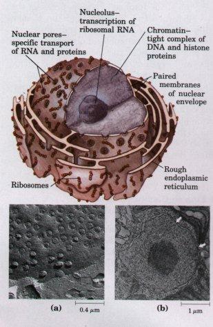
The cytoplasm-like material within the nucleus is called nucleoplasm. It contains chromatin which is made up of coils of DNA bound to proteins. During division the chromatin condenses to form the chromosomes but these are rarely, if ever, visible in a non-dividing cell. The denser, more darkly staining areas of chromatin are called heterochromatin. (Fig. 5)
W

The functions of a nucleus are:
1. To contain the genetic material of a cell in the form of chromosomes.
2. To act as a control centre for the activities of a cell.
3. To carry the instructions for the synthesis of proteins in the nuclear DNA
4. To be involved in the production of ribosomes and RNA.
5. In-cell division.
Figure 5. Chromosomes are visible in the electron microscope during mitosis. Shown here is one of the 46 human chromosomes. Every chromosome is composed of two chromatids; each consisting of tightly folded chromatin fibers. Each chromatin fiber is in turn formed by the packaging of a DNA molecule wrapped about histone proteins to form a series of nucleosomes.