Добавил:
Опубликованный материал нарушает ваши авторские права? Сообщите нам.
Вуз: Предмет: Файл:

Molecular Fluorescence

.pdf
Скачиваний:
71
Добавлен:
15.08.2013
Размер:
7.26 Mб
Скачать

Fig. 11.15. Fluorescence decay of a single molecule of rhodamine 110. The estimated lifetime is 3:9 G0:6 ns. Repetitive measurements on several hundreds of single molecules lead to a value of 3:7 G0:1 ns, in

11.4 Single-molecule fluorescence spectroscopy 375

excellent agreement with the lifetime measured in bulk solutions (3:8 G0:1 ns) (adapted from Wilkerson et al., 1993, Appl. Phys. Lett. 62, 2030).

the observation of the dynamics of a single fluorescent molecule interacting with guanine on a DNA strand.

Under the same optical configuration, FCS (Fluorescence Correlation Spectroscopy) measurements (see Section 11.3) can be carried out on samples at the singlemolecule level under conditions where the average number of fluorescent molecules in the excitation volume is less than 1. It should be noted that at low fluorophore concentrations, the time required to obtain satisfactory statistics for the fluctuations may become problematic in practical applications (e.g. for a concentration of 1 fM, a fluorophore crosses a confocal excitation volume every 15 min).

Several interesting applications to DNA molecules have been reported (e.g. hybridization, replication, detection of single point mutations, etc.). Single-molecule FCS was also used to study proteins and enzymes (e.g. Green Fluorescent Proteins (GFP), interactions of proteins with carbohydrates, conformational change of Hþ- ATPase upon binding to nucleotides, etc.). Finally, single-molecule FCS is the method of choice for drug screening (Buehler et al., 2001).

Two-photon excitation (TPE) fluorescence microscopy (Section 11.2.1.2) can be applied to the detection of single molecules in solution. By comparison with one-

376 11 Advanced techniques in fluorescence spectroscopy

Box 11.3 DNA sequencing using single-molecule detection of fluorescently labeled nucleotides

After complete sequencing of the human genome using Sanger’s enzymatic chain termination method and automated DNA sequencing machines, it is of interest to develop alternative methods that are more e cient and more accurate in order to understand the function of each gene and the corresponding health implications. This requires investigation of genetic variations in di erent

Fig. B11.3.1. Principle of a flow-based method to sequence single fragments of DNA. A: synthesis of the complementary strand with nucleotides labeled with fluorophores. B: attachment of this strand to a

microsphere by an avidin (Av)–biotin (B) bond and suspension in a flowing sample stream. C: sequential cleavage by an exonuclease and detection (adapted from Keller et al.c)).

11.4 Single-molecule fluorescence spectroscopy 377

cell types, individuals and organismsa). Fluorescence-based detection of single molecules is one of these methods.

In the early 1990s, Keller and coworkers proposed a very clever method to sequence single fragments of DNAb,c). The principle is illustrated in Figure B11.3.1. A DNA strand is replicated by a polymerase using nucleotides linked to a fluorophore via a linker arm. The fluorescently tagged DNA strand is attached to a support (e.g. a latex bead) and suspended in a flowing sample stream. The DNA bases are then sequentially cleaved by an exonuclease enzyme. The released labeled nucleotides are detected and identified by their fluorescence signature. The DNA sequence can thus be determined by the order in which the labeled nucleotides pass through the laser beam. This method has the potential for reading long DNA sequences (A104 bases) in contrast to gel-based techniques (< 103 bases). The rate can reach several hundred bases per second.

When the nucleotides are labeled with di erent fluorophores, they are identified by their spectral characteristics. Alternatively, the same fluorophore can be used and distinction is made on the basis of di erent lifetimes as a result of di erent interactions between the nucleotide and the fluorophore.

The use of sub-micrometer channels and detection by confocal fluorescence microscopy is an interesting alternative, which should allow precise control of the movement of single molecules by electrokinetic or electro-osmotic forcesa).

a)Neumann M., Herten D.-P. and Sauer M. (2001) in: Valeur B. and Brochon J. C. (Eds), New Trends in Fluorescence Spectroscopy. Applications to Chemical and Life Sciences, Springer-Verlag, Berlin, pp. 303–29.

b)Ambrose W. P., Goodwin P. M., Jett J. H.,

Johnson M. E., Martin J. C., Marrone B. L., Schecker J. A., Wilkerson C. W. and Keller R. A. (1993) Ber. Bunsenges. Phys. Chem. 97, 1535.

c)Keller R. A., Ambrose W. P., Goodwin P. M., Jette J. H., Martin J. C. and Wu M. (1996) Appl. Spectrosc. 50, 12A.

photon confocal detection, single-molecule detection by TPE is more sensitive because TPE’s ability to suppress background is better, and the background is smaller than in the UV. The advantages of TPE have been exploited in FCS (Chen et al., 2001).

Single molecules can be detected by NSOM (Near-field Scanning Optical Microscopy; see Section 11.2.1.3) with the advantages of (i) higher spatial resolution over far-field techniques, (ii) reduced photobleaching, (iii) simultaneous information of the surrounding of the molecule obtained from force mapping (Dunn, 1999), (iv) possible information on the orientation of the fluorophore transition moment. However, lifetime measurements on single molecules are perturbed by the nearby metal-coated tip. The e ect of the latter on spectral measurements is generally negligible at room temperature and such measurements can reveal new insights into sample properties.

Finally, the choice between far-field and near-field techniques largely depends on the application. When spatial resolution is not critical, far-field techniques are preferred, especially for studying the photophysical properties of single molecules in

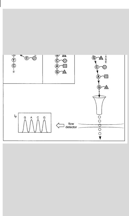
378 11 Advanced techniques in fluorescence spectroscopy

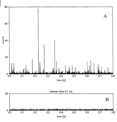
Fig. 11.16. Detection of single molecules of Rhodamine 6G by confocal fluorescence microscopy. A: solution of Rhodamine 6G 2 10 12 M in water; B: pure water (reproduced with permission from Mets and Rigler, 1994, J. Fluorescence 4, 259).

samples where coverage can be controlled. In fact, the advantages of these techniques are the enhanced signal-to-noise ratio and the higher speed of data acquisition.

11.5

Bibliography

Ambrose W. P., Goodwin P. M., Jett J. H.,

Bashe´ T., Moerner W. E.,

Orrit M. and

Van Horden A., Werner J. H. and Keller

Wild U. P. (Eds) (1996)

Single-Molecule

R. A. (1999) Single Molecule Fluorescence

Optical Detection, Imaging and Spectroscopy,

Spectroscopy at Ambient Temperature,

VCH, Weinheim.


Chem. Rev. 99, 2929–56.

Buehler C., Stoeckli K. and Auer M. (2001)

The Integration of Single Molecule Detection Technologies into Miniaturized Drug Screening: Current Status and Future Perspectives, in: Valeur B. and Brochon

J. C. (Eds), New Trends in Fluorescence Spectroscopy. Applications to Chemical and Life Sciences, Springer-Verlag, Berlin, pp. 331–79.

Chen Y., Mu¨ller J. D., Eid J. S. and Gratton E. (2001) Two-Photon Fluorescence Fluctuation Spectroscopy, in: Valeur B. and Brochon J. C. (Eds) New Trends in Fluorescence Spectroscopy. Applications to Chemical and Life Sciences, Springer-Verlag, Berlin, pp. 276–96.

Davidson R. S. (1996) Application of Fluorescence Microscopy to a Study of Chemical Problems, Chem. Soc. Rev. 241–53.

Dunn R. C. (1999) Near-Field Scanning Optical Microscopy, Chem. Rev. 99, 2891– 927.

Ehrenberg M. and Rigler R. (1976)

Fluorescence Correlation Spectroscopy Applied to Rotational Di usion of Macromolecules, Quart. Rev. Biophys. 9, 69–81.

Elson E. L. (1985) Fluorescence Correlation Spectroscopy and Photobleaching Recovery,

Ann. Rev. Phys. Chem. 36, 379–406. Gadella T. W. J., Jovin T. M. and Clegg R.

M. (1993) Fluorescence Lifetime Imaging Microscopy (FLIM): Spatial Resolution of Microstructures on the Nanosecond Time Scale, Biophys. Chem. 48, 221–39.

Goodwin P. M., Ambrose W. P. and Keller

R. A. (1996) Single-Molecule Detection in Liquids by Laser-Induced Fluorescence, Acc. Chem. Res. 29, 607–13.

Keller R. A., Ambrose W. P., Goodwin P. M., Jette J. H., Martin J. C. and Wu M. (1996) Single-Molecule Fluorescence Analysis in Solution, Appl. Spectrosc. 50, A12–32.

Kotyk A. (Ed.) (1999) Fluorescence Microscopy and Fluorescent Probes (Proceedings of the Third Conference held in Prague on June 20–23, 1999), Espero Publishing, Prague.

Herman B., Wang X. F., Wodnicki P., Perisamy A., Mahajan N., Berry G. and Gordon G. (1999) Fluorescence Lifetime Imaging Microscopy, in: Rettig W. et al. (Eds), Applied Fluorescence in Chemistry, Biology and Medicine, Springer-Verlag, Berlin, pp. 491–507.

11.5 Bibliography 379

Hink M. and Visser A. J. W. G. (1999) Characterization of Membrane Mimetic Systems with Fluorescence, in: Rettig W. et al. (Eds), Applied Fluorescence in Chemistry, Biology and Medicine, Springer-Verlag, Berlin, pp. 101–18.

Lakowicz J. R. and Szymacinski H. (1996)

Imaging Applications of Time-Resolved Fluorescence Spectroscopy, in: Wang X. F. and Herman B. (Eds), Fluorescence Imaging Spectroscopy and Microscopy, Chemical Analysis Series, Vol. 137, John Wiley & Sons, New York, pp. 273–311.

Lemasters J. J. (1996) Confocal Microscopy of Single Living Cells, in: Wang X. F. and Herman B. (Eds), Fluorescence Imaging Spectroscopy and Microscopy, Chemical Analysis Series, Vol. 137, John Wiley & Sons, New York, pp. 157–77.

Madge D. (1976) Chemical Kinetics and Applications of Fluorescence Correlation Spectroscopy, Quart. Rev. Biophys. 9, 35–47.

Mialocq J.-C. and Gustavsson T. (2001)

Investigation of Femtosecond Chemical Reactivity by Means of Fluorescence UpConversion, in: Valeur B. and Brochon

J. C. (Eds), New Trends in Fluorescence Spectroscopy. Applications to Chemical and Life Sciences, Springer-Verlag, Berlin, pp. 61–80.

Neumann M., Herten D.-P. and Sauer M. (2001) New Techniques for DNA Sequencing Based on Diode Laser Excitation and Time-Resolved Fluorescence Detection, in: Valeur B. and Brochon

J. C. (Eds), New Trends in Fluorescence Spectroscopy. Applications to Chemical and Life Sciences, Springer-Verlag, Berlin, pp. 303–29.

Petersen N. O. and Elson E. L. (1986) Measurements of Di usion and Chemical Kinetics by Fluorescence Photobleaching Recovery and Fluorescence Correlation Spectroscopy, Methods Enzymol. 130, 454– 84.

Piston D. W. (1996) Two-photon Excitation Microscopy, in: Wang X. F. and Herman B. (Eds), Fluorescence Imaging Spectroscopy and Microscopy, Chemical Analysis Series, Vol. 137, John Wiley & Sons, New York, pp. 253–72.

¨

Rigler R., Widengren J. and Mets U. (1993)

Interactions and Kinetics of Single

380 11 Advanced techniques in fluorescence spectroscopy

Molecules as Observed by Fluorescence

1: Techniques, Plenum Press, New York, pp.

Correlation Spectroscopy, in: Wolfbeis O.

337–78.

S. (Ed.), Fluorescence Spectroscopy. New

Wang X. F., Periasamy A. and Herman B.

Methods and Applications, Springer-Verlag,

(1992) Fluorescence Lifetime Imaging

Berlin, pp. 15–24.

Microscopy (FLIM): Instrumentation and

So P. T. C., French T., Yu W. M., Berland K.

Applications, Crit. Rev. Anal. Chem. 23, 369–

M., Dong C. Y. and Gratton E. (1996)

95.

Two-Photon Fluorescence Microscopy:

Wang X. F., Periasamy A., Wodnicki P.,

Time-Resolved and Intensity Imaging, in:

Gordon G. W. and Herman B. (1996)

Wang X. F. and Herman B. (Eds),

Time-Resolved Fluorescence Lifetime

Fluorescence Imaging Spectroscopy and

Imaging Microscopy: Instrumentation and

Microscopy, Chemical Analysis Series, Vol.

Biomedical Applications, in: Wang X. F.

137, John Wiley & Sons, New York, pp.

and Herman B. (Eds), Fluorescence Imaging

351–74.

Spectroscopy and Microscopy, Chemical

Tan W. and Kopelman R. (1996) Nanoscale

Analysis Series, Vol. 137, John Wiley &

Imaging and Sensing by Near-Field Optics,

Sons, New York, pp. 313–50.

in: Wang X. F. and Herman B. (Eds),

Webb W. W. (1976) Applications of

Fluorescence Imaging Spectroscopy and

Fluorescence Correlation Spectroscopy,

Microscopy, Chemical Analysis Series, Vol.

Quart. Rev. Biophys. 9, 49–68.

137, John Wiley & Sons, New York, pp.

¨

Widengren J., Mets U. and Rigler R. (1995)

407–75.

Fluorescence Correlation Spectroscopy of

Thompson N. L. (1991) Fluorescence

Triplet States in Solution: A Theoretical and

Correlation Spectroscopy, in: Lakowicz J. R.

Experimental Study, J. Phys. Chem. 99,

(Ed.), Topics in Fluorescence Spectroscopy, Vol.

13368–79.

Molecular Fluorescence: Principles and Applications. Bernard Valeur

> 2001 Wiley-VCH Verlag GmbH ISBNs: 3-527-29919-X (Hardcover); 3-527-60024-8 (Electronic)

381

Epilogue

Dans la phase initiale de la de´marche (. . .), le scientifique fonctionne par l’imagination, comme l’artiste. Apre`s seulement, quand interviennent l’e´preuve critique et l’expe´rimentation, la science se se´pare de l’art

. . .

[In the initial phase of the process (. . .), the scientist works through the imagination, as does the artist. Only afterwards, when critical testing and experimentation come into play, does science diverge from art . . .]

F. Jacob, 1997

Соседние файлы в предмете Химия