

Molecular Fluorescence
.pdf9.2 Determination of distances at a supramolecular level using RET 255
Box 9.1 Recovery of distance distributions in a triantennary glycopeptidea)
The flexibility of the sugar linkages in glycoproteins results in multiple conformations that can be detected by time-resolved fluorescence experiments. Figure B9.1.1 shows an example of a triantennary glycopeptide labeled with a
Fig. B9.1.1. Distance distribution of a triantennary
glycopeptide reflecting the multiple conformations (adapted from Wu et al.a)).

256 9 Resonance energy transfer and its applications
naphthyl-2-acetyl donor and a dansylethylenediamine acceptor. The fluorescence decay curves were found to be satisfactorily fitted with a bimodal Lorentzian distribution, which suggests that the branch labeled with the acceptor can fold back towards the donor. The two conformations di er in donor–acceptor distance by about 10 A˚ .
Interestingly, one of the conformers obtained from data analysis corresponds to the bound conformation of the same chain in a biantennary oligosaccharide– lectin complex from X-ray di raction.
Resonance energy transfer is thus a powerful tool for understanding the functions of glycoproteins.
a)Wu P. G., Rice K. G., Brand L. and Lee Y. C. (1991) Proc. Nat. Acad. Sci. USA 88, 9355–9.
generally assumed to be Gaussian, but this assumption is no longer valid in the case of short chains.
As outlined in Section 9.2.1, orientational heterogeneity may a ect the determination of a distance distribution, especially in the case of static orientation. Timeresolved fluorescence experiments provide an ‘apparent’ average distance and an ‘apparent’ distance distribution containing contributions from both distance and orientation (Wu and Brand, 1992).
A good example of a distribution of distances that was recovered from timeresolved measurements is presented in Box 9.1.
Distance distributions can be determined when the interchromophoric distance does not change significantly during the donor lifetime. Otherwise, energy transfer is enhanced by translational di usion of the donor and acceptor moieties towards each other. Then, information on relative di usion coe cients of chain ends in oligopeptides (Katchalski-Katzir et al., 1981) or polymers can be obtained.
9.3
RET in ensembles of donors and acceptors
We have considered so far the energy transfer from a donor to a single acceptor. Extension to ensembles of donor and acceptor molecules distributed at random in an infinite volume will now be considered, paying special attention to the viscosity of the medium. Then, the e ect of dimensionality and restricted geometry will be examined. Homotransfer among the donors or among the acceptors will be assumed to be negligible.
9.3.1
RET in three dimensions. E ect of viscosity
The viscosity of the medium plays an important role in the dynamics of energy transfer because the mean distance di used by the donor and acceptor relative to

9.3 RET in ensembles of donors and acceptors 257
each other during the excited-state lifetime of the donor tD0 is ð6DtD0 Þ1=24), in which D is the mutual di usion coe cient of the donor and acceptor (given by Eq. 4.12), depends on viscosity. This di usion distance is thus to be compared with the mean distance r between donors and acceptors, so that the criterion will be 6DtD0 =r2. Three regimes can be distinguished:
1) 6DtD0 /r 2 f1: the donor and acceptor cannot significantly di use during the excited-state lifetime of the donor. This case is called the static limit. Distinction according to the mechanism of tranfer should be made.
(a) When energy transfer from a donor to nearby acceptors can occur via the exchange mechanism (see Section 4.6.3), the Perrin model provides an approximate description. As explained in Section 4.2.3, this model is based on the existence of a sphere of e ective quenching of the donor in which transfer from the donor to the acceptor occurs with 100% e ciency. If the acceptor lies beyond this sphere, there is no transfer from the donor. Perrin’s equation can then be written as
I0
D ¼ expðVqNa½A&Þ ð9:26Þ
ID
where Vq is the volume of the quenching sphere of the donor, and Na is Avogadro’s number. A plot of lnðID0 =IDÞ versus [A] yields Vq.
(b) When energy transfer is possible via the Fo¨rster dipole–dipole mechanism, transfer occurs from a donor to acceptors that are at a distance less than about 2R0 (R0: Fo¨rster critical radius). An analytical expression for the fluorescence decay of the donor in the presence of an ensemble of acceptors can be obtained under the following assumptions:
. the acceptors are randomly distributed in three dimensions;
. translational di usion is slow compared to the rate of transfer;
.rotational motion of both donor and acceptor molecules is much faster than transfer and is unrestricted, so that the orientation factor is set equal to 2=3.
The survival probability GsðtÞ of the donor molecule (i.e. the probability that when excited at t ¼ 0, it is still excited at time t) is obtained by summation over all possible rate constants kT (given by Eq. 9.1), each corresponding to a given donor– acceptor distance r. For a donor molecule surrounded with n acceptor molecules distributed at random in a spherical volume whose radius is much larger than the Fo¨rster critical radius R0, GsðtÞ is given by
dGs |
1 |
X |
|
1 1 |
X |
R0 |
6 |
|
|||||||
|
n |
|
|
n |
|
|
|||||||||
|
|
|
|
kTi |
Gs |
|
|
|
|
|
|
|
|
Gs |
9:27 |
dt |
¼ "tD0 þ i |
1 |
# |
¼ "tD0 þ tD0 |
i |
¼ |
1 ri |
# |
ð Þ |
||||||
|
|
|
|
¼ |
|
|
|
|
|
|
|
|
|
|
|
4)According to classical equations of transla-tional Brownian motion in three dimensions, the mean di usion distance of a particle during time t is ð6DtÞ1=2.
258 9 Resonance energy transfer and its applications
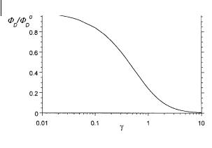
Fig. 9.2. Variations in the relative fluorescence intensity as a function of the parameter g according to Eq. (9.31).
The solution of this di erential equation, with the initial condition Gð0Þ ¼ 1, is
|
|
|
t |
|
t |
X |
R0 |
6 |
|
|
|
|
|
|
||||
Gs t |
exp |
|
|
|
|
|
|
|
|
|
|
|
|
|
|
|
|
|
ð Þ ¼ |
|
" tD0 |
tD0 i |
¼ |
1 ri |
# |
|
|
|
|
|
|||||||
|
|
|
t |
|
Y |
|
|
|
|
t |
R0 |
6 |
|
|
|
|||
|
|
|
|
n |
|
|
|
|
|
|
|
|
||||||
¼ |
|
|
|
! n 1 |
|
|
" |
|
|
|
|
# |
ð |
Þ |
||||
|
tD0 |
|
|
tD0 |
ri |
|||||||||||||
|
exp |
|
|
|
|
|
|
exp |
|
|
|
|
|
|
|
|
9:28 |
|
¼
The fluorescence of the donor is proportional to the average hGsðtÞi of this quantity. Fo¨rster obtained in this way the following relation:
|
|
|
|
|
2 |
|
|
|
|
1=23 |
|
|
|
ð Þ ¼ |
ð Þ |
|
4 |
t |
|
|
t |
! |
ð |
Þ |
|
|
|
|
|
|||||||||
|
|
tD0 |
|
tD0 |
5 |
|||||||
|
iD t |
iD 0 |
exp |
|
|
|
2g |
|
|
|
9:29 |
|
|
|
|
|
|
|
|
|
|
|
|
|
|
where g is given by |
|
|
|
|
|
|
ð9:30Þ |
|||||
|
g ¼ 2 CA 3 pR30 |
|
|
|
|
|
|
|||||
|
|
pp |
4 |
|
|
|
|
|
|
|
|
|
CA is the concentration of acceptors (expressed in number of molecules per A˚ 3). CA 43 pR30 represents the average number of acceptor molecules in a sphere of radius R0. Calculation of the integral of hGsðtÞi leads to the following expression for the ratio of the fluorescence quantum yields in the presence and absence of energy transfer:
|
FD0 ¼ |
|
|
|
ð |
Þ½ |
|
|
|
ð |
|
Þ& |
|
|
|
ð |
Þ |
|
|
FD |
1 |
|
ppg exp g2 |
|
1 |
|
erf |
|
g |
|
|
|
|
|
9:31 |
||
|
|
|
|
|
|
|
2 |
g |
2 |
|
||||||||
|
|
|
|
|
|
|
|
|
|
|
|
|
|
|
||||
|
|
|
|
|
|
|
|
|
|
|
|
|
|
|
|
|||
|
|
|
|
|
|
|
|
|
|
|
|
|
|
|
ð0 e u du. |
|
||
|
|
|
|
|
|
|
|
|
|
|
|
|
|
|
|
|||
where erf ðgÞ is the error function erf ðgÞ ¼ pp |
|
|||||||||||||||||
The variation of FD=FD0 versus g are shown in Figure 9.2.

9.3 RET in ensembles of donors and acceptors 259
2) 6DtD0 /r 2 g1: the mean distance di used by the donor and acceptor relative to each other during the excited-state lifetime of the donor is much larger than the mean distance R between donors and acceptors. Two cases are to be considered according to the mechanism of energy transfer:
(a) In the case of the exchange mechanism, energy transfer occurs via a collisional process and thus obeys Stern–Volmer kinetics, the transfer rate constant being that of the mutual di usional rate constant k1 of the donor and acceptor (see Section 4.2.1):
I0 |
t0 |
0 |
|
|
|
D |
|
D |
|
|
|
|
¼ |
|
¼ 1 þ k1tD |
½A& |
ð9:32Þ |
ID |
tD |
||||
where [A] is the acceptor concentration.
(b) In the case of the Fo¨rster mechanism, energy transfer from donors occurs not only to acceptors that are within a distance about 2R0 at the instant of excitation but also to acceptors that come within this distance during the donor excitedstate lifetime tD0 . The limit corresponding to 6DtD0 =r2 g1 is called the rapid di u- sion limit.
For an ensemble of donor and acceptor molecules distributed at random in an infinite volume, it is easy to calculate the sum of the rate constants for transfer from donor to all acceptors because all donors of this ensemble are identical in the rapid di usion limit:
y 1 |
|
R0 |
6 |
4 |
1 |
R0 |
3 |
|
||||
kT ¼ CA ðRc |
|
|
|
|
4pr2 dr ¼ CA |
|
pR30 |
|
|
|
|
ð9:33Þ |
tD0 |
r |
3 |
tD0 |
Rc |
||||||||
|
|
|
|
|
|
|
|
|
|
|
˚ 3 |
) and Rc is the distance of |
where CA is the concentration of acceptors (molecules/A |
||||||||||||
closest approach.
It should be emphasized that, because small molecules in usual solvents have di usion coe cients <10 5 cm2 s 1, the rapid di usion limit can be attained only for donors with lifetimes of @1 ms. This is the case for lanthanide ions; for instance, the lifetime of Tb3þ chelated to dipicolinate is 2.2 ms. Stryer and coworkers (1978) showed that using Tb3þ as a donor and rhodamine B as an acceptor, the concentration of rhodamine B resulting in 50% transfer was 6:7 10 6 M, which is three orders of magnitude less than the concentration corresponding to 50% transfer in the static limit.
It is interesting to note that energy transfer in the rapid di usion limit is sensitive to the distance of closest approach of the donor and acceptor. Based on this observation, some interesting applications in biology have been described, such as the measurement of the distance at which an acceptor is buried in biological macromolecules and membrane systems.
3) 6DtD0 /r 2 A1: the mean distance di used by the donor and acceptor relative to each other during the excited-state lifetime of the donor is comparable to the mean

260 9 Resonance energy transfer and its applications
distance r between donors and acceptors. This case is very complex. Among the various approaches, one of the most successful is that due to Go¨sele and coworkers (1975) (see Box 4.1 of Chapter 4) who obtained the following approximate solution:
|
|
|
|
|
|
2 |
|
|
|
|
|
1=23 |
|
|
|
ð Þ ¼ |
ð Þ |
4 |
t |
|
|
t |
! |
ð |
Þ |
||||
|
|
|
||||||||||||
|
tD0 |
|
tD0 |
5 |
||||||||||
iD t |
|
|
iD |
0 |
exp |
|
|
|
2Bg |
|
|
|
9:34 |
|
where the parameter B is given by |
|
|
|
|||||||||||
|
|
1 |
|
5:4x þ 4:00x2 |
|
3=4 |
|
|
|
|
||||
B |
|
|
|
|
|
|
|
9:35 |
||||||
¼ |
" |
|
þ |
|
|
|
ð |
|||||||
|
|
1 þ 3:34x |
# |
|
|
|
Þ |
|||||||
with x ¼ DðR60=tD0 Þ 1=3t2=3 where D is the mutual di usion coe cient and g is given by Eq. (9.30).
Finally, as described in Box 4.1 of Chapter 4, an exact numerical solution of the di usion equation (based on Fick’s second law with an added sink term that falls o as r 6) was calculated by Butler and Pilling (1979). These authors showed that, even for high values of R0 (A60 A˚ ), large errors are made when using the Fo¨rster equation for di usion coe cients > 10 5 cm2 s 1. Equation (9.34) proposed by Go¨sele et al. provides an excellent approximation.
9.3.2
E ects of dimensionality on RET
Equation (9.29) for Fo¨rster kinetics is valid for randomly distributed acceptors in an infinite volume, i.e. in three dimensions. If the dimension is not 3, but 1, or 2, Eq. (9.29) must be rewritten in a more general form:
|
|
|
|
|
|
2 |
|
|
|
|
d=63 |
|
|
|
|
|
|
ð Þ ¼ |
ð Þ |
|
4 |
t |
|
|
t |
! |
|
ð |
Þ |
||
|
|
|
|
|
|
||||||||||
|
|
|
tD0 |
|
tD0 |
5 |
|
||||||||
|
iD t |
|
iD 0 exp |
|
|
|
|
2g |
|
|
|
|
9:36 |
||
|
|
|
|
|
|
|
|
|
|||||||
where d is the Euclidian dimension (1, 2 or 3) and g is given by |
|
|
|||||||||||||
|
g |
¼ |
Gð1 d=6Þ |
C |
V Rd |
|
|
|
|
ð |
9:37 |
||||
|
|
|
2 |
|
A |
d |
0 |
|
|
|
|
Þ |
|||
In this expression, Vd is the volume of the unit sphere of dimension d ðV1 ¼ 2;
V2 ¼ p; V3 ¼ 4p=3Þ. CAVdR0d represents the number of acceptors in the sphere of |
||||||
for d ¼ 2, Gð2=3Þ ¼ 1:35417 . . . ; for d ¼ 1, Gð5=6Þ ¼ 1:12878. |
¼ |
|
ð |
|
Þ ¼ |
|
dimension d and of radius R0. G is the gamma function: for d |
|
3, |
G |
1=2 |
|
pp; |
Figure 9.3 shows the e ect of dimensionality on the decay of the donor using Eqs (9.36) and (9.37).
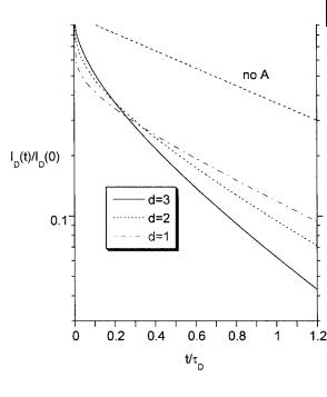
9.3 RET in ensembles of donors and acceptors 261
Fig. 9.3. Effect of dimensionality on the fluorescence decay of the donor. The value taken for g (Eq. 9.37) is Gð1 d=6Þ, i.e. for CAVdR0d ¼ 1 (average of 1 acceptor in the sphere of dimension d and of radius R0).
Only a few studies of RET in one dimension – between dyes intercalated into DNA – have been reported.
RET in two dimensions has been studied in monolayers, Langmuir–Blodgett films and phospholipid bilayers of vesicles. In particular, RET provides a very useful tool for the investigation of biological membranes, as exemplified in Box 9.2.
In media of fractal structure, non-integer d values have been found (Dewey, 1992). However, it should be emphasized that a good fit of donor fluorescence decay curves with a stretched exponential leading to non-integer d values have been in some cases improperly interpreted in terms of fractal structure. An ‘apparent fractal’ dimension may not be due to an actual self-similar structure, but to the e ect of restricted geometries (see Section 9.3.3). Another cause of non-integer values is a non-random distribution of acceptors.
9.3.3
E ects of restricted geometries on RET
Analytical expressions for the fluorescence decay in the case of RET between donor and acceptor molecules randomly distributed in various models of restricted geometries (spheres, cylinders, etc.) that mimic simple pores have been established by Klafter and Blumen (1985). The donor decay can be written in a form similar to that of Eq. (9.36):

9.3 RET in ensembles of donors and acceptors 263
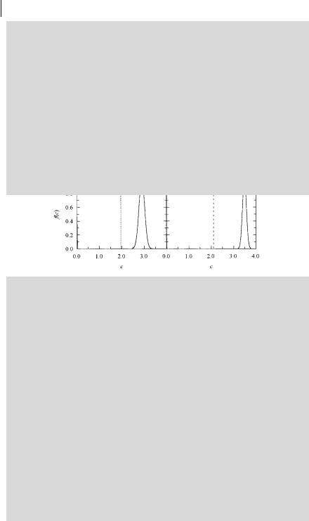
C ¼ Gð2=3ÞCApR20
The recovery of f ðCÞ is an ill-conditioned problem and it is reasonable to take a Gaussian distribution. Moreover, when probes experience two distinct environments (as expected for instance for gel/fluid heterogeneity), a sum of two Gaussian curves should be adequate for data analysisa):
f |
|
C |
|
A exp |
ð C C1 |
Þ2 |
|
h exp |
ð C C2 |
Þ2 |
ð |
Þ ¼ |
2s12 |
|
þ |
2s22 |
# |
||||
|
|
" |
|
|
An investigation of large unilamellar vesicles of dipalmitoylphosphatidylcholine using octadecyl rhodamine B (ORB) (donor) and 1,10,3,3,30,30-hexamethylindo- tricarbocyanine [DiIC1(7)] (acceptor) was carried out by Prieto and coworkersb). In the fluid phase (50 C), for moderate (1%) acceptor concentrations, donors and acceptors are essentially randomly distributed, and data analysis shows that the acceptor concentration distribution curves are well described by a single narrow Gaussian. In contrast, for the gel phase (25 C), a sum of two Gaussians is required to fit the decay curves, as shown in Figure B9.2.1. Consequently, in the gel phase, acceptors are partially segregated into a pseudo (defect) phase and donors are more randomly dispersed in the gel phase. These conclusions are supported by Monte Carlo simulations and additional photophysical measurements (steady-state energy transfer, fluorescence self-quenching in steady and transient states, and excitation energy transport).
a)Loura L. M. S., Fedorov A. and Prieto M. (2000) J. Phys. Chem. B 104, 6920.
b)Loura L. M. S. and Prieto M. (2000) J. Phys. Chem. B 104, 6911.
2! !d=63
ð Þ ¼ |
ð Þ |
4 |
t |
|
|
|
|
d |
t |
|
ð Þ |
||
tD0 |
|
|
|
6 |
|
tD0 |
5 |
||||||
iD t |
iD 0 exp |
|
|
|
A0G |
1 |
|
|
|
|
|
|
9:38 |
but d is now an ‘e ective’ or ‘apparent’ dimension and A0 is related to the surface concentration of acceptor.
Deviations from the Fo¨rster decay (Eq. 9.29) arise from the geometrical restrictions. In the case of spheres, the restricted space results in a crossover from a threedimensional Fo¨rster-type behavior to a time-independent limit. In an infinite cylinder, the cylindrical geometry leads to a crossover from a three-dimensional to a one-dimensional behavior. In both cases, the geometrical restriction induces a slower relaxation of the donor.

264 9 Resonance energy transfer and its applications
RET provides a spectroscopic method of probing local pore geometries. Levitz et al. (1988) critically evaluated the application of RET in probing the morphology of porous solids (e.g. silica gels).
9.4
RET between like molecules. Excitation energy migration in assemblies of chromophores
We have considered so far non-radiative energy transfer from a donor to an acceptor that is a di erent molecule (heterotransfer). Energy transfer between like molecules is also possible (homotransfer) if there is some overlap between the absorption and fluorescence spectra.
D þ D ! D þ D
In an assembly of chromophores, the process can repeat itself so that the excitation migrates over several molecules; it is called excitation transport, or energy migration.
Homotransfer does not cause additional de-excitation of the donor molecules, i.e. does not result in fluorescence quenching. In fact, the probability of de-excitation of a donor molecule does not depend on the fact that this molecule was initially excited by absorption of a photon or by transfer of excitation from another donor molecule. Therefore, the fluorescence decay of a population of donor molecules is not perturbed by possible excitation transport among donors. Because the transition dipole moments of the molecules are not parallel (except in very rare cases), the polarization of the emitted fluorescence is a ected by homotransfer and information on the kinetics of excitation transport is provided by the decay of emission anisotropy.
9.4.1
RET within a pair of like chromophores
For pairs of like chromophores at a fixed distance and with random and uncorrelated static orientations, the decay of emission anisotropy of the indirectly excited chromophore varies with time, tending to zero (Berberan-Santos and Valeur, 1991) in contradiction to earlier works where it was reported to be 4% of that of the directly excited chromophore. Therefore, because the probability that emission arises from the directly excited chromophore is 1=2, the decay of emission anisotropy of the latter levels o at r0/2. This can be generalized to an ensemble of n chromophores (with random and uncorrelated static orientations): the decay of emission anisotropy of the directly excited chromophore levels o at r0=n.
Consequently, homotransfer is fully characterized by the survival probability GsðtÞ representing the average probability that an initially excited molecule is still excited at time t. From these relations, the emission anisotropy can be directly expressed as rðtÞ ¼ r0GsðtÞ.