

Molecular Fluorescence
.pdfMolecular Fluorescence: Principles and Applications. Bernard Valeur
> 2001 Wiley-VCH Verlag GmbH
ISBNs: 3-527-29919-X (Hardcover); 3-527-60024-8 (Electronic)
3
1
Introduction
From the discovery of the fluorescence of Lignum Nephriticum (1965) to fluorescence probing of the structure and dynamics of matter and living systems at a molcular level
. . . ex arte calcinati, et illuminato aeri seu solis radiis, seu flammae fulgoribus expositi, lucem inde sine calore concipiunt in sese; . . .
[. . . properly calcinated, and illuminated either by sunlight or flames, they conceive light from themselves without heat; . . .]
Licetus, 1640 (about the Bologna stone)
1.1
What is luminescence?
Luminescence is an emission of ultraviolet, visible or infrared photons from an electronically excited species. The word luminescence, which comes from the Latin (lumen ¼ light) was first introduced as luminescenz by the physicist and science historian Eilhardt Wiedemann in 1888, to describe ‘all those phenomena of light which are not solely conditioned by the rise in temperature’, as opposed to incandescence. Luminescence is cold light whereas incandescence is hot light. The various types of luminescence are classified according to the mode of excitation (see Table 1.1).
Luminescent compounds can be of very di erent kinds:
.organic compounds: aromatic hydrocarbons (naphthalene, anthracene, phenanthrene, pyrene, perylene, etc.), fluorescein, rhodamines, coumarins, oxazines, polyenes, diphenylpolyenes, aminoacids (tryptophan, tyrosine, phenylalanine),
etc.
.inorganic compounds: uranyl ion (UOþ2 ), lanthanide ions (e.g. Eu3þ, Tb3þ), doped glasses (e.g. with Nd, Mn, Ce, Sn, Cu, Ag), crystals (ZnS, CdS, ZnSe, CdSe, GaS, GaP, Al2O3/Cr3þ (ruby)), etc.
4 1 Introduction
Tab. 1.1. The various types of luminescence
Phenomenon |
Mode of excitation |
|
|
Photoluminescence (fluorescence, |
Absorption of light (photons) |
phosphorescence, delayed fluorescence) |
|
Radioluminescence |
Ionizing radiation (X-rays, a, b, g) |
Cathodoluminescence |
Cathode rays (electron beams) |
Electroluminescence |
Electric field |
Thermoluminescence |
Heating after prior storage of energy |
|
(e.g. radioactive irradiation) |
Chemiluminescence |
Chemical process (e.g. oxidation) |
Bioluminescence |
Biochemical process |
Triboluminescence |
Frictional and electrostatic forces |
Sonoluminescence |
Ultrasounds |
|
|
.organometallic compounds: ruthenium complexes (e.g. Ru(biPy)3), complexes with lanthanide ions, complexes with fluorogenic chelating agents (e.g. 8-hydroxy- quinoline, also called oxine), etc.
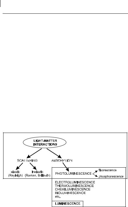
Fluorescence and phosphorescence are particular cases of luminescence (Table 1.1). The mode of excitation is absorption of a photon, which brings the absorbing species into an electronic excited state. The emission of photons accompanying deexcitation is then called photoluminescence (fluorescence, phosphorescence or delayed fluorescence), which is one of the possible physical e ects resulting from interaction of light with matter, as shown in Figure 1.1.
Fig. 1.1. Position of fluorescence and phosphorescence in the frame of light–matter interactions.
|
|
1.2 A brief history of fluorescence and phosphorescence |
5 |
|
|
|
|||
Tab. 1.2. Early stages in the history of fluorescence and phosphorescencea) |
||||
|
|
|
|
|
Year |
Scientist |
Observation or achievement |
||
|
|
|
|
|
1565 |
N. Monardes |
Emission of light by an infusion of wood Lignum Nephriticum |
||
|
|
(first reported observation of fluorescence) |
||
1602 |
V. Cascariolo |
Emission of light by Bolognese stone (first reported observation of |
||
|
|
phosphorescence) |
||
1640 |
Licetus |
Study of Bolognese stone. First definition as a non-thermal light |
||
|
|
emission |
||
1833 |
D. Brewster |
Emission of light by chlorophyll solutions and fluorspar crystals |
||
1845 |
J. Herschel |
Emission of light by quinine sulfate solutions (epipolic dispersion) |
||
1842 |
E. Becquerel |
Emission of light by calcium sulfide upon excitation in the UV. |
||
|
|
First statement that the emitted light is of longer wavelength |
||
|
|
than the incident light |
||
1852 |
G. G. Stokes |
Emission of light by quinine sulfate solutions upon excitation in |
||
|
|
the UV (refrangibility of light) |
||
1853 |
G. G. Stokes |
Introduction of the term fluorescence |
||
1858 |
E. Becquerel |
First phosphoroscope |
||
1867 |
F. Goppelsro¨der |
First fluorometric analysis (determination of Al(III) by the |
||
|
|
fluorescence of its morin chelate) |
||
1871 |
A. Von Baeyer |
Synthesis of fluorescein |
||
1888 |
E. Wiedemann |
Introduction of the term luminescence |
||
|
|
|
|
|
a)More details can be found in:
Harvey E. N. (1957) History of Luminescence, The American Philosophical Society, Philadelphia.
O’Haver T. C. (1978) The Development of Luminescence Spectrometry as an Analytical Tool, J. Chem. Educ. 55, 423–8.
1.2
A brief history of fluorescence and phosphorescence
It is worth giving a brief account of the early stages in the history of fluorescence and phosphorescence (Table 1.2), paying special attention to the origin of these terms.
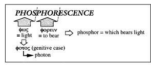
The term phosphorescence comes from the Greek: fov ¼ light (genitive case: f!t!v ! photon) and f!rein ¼ to bear (Scheme 1.1). Therefore, phosphor means ‘which bears light’. The term phosphor has indeed been assigned since the Middle
Scheme 1.1

61 Introduction
Ages to materials that glow in the dark after exposure to light. There are many examples of minerals reported a long time ago that exhibit this property, and the most famous of them (but not the first one) was the Bolognian phosphor discovered by a cobbler from Bologna in 1602, Vincenzo Cascariolo, whose hobby was alchemy. One day he went for a walk in the Monte Paterno area and he picked up some strange heavy stones. After calcination with coal, he observed that these stones glowed in the dark after exposure to light. It was recognized later that the stones contained barium sulfate, which, upon reduction by coal, led to barium sulfide, a phosphorescent compound. Later, the same name phosphor was assigned to the element isolated by Brandt in 1677 (despite the fact that it is chemically very di erent) because, when exposed to air, it burns and emits vapors that glow in the dark.
In contrast to phosphorescence, the etymology of the term fluorescence is not at all obvious. It is indeed strange, at first sight, that this term contains fluor which is not remarked by its fluorescence! The term fluorescence was introduced by Sir George Gabriel Stokes, a physicist and professor of mathematics at Cambridge in the middle of the nineteenth century. Before explaining why Stokes coined this term, it should be recalled that the first reported observation of fluorescence was made by a Spanish physician, Nicolas Monardes, in 1565. He described the wonderful peculiar blue color of an infusion of a wood called Lignum Nephriticum. This wood was further investigated by Boyle, Newton and others, but the phenomenon was not understood.
In 1833, David Brewster, a Scottish preacher, reported1) that a beam of white light passing through an alcoholic extract of leaves (chlorophyll) appears to be red when observed from the side, and he pointed out the similarity with the blue light coming from a light beam passing through fluorspar crystals. In 1845, John Herschel, the famous astronomer, considered that the blue color at the surface of solutions of quinine sulfate and Lignum Nephriticum was ‘a case of superficial color presented by a homogeneous liquid, internally colorless’. He called this phenomenon epipolic dispersion, from the Greek epip!lh ¼ surface2). The solutions observed by Herschel were very concentrated so that the majority of the incident light was absorbed and all the blue color appeared to be only at the surface. Herschel used a prism to show that the epipolic dispersion could be observed only upon illumination by the blue end of the spectrum, and not the red end. The crude spectral analysis with the prism revealed blue, green and a small amount of yellow light, but Herschel did not realize that the superficial light was of longer wavelength than the incident light.
The phenomena were reinvestigated by Stokes, who published a famous paper entitled ‘On the refrangibility of light’ in 18523). He demonstrated that the phenomenon was an emission of light following absorption of light. It is worth describing one of Stokes’ experiments, which is spectacular and remarkable for its
1) |
Brewster D. (1833) Trans. Roy. Soc. Edinburgh |
& 147–153. |
|
12, 538–45. |
3) Stokes G. G. (1852) Phil. Trans. 142, 463–562. |
2) |
Herschel J. F. W. (1945) Phil. Trans. 143–145 |
|
1.2 A brief history of fluorescence and phosphorescence 7
Scheme 1.2
simplicity. Stokes formed the solar spectrum by means of a prism. When he moved a tube filled with a solution of quinine sulfate through the visible part of the spectrum, nothing happened: the solution simply remained transparent. But beyond the violet portion of the spectrum, i.e. in the non-visible zone corresponding to ultraviolet radiations, the solution glowed with a blue light. Stokes wrote: ‘It was certainly a curious sight to see the tube instantaneously light up when plunged into the invisible rays; it was literally darkness visible.’ This experiment provided compelling evidence that there was absorption of light followed by emission of light. Stokes stated that the emitted light is always of longer wavelength than the exciting light. This statement becomes later Stokes’ law.
Stokes’ paper led Edmond Becquerel, a French physicist4), to ‘re´clamation de priorite´’ for this kind of experiment5). In fact, Becquerel published an outstanding paper6) in 1842 in which he described the light emitted by calcium sulfide deposited on paper when exposed to solar light beyond the violet part of the spectrum. He was the first to state that the emitted light is of longer wavelength than the incident light.
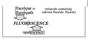
In his first paper3), Stokes called the observed phenomenon dispersive reflexion, but in a footnote, he wrote ‘I confess I do not like this term. I am almost inclined to coin a word, and call the appearance fluorescence, from fluorspar, as the analogous term opalescence is derived from the name of a mineral.’ Most of the varieties of fluorspar or fluorspath (minerals containing calcium fluoride (fluorite)) indeed exhibit the property described above. In his second paper7), Stokes definitely resolved to use the word fluorescence (Scheme 1.2).
We understand now why fluorescence contains the term fluor, but what is the origin of fluorspar or fluorspath and why are these materials fluorescent? Spar (in English) and spath (in German) were the names given in the eighteenth century8) to ‘stones’ that are more or less transparent and crystallized with a lamellar texture. Because these materials can be easily melted, and some of them can help to melt
4)Edmond Becquerel is the father of Henri Becquerel, who discovered radioactivity. Edmond Becquerel invented the famous phosphoroscope that bears his name. He was Professor at the Museum National d’Histoire Naturelle and at the Conservatoire National des Arts et Me´tiers in Paris.
5)In Cosmos (1854) 3, 509–10.
6)Becquerel E. (1842) Annales de Chimie et Physique (3) 9, 257–322.
7)Stokes G. G. (1853) Phil. Trans. 143, 385–96.
8)Macquer P. J. (1779) Dictionnaire de Chymie, p. 462.

81 Introduction
other materials, many mineralogists and metallurgists employed the word fluor 9) in order to express some fluidity ( fluere ¼ to flow in Latin). This is the origin of the name of the element fluor, isolated by Moissan in 1886, although there is no direct relationship between the Latin origin of this element and fluidity. Many spaths are known to be colored because of the presence of small amounts of impurities, which explains the fluorescence properties because fluorite itself is not fluorescent. The blue and red fluorescences are due to divalent and trivalent europium ions, respectively. Yttrium and Dysprosium can also be present and yield a yellow fluorescence10). Other possible fluorescent impurities may exist but they were found to be di cult to characterize.
The di erence between the Stokes and Becquerel experiments described above is that quinine sulfate is fluorescent whereas calcium sulfide is phosphorescent, but both species are relevant to photoluminescence. In the nineteenth century, the distinction between fluorescence and phosphorescence was made on an experimental basis: fluorescence was considered as an emission of light that disappears simultaneously with the end of excitation, whereas in phosphorescence the emitted light persists after the end of excitation. Now we know that in both cases emission lasts longer than excitation and a distinction only based on the duration of emission is not sound because there are long-lived fluorescences (e.g. uranyl salts) and short-lived phosphorescences (e.g. violet luminescence of zinc sulfide). The first theoretical distinction between fluorescence and phosphorescence was provided by Francis Perrin11): ‘if the molecules pass, between absorption and emission, through a stable or unstable intermediate state and are thus no longer able to reach the emission state without receiving from the medium a certain amount of energy, there is phosphorescence’. Further works clarified the distinction between fluorescence, delayed-fluorescence and phosphorescence12).
In addition to this clarification, many other major events in the history of fluorescence occurred during the first half of the twentieth century. The most important are reported in Table 1.3, together with the names of the associated scientists. It is remarkable that the period 1918–35 (i.e. less than 20 years) was exceptionally fecund for the understanding of the major experimental and theoretical aspects of fluorescence and phosphorescence.
1.3
Fluorescence and other de-excitation processes of excited molecules
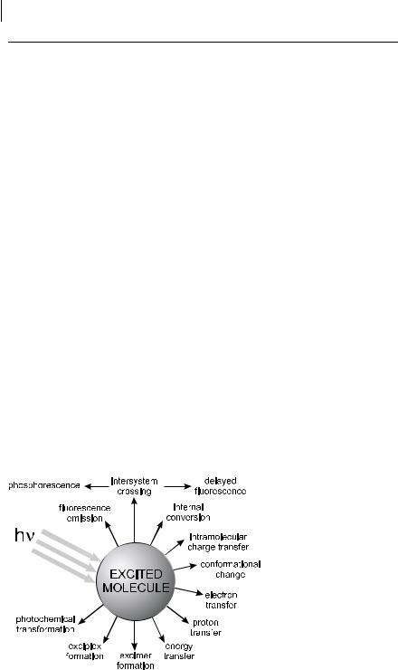
Once a molecule is excited by absorption of a photon, it can return to the ground state with emission of fluorescence, but many other pathways for de-excitation are also possible (Figure 1.2): internal conversion (i.e. direct return to the ground state
9)Macquer P. J. (1779) Dictionnaire de Chymie, p. 464.
10)Robbins M. (1994) Fluorescence. Gems and Minerals under Ulraviolet Light, Geoscience Press.
11)Perrin F. (1929) Doctoral thesis, Paris;
Annales de Physique 12, 2252–4.
12)Nickel B. (1996) Pioneers in Photochemistry. From the Perrin Diagram to the Jablonski Diagram, EPA Newsletter 58, 9–38.

1.3 Fluorescence and other de-excitation processes of excited molecules 9
Tab. 1.3. Milestones in the history of fluorescence and phosphorescence during the first half of the twentieth centurya)
Year |
Scientists |
Observation or achievement |
|
|
|
1905, |
E. L. Nichols and |
First fluorescence excitation spectrum of a dye |
1910 |
E. Merrit |
|
1907 |
E. L. Nichols and |
Mirror symmetry between absorption and fluorescence |
|
E. Merrit |
spectra |
1918 |
J. Perrin |
Photochemical theory of dye fluorescence |
1919 |
Stern and Volmer |
Relation for fluorescence quenching |
1920 |
F. Weigert |
Discovery of the polarization of the fluorescence emitted by |
|
|
dye solutions |
1922 |
S. J. Vavilov |
Excitation-wavelength independence of the fluorescence |
|
|
quantum yield |
1923 |
S. J. Vavilov and |
First study of the fluorescence polarization of dye solutions |
|
W. L. Levshin |
|
1924 |
S. J. Vavilov |
First determination of fluorescence yield of dye solutions |
1924 |
F. Perrin |
Quantitative description of static quenching (active sphere |
|
|
model |
1924 |
F. Perrin |
First observation of alpha phosphorescence (E-type delayed |
|
|
fluorescence) |
1925 |
F. Perrin |
Theory of fluorescence polarization (influence of viscosity) |
1925 |
W. L. Levshin |
Theory of polarized fluorescence and phosphorescence |
1925 |
J. Perrin |
Introduction of the term delayed fluorescence |
|
|
Prediction of long-range energy transfer |
1926 |
E. Gaviola |
First direct measurement of nanosecond lifetimes by phase |
|
|
fluorometry (instrument built in Pringsheim’s laboratory) |
1926 |
F. Perrin |
Theory of fluorescence polarization (sphere). |
|
|
Perrin’s equation |
|
|
Indirect determination of lifetimes in solution. |
|
|
Comparison with radiative lifetimes |
1927 |
E. Gaviola and |
Demonstration of resonance energy transfer in solutions |
|
P. Pringsheim |
|
1928 |
E. Jette and W. |
First photoelectric fluorometer |
|
West |
|
1929 |
F. Perrin |
Discussion on Jean Perrin’s diagram for the explanation of |
|
|
the delayed fluorescence by the intermediate passage |
|
|
through a metastable state |
|
|
First qualitative theory of fluorescence depolarization by |
|
|
resonance energy transfer |
1929 |
J. Perrin and |
Sensitized dye fluorescence due to energy transfer |
|
Choucroun |
|
1932 |
F. Perrin |
Quantum mechanical theory of long-range energy transfer |
|
|
between atoms |
1934 |
F. Perrin |
Theory of fluorescence polarization (ellipsoid) |
1935 |
A. Jablonski |
Jablonski’s diagram |
1944 |
Lewis and Kasha |
Triplet state |
1948 |
Th. Fo¨rster |
Quantum mechanical theory of dipole–dipole energy transfer |
|
|
|
a)More details can be found in the following references: Nickel B. (1996) From the Perrin Diagram to the Jablonski
Diagram. Part 1, EPA Newsletter 58, 9–38.
101 Introduction
Tab 1.3 (cont.)
Nickel B. (1997) From the Perrin Diagram to the Jablonski Diagram. Part 2, EPA Newsletter 61, 27–60.
Nickel B. (1998) From Wiedemann’s discovery to the Jablonski Diagram EPA Newsletter 64, 19–72.
Berberan-Santos M. N. (2001) Pioneering Contributions of Jean and Francis Perrin to Molecular Fluorescence, in: Valeur B. and Brochon J. C. (Eds), New Trends in Fluorescence Spectroscopy. Applications to Chemical and Life Sciences, Springer-Verlag, Berlin, pp. 7–33.
without emission of fluorescence), intersystem crossing (possibly followed by emission of phosphorescence), intramolecular charge transfer and conformational change. These processes will be the subject of Chapter 3. Interactions in the excited state with other molecules may also compete with de-excitation: electron transfer, proton transfer, energy transfer, excimer or exciplex formation. These intermolecular photophysical processes will be described in Chapter 4.
These de-excitation pathways may compete with fluorescence emission if they take place on a time-scale comparable with the average time (lifetime) during which the molecules stay in the excited state. This average time represents the experimental time window for observation of dynamic processes. The characteristics of fluorescence (spectrum, quantum yield, lifetime), which are a ected by any excitedstate process involving interactions of the excited molecule with its close environment, can then provide information on such a microenvironment. It should be noted that some excited-state processes (conformational change, electron transfer, proton transfer, energy transfer, excimer or exciplex formation) may lead to a fluorescent species whose emission can superimpose that of the initially excited molecule. Such an emission should be distinguished from the ‘primary’ fluorescence arising from the excited molecule.
The success of fluorescence as an investigative tool in studying the structure and dynamics of matter or living systems arises from the high sensitivity of fluoro-
Fig. 1.2. Possible de-excitation pathways of excited molecules.
1.4 Fluorescent probes 11
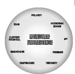
Fig. 1.3. Various parameters influencing the emission of fluorescence.
metric techniques, the specificity of fluorescence characteristics due to the microenvironment of the emitting molecule, and the ability of the latter to provide spatial and temporal information. Figure 1.3 shows the physical and chemical parameters that characterize the microenvironment and can thus a ect the fluorescence characteristics of a molecule.
1.4
Fluorescent probes
As a consequence of the strong influence of the surrounding medium on fluorescence emission, fluorescent molecules are currently used as probes for the investigation of physicochemical, biochemical and biological systems. A large part of this book is devoted to the use of so-called fluorescent probes.
It is worth recalling that other types of probes are used in practice: for example, radioactive tracers, with their well-known drawback of their radioactivity, and EPR (electronic paramagnetic resonance) probes that provide information mainly on molecular mobility. In contrast to these probes, which are used in rather limited fields of applications, fluorescent probes can o er a wealth of information in various fields, as shown in Table 1.4. The various examples described in this book will demonstrate their outstanding versatility.
Fluorescent probes can be divided into three classes: (i) intrinsic probes; (ii) extrinsic covalently bound probes; and (iii) extrinsic associating probes. Intrinsic probes are ideal but there are only a few examples (e.g. tryptophan in proteins). The advantage of covalently bound probes over the extrinsic associating probes is that the location of the former is known. There are various examples of probes covalently

12 1 Introduction
Tab. 1.4. Information provided by fluorescent probes in various fields
Field |
Information |
|
|
Polymers |
dynamics of polymer chains; microviscosity; free volume; |
|
orientation of chains in stretched samples; miscibility; phase |
|
separation; di usion of species through polymer networks; |
|
end-to-end macrocyclization dynamics; monitoring of |
|
polymerization; degradation |
Solid surfaces |
nature of the surface of colloidal silica, clays, zeolites, silica gels, |
|
porous Vycor glasses, alumina: rigidity, polarity and |
|
modification of surfaces |
Surfactant solutions |
critical micelle concentration; distribution of reactants among |
|
particles; surfactant aggregation numbers; interface properties |
|
and polarity; dynamics of surfactant solutions; partition |
|
coe cients; phase transitions; influence of additives |
Biological membranes |
fluidity; order parameters; lipid–protein interactions; translational |
|
di usion; site accessibility; structural changes; membrane |
|
potentials; complexes and binding; energy-linked and light- |
|
induced changes; e ects of additives; location of proteins; |
|
lateral organization and dynamics |
Vesicles |
characterization of the bilayer: microviscosity, order parameters; |
|
phase transition; e ect of additives; internal pH; permeability |
Proteins |
binding sites; denaturation; site accessibility; dynamics; |
|
distances; conformational transition |
Nucleic acids |
flexibility; torsion dynamics; helix structure; deformation due to |
|
intercalating agents; photocleavage; accessibility; |
|
carcinogenesis |
Living cells |
visualization of membranes, lipids, proteins, DNA, RNA, surface |
|
antigens, surface glycoconjugates; membrane dynamics; |
|
membrane permeability; membrane potential; intracellular pH; |
|
cytoplasmic calcium, sodium, chloride, proton concentration; |
|
redox state; enzyme activities; cell–cell and cell–virus |
|
interactions; membrane fusion; endocytosis; viability, cell cycle; |
|
cytotoxic activity |
Fluoroimmunochemistry |
fluoroimmunoassays |
|
|
attached to surfactants, polymer chains, phospholipids, proteins, polynucleotides, etc. A selection of them is presented in Figure 1.4. In particular, the anthroyloxy stearic acids with the anthracene moiety attached in various positions of the paraffinic chain allows micellar systems or bilayers to be probed at various depths.
Protein tagging can be easily achieved by means of labeling reagents having proper functional groups: covalent binding is indeed possible on amino groups (with isothiocyanates, chlorotriazinyl derivatives, hydroxysuccinimido active esters), and on sulfhydryl groups (with iodoacetamido and maleimido functional groups). Fluorescein, rhodamine and erythrosin derivatives with these functional groups are currently used.
Owing to the di culty of synthesis of molecules or macromolecules with covalently bound specific probes, most of the investigations are carried out with non-