

- •VOLUME 5
- •CONTRIBUTOR LIST
- •PREFACE
- •LIST OF ARTICLES
- •ABBREVIATIONS AND ACRONYMS
- •CONVERSION FACTORS AND UNIT SYMBOLS
- •NANOPARTICLES
- •NEONATAL MONITORING
- •NERVE CONDUCTION STUDIES.
- •NEUROLOGICAL MONITORS
- •NEUROMUSCULAR STIMULATION.
- •NEUTRON ACTIVATION ANALYSIS
- •NEUTRON BEAM THERAPY
- •NEUROSTIMULATION.
- •NONIONIZING RADIATION, BIOLOGICAL EFFECTS OF
- •NUCLEAR MAGNETIC RESONANCE SPECTROSCOPY
- •NUCLEAR MEDICINE INSTRUMENTATION
- •NUCLEAR MEDICINE, COMPUTERS IN
- •NUTRITION, PARENTERAL
- •NYSTAGMOGRAPHY.
- •OCULAR FUNDUS REFLECTOMETRY
- •OCULAR MOTILITY RECORDING AND NYSTAGMUS
- •OCULOGRAPHY.
- •OFFICE AUTOMATION SYSTEMS
- •OPTICAL FIBERS IN MEDICINE.
- •OPTICAL SENSORS
- •OPTICAL TWEEZERS
- •ORAL CONTRACEPTIVES.
- •ORTHOPEDIC DEVICES MATERIALS AND DESIGN OF
- •ORTHOPEDICS PROSTHESIS FIXATION FOR
- •ORTHOTICS.
- •OSTEOPOROSIS.
- •OVULATION, DETECTION OF.
- •OXYGEN ANALYZERS
- •OXYGEN SENSORS
- •OXYGEN TOXICITY.
- •PACEMAKERS
- •PAIN SYNDROMES.
- •PANCREAS, ARTIFICIAL
- •PARENTERAL NUTRITION.
- •PERINATAL MONITORING.
- •PERIPHERAL VASCULAR NONINVASIVE MEASUREMENTS
- •PET SCAN.
- •PHANTOM MATERIALS IN RADIOLOGY
- •PHARMACOKINETICS AND PHARMACODYNAMICS
- •PHONOCARDIOGRAPHY
- •PHOTOTHERAPY.
- •PHOTOGRAPHY, MEDICAL
- •PHYSIOLOGICAL SYSTEMS MODELING
- •PICTURE ARCHIVING AND COMMUNICATION SYSTEMS
- •PIEZOELECTRIC SENSORS
- •PLETHYSMOGRAPHY.
- •PNEUMATIC ANTISHOCK GARMENT.
- •PNEUMOTACHOMETERS
- •POLYMERASE CHAIN REACTION
- •POLYMERIC MATERIALS
- •POLYMERS.
- •PRODUCT LIABILITY.
- •PROSTHESES, VISUAL.
- •PROSTHESIS FIXATION, ORTHOPEDIC.
- •POROUS MATERIALS FOR BIOLOGICAL APPLICATIONS
- •POSITRON EMISSION TOMOGRAPHY
- •PROSTATE SEED IMPLANTS
- •PTCA.
- •PULMONARY MECHANICS.
- •PULMONARY PHYSIOLOGY
- •PUMPS, INFUSION.
- •QUALITY CONTROL, X-RAY.
- •QUALITY-OF-LIFE MEASURES, CLINICAL SIGNIFICANCE OF
- •RADIATION DETECTORS.
- •RADIATION DOSIMETRY FOR ONCOLOGY
- •RADIATION DOSIMETRY, THREE-DIMENSIONAL
- •RADIATION, EFFECTS OF.
- •RADIATION PROTECTION INSTRUMENTATION
- •RADIATION THERAPY, INTENSITY MODULATED
- •RADIATION THERAPY SIMULATOR
- •RADIATION THERAPY TREATMENT PLANNING, MONTE CARLO CALCULATIONS IN
- •RADIATION THERAPY, QUALITY ASSURANCE IN
- •RADIATION, ULTRAVIOLET.
- •RADIOACTIVE DECAY.
- •RADIOACTIVE SEED IMPLANTATION.
- •RADIOIMMUNODETECTION.
- •RADIOISOTOPE IMAGING EQUIPMENT.
- •RADIOLOGY INFORMATION SYSTEMS
- •RADIOLOGY, PHANTOM MATERIALS.
- •RADIOMETRY.
- •RADIONUCLIDE PRODUCTION AND RADIOACTIVE DECAY
- •RADIOPHARMACEUTICAL DOSIMETRY
- •RADIOSURGERY, STEREOTACTIC
- •RADIOTHERAPY ACCESSORIES
392 POROUS MATERIALS FOR BIOLOGICAL APPLICATIONS
BIBLIOGRAPHY
1.Bower DI. An Introduction to Polymer Physics. Cambridge: Cambridge University Press; 2002.
2.Fried JR. Polymer Science and Technology. 2nd ed. NJ: Pearson Education Inc.; 2003.
3.Lakes R, Park J. Biomaterials: an Introduction. 2nd ed., New York: Plenum; 1992.
4.Ratner B, Hoffman A, Schoen F, Lemons J. Biomaterials Science: An Introduction to Materials in Medicine. 2nd ed. Burlington, (MA): Academic Press; 2004.
5.Lanza R, Langer R, Vacanti J. Principles of Tissue Engineering. 2nd ed. Burlington, (MA): Academic Press; 2000.
6.Agrawal CM, Ray RB. Biodegradable Polymeric Scaffolds for Musculoskeletal Tissue Engineering Hoboken, (NJ): John Wiley & Sons, Inc.; 2001.
7.Liu X, Ma PX. Polymeric scaffolds for bone tissue engineering. Ann Biomed Eng 2004;32:477–486.
8.Smith LA, Ma PX. Nano-fibrous scaffolds for tissue engineering. Colloids and Surfaces B: Biointerf 2004;39:125–131.
9.Langer R. New methods of drug delivery. Science 1990;249:1527–1533.
10.Hoffman AS, Hydrogels for biomedical applications. Adv Drug Deliv Rev 2002;54:3–12.
11.Brannon-Peppas L. Polymers in controlled drug delivery. Med Plast Biomater 1997;4:34–44.
12.Qiu Y, Park K. Environment-sensitive hydrogels for drug delivery. Adv Drug Deliv Rev 2001;53:321–339.
13.Kikuchi A, Okano T. Pulsatile drug release control using hydrogels. Adv Drug Deliv Rev 2002;54:53–77.
See also BIOMATERIALS: POLYMERS; BIOMATERIALS, TESTING AND STRUCTURAL PROPERTIES OF.
POLYMERS. See BIOMATERIALS: POLYMERS.
PRODUCT LIABILITY. See CODES AND REGULATIONS:
MEDICAL DEVICES.
PROSTHESES, VISUAL. See VISUAL PROSTHESES.
PROSTHESIS FIXATION, ORTHOPEDIC. See
ORTHOPEDICS, PROSTHESIS FIXATION FOR.
POROUS MATERIALS FOR BIOLOGICAL APPLICATIONS
GRACE E. PARK
THOMAS J. WEBSTER
Purdue University
West Lafayette, Indiana
INTRODUCTION
Porous materials have received much attention in the scientific community because of their ability to interact with biological ions and molecules not only at their surfaces, but also throughout their bulk (1). Because of this intrigue, traditional applications of porous materials have involved catalysis, bioseparations, adsorption of select species, and ion exchange (1). As the tissue engineering field has emerged due to the continuous need for better implan-
table materials, porous materials have also found their niche in regenerative medicine. Specifically, porous materials have been employed as implants for various parts of the body (e.g., bone, cartilage, vasculature, central and peripheral nervous systems, bladder, and skin) either as stand-alone regenerative devices or as drug delivery vehicles to promote tissue growth. Problems associated with current implants and the need for better porous biomaterials in numerous anatomical locations are described below.
Most significantly, estimated annual U.S. healthcare costs related to tissue loss or to organ failure surpassed $400 billion in 1997 (2). An estimated 11 million people in the United States. have received at least one medical implant device; specifically, orthopedic implants (including fracture, fixation, and artificial joint devices) constitute the majority of these and accounted for 51.3% of all implants in 1992 (3). Among joint-replacement procedures, hip and knee surgeries represented 90% of the total and in 1988 were performed 310,000 times in the United States alone
(3). Implanting an orthopedic material can be a costly procedure involving considerable patient discomfort, both of which can increase if surgical revisions become necessary after an orthopedic or dental implant is rejected by the host tissue, is insufficiently integrated into juxtaposed bone, and/or fails under physiological loading conditions. Unfortunately, the average lifetime of an orthopedic implant is only 15 years due to many factors including the lack of osseointegration into surrounding bone. Current metallic implants are for the most part nonporous with subsequent poor surface properties to promote new bone ingrowth quickly.
The reason for such a high number of implanted orthopedic–musculoskeletal devices stems from numerous bone diseases. For example, approximately one out of seven Americans suffer from some form of arthritis, which is an inflammatory condition due to wear and tear in the joint
(4). The cost of arthritis and rheumatic diseases reaches $86.2 billion a year, according to a study by the Arthritis Foundation and the National Institutes of Arthritis and Musculoskeletal and Skin Diseases (NIAMS) (5). Although a very common disease, repairing damaged articular cartilage is challenging due to its limited ability to self-repair as a result of its avascularity. In fact, one of the most popular surgical techniques to repair cartilage is not through the use of a biomaterial, but rather is a surgical technique that further injures cartilage to induce scar tissue formation. This scar tissue is intended to serve as new cartilage, but since it does not match the mechanical properties of cartilage tissue, patients receiving such treatments usually suffer from additional complications and pain after only 5 years of this procedure.
The story is not any better for vascular diseases requiring biomaterial intervention. Specifically, the leading cause of death in the United States is vascular disease (including atherosclerosis), affecting 58 million people
(6). Atherosclerosis, which is hardening of the arteries, is caused by accumulation of cholesterol, fatty molecules, and other substances inside the vessel wall as the lumen becomes gradually narrower. Consequently, complete blockage of the lumen may result, inhibiting the blood flow
through that blood vessel. Treatments for these conditions require the use of a vascular graft, initially seeking autologous (or taken from an individual’s own tissue) materials. For those patients receiving a synthetic vascular graft, success rates for vessels < 7 mm approaches only 25% after 5 years. Current biomaterials used as small diameter vascular grafts are usually nonporous and result in the eventual reaccumulation of undesirable substances that clog the vessel lumen to block blood flow.
Neurological problems also necessitate the use of biomaterials. For example, Parkinson’s disease, Huntington’s disease, Alzheimer’s disease, and epilepsy prevail as common central nervous system (CNS) degenerative pathologies, especially targeting the aging population. While most of these diseases may cause a form of dementia (a mental deterioration), Alzheimer’s disease involves the loss of nerve cells related to memory and mental functions, whereas Parkinson’s and Huntington’s disease affect the mind and body. Among these, > 1.5 million Americans have been affected by Parkinson’s disease (6) and 24.4 million people are diagnosed with Alzheimer’s disease and stroke, costing > $174 billion annually (7). These diseases, however, account for only those affecting a portion of the CNS: the brain. Equally as troubling are spinal cord disfunctions. Spinal cord injuries can seriously cause damage to a person’s quality of life, contributing to 200,000 Americans with this disability and expenses of up to $250,000 a year per individual as reported in 1996 (8). Various treatment methods, such as the use of pharmaceutical agents, electrical stimulation probes, and bridges or conduits to physically connect damaged regions of the spinal cord have been developed and improved. However, few clinically approved porous biomaterials are available for treating peripheral and central nerve damage. This is despite the fact that pores in biomaterials could be very useful for guiding nerve fibers through damaged tissues.
Bladder is another organ that could benefit from the use of porous biomaterials. Urinary cancer stands as one of the most common forms of bladder disease, which is the second most common malignancy of the genitourinary tract and the fourth leading cause of cancer among American men
(9). Conventional treatment methods include the resection of the cancerous portion of the bladder wall in conjunction with intravesical immunochemotherapy (10). However, these treatments have been less than successful due to local and systemic toxicity of chemotherapy agents (11) and possible reoccurrence of the cancer (12–14). The best approach to resolve these problems is to completely remove the bladder wall, which clearly leads to the need for a replacement porous biomaterial with highly effective designs matching the material and mechanical properties of the native bladder tissue.
The above statistics highlight the current state of diseases in numerous organs and the potential effect porous biomaterials could have in treating these ailments. It is currently believed that porous biomaterials may be the solution to healing these damaged organs if designed appropriately. The next section will emphasize the features a successful porous biomaterial should have for regenerating tissues.
POROUS MATERIALS FOR BIOLOGICAL APPLICATIONS |
393 |
FEATURES OF THE NEXT GENERATION OF SUCCESSFUL POROUS BIOMATERIALS
An ideal porous scaffold for regenerating the tissues–organs mentioned in the previous section should have these characteristics (15,16): a highly porous three-dimensional (3D) interconnected network of pores for cell infiltration and subsequent tissue ingrowth; biodegradable or bioresorbable in a controllable manner with a rate that matches that of tissue growth; appropriate surface chemistry to promote desirable cell adhesion, proliferation, and differentiation; permeable for transporting sufficient amount of nutrients and metabolic waste to and from cells; mechanical properties that match that of the tissues surrounding the biomaterial in situ; and ease of processibility for various desired shapes and sizes to match specific tissue abnormality.
Several studies have confirmed that biomaterial pore size, interconnectivity, and permeability (among other properties) play a crucial role in tissue repair (17–19). Specifically, from the aforementioned list, in the following sections surface, mechanical, degradation, porosity, pore size, and pore interconnectivity properties important for the success of porous biomaterials are elaborated.
Surface Properties
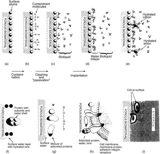
Porous Biomaterial Surface Interactions With the Biological Milieu. Assuming that the porous biomaterial has a clean surface after synthesis (Fig. 1a), the surface will be contaminated with various substances in air (e.g., hydrocarbons, sulfur, and nitrogen compounds) immediately before implantation (Fig. 1a and b) (20). Sterilization and/or introducing coatings can remove or reduce the level of contaminants. The initial interaction between an implant and the biological milieu In vivo occurs with water molecules (Fig. 1d) as a monoor bilayer forms on the surface depending on the porous biomaterial’s surface hydrophilicity (or binding strength of water molecules to the implant surface) (21). Water layers form within nanoseconds as other ions contained in body fluids (e.g., Cl and Naþ) interact with the adsorbed water molecules depending on the porous biomaterial surface chemistry (Fig. 1e). It is also possible that water interactions containing ions can penetrate the bulk porous biomaterial. Subsequently, proteins adsorb to their surfaces via initial adsorption, and then possible protein conformational changes or denaturation occurs (Fig. 1f). Replacement of these initial proteins with other proteins contained in bodily fluids may occur when biomolecules with stronger binding affinities approach the surface at a later time. Final conformations of the adsorbed proteins may differ from what occurred initially (Fig. 1g). Cells then interact with or bind to the adsorbed proteins on the porous biomaterial surface (Fig. 1h). The type of cells attached to proteins adsorbed on material surfaces and their subsequent activities will determine the tissue formed on the surface (Fig. 1i).
Protein Interactions with Porous Biomaterials
Protein Structure. Clearly, as just mentioned, one of the key events that will determine porous biomaterial success or failure is initial protein adsorption. To further explore
394 POROUS MATERIALS FOR BIOLOGICAL APPLICATIONS
Figure 1. Schematic of the porous biomaterial–tissue interface. (Adapted from Ref. 20) (a) An initially clean porous biomaterial surface possesses surface atoms. (b) A porous biomaterial surface is contaminated with molecules from the ambient environment. (c) The surface is cleaned and passivated by saturation of dangling bonds. (d) A water bilayer forms immediately after implantation. (e) Hydrated ions (e.g., Naþ, and Cl , and Ca2þ) are incorporated into the water layer. (f) Proteins adsorb onto the surface depending on their concentration and size as well as properties of the porous biomaterial surface. (g) Various types of proteins adsorb to the surface at different conformations. (h) Cells bind to the proteins that adsorbed on the porous biomaterial surface. (i) Activity of the cells at the interface determines the type of tissue formed at that site.
this, first protein structure must be discussed. There are four levels of protein structure: primary, secondary, tertiary, and quaternary structures. It is important to understand how these different types of protein structures influence initial interactions with surfaces and consequently control cellular adhesion. The primary structure of a protein is its linear sequence of amino acids. Each amino acid is linked to another through peptide bonds. Some amino acids have side chains that are charged or are neutral. Those of particular importance in aqueous solutions exhibit polar characteristics. Other amino acids change their properties depending on the pH of the solution they reside in. Therefore, it should not be surprising that proteins exist with a wide range of properties as
shown in Table 1. Table 1 describes the diverse nature of proteins in terms of size, shape, stability, and surface activity. To emphasize this diversity in protein properties, note the different interactions of albumin compared to fibrinogen on polyethylene (Table 1). Albumin is a cell nonadhesive protein while fibrinogen adsorption enhances a series of events leading to blood clot formation, a common problem of porous biomaterials in vascular applications.
Secondary protein structure consists of ordered structures in the protein chain. Two main secondary structures of proteins are the a-helix and b-pleated sheet. The degree of these structures may vary in a single protein and they are controlled by hydrogen-bonding mechanisms, which
|
|
|
|
|
POROUS MATERIALS FOR BIOLOGICAL APPLICATIONS |
395 |
||
Table 1. Diverse Properties of Proteins a |
|
|
|
|
|
|
||
Protein |
Function |
Location |
Size, kDa |
Shape, nm |
Stability |
Surface Activity |
|
|
|
|
|
|
|
|
|
|
|
Albumin |
Carrier |
Blood |
65 |
4.2 |
14.1 |
Denatures |
Low on polyethylene |
|
|
|
|
|
46.0 6. |
at 60 8C |
|
|
|
Fibrinogen |
Clotting |
Blood |
340 |
Denatures |
High on polyethylene |
|
||
|
|
|
|
(trinodular |
at 56 8C |
|
|
|
|
|
|
|
string) |
|
|
|
|
IgG |
Antibody |
Blood |
165 |
T-shaped |
|
Low on polyethylene |
|
|
Lysozyme |
Bacterial |
Tear; |
14.6 |
4.5 |
3.0 |
DGn ¼ |
High on negatively |
|
|
lysis |
hen egg |
|
(globular) |
14 kcl mol 1 |
charged surfaces |
|
|
Hemoglobin |
Oxygen |
Red blood |
65 |
5.5 |
(spherical) |
Normal form |
Very high on |
|
|
carrier |
cells |
|
|
|
|
polyethylene |
|
Hemoglobin S |
Oxygen |
Sickle red |
65 |
5.5 |
(spherical) |
Less than |
Much higher |
|
|
carrier |
blood cells |
|
|
|
hemoglobin |
air–water activity |
|
|
|
|
|
|
|
|
than hemoglobin |
|
Myoglobin |
Oxygen |
Muscle |
16.7 |
4.5 3.5 |
DGn ¼ 12 kcl mol 1 |
|
carrier |
|
|
2.5 spherical) |
|
Collagen |
Matrix |
Tissue |
285 |
300.0 1.5 |
melts at 39 8C |
|
factor |
|
|
(triple helical |
|
|
|
|
|
rod) |
DGn ¼ 8.8 kcl mol 1 |
Bacteriorhodopsin |
Membrane |
|
26 |
3.0–4.0 long |
|
|
protein |
|
|
|
denatures at 55 8C |
Tryptophan |
Enzyme |
|
27 |
|
DGn ¼ 16.8 kcl mol 1 |
Synthase |
|
|
|
|
|
alpha |
|
|
|
|
|
Subunit |
|
|
|
|
|
(wild type) |
|
|
|
|
|
Tryptophasn |
Enzyme |
|
27 |
|
|
Synthase |
|
|
|
|
|
Variant |
|
|
|
|
|
alpha Subunit |
|
|
|
|
|
High at cell membrane
High air–water activity compared to ovalbumin
Much less active at air–water interface than wild type
aSee Ref. 22.
are electrostatic attractions between oxygen of one chemical group and hydrogen of another chemical group.
Tertiary protein structures are the overall 3D shape of the protein that can be quite ordered or extremely complicated. The tertiary structure of proteins is a consequence of its primary structure as it depends on the spontaneous interactions between different amino acids and, under aqueous conditions, the spontaneous interactions between amino acids and water. There are four main interactions among residues of amino acids that contribute to the tertiary structure of proteins, each with different strengths: covalent, ionic, hydrogen, and van der Waals bonds. Of these interactions, covalent bonds are the strongest, ionic bonds are also strong (occurring between chemical groups with opposite charges), and van der Waals forces resulting from interactions between hydrophobic molecules are the weakest. However, the most influential bonds on protein tertiary structure are the weakest bonds: hydrogen bonds and van der Waals bonds. This is true since, compared to covalent and ionic bonds, these weaker bonds have many more opportunities for interacting in protein tertiary structure. In addition, because proteins exist in aqueous media, residues of amino acids must interact with water, which is a highly polar compound that forms strong hydrogen bonds. Therefore, the most stable structure of proteins in aqueous media is globular, having hydrophobic areas in the center and hydrophilic areas in the outer layer. Thus, although a generalization, it is possible that the adsorption of proteins to a porous bioma-
terial surface will be influenced by the presence of these hydrophilic amino acids on the outside of proteins in solution. However, when proteins come in contact with solid surfaces (e.g., porous biomaterials), protein structure will drastically change.
Only proteins that posses numerous subunits have quaternary structure. How these subunits interact will determine the quaternary structure of the protein. Interactions between amino acids on the exterior of the tertiary structure (mostly hydrophilic) will influence the quaternary structure, but certainly some hydrophobic interactions will also occur at the surface and impact quaternary structure.
Under certain extreme conditions (e.g., conditions that are outside of the physiological range or outside the range of 0–45 8C, pH 5–8, and in aqueous solutions of 0.15 M ionic strength), proteins may loose their normal structure (23). In other words, under such conditions, the spherical or globular tertiary structure most soluble proteins assume in aqueous media will unfold or denature. The structure of denatured proteins has been described as a random coil structure similar to those found in synthetic polymers (23). Since the structure of the protein has changed from that of a hydrophilic–hydrophobic exterior–interior to a more random arrangement, often times denatured proteins loose their solubility, become less dense (folded protein structures have densities of 1.4 g cm 3), and loose their bioactivity (23). Although there have been many examples of protein denaturation in solution, in general, only few

396 POROUS MATERIALS FOR BIOLOGICAL APPLICATIONS
cases of full protein denaturation on porous biomaterial surfaces have been reported (23). That is, generally, proteins adsorbed at the solid–liquid interface are not fully denatured and retain some degree of structure necessary to mediate cell adhesion.
Protein Interactions Mediated by Surfaces. Soluble proteins present in biological fluids (e.g., blood plasma) are the type of proteins that are involved in immediate adsorption to surfaces (24). In contrast, insoluble proteins that comprise tissues (like collagen and elastin) are not normally free to diffuse to a solid surface; these proteins may, however, appear on solid surfaces of implantable devices due to synthesis and deposition by cells (23). As mentioned, in seconds to minutes, a monolayer of adsorbed protein will form on solid surfaces (23). The concentration of proteins adsorbed on a material surface is often 1000 times greater than in the bulk phase (23). Thus, extreme competition exists for protein adsorption due to a limited space available on the surface. Because of their diverse properties just described, proteins do not absorb indiscriminately to every material surface; that is, complimentary properties of the surface and of the protein as well as the relative bulk concentration of each protein determine the driving forces for adsorption (25,26). Moreover, this initial interaction is extremely important since some proteins are not free to rotate once adsorbed to material surfaces due to multiple bonding mechanisms. Thus, immediately upon adsorption, proteins are somewhat fixed in a preferred orientation or bioactivity to the bulk media that contains cells (23). Some porous biomaterial surface properties that have influenced protein adsorption events include chemistry (i.e., ceramic versus polymer), wettability (i.e., hydrophilicity compared to hydrophobicity), roughness, and charge as will be discussed later.
One of the major differences between a flat two-dimen- sional (2D) substrate surface and that of a 3D porous material is tortuosity. Clearly, protein interactions are
much different on materials due to tortuosity. Specifically, a curved porous surface allows for greater surface area, enhanced interactions between adjacent electrons of the atoms on the surface of the pores, increased localization of point charges, and the potential for greater surface energy due to a larger juxtaposition of localized surface defects. Collectively, all of these differences between a nonporous and porous biomaterial provide for a much more complex environment for interactions between proteins and pore surfaces. It is the challenge of the porous biomaterial community to understand this challenge and thus design scaffolds that control select protein interactions.
Protein-Mediated Cell Adhesion. Interactions of proteins (both their adsorption and orientation or conformation) on porous biomaterials mediate cell adhesion. These interactions lead to extreme consequences for the ultimate function of an implanted device (27,28). An example of the importance of protein orientation for the adhesion of cells is illustrated in Fig. 2. A typical cell is pictured in this figure with integrin receptors that bind to select amino acid sequences exposed once a protein adsorbs to a surface (Fig. 1h). It is the ability of the cell to recognize such exposed amino acids that will determine whether a cell adheres or not. For example, many investigators are designing porous biomaterials to be more cytocompatible. However, it is the adhesion of select cells that must be emphasized. That is, many attempts have been made to immobilize select cell adhesive epitopes in proteins (e.g., the amino acid sequence arginine-glycine-aspartic acid or RGD) onto polymeric tissue engineering scaffolds. But, once implanted into bone, not only do desirable osteoblasts adhere, but so do undesirable fibroblasts (cells that contribute to soft not bony tissue juxtaposition).
Not only will cell adhesion be influenced by the exposure of amino acids in adsorbed proteins, but so will subsequent cell functions (e.g., extracellular matrix deposition). This is true since for anchorage-dependent cells, adhesion is a
|
Cytoskeleton |
|
|
Cell |
|
|
|
|
Integrin |
|
|
Adhesive peptide sequence of |
|
|
|
protein |
α |
|
|
(for example Arginine-Glycine- |
–Ca2± |
β |
|
RGD |
|||
Aspartic Acid (RGD)) |
|
||
|
|
||
|
Fibronectin |
|
Cell
Membrane
Integrin receptors
Proteins
(for example : vitronectin, fibronectin, collagen, etc.)
Substrate
Surface properties affecting protein conformation/bioactivity:
Chemistry, wettability; topography; surface energy; etc.
Figure 2. Influence of protein conformation on cell integrin binding. Cell adhesion and its subsequent activity will be determined by the type of integrins that the cell uses to adhere to adsorbed proteins. (Adapted and redrawn from Ref. 35.)
crucial prerequisite for subsequent cell functions. Moreover, specific intracellular messages that control subsequent cell functions are transferred inside the cell depending on which integrin receptors are utilized by the cell to adhere to adsorbed proteins. For example, a recent study by Price et al. (29), demonstrated that new bone growth was promoted when osteoblasts adhere via heparin sulfate proteoglycan binding mechanisms (as opposed to RGD) to vitronectin adsorbed on porous ceramic scaffolds.
In this manner, it is clear that cells interact with their external environment through mechanical, electrical, and chemical signals transmitted through the cell membrane. As mentioned, cell adhesion is established through cellbinding regions of extracellular matrix proteins and respective cell-membrane-intercalated receptors (i.e., integrins) among other mechanisms. Integrins are a family of transmembrane heterodimeric glycoproteins that are receptors for specific epitopes of extracellular matrix proteins and for other cell-surface molecules (30). Integrins exist as a dimer complex composed of an a-subunit (120– 180 kDa) noncovalently associated with a b-subunit (90– 110 kDa) (31). Several of these integrins have been identified that are concentrated at loci, called focal adhesion sites, of close proximity between cells and extracellular matrices on substrates (31). Focal adhesion sites are points of aggregation of, and are physically associated with, intracellular cytoskeletal molecules that control, direct, and modulate cell function in response to extracellular signals (32).
However, integrin–protein interactions are not the only mechanisms by which cells adhere. Several articles suggested that In vivo (6) and In vitro (33,34) osteoblasts (bone-forming cells) attach to an implanted material through cell membrane heparin sulfate proteoglycan interactions with, for example, heparin-binding sites on fibronectin and collagen. Moreover, Nakamura and Ozawa (6) immunohistochemically detected heparin sulfate on the membranes of osteoblasts attached to bone matrix.
Whatever the method of cell attachment, protein orientation will alter from surface to surface, since neither proteins nor materials are homogeneous in properties or structure on the exterior. The existence of protein regions that are largely acidic–basic or hydrophilic–hydrophobic or have select amino acids exposed to the media will greatly influence how that protein adsorbs to a surface and, thus, its orientation. Similarly, ceramics, metals, polymers, and composites thereof have vastly different chemistries and atomic bonding mechanisms (i.e., ionic, metallic, and covalent) to influence protein interactions. The initial interactions between proteins important for cell functions and the design of better porous biomaterials is emphasized in the next section.
Design of Better Porous Biomaterial Surfaces. As mentioned, not only do properties of proteins determine the degree of their interactions with surfaces, but properties of the media and surface (specifically, wettability, surface energy, chemistry, roughness, etc.) also influence the degree of protein interactions (35). Clearly, altering surface properties to control such protein events for mediating
POROUS MATERIALS FOR BIOLOGICAL APPLICATIONS |
397 |
cell function leading to tissue regeneration is at the heart of the research of many biomaterial scientists and engineers. Surface properties are so important because proteins have relatively large sizes and correspondingly large numbers of charged amino acid residues of different acidity/basicity well distributed on their exteriors. The polyelectrolytic property of proteins provides for exciting design criteria in surfaces to maximize or minimize specific protein interaction. Not surprisingly, at a neutral or slightly charged surface and at a pH in which the net charge on the protein is minimal, most proteins will exhibit maximum adsorption (23). For surfaces with a large net charge, initial protein interactions will be dominated by the degree of the opposite charge on the surface (23,35).
Consideration of the spatial organization of amino acids can be used in the design of surfaces to enhance protein interactions (36). As previously mentioned, for some proteins, hydrophilic and hydrophobic amino acids are present primarily on the exterior and interior, respectively. This spatial arrangement has a direct consequence on the initial interactions of these proteins with surfaces. For example, a surface that initiates interactions with the exterior hydrophilic amino acid residues in that type of a protein may promote its adsorption. In contrast, for the interior hydrophobic amino acid residues to interact with material surfaces, which may contain desirable cell adhesive epitopes (e.g., RGD), the soluble protein may have to unfold or loose tertiary structure. For this reason, one approach to increase the adsorption of a protein whose external amino acids are largely hydrophilic, would be to design a material surface which exhibits polar properties. The same can be said for any type of protein; that is, through an understanding of the amino acids that reside on the protein exterior when in the appropriate biological milieu, a complimentary surface can be designed. It is important to note, though, that this is a generalization as many proteins have a diverse collection of hydrophilic–hydrophobic amino acids externally that must be considered. In addition, as previously mentioned, proteins adsorb to surfaces in a competitive manner in which the adsorption of one protein will influence that of another.
Several studies have confirmed these speculations that properties (chemistry, charge, topography, etc.) of porous biomaterial surfaces dictate select interactions (type, concentration, and conformation–bioactivity, etc.) of proteins (24,37–40). It has been reported in the literature that changes in the type and concentration (up to 2100, 84, and 53% for albumin (40), fibronectin (41), and vitronectin (34), respectively) of protein adsorption on material surfaces depends on material surface properties, such as chemistry (i.e., polymer, metal, or ceramic), hydrophili- city–hydrophobicity, roughness, and surface energy. Consequently, since protein interactions can be controlled on porous biomaterial surfaces, so can cell adhesion. For example, a common porous biomaterial [poly(lactic-co- glycolic acid) or PLGA] has been modified to increase the adsorption of vitronectin and fibronectin through NaOH treatments (42–44). Since both vitronectin and fibronectin mediate osteoblast, vascular cell, and bladder cell adhesion, these NaOH treated PLGA scaffolds have found a home in numerous tissue engineering applications.
398 POROUS MATERIALS FOR BIOLOGICAL APPLICATIONS
However, for the field of porous biomaterials to advance even further, instead of broadly speaking of protein adsorption on surfaces, researchers need to investigate and design succinct regions of surfaces to promote protein adsorption considering the complexities of their properties. Only when porous biomaterials are considered from the context of protein interactions necessary for desirable cell interactions, will better tissue engineering materials be formulated.
Mechanical Properties and Degradation Byproducts
Although porous biomaterial surface properties determine cell attachment, mechanical strength of the scaffold and the mechanical environment it provides plays an equally important role in enhancing subsequent cell functions leading to tissue growth (45). Mechanical forces felt by cell membrane molecules are interconnected to the cytoskeleton that can influence messenger ribonucleic acid (mRNA) and subsequent synthesis of intracellular proteins (all the way to the nucleus where gene expression can be changed). It is for these reasons that mechanical properties must also be carefully controlled in porous biomaterials. For example, a study of various mechanical stimuli placed on equine articular chondrocytes within nonwoven polyglycolic acid (PGA) mesh scaffolds indicated that when the stimuli were removed, after a period of 1 week, the mechanical integrity of the resulting tissue construct was lost (46). This result implies that the mechanical stimuli applied to cells within a porous biomaterial may influence the biomechanical functionality of the regenerated tissue.
Although most agree that the mechanical properties of a porous biomaterial should match those of the physiological tissue they are intending to replace, the specific parameters and values desirable in these studies vary. For bone tissue engineering, for example, Yaszemski et al. (47) stated that scaffolds should possess mechanical stiffness matching the low range values of trabecular bone (50–100 MPa), whereas Hutmacher’s design principle (15,48) suggests matching the native tissue stiffness (10–1500 Mpa for trabecular bone (49)). Clearly, this wide range in mechanical values can provide for much different porous biomaterial efficacies and a consensus needs to be established.
Once deciding on the optimal mechanical properties needed in scaffold structures, there are numerous design parameters that can be exploited to match such values. For example, for a fibrous mesh, a decrease in fiber diameter increases mechanical strength due to an increase in fiber density (50). Obviously, increasing percent porosity and the diameters of individual pores can also be used to decrease the strength of scaffolds to match desired values. These properties not only influence inherent mechanical properties of scaffolds, but they can also be used to manipulate cell functions.
Specifically, Maroudas postulated that the scaffold surface rigidity or stiffness enhances cell adhesiveness and cell spreading (51). Pelham and Wang (52) have shown that focal adhesion contacts in cells and their migration on acrylamide gels are controlled by scaffold flexibility. They also suggested that tyrosine phosphorylation might be
involved, activated by local tension at cell adhesion sites (53). Recently, Ohya et al. (54) studied the effects of hydrogel mechanical properties on cell adhesiveness and found that the higher the strength of the hydrogel formulation, the greater the capability to withstand cell traction forces, thereby resulting in greater cell spreading. These authors also noticed that cells preferred to adhere to stiffer regions within the hydrogel.
Common pore shapes in porous biomaterials include tube-like, spherical, and randomly spaced shapes. Differences in cell attachment, growth, migration, and matrix deposition by cells have all been observed depending on pore structure. Specifically, certain cell types prefer a select pore structure in accordance to their physiological matrix environment. For example, orthopedic tissue engineering scaffolds should have spherical pores with a high porosity to allow for immediate bone ingrowth, while maintaining the mechanical strength and integrity necessary due to their harsh mechanical environments In vivo (36). Porous biomaterial pore shapes are critically related to pore interconnectivity. Not only does pore interconnectivity in a porous biomaterial affect nutrient–waste diffusion, but it also influences cell growth. Bignon et al. (55) observed that the density of pore interconnections determines cellular colonization rates; meaning that the larger the macropores (within limits), the fewer pore interconnections that have to be transversed by the cells thus resulting in higher colonization rates. Of course, guided cell growth or migration is possible through deliberate pore shape and interconnectivity. For example, tube-like or fibrous pore shapes may promote neurite extension from neurons in specific directions. Studies have also shown that cells prefer discontinuities within a porous material in terms of growth and migration; clearly pores provide such discontinuities (56–58).
In addition, maintaining mechanical strength and structural integrity of porous biomaterials are crucial because scaffolds may be crushed when implanted or may degrade over time. Mechanical properties are especially important to characterize when they change over time. A thorough knowledge of the degradation process of the porous materials of interest (including degradation byproducts) should be mapped in order to control the mechanical stability and the degradation rate until the native tissue is formed at the site of implantation.
For porous biomaterials, a range of biodegradation choices exist, from nondegradable metals to degradable ceramics and polymers. Importantly as well, degradation rates of porous materials have in some cases been shown to be faster compared to solid block polymers (59,60) because acidic byproducts become trapped inside the bulk as they degrade, therefore causing an autocatalytic effect. Of course, trapping of acidic byproducts in polymeric scaffolds can have detrimental consequences on cell health. Porous degradable polymers, such as PGA, polylactic acid, PLGA, and polycaprolactone (PCL) degrade via nonenzymatic random hydrolytic breaking of ester linkages. Sung et al. (61) studied the degradation of PLGA and PCL scaffolds In vitro and In vivo. They found a significant decrease in the molecular weight of these polymers within 1 month In vitro and, as expected, at a much faster rate In vivo.
Specifically, the influence of acidic byproducts from these polymeric scaffolds on cell health was investigated by measuring the pH of the media in which the polymers resided compared to the media in which tissue culture polystyrene (TCPS) was cultured. Changes in the media pH occurred only for PLGA (reducing it by 5) whereas no significant changes were measured during TCPS or PCL culture for up to 28 days (61).
Moreover, an In vivo study by Hedberg et al. (62) determined that soluble acidic products from degradable polymers lead to an increased recruitment of inflammatory cells compared to that induced by the scaffold itself. This was evidenced by the fact that a minimal inflammatory response was observed at the site of bone growth juxtaposed to the surface of polymeric scaffolds, whereas a major inflammatory response was observed in the scaffold where there was significant degradation. However, Sung et al. (61) suggested that an inflammatory response can be beneficial towards angiogenesis that is highly desirable to remove harmful degradation products from the interior of a polymer scaffold. This clearly demonstrates the need for controlling polymer degradation products in order to elicit a desirable response from host tissue (63). Collectively, such studies highlight the necessity for a better understanding of material degradation products on cell health.
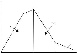
In addition, according to Wu and Ding (64), the molecular weight of a porous PLGA scaffold decreases during degradation, which not only creates a more acidic local environment, but also leads to other changes. In their study, degradation was divided into three stages, marking distinct characteristics in mechanical properties (Fig. 3). In the first stage (I), the mechanical strength increased as the porous scaffold dimensions decreased while the weight remained constant; this can be interpreted simply as the change of porous biomaterial dimensions resulting in mechanical property increases. Increased elastic modulus of porous PLGA scaffolds with degradation time was also observed in another study by Zhang and Ma (65) who contributed this to decreased porosity of the foams with time. In the second stage (II), a dramatic decrease in mechanical properties were observed, which was correlated with an increased presence of low molecular weight
strength |
Scaffold |
|
Increased presence of |
dimension |
|
low molecular weight |
|
|
|
||
Mechanical |
decrease |
|
degradation products |
|
II |
Full degradation |
|
|
|
I |
|
III 
Time
Figure 3. Three stages of mechanical strength degradation in porous biomaterials. (Adapted and redrawn from Ref. 64.)
POROUS MATERIALS FOR BIOLOGICAL APPLICATIONS |
399 |
degradation products (64). The third stage (III) was characterized by the breakdown of the scaffold’s structural integrity and associated rapid weight loss due to pH decreases from acidic degradation products. Understanding of these three distinct phases of mechanical property degradation for every proposed porous biomaterial is imperative. In addition, more studies are needed that correlate cell function at each stage of mechanical property changes in porous biomaterials as they degrade.
The Role of Porosity, Pore Size, and Interconnectivity
Among other properties (e.g., the aforementioned mechanical properties), porosity can also influence how cells behave in a scaffold. Open pore structures are desirable in most tissue engineering applications, providing enhanced cell seeding, cell attachment, proliferation, extracellular matrix production, and tissue ingrowth. For example, for orthopedic applications, both In vitro and In vivo studies demonstrated exceptional osteoblast proliferation and differentiation leading to new bone growth in PLGA foams with 90% porosity (66,67). In addition, a study by Sherwood et al. (68) reported that osteochondral composite scaffolds with 90% porosity at the cartilage portion allowed full incorporation of the chondrocytes (cartilagesynthesizing cells) into the scaffold.
Permeability, or high interconnectivity of pores is a crucial property for a porous biomaterial due to its influence on cellular communication (16), adaptation to mechanical stimulation (45), and prevention of the accumulation of acidic degradation byproducts (69,70). It also allows for uniform cell seeding and distribution, as well as proper diffusion of nutrients and metabolic wastes. Studies have shown that when tissues become thicker than 100–200 mm, the oxygen supply to cells becomes limited in a static environment (71,72). Thus, interconnectivity of pores is an extremely important design consideration to increase tissue growth into porous biomaterials.
In addition, as mentioned in the section above, the increased tortuosity present in porous biomaterials will influence protein interactions and, thus, manipulate cellular functions. Specifically, because of altered initial protein interactions, certain cell types (e.g., chondrocytes) perform much better on porous compared to flat (or nonporous) biomaterials (42). Moreover, macroporosity (pore diameters > 50 mm) influences the type of cells adhering to a polymeric scaffold. For example, large pores (100– 200 mm diameter) have been shown to enhance bone ingrowth compared to smaller pores (10–75 mm diameter) in which undesirable fibrous soft tissue formation has been observed (73). Yuan et al. (74) added that pore sizes < 10 mm promotes bone ingrowth due to optimal initial protein adsorption events possibly because of their greater surface areas. Furthermore, Bignon et al. (55) demonstrated greater cell spreading on biomaterials with micro (pore diameters < 10 mm) compared to macroporosity. Importantly as well, pore wall roughness is influenced by pore size that may be providing greater roughness to promote cell functions. Studies are needed to carefully control pore wall roughness to make accurate comparisons between scaffolds of various degrees of pore sizes. Since
400 POROUS MATERIALS FOR BIOLOGICAL APPLICATIONS
very small topographical changes have been shown to alter cell functions (75), surface roughness values in the nanometer regime could also be incorporated into porous biomaterials regardless of their pore sizes to significantly enhance protein–surface and protein–cell interactions (42,76). Fabrication methods that can provide for the manipulation of pore properties is further emphasized here.
POROUS BIOMATERIAL FABRICATION METHODS
Various methods for fabricating porous biomaterials have been explored to date. Examples for forming polymeric porous scaffolds include solvent casting with particulate leaching, gas-foaming processes, emulsion freeze drying, freeze-extraction, electrospinning, rapid-prototyping, and thermally induced phase separation. Although polymers receive the most attention in porous biomaterial applications, porous ceramics, and metals have been recently receiving much attention. This is mostly because ceramics and metals have a long history of implantation, so methods that can improve their cytocompatibility properties (e.g., by creating pores) are highly desirable. For ceramic and metallic porous biomaterials, electrophoretic deposition, salt leaching, microsphere (polymer) melt out, and annodization have been commonly employed. These methods will be briefly described in the following sections.
Cellular Solids
Current methods, such as solvent casting, gas foaming, vacuum drying, and thermally induced phase separation (TIPS) in conjunction with particulate leaching techniques create cellular solids (77). These methods can create porous constructs easily and in an inexpensive manner (78,79). In solvent casting, a pellet or powder form of a polymer is dissolved in a solvent. Then, water-soluble salt particles (e.g., sodium chloride, sodium citrate) or other particulate materials [e.g., gelatin, paraffin (79)] are added to the polymer solution. The solvent is removed through evaporation or lyophilization and then particles are leached out through the use of water or another solvent (depending on the particle chemistry) to create the desired porous structure. The advantages of these methods include simplicity and the ability to control pore size and porosity. However, the pore shape is limited to the shape of the porogen and the pore interconnectivity is poor; thus, the porogen may not be completely removed from the construct (80). Furthermore, uneven dispersion or settling of the particles within the constructs may occur. Lastly, these first generation approaches rarely provided the succinct spatial ability to control protein adsorption necessary for the next generation of more successful biomaterials.
For the gas-foaming process, a gas, usually carbon dioxide (CO2), is utilized instead of using an organic solvent at high pressures to create a highly porous structure (80–82). Again, these techniques are easy to implement and are inexpensive. However, a polymer with highly amorphous fractions can be processed with this technique even though the interconnectivity of the pores is very low, only 10–30% (18).
Thermally induced phase separation produces a highly porous material using a solvent at elevated temperatures followed by lowering the temperatures to separate the solution into liquid–liquid or solid–liquid phases. Then, the unwanted solvent is removed through sublimation (65,83). Although high mechanical strength may be obtained with this technique, the pore size created with TIPS normally ranges from 10–100 mm, which does not satisfy the permeability requirements for the removal and entry of cellular wastes and nutrients, respectively.
The emulsion freeze-drying method was developed by Whang et al. (84). A porous structure is obtained through homogenization of a polymer, organic solvent, and water mixture; rapidly cooling the mixture to maintain the liquid state structure; and then removing the solvent, and water by freeze-drying (80). In Whang’s study, 90% or greater porosity and up to 200 mm diameter pores were created. However, this method is user and technique sensitive, meaning that pore structures and associated interconnectivities greatly depend on the processing method. The freeze-extraction method is a modified version of the freeze-drying technique, in which the solvent in the frozen polymer solution is replaced with a nonsolvent at temperatures below the freezing point of the polymer solution. This procedure removes the solvent before the drying stage (85).
Electrospinning Technique
In electrospinning, an electric field directs polymer fibers to form and deposit onto a substrate of interest (86,87). Specifically, an electric potential is applied as the polymer solution is injected, which ultimately forms an electrically charged jet of polymer landing on the target substrate. The solvent evaporates and porous polymer fibers are formed. Fibrous polymer scaffolds with diameters of several hundred nanometers can be fabricated using this method, thus, simulating the physiological fibrous structure of such proteins like collagen that comprise tissues. Only films and cylindrical shapes of the porous material have been created through this technique, therefore, further investigations are needed. But in addition to creating biologically inspired nanometer fibers an advantage of this process is its ability to coat an existing implant material. Thus, this technique could be used to modify the surface properties of currently used implant materials to promote cell functions.
Rapid Prototyping
Rapid prototyping is a computer-guided manufacturing system that can produce complex designs rapidly. One of the prototyping techniques is called 3D printing (3-DP) and it has been used to fabricate biodegradable polymer scaffolds for tissue engineering purposes (88). This technique produces porous biomaterials by ink-jet printing a binder onto sequential powder layers. Importantly, growth factors, proteins, cells, and other biological factors can be incorporated into the porous biomaterial without risking inactivation because the process is performed at room temperature. However, a disadvantage of this process so far includes porous biomaterial size limitations (due to the size of the ink jet). This can also limit the creation of desirable fine details or nanostructures on the polymer.
Microsphere Burnt Out
The microsphere burnt out method is similar to the previously described salt leaching method except that polymer microspheres are utilized instead of a salt porogen. This method is useful for ceramic materials that require a sintering process at very high temperatures (approaching 10008C) at which point the polymer melts. As a very simple and easy method, it also has the disadvantages of the need for large amounts of the microspheres to create high pore interconnectivity; this results in poor mechanical properties.
Electrophoretic Deposition
Another attractive method for creating porous ceramics is electrophorectic deposition (or EPD). Due to a relatively simple setup and accommodation of complex designs and shapes, EPD has received much attention for processing fine particles, especially for coating applications (89). For this process, Ma used a graphite cathode and a stainless steel anode in the EPD cell while a current was applied to induce deposition of the particles onto a designated material. In this study, hydroxyapatite 3D porous biomaterials were fabricated. Hydroxyapatite is the main inorganic component of bone and, thus, has experienced wide spread use in orthopedic applications. This simple powder consolidation method requires no additives and high pore interconnectivity can be achieved with sufficient mechanical strength. However, this process can be costly when designing a large sample.
Anodization
Although not many methods exist to create porous metals, anodization is one that is gaining in popularity. Anodization involves the application of a voltage to a metal submerged in an electrolyte solution. Anodization has been used to create various pore sizes (from 10 nm to 1 mm) and shapes on two popular orthopedic metal chemistries: titanium and aluminum. In both cases, compared to respective unanodized metals, increased osteoblast functions have been reported on anodized titanium and aluminum (90,91). In addition, a study by Chang et al. (90) demonstrated that under certain anodization conditions porous nanotubes were created in titanium that further increased osteoblast adhesion. Although more testing is required, these studies highlight the fact that anodization is a fast and inexpensive method for creating pores in metals necessary for promoting bone growth.
Chemical Vapor Deposition
Another technique used to create porous metals is chemical vapor deposition. Chemical vapor deposition has been mostly used to fabricate porous tantalum for orthopedic applications. Tantalum is a new metal to the orthopedic field that possesses exception cytocompatibility properties. Tantalum porous biomaterials have been synthesized using vitreous carbon as the skeleton structure material (92). Tantalum was then coated onto the template and the template was removed by either chemical or heat treatments. Chemical vapor deposition is a common technique
POROUS MATERIALS FOR BIOLOGICAL APPLICATIONS |
401 |
used in the coating industry and can easily be utilized for the fabrication of porous materials as long as a template or a mold is provided.
FUTURE DIRECTIONS IN THE DESIGN OF MORE EFFECTIVE POROUS BIOMATERIALS
Although there are numerous avenues, investigators are pursuing to improve the efficacy of porous biomaterials, one approach that involves the incorporation of nanotechnology seems to be working. Nanotechnology embraces a system whose core of materials is in the range of nanometers (1 nm). The application of nanomaterials for medical diagnosis, treatment of failing organ systems, or prevention and cure of human diseases can generally be referred to as nanomedicine. The commonly accepted concept refers to nanomaterials as that material with the basic structural unit in the range of 1–100 nm (nanostructured), crystalline solids with grain sizes 1–100 nm (nanocrystals), extremely fine powders with an average particle size in the range 1–100 nm (nanopowders), and fibers with a diameter in the range 1–100 nm (nanofibers). There have been many attempts to improve health through the use of nanotechnology, but perhaps the closest to clinical applications involves nanostructured biomaterials.
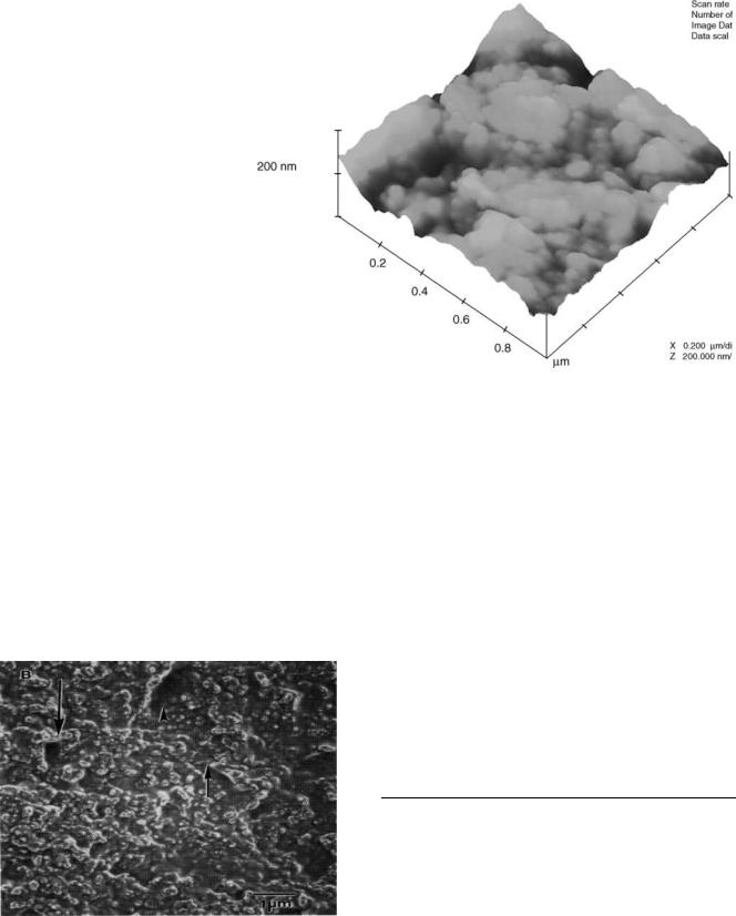
The greatest advantage of nanobiomedical implants in a biological context centers on scientific activities that seek to mimic the nanomorphology that proteins create in natural tissues. As seen in Figs. 4 and 5, bone and vascular tissue possesses numerous nanometer surface features due to the presence of entities like collagen and other proteins (93). Dimensions of some additional proteins found in the extracellular matrix of numerous tissues are found in Table 2 (94). As can be seen, the fundamental dimensions of these proteins (and all proteins) are in the nanometer regime. Clearly, when assembled into an extracellular matrix that comprises a tissue, these proteins provide a diverse surface with numerous nanostructured features for cellular interactions. Since some of these proteins are also soluble and present in bodily fluids, they will initially adsorb to implanted materials to provide for a highly nanostructured surface roughness for cellular interactions. It is for these reasons that cells of our body are accustomed to interacting with nanostructured surfaces. This is in stark contrast to most conventional porous biomaterials that are smooth at the nanoscale.
Aside from mimicking the surface roughness of natural tissues, there are other more scientific reasons to consider porous nanostructured biomaterials for tissue regeneration. Specifically, surface properties (e.g., area, charge, and topography) depend on the surface feature sizes of a material (95,96). In this respect, nanophase materials that, by their very nature, possess higher areas with increased portions of defects [e.g., edge/corner sites and grain or particle boundaries (95,96)] have special advantageous properties that are being exploited by porous biomaterial scientists for applications involving proteins and cells. As mentioned, proteins have complex structures and charges. Thus, surfaces with biologicallyinspired nanometer roughness provide control over protein interactions that were not
402 POROUS MATERIALS FOR BIOLOGICAL APPLICATIONS
Figure 4. An AFM image of the surface of bovine cortical bone. Numerous nanometer features of bone duplicated in porous biomaterials are showing progress in orthopedic applications.
possible with conventional porous materials. Advances of porous nanostructured biomaterials pertinent to orthopedic, cartilage, vascular, central and peripheral nervous systems, and bladder applications will be briefly discussed in the sections below.
Orthopedic Applications
Nanophase ceramics (including alumina, titania, and hydroxyapatite), metals (e.g., titanium, titanium aluminum alloys, and cobalt chromium alloys), polymers (specifically, PLGA, polyether urethane, and polycaprolactone), and composites thereof have been explored for orthopedic applications (22,97,98). For these studies, nanophase surface features in ceramics and metals were created by using
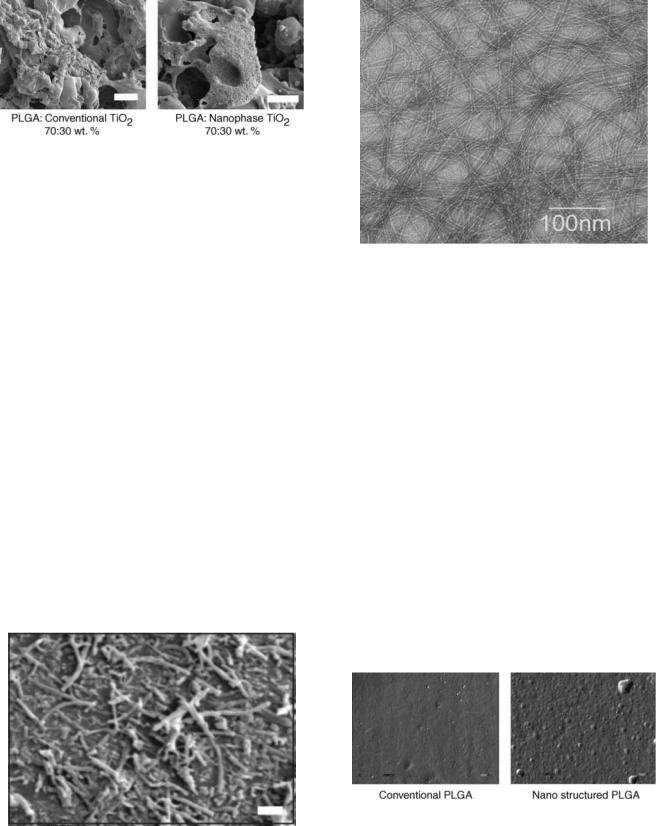
Figure 5. Cast replica of vascular tissue demonstrating nanometer roughness. (Adapted from Ref. 93.) Vascular tissue has numerous irregular nanometer features that when duplicated in porous biomaterials show progress in vascular applications.
constituent nanometer particles, whereas nanostructured polymers were created using chemical etching techniques. In all of these studies, regardless of the manner in which the materials were synthesized, results indicated that nanophase materials enhanced osteoblast functions (e.g., attachment, proliferation, production of extracellular matrix proteins, and deposition of bone) compared to their respective conventional formulations (Fig. 6).
In addition, other porous biomaterials with nanostructured surface features [e.g., carbon nanotubes in polymer composites (Fig. 7) and porous helical rosette carbon nanotubes (Fig. 8)], increased osteoblast functions over conventionally used PLGA scaffolds (99,100). Interestingly, as opposed to conventional porous biomaterials, helical rosette carbon nanotubes self-assemble into a porous biomaterial, which when heated to temperatures only slightly above body temperature solidify (100); thus, these materials could be formulated immediately before implantation to match the dimensions of any bony defect. These novel porous helical rosette nanotubes also allow for optimal pore interconnectivity for the transfer of nutrients and waste to and from cells (100).
Table 2. Nanometer Dimensions of Extracellular Matrix
Proteinsa
Protein |
Characteristic Dimensions |
|
|
Fibronectin |
Dimmer of two identical subunits; 60–70 nm long; |
|
2–3 nm wide |
Vitronectin |
Linear molecule 15 nm long |
Laminin |
Cruciform configuration with one 50 nm long arm |
|
and two 35 nm short arms; total length 50 nm; |
|
total width 70 nm |
Collagen |
Triple helical linear protein consisting of 2 a(1)- |
|
chains and one a(2); 300 nm long; 0.5 nm wide; |
|
67 nm periodicity |
|
|
aSee Ref. 94. |
|
POROUS MATERIALS FOR BIOLOGICAL APPLICATIONS |
403 |
Figure 6. The scanning electron microscopy (SEM) images of PLGA composites containing either conventional or nanophase titania. Increased bone regeneration has been measured in polymer composites containing nanometer compared to conventional ceramics. Bar ¼ 10 mm.
Cartilage Applications
Such pore interconnectivity is also crucial for cartilage forming cells, chondrocytes, since chondrocytes reside far apart from each other and their main communication is through their extracellular matrix. Recently, a porous biomaterial matrix fabricated via solvent casting and particulate leaching to create nanometer surface roughness was tested for cartilage applications (42). The polymer used was PLGA and it was modified to possess nanometer surface features through soaking for 10 min in 10 N NaOH (Fig. 9). Compared to conventional PLGA, results showed increased chondrocyte adhesion, proliferation, and synthesis of a cartilage extracellular matrix (as noted by collagen and glycosaminoglycan synthesis) (42).
Vascular and Bladder Applications
Not only do osteoblasts and chondrocytes interact better with nanophase materials, but so do other cells such as vascular (including endothelial and vascular smooth muscle cells) and bladder cells. For example, Miller et al. (43) and Thapa et al. (44) created nanometer surface features on PLGA films by developing novel molds of NaOH treated PLGA (Fig. 10). When compared to PLGA films without nanometer surface features, vascular smooth muscle cell,
Figure 8. The transmission electron microscopy (TEM) micrograph of porous helical rosette carbon nanotubes. Individual outertube diameters are 4.6 0.09 nm. Increased bone regeneration has been measured in helical rosette nanotubes compared to currently used titanium implants.
endothelial cell, and bladder smooth muscle cell functions were enhanced on the nanostructured PLGA. For bladder applications, Pattison et al. (76) created NaOH induced nanofeatures onto 3D PLGA scaffolds and also observed greater bladder smooth muscle cell adhesion, proliferation, and collagen synthesis. Their studies have further demonstrated increased fibronectin and vitronectin adsorption on nanostructured PLGA compared to conventional PLGA, thus, providing a key mechanism for why vascular and bladder cell adhesion is enhanced on nanostructured PLGA surfaces (43). In addition, PCL and polyurethane have been modified to possess nanostructured surface features by NaOH and HNO3 treatments, respectively; increased vascular and bladder cell functions have been measured on these treated compared to nontreated polymers (43,44). Such studies highlight the versatility of modifying numerous polymers to possess nanostructured features for enhanced vascular and bladder applications.
Figure 7. The SEM image of a polyether urethane composite containing nanophase carbon fibers. Increased bone regeneration has been measured in polymer composites containing nanometer compared to conventional carbon fibers. Bar ¼ 1 mm.
Figure 9. The SEM images of PLGA possessing conventional and nanoscale surface roughness. Nanoscale surface roughness was created by fabricating molds of PLGA etched in 10 N NaOH for 10 min. Increased functions of chondrocytes, vascular cells, and bladder cells have been measured on polymers with nanoscale compared to conventional surface features. Bar ¼ 1 mm.
404 POROUS MATERIALS FOR BIOLOGICAL APPLICATIONS
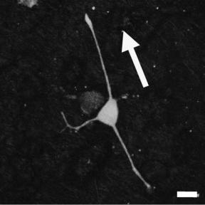
Figure 10. Fluorescent microscopy image of neuron axon alignment corresponding with aligned carbon nanofibers in polyether urethane composites. The arrow indicates carbon nanofiber alignment. Bar ¼ 20 mm.
Central and Peripheral Nervous System Applications
Finally, McKenzie et al. (101) also provided evidence that biomaterials created to have numerous nanometer features can decrease glial scar tissue formation while at the same time increase interactions with neurons. These materials were created by combining carbon nanofibers to polyether urethane. In addition, these investigators have aligned carbon nanofibers in such porous structures to control the direction of axon extension from neurons. In this manner, such porous nanostructured biomaterials could be used to regenerate electrical activity in damaged areas of the brain.
CONCLUSIONS
All these indications point to the conclusion that a successful implantable porous biomaterial should possess properties and structures that simulate the formation of an extracellular matrix similar to that of the target organ it intends to replace. Equally as important for porous biomaterials are appropriate mechanical strength, mechanical structural integrity, degradation rate, permeability, porosity, pore structure, pore interconnectivity, surface energy, surface roughness, and surface chemistry. These all play a role in the function of an optimal porous biomaterial to regenerate tissue. Importantly, to date, to address some of these material properties, several processing techniques have been developed. Although much more needs to be learned concerning the most important aspects of tissue regeneration on porous biomaterials, proper attachment of the appropriate cell is crucial. This is mediated by initial protein interactions that must be the focus of future endeavors to design more effective porous biomaterials. Recent evidence has been provided that porous biomaterials with
nanostructured surface roughness might just control initial protein interactions pertinent for enhancing cell functions necessary to improve the efficacy of orthopedic, cartilage, vascular, central and peripheral nervous system, and bladder applications.
BIBLIOGRAPHY
1.Davis ME. Ordered porous materials for emerging applications [Review]. Nature (London) 2002;417(6891):813–821.
2.Niklason LE, Langer R. Advances in tissue engineering of blood vessels and other tissues. Transplant Immunol 1997;5:303–306.
3.Praemer A, Furner S, Rice SD. Musculoskeletal Conditions in the United States, Park Ridge, IL: American Academy of Orthopaedic Surgery; 1992.
4.D’ Angelo K, Austin T. The moving target: Understanding why arthritis patients do not consider total joint replacement. American Academy of Orthopaedic Surgeons: News Release July 6, 2004.
5.Arthritis Today. 52 Ways to bite back–5 astounding figures. The Arthritis Foundation. Available at http://www.arthritis. org/resources/arthritistoday/2003_archives/ 2003_09_10_51_ways_5figures.asp [2005, March 31].
6.American Parkinson’s Disease Association. Available at http://www.apdaparkinson.org/user/AboutParkinson.asp. [2005, March 31]. Nakamura H, Ozawa H. Immunohistochemical localization of peharan sulfate proteoglycan in rat
tibiae. J Bone Mineral Res 1994;9:1289–1299.
7. National Center for Chronic Disease Prevention. Available at www.cdc.gov/diabetes/pubs/costs/figure.htm figure1, June 1998.
8.National Institute of Health. Spinal cord injury: emerging concepts. NIH Proc Sept 30–Oct. 1, 1996.
9.Greenlee RT, Murray T, Bolden S, Wingo PA. Cancer statistics, 2000. CA Cancer J Clin 2000;50:7–33.
10.Melekos MD, Moutzouris GD. Intravesical therapy of superficial bladder cancer. Curr Pharm Des 2000;6:345–359.
11.Highly MS, Oosterom AT, Maes RA, De Bruijn EA. Intravesical drug delivery. Pharmacokinetic and clinical considerations. Clin Pharmacokinet 1999;37:59–73.
12.Dalbagni G, Herr HW. Current use and questions concerning intravesical bladder cancer group for superficial bladder cancer. Urol Clin North Am 2000;27:137–146.
13.Bianco FJ Jr, et al. Matrix metalloproteinase-9 expression in bladder washes from bladder cancer patients predicts pathological stage and grade. Clin Cancer Res 1998;4:3011–3016.
14.Lebret T, et al. Recurrence, progression and success in stage Ta grade 3 bladder tumors treated with low dose bacillus Calmette-Guerin instillations. J Urology 2000;163:63–67.
15.Hutmacher DW. Scaffold design and fabrication technologies for engineering tissues–state of the art and future perspectives [Review]. J Biomater Sci Polym Ed 2001;12(1):107–124.
16.Sander EA, et al. Solvent effects on the microstructure and properties of 75/25 poly(D,L-lactide-co-glycolide) tissue scaffolds. J Biomed Mater Res Part A 2004;70A(3):506–513.
17.Holy CE, Fialkov JA, Davies JE, Shoichet MS. Use of a biomimetic strategy to engineer bone. J Biomed Mater Res 2003;65A:447–453.
18.Peters MC, Mooney DJ. Synthetic extracellular matrices for cell transplantation. Mater Sci Forum 1997;250:43–52.
19.Gomes ME, et al. Effect of flow perfusion on the osteogenic differentiation of bone marrow stromal cells cultured on starch-based three dimensional scaffolds. J Biomed Mater Res 2003;67A:87–95.
20.Kasemo B, Gold J. Implant surfaces and interface processes [Review]. Adv Dental Res 1999;13:8–20.
21.Vogler EA. Structure and reactivity of water at biomaterial surfaces. Adv Colloid Interface Sci 1998;74:69–117.
22.Webster TJ, Hellenmeyer EL, Price RL. Increased osteoblast functions on theta+delta nanofiber alumina. Biomaterials 2005;26(9):953–960.
23.Horbett TA. Proteins: structure, properties and adsorption to surfaces. In: Ratner BD, Hoffman AS, Schoen AS, Lemmons JE, editors. Biomaterials Science: An Introduction to Materials in Medicine. New York: Academic Press; 1996. p 133.
24.Horbett TA. Chapter 13 Principles underlying the role of adsorbed plasma proteins in blood interactions with foreign materials. Cardiovas Pathol 1993;2(137S):137–148.
25.Hlady V, Buijs J. Protein adsorption on solid surfaces. Curr Opin Biotechnol 1996;7:72–77.
26.Norde W. Driving forces for protein adsorption at solid surfaces. Macromol Symp 1996;103:5–18.
27.Webster TJ, et al. Enhanced functions of osteoblasts on nanophase ceramics. J Biomed Mater Res 2000;51:475.
28.Horbett TA. Techniques for protein adsorption studies. In: Williams DF, editor. Techniques of Biocompatibility Testing, Boca Raton, FL: CRC Press; 1986. p 183.
29.Price RL, Haberstroh KM, Webster TJ. Improved osteoblast viability in the presence of smaller nanometer dimensions carbon fibres. Nanotechnology 2005;15(8):892–900.
30.Kramer RH, Enenstein J, Waleh NS. Integrin structure and ligand specificity in cell matrix interactions. In: Rohrbach DJ, Timpl R, editors. Molecular and Cellular Aspects of Basement Membranes, New York: Academic Press; 1993. p 239–258.
31.Hynes RO. Integrins: versatility, modulation, and signaling in cell adhesion. Cell 1992;69:11–25.
32.Schwartz MA. Transmembrane signaling by integrins. Trends Cell Biol 1992;2:304–308.
33.Puleo DA, Bizios R. Mechanisms of fibronectin-mediated attachment of osteoblasts to substrates In vitro. Bone Mineral 1992;18:215–226.
34.Dalton BA, et al. Polymer surface chemistry and bone cell migration. J Biomater Sci Polym Ed 1998;9(8):781–799.
35.Schakenraad JM. Cell: their surfaces and interactions with materials. In: Ratner BD, Hoffman AS, Schoen AS, Lemmons JE, editors. Biomaterials Science: An Introduction to Materials in Medicine, New York: Academic Press; 1996. p 141.
36.Webster TJ. Nanophase ceramics: the future orthopedic and dental implant material. In: Ying JY, editor. Advances in Chemical Engineering, Vol. 27, New York: Academic Press; 2001. p 125.
37.Sinha RK, Tuan RS. Regulation of human osteoblast integrin expression by orthopedic implant materials. Bone 1996; 18(5):451–457.
38.Davies JE. The importance and measurement of surface charge species in cell behavior at the biomaterial interface. In: Ratner BD, editor. Surface Characterization of Biomaterials: Progress in Biomedical Engineering, Vol. 6, New York: Elsevier; 1988. p 219.
39.Brunette PM. The effect of surface topography of cell migration and adhesion. In: Ratner BD, editor. Surface Characterization of Biomaterials: Progress in Biomedical Engineering, Vol. 6, New York: Elsevier; 1988.p 203.
40.Luck M, et al. Analysis of plasma protein adsorption on polymeric nanoparticles with different surface characteristics. J Biomed Mater Res 1998;39:478–485.
41.Degasne I, et al. Effects of roughness, fibronectin and vitronectin on attachment, spreading, and proliferation of human osteoblast-like cells (Saos-2) on titanium surfaces. Calcif Tissue Int 1999;64(6):499–507.
42.Park GE, Pattison MA, Park K, Webster TJ. Accelerated chondrocyte functions on NaOH-treated PLGA scaffolds. Biomaterials 2005;26(16):3075–3082.
POROUS MATERIALS FOR BIOLOGICAL APPLICATIONS |
405 |
43.Miller DC, Haberstroh KM, Webster TJ. Mechanism(s) of increased vascular cell adhesion on nanostructured poly(lactic- co-glycolic acid) films, J Biomed Mat Res 2005;73(4):476–484.
44.Thapa A, Miller DC, Webster TJ, Haberstroh KM. Nanostructured polymers enhance bladder smooth muscle cell function. Biomaterials 2003;24(17):2915–2926.
45.Agrawal CM, Ray RB. Biodegradable polymer scaffolds for musculoskeletal tissue engineering. J Biomed Mater Res 2001;55:141–150.
46.Carver SE, Heath CA. Influence of intermittent pressure, fluid flow, and mixing on the regenerative properties of articular chondrocytes. Biotechnol Bioeng 1999;65:274–281.
47.Yaszemski MJ, et al. Evolution of bone transplantation: molecular, cellular, and tissue strategies to engineer scaffold human bone. Biomaterials 1995;17:175–185.
48.Hutmacher DW. Scaffolds in tissue engineering bone and cartilage. Biomaterials 2000;21:2925–2943.
49.Goulet RW, et al. The relationship between the structural and orthogonal compressive properties of trabecular bone. J Biomech 1994;27:375–389.
50.Kwon IK, Kidoaki S, Matsuda T. Electrospun nanoto microfiber fabrics made of biodegradable copolyesters: structural characteristics, mechanical properties and cell adhesion potential. Biomaterials 2005;26(18):3929–3939.
51.Maroudas NG. Chemical and mechanical requirements for fibroblast adhesion, Nature (London) 1973;244:363–364.
52.Pelham RJ, Wang YL. Cell locomotion and focal adhesions are regulated by substrate flexibility. Proc Natl Acad Sci USA 1997;94:13661–13665.
53.Katz BZ, et al. Physical state of the extracellular matrix regulates the structure and molecular composition of cellmatrix adhesions. Mol Biol Cell 2000;11:1047–1060.
54.Ohya S, Kidoaki S, Matsuda T. Poly(N-isopropylacrylamide) (PNIPAM)-grafted gelatin hydrogel surfaces: interrelationship between microscopic structure and mechanical property of surface regions and cell adhesiveness. Biomaterials 2005;26:3105–3111.
55.Bignon A, et al. Effect of microand macroporosity of bone substitutes on their mechanical properties and cellular response. J Mater Sci Mat Med 2003;14:1089–1897.
56.Wilkinson CDW, et al. The use of materials patterned on a nanoand micro-metric scale in cellular engineering. Mater Sci Eng 2001;19:263.
57.Clark P, et al. Topographical control of cell behaviour. II. Multiple grooved substrata. Development 1999;108:635.
58.Tranquillo RT. Self-organisation of tissue equivalents: the nature and role of contact guidance. Biochem Soc Symp 1999;65: 27.
59.Li SM, Garreau H, Vert M. Structure-property relationships in the case of the degradation of massive poly(hydroxyl acid) in aqueous media,part 2:degradation of lactide–glycolide copolymers. J Mater Sci Mater Med 1990;1:131–139.
60.Grizzi I, Garreau H, Li S, Vert M. Hydrolytic degradation of devices based on poly(d,l-lactic acid): size dependence. Biomaterials 1995;16:305–311.
61.Sung HJ, Meredith C, Johnson C, Galis ZS. The effect of scaffold degradation rate on three-dimensional cell growth and angiogenesis. Biomaterials 2004;25:5735–5742.
62.Hedberg EL, et al. In vivo degradation of porous poly(propylene fumarate)/poly(DL-lactic-co-glycolic acid) composite scaffolds. Biomaterials 2005;26:4616–4623.
63.Perugini P, et al. PLGA microspheres for oral osteopenia treatment: preliminary ‘‘In vitro’’/‘‘In vivo’’ evaluation. Int J Pharm 2003;256:153–160.
64.Wu L, Ding J. In vitro degradation of three-dimensional porous poly(D,L-lactide-co-glycolide) scaffolds for tissue engineering, Biomaterials 2004;25:5821–5830.