

Kluwer - Handbook of Biomedical Image Analysis Vol
.1.pdfLevel Set Segmentation of Biological Volume Datasets |
465 |
(a) |
(c) |
(b) |
(d) |
Figure 8.24: 3D results: (a) surface initialization, (b) final surface estimated after 150 iterations, (c) a portion of the initial surface enlarged, and (d) the corresponding portion in the final surface.
often fails to show up in the FBP reconstruction, does appear quite regularly in hand-segmentations of the same datasets.
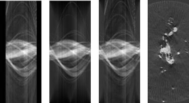
8.6.5.2 Sinogram Extrapolation
The fitting of surfaces to this data is a simplification. It is justified in the context of segmentation, but there are underlying inhomogeneities in the density of this specimen, which could be indicative of relevant structures. Thus for some applications direct visualization of the measured data, by volume rendering, offers advantages over the segmented surfaces. We propose to use the surface estimation algorithm as a mechanism for estimating the missing data in the sinograms.
Figures 8.25(a) and (b) show the input sinogram and the sinogram of the estimated model (for one slice) of the TEM dendrite data. The estimated sinogram demonstrates that the surface estimation method recovers the missing information in a reasonable way. Thus, we combine the sinograms from the model with original sinograms to produce a “full” sinogram that still contains all of the
Level Set Segmentation of Biological Volume Datasets |
467 |
(a) |
(c) |
(b) |
(d) |
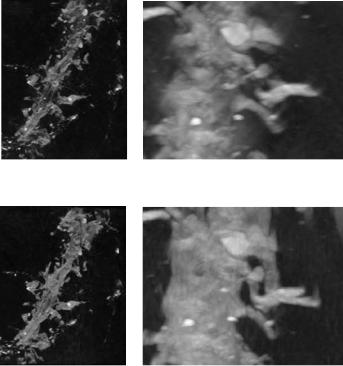
Figure 8.26: Sinogram extrapolation results: (a) MIP volume rendering of volume reconstructed from original sinograms, (b) MIP volume rendering of volume reconstructed from augmented (extrapolated) sinograms, (c) a portion of original MIP enlarged, and (d) the corresponding portion in augmented MIP enlarged.
applications. Several standard volume processing algorithms have been incorporated into the framework in order to segment datasets generated from MRI, CT, and TEM scans. A technique based on moving least-squares has been developed for segmenting multiple nonuniform scans of a single object. New scalar measures have been defined for extracting structures from diffusion tensor MRI scans. Finally, a direct approach to the segmentation of incomplete tomographic data using density parameter estimation is described. These techniques, combined with level set surface deformations, allow us to segment many different types of biological volume datasets.
468 |
Breen, Whitaker, Museth, and Zhukov |
8.8 Acknowledgements
Several people provided valuable technical assistance and support to our work. They are Dr. Alan Barr, Dr. Jason Wood, Dr. John Wood, Dr. Cyrus Papan, Dr. Russ Jacobs, Dr. Scott Fraser, Dr. J. Michael Tyszka, Dr. Miriam Scadeng, Dr. David Dubowitz, Dr. Eric Ahrens, Dr. Mark Ellisman, Dr. Maryanne Martone, Dr. Chris Johnson, and Dr. Mark Bastin. Datasets were provided by Caltech Biological Imaging Center (e.g. Fig. 8.8), National Center for Microscopy and Imaging Research (e.g. Fig. 8.7, funded by NIH grant P41-RR04050), Caltech Multi-Res Modeling Group (Fig. 8.9 (top)), Stanford Computer Graphics Laboratory (Fig. 8.9 (top)), Childrens Hospital—Los Angeles (Fig. 8.10), University of Utah’s SCI Institute (e.g. Fig. 8.14), and the University of Edinburgh, UK (Fig. 8.16).
This work was supported by National Science Foundation grants ASC-89- 20219, ACI-9982273, ACI-0083287, and ACI-0089915, the Office of Naval Research Volume Visualization grant N00014-97-0227, the National Institute on Drug Abuse and the National Institute of Mental Health, as part of the Human Brain Project, the National Library of Medicine “Insight” Project N01-LM-0-3503, and the Caltech SURF Program.

Level Set Segmentation of Biological Volume Datasets |
469 |
Bibliography
[1]Drebin, R., Carpenter, L., and Hanrahan, P., Volume rendering, In: Proceedings SIGGRAPH 88 Conference, pp. 65–74, 1988.
[2]Levoy, M., Display of surfaces from volume data, IEEE Comput. Graph. Appl., Vol. 9, No. 3, pp. 245–261, 1990.
[3]Laur, D. and Hanrahan, P., Hierarchical splatting: A progressive refinement algorithm for volume rendering, In: SIGGRAPH ’91 Proceedings, Sederberg, T. W., ed., pp. 285–288, 1991.
[4]Parker, S., Parker, M., Livnat, Y., Sloan, P., Hansen, C., and Shirley, P., Interactive Ray Tracing for volume visualization, IEEE Trans. Vis. Comput. Graph., Vol. 5, No. 3, pp. 238–250, 1999.
[5]Leventon, M., Faugeraus, O., Grimson, W., and Wells, W. III, Level set based segmentation with intensity and curvature priors, In: Workshop on Mathematical Methods in Biomedical Image Analysis Proceedings, pp. 4–11, 2000.
[6]Malladi, R., Sethian, J., and Vemuri, B., Shape modeling with front propagation: A level set approach, IEEE Trans. Pattern Anal. Mach. Intell., Vol. 17, No. 2, pp. 158–175, 1995.
[7]Sethian, J., Level Set Methods and Fast Marching Methods, 2nd edn., Cambridge University Press, Cambridge, UK, 1999.
[8]Staib, L., Zeng, X., Schultz, R., and Duncan, J., Shape constraints in deformable models, In: Handbook of Medical Imaging, Bankman, I., ed., Academic Press, New York, Chapter 9, pp. 147–157, 2000.
[9]Wu, Z., Chung, H.-W., and Wehrli, F. W., A Bayesian approach to subvoxel tissue classification in NMR microscopic images of trabecular bone, J. Comput. Assist. Tomogr., Vol. 12, No. 1, pp. 1–9, 1988.
[10]Kao, Y.-H., Sorenson, J. A., and Winkler, S. S., MR image segmentation using vector decomposition and probability techniques: A general model and its application to dual-echo images, Magn. Reson. Med., Vol. 35, pp. 114–125, 1996.
470 |
Breen, Whitaker, Museth, and Zhukov |
[11]Cline, H. E., Lorensen, W. E., Kikinis, R., and Jolesz, F., Threedimensional segmentation of MR images of the head using probability and connectivity, J. Comput. Assist. Tomogr., Vol. 14, No. 6, pp. 1037– 1045, 1990.
[12]Laidlaw, D. H., Fleischer, K. W., and Barr, A. H., Partial-volume Bayesian classification of material mixtures in MR volume data using voxel histograms, IEEE Trans. Med. Imaging, Vol. 17, No. 1, pp. 74–86, 1998.
[13]Johnson, V. E., A framework for incorporating structural prior information into the estimation of medical images, In: Information Processing in Medical Imaging (IPMI’93), Barrett, H. H. and Gmitro, A. F., eds., No. 687 In Lecture Notes in Computer Science, Springer-Verlag, Berlin,
pp.307–321, 1993.
[14]Marr, D. and Hildreth, E., Theory of Edge Detection, Proc. R. Soc. London, Vol. B, No. 207, pp. 187–217, 1980.
[15]Marr, D., Vision, Freeman, San Francisco, 1982.
[16]Canny, J., A computational approach to edge detection, IEEE Trans. Pattern Anal. Mach. Intell., Vol. 8, No. 6, pp. 679–698, 1986.
[17]Cootes, T., Hill, A., Taylor, C., and Haslam, J., The use of active shape models for locating structures in medical images, In: Information Processing in Medical Imaging (IPMI’93), Barrett, H. H. and Gmitro, A. F., eds., No. 687 In Lecture Notes in Computer Science, Springer-Verlag, Berlin, pp. 33–47, 1993.
[18]Stetten, G. and Pizer, S., Medial node models to identify and measure objects in real-time 3D echocardiography, IEEE Trans. Med. Imaging, Vol. 18, No. 10, pp. 1025–1034, 1999.
[19]Wood, Z., Desbrun, M., Schroder,¨ P., and Breen, D., Semi-regular mesh extraction from volumes, In: Proceedings of IEEE Visualization 2000,
pp.275–282, 2000.
[20]Miller, J., Breen, D., Lorensen, W., O’Bara, R., and Wozny, M., Geometrically deformed Models: A method for extracting closed geometric models from volume data, In: SIGGRAPH ’91 Proceedings, pp. 217– 226, 1991.
Level Set Segmentation of Biological Volume Datasets |
471 |
[21]Pentland, A. P., Perceptual organization and the representation of natural form, Artif. Intell., Vol. 28, pp. 293–331, 1986.
[22]Terzopoulos, D. and Metaxas, D., Dynamic 3D models with local and global deformations: Deformable superquadrics, IEEE Trans. Pattern Anal. Mach. Intell., Vol. 13, No. 7, pp. 703–714, 1991.
[23]Gupta, A. and Bajcsy, R., Volumetric segmentation of range images of 3D objects using superquadric models, CVGIP: Image Underst., Vol. 58, No. 3, pp. 302–326, 1993.
[24]Muraki, S., Volumetric shape description of range data using “Blobby Model,” In: SIGGRAPH ’91 Proceedings, Sederberg, T. W., ed., pp. 227– 235, 1991.
[25]Szeliski, R., Tonnesen, D., and Terzopoulos, D., Modeling surfaces of arbitrary topology with dynamic particles, In: Proc. Fourth Int. Conf. on Comp. Vision (ICCV’93), pp. 82–87, IEEE Computer Society Press, Berlin, 1993.
[26]McInerney, T. and Terzopoulos, D., A dynamic finite element surface model for segmentation and tracking in multidimensional medical images with application to cardiac 4D image analysis, Comput. Med. Imaging Graph., Vol. 19, No. 1, pp. 69–83, 1995.
[27]Park, J., Metaxas, D., Young, A. A., and Axel, L., Deformable models with parameter functions for cardiac motion analysis from tagged MRI data, IEEE Trans. Med. Imaging, Vol. 15, No. 3, pp. 278–289, 1996.
[28]DeCarlo, D. and Metaxas, D., Shape evolution with structural and topological changes using blending, IEEE Trans. Pattern Anal. Mach. Intell., Vol. 20, No. 11, pp. 1186–1205, 1998.
[29]Ramamoorthi, R. and Arvo, J., Creating generative models from range images, In: SIGGRAPH ’99 Proceedings, pp. 195–204, 1999.
[30]Osher, S. and Sethian, J., Fronts propagating with curvature-dependent speed: Algorithms based on Hamilton–Jacobi formulations, J. Comput. Phys., Vol. 79, pp. 12–49, 1988.
[31]Osher, S. and Fedkiw, R., Level Set Methods and Dynamic Implicit Surfaces, Springer, Berlin, 2002.
472 |
Breen, Whitaker, Museth, and Zhukov |
[32]Sethian, J., A fast marching level set method for monotonically advancing fronts, In: Proceedings of the National Academy of Science, Vol. 93 of 4, pp. 1591–1595, 1996.
[33]Tsitsiklis, J., Efficient algorithms for globally optimal trajectories, IEEE Trans. Autom. Control, Vol. 40, No. 9, pp. 1528–1538, 1995.
[34]Adalsteinsson, D. and Sethian, J. A., A fast level set method for Propagating interfaces, J. Comput. Phys., Vol. 118, No. 2, pp. 269–277, 1995.
[35]Peng, D., Merriman, B., Osher, S., Zhao, H.-K., and Kang, M., A PDEbased fast local level set method, J. Comput. Phys., Vol. 155, pp. 410– 438, 1999.
[36]Whitaker, R., A level-set approach to 3D reconstruction from range data, Int. J. Comput. Vis., Vol. 29, No. 3, pp. 203–231, 1998.
[37]Whitaker, R., Breen, D., Museth, K., and Soni, N., Segmentation of biological datasets using a level-set framework, In: Volume Graphics 2001, Chen, M. and Kaufman, A., eds., Springer, Vienna, pp. 249–263, 2001.
[38]van den Boomgaard, R. and Smeulders, A. W. M., The morphological structure of images, the differential equations of morphological scalespace, IEEE Trans. Pattern Anal. Mach. Intell., Vol. 16, No. 11, pp. 1101–1113, 1994.
[39]Maragos, P., Differential morphology and image processing, IEEE Trans. Image Process., Vol. 5, No. 6, pp. 922–937, 1996.
[40]Requicha, A. and Voelcker, H., Boolean operations in solid modeling: Boundary evaluation and merging algorithms, Proc. IEEE, Vol. 73, No. 1, pp. 30–44, 1985.
[41]Whitaker, R. T., Volumetric deformable models: Active blobs, In: Visualization in Biomedical Computing, Robb, R. A., ed., SPIE, Mayo Clinic, Rochester, MN, pp. 122–134, 1994.
[42]Sapiro, G., Geometric Partial Differential Equations and Image Analysis, Cambridge University Press, Cambridge, UK, 2001.
Level Set Segmentation of Biological Volume Datasets |
473 |
[43]Museth, K., Breen, D., Zhukov, L., and Whitaker, R., Level set segmentation from multiple non-uniform volume datasets, In: Proc. IEEE Visualization Conference, pp. 179–186, 2002.
[44]Shepard, D., A two-dimensional interpolation function for irregularly spaced points, In: Proc. ACM Nat. Conf., pp. 517–524, 1968.
[45]Lancaster, P. and Salkauskas, K., Surfaces generated by moving least squares methods, Math. Comput., Vol. 37, pp. 141–159, 1981.
[46]Farwig, R., Multivariate interpolation of arbitrarily spaced data by moving least-squares methods, J. Comput. Appl. Math., Vol. 16, pp. 79–93, 1986.
[47]Zhao, H.-K., Osher, S., and Fedkiw, R., Fast surface reconstruction using the level set method, In: Proc. 1st IEEE Workshop on Variational and Level Set Methods, pp. 194–202, 2001.
[48]Turk, G. and Levoy, M., Zippered polygon meshes from range images, In: Proc. of SIGGRAPH ’94, pp. 311–318, ACM SIGGRAPH, 1994.
[49]Curless, B. and Levoy, M., A volumetric method for building complex models from range images, In: Proc. SIGGRAPH ’96, pp. 303–312, 1996.
[50]Tamez-Pena, J., Totterman, S., and Parker, K., MRI isotropic resolution reconstruction from two orthogonal scans, In: Proc. SPIE Medical Imaging, Vol. 4322, pp. 87–97, 2001.
[51]Goshtasby, A. and Turner, D. A., Fusion of short-axis and longaxis cardiac MR images, In: IEEE Workshop on Mathematical Methods in Biomedical Image Analysis, San Francisco, pp. 202–211, 1996.
[52]Brejl, M. and Sonka, M., Directional 3D Edge Detection in anisotropic data: Detector design and performance assessment, Comput. Vis. Image Underst., Vol. 77, pp. 84–110, 2000.
[53]Haralick, R. M. and Shapiro, L. G., Computer and Robot Vision, Addison-Wesley, Reading, MA, 1991.
474 |
Breen, Whitaker, Museth, and Zhukov |
[54]Press, W., Flannery, B., Teukolsky, S., and Vetterling, W., Numerical Recipes in C, 2nd edn., Cambridge University Press, New York, NY, 1992.
[55]Basser, P. J., Mattielo, J., and Bihan, D. L., Estimation of the effective self-diffusion tensor from the NMR spin echo, J. Magn. Reson., B, Vol. 103, No. 3, pp. 247–254, 1994.
[56]Basser, P. J., Mattielo, J., and Bihan, D. L., MR diffusion tensor spectroscopy and imaging, Biophys. J., Vol. 66, No. 1, pp. 259–267, 1994.
[57]Basser, P. J. and Pierpaoli, C., Microstructural and physiological features of tissues elucidated by quantitative-diffusion-tensor MRI, J. Magn. Reson., B, Vol. 111, No. 3, pp. 209–219, 1996.
[58]Westin, C.-F., Peled, S., Gudbjartsson, H., Kikinis, R., and Jolesz, F. A., Geometrical diffusion measures for MRI from tensor basis analysis, In: Proceedings ISMRM 5th Annual Meeting, p. 1742, 1997.
[59]Peled, S., Gudbjartsson, H., Westin, C., Kikinis, R., and Jolesz, F., Magnetic resonance imaging shows orientation and asymmetry in white matter fiber tracts, Brain Res., Vol. 780, pp. 27–33, 1998.
[60]Basser, P. and Pajevic, S., Statistical artifacts in diffusion tensor MRI caused by background noise, Magn. Reson. Med., Vol. 44, pp. 41–50, 2000.
[61]Ulug, A. and van Zijl, P., Orientation-independent diffusion imaging without tensor diagonalization: Anisotropy definitions based on physical attributes of the diffusion ellipsoid, J. Magn. Reson. Imaging, Vol. 9, pp. 804–813, 1999.
[62]Laidlaw, D., Ahrens, E., Kremers, D., Avalos, M., Jacobs, R., and Readhead, C., Visualizing diffusion tensor images of the mouse spinal cord, In: Proceedings IEEE Visualization ’98, pp. 127–134, 1998.
[63]Kindlmann, G. and Weinstein, D., Hue-balls and lit-tensors for direct volume rendering of diffusion tensor fields, In: Proc. IEEE Visualization ’99, pp. 183–189, 1999.