Kluwer - Handbook of Biomedical Image Analysis Vol
.3.pdf
Inter-Subject Non-Rigid Registration |
319 |
Table 8.2: Numerical evaluation of the multimodal registration method on simulated data. The overlapping measures (specificity, sensitivity, and total performance) are computed after rigid registration and at each grid level of the non-rigid registration process
Registration |
Overlap measure |
Grey matter |
White matter |
|
|
|
|
Rigid |
sensibility |
74.7% |
76.6% |
|
specificity |
93.0% |
92.8% |
|
Total performance |
87.0% |
87.6% |
Non-rigid |
sensibility |
84.7% |
86.0% |
grid level 7 |
specificity |
97.2% |
96.2% |
|
Total performance |
93.2% |
92.9% |
Non-rigid |
sensibility |
86.6% |
86.8% |
grid level 6 |
specificity |
98.5% |
97.3% |
|
Total performance |
94.6% |
93.9% |
Non-rigid |
sensibility |
87.5% |
87.0% |
grid level 5 |
specificity |
98.9% |
98.0% |
|
Total performance |
95.8% |
95.3% |
|
|
|
|
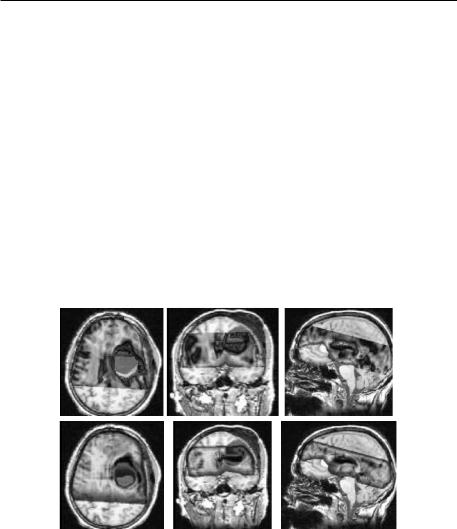
Figure 8.22: Results of clinical data. Top: results of the rigid registration. The multiple artefacts are visible: distortions, signal saturation, signal drops. Bottom: Results of the non-rigid registration. The registration is more accurate, in particular, for the ventricles and for the cyst. The data are courtesy of “laboratoire IDM, Hopital de Pontchaillou, Rennes”.
320 |
Hellier |
8.4 Conclusion
This chapter has presented an overview of the classification of non-rigid registration methods with particular focus on non-rigid registration of brains of different subjects. Methods can be broadly classified into two groups: geometric methods that rely on the extraction and matching of sparse features (points, curves, surfaces), and photometric (or intensity-based) methods that rely on image luminance directly.
Geometric methods reduce the dimensionality of the problem and are consistent in the vicinity of features used for registration. However, the registration might be incorrect far from used features. Photometric methods use all the available information present in the volume but lead to a complex problem involving a very large number of variables.
We have presented here the Romeo algorithm (Robust Multigrid Elastic registration based on optical flow) that refers to photometric methods. Romeo uses the optical flow as a similarity measure and relies on an efficient multiresolution and multigrid optimization scheme. Robust estimators are introduced to limit the effect of erroneous data and to preserve discontinuities of the deformation field when needed. Prior to the non-rigid registration step, two preprocessing steps are performed: a rigid registration by maximization of mutual information and an intensity correction so that the luminance of the volumes to be registered are comparable. An extension to multimodal data has been presented. The multiresolution and multigrid framework is flexible enough to be adapted to multimodal similarity measures such as mutual information for instance.
It has been shown that photometric methods fail in matching cortical structures such as cortical sulci, for instance [74]. This can be explained by the high variability of cortical structures among subjects. Anatomists have pointed out [103] that cortical sulci of different subjects are very different in shapes. This has motivated mixed approaches where a photometric registration method incorporates sparse anatomical structures so as to drive the registration process [22, 29, 36, 68, 73, 79, 141].
In this context, it must be noted that validation is difficult and should be investigated further. Validation of non-rigid registration methods on anatomical structures have been conducted [74, 119]. However, the impact of non-rigid
Inter-Subject Non-Rigid Registration |
321 |
registration methods on functional data is still unknown. As a matter of fact, since these methods are dedicated to anatomical and functional normalization, it would be interesting to know how much of the intersubject functional variability can be understood and compensated by anatomical non-rigid registration. This is a challenging research subject that requires a better knowledge about the relationship between the anatomy of the brain and its functional organization.
Questions
1.What are photometric and geometric registration methods? How can these methods be compared?
2.What is optical-flow?
3.What is the aperture problem? How can it be solved?
4.What are the advantages and drawbacks of optical-flow based registration?
5.What are robust estimators?
6.What are the advantages and drawbacks of robust M-estimators?
7.How useful is a multiresolution scheme?
8.How should the Gaussian standard deviation be chosen for building the multiresolution pyramid?
9.What is a multigrid optimization scheme?
10.What are the different options to regularize the deformation field?

322 |
Hellier |
Bibliography
[1]Ashburner, J., Andersson, J., and Friston, K. J., Image registration using a symetric prior in three dimensions. Human Brain Mapping, Vol. 9, pp. 212–225, 1998.
[2]Ashburner, J. and Friston, K., Nonlinear Spatial Normalization using basis functions. Human Brain Mapping, Vol. 7, No. 4, pp. 254–266, 1999.
[3]Cuadra, M. Bach, Platel, B., Solanas, E., Butz, T., and Thiran, J. P., Validation of tissue modelization and classification techniques in T1-weighted MR brain images, In: Proc. of Medical Image Computing and ComputerAssisted Intervention, Dohi, T. and Kikinis, R., ed., No. 2488 In Lecture Notes in Computer Science, pp. 290–297, Springer, Tokyo, September 2002.
[4]Bajcsy, R. and Broit, C., Matching of deformed images, In: Proc. International Conference on Pattern Recognition, Vol. 1, pp. 351–353, IEEE, New York, Munich, West Germany, Oct. 1982.
[5]Bajcsy, R. and Kovacic, S., Multiresolution elastic matching, Computer Vision, Graphics, Image Processing, Vol. 46, pp. 1–21, 1989.
[6]Barr, A., Global and local deformations of solid primitives, Computer Graphics (SIGGRAPH), Vol. 18, no. 3, pp. 21–30, 1984.
[7]Barron, J., Fleet, D., Beauchemin, S., and Burkitt, T., Performance of optical flow techniques, In: Proc. Conference on Computer Vision Pattern Recognition, pp. 236–242, Champaign, Illinois, Jun. 1992.
[8]Bascle, B., Contributions et applications des modeles` deformables´ en vision par ordinateur, Ph.D. thesis, Universite´ de Nice Sophia-Antipolis, Sep. 1994.
[9]Battiti, R., Amaldi, E., and Koch, C., Computing optical flow over multiple scales: an adaptive coarse-to-fine strategy, International Journal of Computer Vision, Vol. 6, No. 2, pp. 133–146, 1991.
[10]Beauchemin, S. and Barron, J., The computation of optical flow, ACM Computing Surveys, Vol. 27, No. 3, pp. 433–467, 1995.
Inter-Subject Non-Rigid Registration |
323 |
[11]Bergen, J., Anadan, P., Hanna, K., and Hingorani, R., Hierarchical modelbased motion estimation, In: Proc. European Conference on Computer Vision, pp. 5–10, 1991.
[12]Besl, P. and McKay, N., A method for registration of 3D shapes, IEEE Trans. Pattern Analysis and Machine Intelligence, Vol. 14, No. 2, pp. 239– 255, 1992.
[13]Black, M. and Anandan, P., The robust estimation of multiple motions: parametric and piecewise-smooth flow fields, Computer Vision and Image Understanding, Vol. 63, No. 1, pp. 75–104, 1996.
[14]Black, M. and Rangarajan, A., On the unification of line processes, outlier rejection, and robust statistics with application in early vision, International Journal of Computer Vision, Vol. 19, No. 1, pp. 57–91, 1996.
[15]Blake, A. and Isard, M., Active contours. Springer, 1998.
[16]Bookstein, F., Principal warps: Thin-plate splines and the decomposition of deformations, IEEE Transactions on Pattern Analysis and Machine Intelligence, Vol. 11, No. 6, pp. 567–585, 1989.
[17]Bro-Nielsen, M. and Gramkow, C., Fast fluid registration of medical images, In: Proc. Visualization in Biomedical Computing, Hohne, K-. H. and Kikinis, R., ed., No. 1131 In Lecture Notes in Computer Science, pp. 267–276, Springer, Sep. 1996.
[18]Broit, C., Optimal registration of deformed images, Ph.D. Dissertaition, Department of Computer and Information Science, University of Pennsylvania, Philadelphia, 1981.
[19]Brown, L. F., A survey of image registration techniques, ACM: Computer Surveys, Vol. 24, No. 4, pp. 325–376, 1992.
[20]Burt, J. P., Multiresolution image processing and analysis, Chapter 2 : The pyramide as a structure for efficient computation, pp. 6–38, No. 12 In Springer series in Information Science, Springer-Verlag, 1984.
[21]Cachier, P., Bardinet, E., Dormont, D., Pennec, X., and Ayache, N., Iconic feature based nonrigid registration: the PASHA algorithm, Computer Vision and Image Understanding, Vol. 89, No. 2/3, pp. 272–299, 2003.
324 |
Hellier |
[22]Cachier, P., Mangin, J. F., Pennec, X., Riviere,` D., Papadopoulos-Orfanos, D., Regis,´ J., and Ayache, N., Multisubject non-rigid registration of brain MRI using intensity and geometric features, In: Proc. of Medical Image Computing and Computer-Assisted Intervention, Niessen, W. and Viergever, M., eds., No. 2208 in Lecture Notes in Computer Science,
pp.734–742, 2001.
[23]Celeux, G., Chauveau, D., and Diebolt, J., Stochastic version of the EM algorithm: an experimental study in the mixture context, Journal of Computationnal Statistical Computation and Simulation, Vol. 55,
pp.287–314, 1996.
[24]Charbonnier, P., Blanc-Feraud,´ L., Aubert, G., and Barlaud, M., Deterministic edge preserving regularization in computed imaging, IEEE Transactions Image Processing, Vol. 6, No. 2, pp. 298–311, 1997.
[25]Chen, C., Huang, T., and Arrott, M., Modeling, analysis and vizualisation of left-ventricule shape and motion by hierarchical decomposition, IEEE Transactions on Pattern Analysis and Machine Intelligence, Vol. 16, No. 4, 1994.
[26]Chen, G., Kessler, M., and Pitluck, S., Structure transfer between sets of three dimensionnal medical imaging data, In: VI Conference on Computer Graphics, Vol. 6, pp. 171–177, 1985.
[27]Christensen, G., Rabbit, R., and Miller, M. I., Deformable templates using large deformation kinematics, IEEE Transactions on Image Processing, Vol. 5, No. 10, pp. 1435–1447, 1996.
[28]Christensen, G. and Johnson, H. J., Consistent image registration, IEEE Transactions Medical Imaging, Vol. 20, No. 7, pp. 568–582, 2001.
[29]Chui, H., Rambo, J., Duncan, J., Schultz, R., and Rangarajan, A., Registration of cortical anatomical structures via robust 3D point matching, In: Proc. Information Processing in Medical Imaging, Cuba, et al., ed., No. 1613 In: Lecture Notes in Computer Science, pp. 168–181, Springer Verlag, 1999.
Inter-Subject Non-Rigid Registration |
325 |
[30]Chui, H. and Rangarajan, A., A new point matching algorithm for nonrigid registration, Computer Vision and Image Understanding, Vol. 89, No. 2/3, pp. 114–142, 2003.
[31]Cohen, I. and Herlin, I., Optical flow and phase portrait methods for environmental satellite image sequences, In: Proc. European Conference on Computer Vision, pp. II:141–150, Cambridge, UK, April 1996.
[32]Cohen, L. and Cohen, I., Finite element method for active contour models and ballons for 2D and 3D images, IEEE Transactions of Pattern Analysis and Machine Intelligence, Vol. 15, No. 11, pp. 1131– 1147, 1993.
[33]Collignon, A., Vanderneulen, D., Suetens, P., and Marchal, G., 3D multimodality medical image registration using feature space clustering, In: Proc. of Computer Vision, Virtual Reality and Robotics in Medecine, pp. 195–204, Nice, France, 1995.
[34]Collins, D. L., Zijdenbos, A. P., Kollokian, V., Sled, J. G., Kabani, N. J., Holmes, C. J., and Evans, A. O., Design and construction of a realistic digital brain phantom, IEEE Transactions on Medical Imaging, Vol. 17, No. 3, pp. 463–468, June 1998.
[35]Collins, L. and Evans, A., Animal : validation and applications of nonlinear registration-based segmentation, International Journal of Pattern Recognition Artificial Intelligence, Vol. 8, No. 11, pp. 1271–1294, 1997.
[36]Collins, L., Goualher, Le, G., and Evans, A., Non-linear cerebral registration with sulcal constraints, In: Proc. of Medical Image Computing and Computer-Assisted Intervention, Colchester, A. and Delp, S., eds., No. 1496 in Lecture Notes in Computer Science, pp. 974–985, Springer, October 1998.
[37]Corpetti, T., Memin,´ E., and Perez,´ P., Dense estimation of fluid flows, IEEE Transactions on Pattern Analysis and Machine Intelligence, Vol. 24, No. 3, pp. 365–380, 2002.
[38]Davatzikos, C., Spatial transformation and registration of brain images using elastically deformable models, Computer Vision and Image Understanding, Vol. 66, No. 2, pp. 207–222, 1997.
326 |
Hellier |
[39]Davatzikos, C., Prince, L., and Bryan, N., Image registration based on boundary mapping, IEEE Transactions on Medical Imaging, Vol. 15, No. 1, pp. 111–115, 1996.
[40]Dawant, B., Hartmann, S., Thirion, J. P., Maes, F., Vandermeulen, D., and Demaerel, P., Automatic 3D segmentation of internal structures of the head in MR images using a combination of similarity and free-form transformations: Part I, Methodology and validation on normal subjects, IEEE Transactions on Medical Imaging, Vol. 18, No. 10,
pp.909–916, 1999.
[41]Declerck, J., Subsol, G., Thirion, J. P., and Ayache, N., Automatic retrieval of anatomical structures in 3D medical images, In: Proc. of Computer Vision, Virtual Reality and Robotics in Medicine, Ayache, N., ed., No. 905 in LNCS, pp. 153–162, Springer, Nice, France, April 1995, Electronic version: ftp://ftp.inria.fr/INRIA/publication/RR/RR-2485.ps.gz.
[42]Delaney, A. and Bresler, Y., Globally convergent edge-preserving regularized reconstruction: an application to limited-angle tomography, IEEE Transanction Image Processing, Vol. 7, No. 2, pp. 204–221, 1998.
[43]Delingette, H., Hebert, M., and Ikeuchi, K., Shape representation and image segmentation using deformable models, Image and Vision Computing, Vol. 10, No. 3, pp. 132–144, 1992.
[44]Dempster, A. P., Laird, N. M., and Rubin, D. B., Maximum likelihood for incomplete data via the EM algorithm, Journal of the Royal Statistical Society, Vol. 39, pp. 1–38, 1977.
[45]Deriche, R., Recursively implementing the gaussian and its derivatives, Technical Report 1893, INRIA, http://www.inria.fr/RRRT/RR-1893.html, April 1993.
[46]Devlaminck, V., A functional for compressive and incompressive elastic deformation estimation, IEEE Signal Processing Letters, Vol. 6, No. 7,
pp.162–164, 1999.
[47]Duchon, J., Interpolation des fonctions de deux variables suivant le principe de la flexion des plaques minces, RAIRO Analyse Numerique,´ Vol. 10, pp. 5–12, 1976.
Inter-Subject Non-Rigid Registration |
327 |
[48]Enkelmann, W., Investigations of multigrid algorithms for the estimation of optical flow fields in image sequences, Computer Vision, Graphics, Image Processing, Vol. 43, No. 2, pp. 150–177, 1988.
[49]Evans, A., Collins, L., and Milner, B., An MRI-based stereotaxic atlas from 250 young normal subjects, Society for Neuroscience Abstract, Vol. 18, pp. 408, 1992.
[50]Evans, A. C., Dai, W., Collins, D. L., Neelin, P., and Marrett, T., Warping of computerized 3D atlas to match brain image volumes for quantitative neuroanatomical and functionnal analysis, In: Proc. of the International Society of Optical Engineering: Medical Imaging V, SPIE, 1991.
[51]Feldmar, J. and Ayache, N., Locally affine registration of free-form surfaces, In: Proc. Conference on Computer Vision Pattern Recognition, pp. 496–500, Seattle, June 1994.
[52]Feldmar, J. and Ayache, N., Rigid, affine and locally affine registration of free-form surfaces, International Journal of Computer Vision, Vol. 18, No. 2, pp. 99–119, 1996.
[53]Feldmar, J., Declerck, J., Malandain, G., and Ayache, N., Extension of the ICP algorithm to nonrigid intensity based registration of 3D images, Computer Vision and Image Understanding, Vol. 66, No. 2, pp. 193–206, 1997.
[54]Ferrant, M., Warfield, S., Guttmann, C., Mulkern, R., Jolesz, F., and Kikinis, R., 3D image matching using a finite element based elastic deformation model, In: Proc. of Medical Image Computing and ComputerAssisted Intervention, Taylor, C. and Colchester, A., eds., No. 1679 in Lecture Notes in Computer Science, pp. 202–209, Cambridge, UK, September 1999.
[55]Friston, K. J., Ashburner, J., Frith, C. D., Poline, J. B., Heather, J. D., and Frackowiak, R. SJ., Spatial registration and normalisation of images, Human Brain Mapping, Vol. 2, pp. 165–189, 1995.
[56]Gabrani, M. and Tretiak, O., Surface-based matching using elastic transformation, Pattern Recognition, Vol. 32, pp. 87–97, 1999.
328 |
Hellier |
[57]Gaens, T., Maes, F., Vandermeulen, D., and Suetens, S., Non-rigid multimodal image registration using mutual information, In: Proc. of Medical Image Computing and Computer-Assisted Intervention, No. 1496 in Lecture Notes in Computer Science, pp. 1099–1106, October 1998. Springer Verlag, Boston.
[58]Gee, J., Briquer, Le, L., Barillot, C., and Haynor, D., Probabilistic matching of brain images, In: Proc. Information Processing in Medical Imaging, Bizais, et al., ed., Kluwer Academic Publisher, Brest, Jun. 1995.
[59]Gee, J. C., Reivicj, M., and Bajcsy, R., Elastically deforming 3D atlas to match anatomical brain images, Journal of Computer Assisted Tomography, Vol. 17, No. 2, pp. 225–236, 1993.
[60]Gerlot, P. and Bizais, Y., Image registration: a review and a strategy for medical application, In: Proc. Information Processing in Medical Imaging, De Graaf, C. and Viergever, M., eds., pp. 81–89, Plenum Press, New York, 1988.
[61]Goshtasby, A., Staib, L., Studholme, C., and Terzopoulos, D., eds., Computer Vision and Image Understanding: Special issue on nonrigid image registration, Vol. 89 No. 2/3, Academic Press, February/March 2003.
[62]Keyserlingk, Graf von, D., Niemann, K., and Wasel, J., A quantitative approach to spatial variation of human cerebral sulci, Acta Anatomica, Vol. 131, pp. 127–131, 1988.
[63]Guimond, A., Roche, A., Ayache, N., and Meunier, J., Multimodal brain warping using the demons algorithm and adaptative intensity corrections, IEEE Transactions on Medical Imaging, Vol. 20, No. 1, pp. 58–69, 2001.
[64]Gupta, S. and Bajcsy, R., Volumetric segmentation of range images of 3D objects using superquadrics models, CVGIP: Image Understanding, Vol. 58, No. 3, pp. 302–326, 1993.
[65]Gupta, S. and Prince, J., On variable brightness optical flow for tagged MRI, In: Proc. Information Processing in Medical Imaging, Bizais et al., ed, pp. 323–334, Kluwer Academic Publisher, Brest, June 1995.
