


3
Cell death after irradiation: how, when and why cells die
BRADLY G. WOUTERS
3.1 |
Definitions of cell death |
27 |
Key points |
39 |
3.2 |
How cells die: programmed cell death |
|
Bibliography |
39 |
|
and mitotic catastrophe |
28 |
Further reading |
40 |
3.3 |
When and why cells die after irradiation |
33 |
|
|
|
|
|
|
|
3.1 DEFINITIONS OF CELL DEATH
The successful use of radiation to treat cancer results primarily from its ability to cause the death of individual tumour cells. As discussed in Chapter 2, the biological consequences of irradiation, including cell death, are highly influenced by pathways within the DNA damage response (DDR) system. The DDR determines not only the sensitivity of cells to die following irradiation, but also the type of cell death that occurs, and the timing of cell death. Because the DDR differs among different types of normal and tumour cells (and perhaps even within different populations of tumour cells), the manifestation of cell death can also differ widely among different cell types.
It is important to define what is meant by cell death in the context of radiobiology and cancer therapy. For many years, little attention was paid towards differences in the mechanisms or types of cell death after irradiation or other cancer treatments. This was in part because many of the pathways that influence cell death were unknown and because cell death is typically very hard to assess.
Quantification is complicated by the fact that cells die at various times after irradiation, often after
one or two trips around the cell cycle, and among surviving cells that continue to proliferate. Instead, researchers have focused on assessing clonogenic survival, which is defined as the ability of a cell to proliferate indefinitely after irradiation. This is a much more robust and relevant parameter to assess radiation effect since any cell that retains proliferative capacity can cause failure to locally control the tumour (discussed in more detail in Chapters 4 and 5). Consequently, cell death in the context of radiobiology is generally equated with any process that leads to the permanent loss of clonogenic capacity. This is a rather wide inclusion criterion for cell death, and obviously does not have meaning when applied to terminally differentiated cell types that do not proliferate, such as nerve and muscle cells. For these types of cells, it makes more sense to consider the specific types of cell death that lead to destruction of the cell, or to evaluate how radiation alters the function of these cells. Nonetheless, loss of reproductive capacity is a widely applicable definition for cell death in radiobiology and is highly relevant for the proliferating cells, including those in tumours and in many of the normal tissues of relevance for radiotherapy.

28 Cell death after irradiation
3.2 HOW CELLS DIE: PROGRAMMED CELL DEATH AND MITOTIC CATASTROPHE
It is now clear that cells can die by many different mechanisms following irradiation. Increased attention to the mechanisms of cell death occurred following the discovery of a genetically ‘programmed’ form of cell death known as apoptosis. This form of cell death results in rapid and normally complete destruction and removal of the cell, and is considered as a ‘choice’ made by the cell itself often as a consequence of damage, stress or as a barrier against tumourigenesis. Furthermore, this pathway can be activated directly by the DDR, and is thus a strong determinant of radiation-induced cell death for certain cell types. Since the discovery of apoptosis, several other pathways under genetic control have been identified that can contribute to loss of reproductive capacity after irradiation, including autophagy, senescence, and even necrosis (Okada and Mak, 2004). Each of these pathways can be distinguished at the molecular and morphological level (see Table 3.1) and each can potentially contribute to radiosensitivity in certain cell types and contexts. Importantly, the pathways that control these programmed forms of cell death are differentially activated in different tissue types, and are frequently altered in cancer. Consequently, differential activation of cell death pathways constitutes a main contributor to variation in radiation response among different cells, tumours, and tissues.
In addition to these genetically controlled programmes, a long-recognized contributor to cell death after irradiation is mitotic catastrophe in which cells fail to complete mitosis correctly. Applying our definitions above, mitotic catastrophe can be considered a form of cell death of its own, the so-called mitotic death, when it is severe enough to prevent mitosis completely or alter cell function sufficiently to prevent further proliferation. Mitotic catastrophe may also result in further chromosomal and DNA damage sufficient to activate the other forms of cell death.
Apoptosis
Apoptosis is a highly regulated form of cell death that can be initiated either as a result of conditions
occurring within the cell itself (such as those after DNA damage) or from signals generated externally such as those from a surrounding tissue or immune cell (Taylor et al., 2008). Apoptosis is an essential and normal part of many physiological processes including embryonic development, the immune system, and maintenance of tissue homeostasis. Consequently, alterations in the control of apoptosis contribute to several human diseases, including cancer.
Apoptosis is both morphologically and molecularly distinct from other forms of cell death (see Table 3.1). Morphologically, it is characterized by membrane blebbing, condensation, and digestion of the DNA into small fragments. During this process, cellular contents are also fragmented into many membrane-enclosed apoptotic bodies, which, in vivo, are taken up by phagocytes. This prevents leakage of potentially damaging cellular proteins and destruction of tissue architecture that is a familiar feature of necrosis.
The molecular participants in the apoptotic pathway can be divided into two groups: the sensors and effectors. The sensor molecules are involved in making the decision to initiate apoptosis whereas the effectors are responsible for carrying out that decision. Apoptotic cell death is characterized by the sequential activation of several different enzymes known as caspases. These proteins are initially expressed in an inactive form (procaspase) and are also kept in check by a family of inhibitors of apoptosis (IAP) proteins. Apoptosis begins following the activation of a ‘sensor’ caspase such as caspase 8 or 9, which generates the initial signal to induce apoptosis. These caspases subsequently activate a common set of other ‘effector’ caspases (e.g. caspase 3), which then cleave a large set of cellular proteins leading to the ultimate destruction of the cell.
Apoptosis that initiates from caspase 8 activation is termed the ‘extrinsic’ pathway because it is normally activated upon the binding of an extracellular ligand and subsequent activation of a death receptor present in the cellular membrane. Examples of these death-inducing ligands include tumuor necrosis factor (TNF), TNF-related apoptosis-inducing ligand (TRAIL), and FAS ligand, which bind to the TNF receptor, TRAIL receptor and FAS receptor respectively. This extrinsic pathway of apoptosis is

Table 3.1 The characteristics of different types of cell death*
Morphological changes
Type of cell death |
Nucleus |
Cell membrane |
Cytoplasm |
Biochemical features |
Common detection methods |
Apoptosis |
Chromatin condensation; nuclear |
Blebbing |
Fragmentation |
Caspase-dependent |
Electron microscopy; TUNEL |
|
fragmentation; DNA laddering |
|
(formation of |
|
staining; annexin staining; |
|
|
|
apoptotic bodies) |
|
caspase-activity assays; |
|
|
|
|
|
DNA-fragmentation assays; |
|
|
|
|
|
detection of increased number |
|
|
|
|
|
of cells in sub-G1/G0; detection |
|
|
|
|
|
of changes in mitochondrial |
|
|
|
|
|
membrane potential |
Autophagy |
Partial chromatin condensation; |
Blebbing |
Increased number |
Caspase-independent; |
Electron microscopy; protein- |
|
no DNA laddering |
|
of autophagic |
increased lysosomal |
degradation assays; assays for |
|
|
|
vesicles |
activity |
marker–protein translocation to |
|
|
|
|
|
autophagic membranes |
Necrosis |
Clumping and random |
Swelling; |
Increased vacuolation; |
– |
Electron microscopy; nuclear |
|
degradation of nuclear DNA |
rupture |
organelle degeneration; |
|
staining (usually negative); |
|
|
|
mitochondrial swelling |
|
detection of inflammation and |
|
|
|
|
|
damage in surrounding tissues |
Senescence |
Distinct heterochromatic |
– |
Flattening and |
SA-β-gal activity |
Electron microscopy; SA-β-gal |
|
structure (senescence-associated |
|
increased granularity |
|
staining; growth-arrest assays |
|
heterochromatic foci) |
|
|
|
|
Mitotic catastrophe |
Multiple micronuclei; nuclear |
– |
– |
Caspase-independent |
Electron microscopy; assays for |
|
fragmentation; dicentric |
|
|
(at early stage) |
mitotic markers (MPM2); TUNEL |
|
chromosomes |
|
|
abnormal CDK1/cyclin |
staining |
|
|
|
|
B activation |
|
CDK1, cyclin-dependent kinase 1; SA-β-gal, senescence-associated galactose; TUNEL, terminal deoxynucleotidyl transferase dUTP nick end labelling.
*Adapted from Okada and Mak (2004). Adapted by permission from Macmillan Publishers Ltd.

30 Cell death after irradiation
not induced by radiation to any significant degree, but is a candidate target for combining novel drugs with radiation (see Chapters 21 and 23).
Apoptosis that initiates from caspase 9 is termed the ‘intrinsic’ pathway because it is activated within the cell in response to various forms of cell damage. The activation of caspase 9 is controlled in large part by the balance of proand anti-apoptotic proteins that reside in or near the mitochondria. Under normal conditions this balance is in favour of the anti-apoptotic factors (such as BCL2), and activation of caspase 9 is prevented. Conditions that alter this balance lead to release of cytochrome c and other molecules from the mitochondria into the cytoplasm resulting in formation of a structure known as the apoptosome, and subsequently activation of caspase 9. After irradiation, this balance can be tipped in favour of apoptosis owing, in part, to p53 activation and induction of pro-apoptotic proteins such as BAX and PUMA.
Activation of apoptosis is highly dependent on the balance of the pro and anti-apoptotic proteins and this balance varies widely among different cell types and tumours. This explains why irradiation causes apoptosis only in certain normal tissues, despite the fact that p53 is activated in response to DNA damage in nearly all normal cells. For example, fibroblast cells almost never undergo apoptosis despite demonstrating p53 and BAX induction. In these cells, induction of BAX is not sufficient to initiate release of cytochrome c and thus activation of caspase 9. These cells may have a larger proportion of anti-apoptotic molecules like those from the BCL2 family, or they may have higher levels of the IAP proteins, which block caspase activation. Consequently, apoptosis plays little or no role in the radiosensitivity of these cell types. In contrast other normal cells, such as lymphocytes and thymocytes, readily undergo apoptosis following irradiation. In these cells, p53 induction of BAX is sufficient to cause cytochrome c release from the mitochondria and induction of apoptosis. Thus, the importance of apoptosis and the genes controlling it such as p53 is highly context dependent.
In tumours, an additional mechanism for variation in apoptosis sensitivity arises from the fact that many of the genes that regulate apoptosis are frequently altered in cancer. For example, many tumours show loss of p53 function, and are thus
unable to initiate apoptosis through this pathway. Apoptosis is an important cellular defence against cancer development and loss of apoptotic sensitivity is recognized as an essential hallmark of cancer. Consequently, apoptotic sensitivity is often reduced in cancer compared with normal tissues although it can vary significantly among different tumours. Since radiation and other anticancer agents are capable of activating apoptosis, it has been widely suggested that apoptotic sensitivity is also an important contributor to radiosensitivity. However, this may or may not be correct, depending upon the relative importance of other forms of cell death.
Autophagy
Autophagy is a term that literally means ‘selfeating’ and describes a process in which cells digest parts of their own cytoplasm in order to generate small macromolecules and energy. The molecular basis of autophagy and its relationship to cell survival mechanisms is an active area of current research. Autophagy is controlled by more than 20 known gene products (Atg proteins) which initiate the formation of a double-membrane bound structure that grows and engulfs cytoplasmic components forming cytoplasm-filled vacuoles called autophagasomes (Klionsky, 2007). These fuse with lysosomes to initiate the degradation of the enclosed material into primary components and energy that can be used to fuel metabolism.
Autophagy is activated in response to several different situations, the best characterized of which occurs in response to growth factor or nutrient removal (starvation). This process is regulated by the mammalian target of rapamycin (mTOR) kinase, which is a general sensor of nutrient status integrating upstream signalling pathways that sense energy levels, oxygen and growth factor signalling. In this situation, autophagy is thought to sustain overall survival during times of low nutrient environment by causing the limited digestion of cytoplasmic elements to sustain metabolic processes. As such, one would expect that autophagy promotes cell survival, rather than cell death.
However, in contrast to this pro-survival role, activation of autophagy can also lead to a distinct form of programmed cell death, sometimes

How cells die 31
referred to as type II death (type I being apoptosis). Some aspects of this form of death are morphologically similar to apoptosis, although no caspase activation or DNA cleavage occurs. Autophagy also appears to function as a tumour suppressor, in much the same way that apoptosis does. The Beclin 1 gene is part of a complex required to initiate autophagy, and its loss leads to enhanced cancer development in mice. This gene is also altered in some human cancers, as are several tumour suppressors recently linked to autophagy including p53 and PTEN. These data suggest that autophagy acts in some way as a barrier to cancer formation, likely in part through its ability to promote cell death in transformed cells.
Autophagy has also been observed following treatment with many anti-cancer agents including radiation, suggesting that it may be an important mechanism of cell killing by these agents (Rubinsztein et al., 2007). However, it is not yet clear whether the observed autophagy represents an attempt by the cell for survival or is an induced form of cell death. There also appears to be some relationship between autophagy and apoptosis, because autophagy is more readily observed in cells with defects in apoptosis. Consequently, similar to what has been discussed for apoptosis, the contribution of autophagy to cell death is also expected to be highly cell specific.
Necrosis
It has been said that if apoptosis represents ‘death by suicide’, then necrosis is ‘death by injury’. Necrosis has historically been considered to be an inappropriate or accidental death that occurs under conditions that are extremely unfavourable, such as those incompatible with a critical normal physiological process. Examples of conditions that can activate necrosis include extreme changes in pH, energy loss and ion imbalance. Consequently, necrosis is generally thought of as an uncontrollable, irreversible and chaotic form of cell death. It is characterized by cellular swelling, membrane deformation, organelle breakdown and the release of lysosomal enzymes which attack the cell. These conditions can occur following infection, inflammation or ischaemia. Necrosis is also frequently
observed in human tumours and can be induced following treatment with certain DNA-damaging agents, including radiation.
More recently, a number of studies have suggested that necrosis is also a regulated process that can be modulated. For example, induction of necrosis seems to be dependent on cellular energy stores, such as NAD, and ATP. Furthermore, cell stress and cell signalling including oxidative stress, calcium levels and p53 activation have been shown to influence lysosomal membrane permeability. Permeabilization leads to intracellular acidification and release of various enzymes that can promote necrosis. Although it is not clear how the cell controls necrosis following irradiation, the frequency with which this is observed does vary among different cell types. This suggests that, just as for all the other forms of cell death, cellular pathways control the sensitivity of its activation.
Senescence
Cellular senescence is the term given to the observation that over time normal cells permanently lose their ability to divide. These cells remain present, metabolically intact and may or may not display functional changes. Senescence was first described by Leonard Hayflick in cultured primary cells that exhibit an initial period of exponential growth, followed by a permanent arrest termed replicative senescence or the Hayflick limit (Hayflick, 1965). Replicative senescence is associated with the aging process and correlates with the gradual shortening of telomeres at the ends of chromosomes during the exponential growth period.
In addition to this replicative form of senescence, ‘premature’ senescence can also be elicited by various cellular stresses such as those caused by oncogene activation or by radiation-induced DNA damage (Campisi and d’Adda di Fagagna, 2007). In both situations, the cells enter a permanent cellcycle arrest characterized morphologically by a flattened cytoplasm and increased granularity or biochemically by an increase in senescenceassociated β-galactosidase expression. Senescenceinducing stresses typically do not induce shortening of the telomeres, but instead are controlled by a number of molecular pathways that are only
32 Cell death after irradiation
partially understood. As is the case for replicative senescence, cells that undergo senescence after irradiation are not metabolically ‘dead’, but because they have permanently ceased proliferation they are unable to contribute to tissue or tumour recovery.
The best understood part of accelerated senescence induction involves the activation of cellcycle inhibitor proteins such as those activated by the DDR system after radiation. In some cell types, a transient G1 checkpoint activation owing to p53 induction of the p21 cyclin-dependent kinase inhibitor (CDKI) can lead to a secondary permanent arrest in G0 that is mediated by the CDKI p16 and the retinoblastoma tumour suppressor protein RB. This arrest may also be associated with chromatin changes and widespread gene silencing giving rise to senescent cells characterized by having increased areas of heterochromatin.
In much the same way as apoptosis, the propensity of different cell types and different tumours to undergo senescence is highly variable. Premature senescence occurs frequently in fibroblast cells after irradiation (which do not undergo apoptosis) and likely contributes in part to radiation-induced skin fibrosis. Both premature and replicative senescence also act as potential barriers to cancer development and consequently the pathways that control this process are frequently altered in cancer. However, it would appear that the pathways that control replicative and premature senescence are at least partly distinct since some tumour cells can be induced to undergo radiation-induced senescence although they have clearly acquired mechanisms to prevent replicative senescence. Nonetheless, the two pathways share some common features that may be altered during carcinogenesis. Consequently, there is a wide variation in the ability of cancer cells to initiate senescence after irradiation, depending upon the genetic changes within that individual cancer.
Mitotic catastrophe
Mitotic catastrophe is a term that has evolved over recent years to encompass the type of cell death that results from, or follows, aberrant mitosis. This is morphologically associated with the accumulation of multinucleated, giant cells containing uncondensed chromosomes and with the presence
of chromosome aberrations and micronuclei. This process is thought to occur when cells proceed through mitosis in an inappropriate manner owing to entry of cells into mitosis with unrepaired or misrepaired DNA damage. This is frequently the case in cells following irradiation, which often display a host of different types of chromosome aberrations when they enter mitosis. Death, as defined here by the loss of replicative potential, can occur simply from a physical inability to replicate and separate the genetic material correctly, or to the loss of genetic material associated with this process. This is determined in large part by the types of chromosome aberrations that may be present in irradiated cells.
In addition to acting as a mechanism of cell death, mitotic catastrophe can also serve as a trigger for other cell death pathways, independently of the initial damage cause by irradiation. Thus, mitotic catastrophe which results in cell fusion, polyploidy, or failure to perform cytokinesis may subsequently lead to cell death by apoptosis, senescence, autophagy or necrosis. In this case, the attempt to undergo mitosis leads to the activation of the cell death programme, and not the initial DNA damage that was present prior to mitosis (Chu et al., 2004). The important distinction is that cell death is caused by the mitotic catastrophe, rather than as a direct cellular response to the initial DNA damage itself.
Several checkpoints in G2 and throughout mitosis exist to prevent mitotic catastrophe. These include two genetically distinct G2 checkpoints that are activated by the DDR following radiationinduced DNA damage (discussed in Chapter 2). Cells that show defects in checkpoint activation enter into mitosis prematurely and die through mitotic catastrophe. The failure to prevent entry into mitosis is thought to account for much of the enhanced radiosensitivity observed in ATMdeficient cells. Bypass of these checkpoints permits premature entry into mitosis even if the DNA has not been fully replicated or repaired, leading to an enhancement of mitotic catastrophe. Additional mitotic checkpoints ensure proper spindle assembly and attachment prior to cytokinesis. The spindle checkpoint is regulated by a number of different kinases, including the aurora kinases (A, B and C), polo kinases (PLK1, 2 and 3) as well as the

When and why cells die after irradiation 33
BUB1 and BUBR1 spindle checkpoint kinases. Deregulation of these kinases has been shown to lead to enhanced mitotic catastrophe. Many of the genes involved in the DDR and mitotic checkpoints are altered during cancer and, consequently, the propensity to undergo mitotic catastrophe can also vary significantly among different tumours.
not imply that it would not have died by some other pathway if apoptosis had been disabled. In this regard, it is perhaps less important to consider how cells die after irradiation, but rather why cells die after irradiation. For this consideration it is possible to broadly classify cell death mechanisms into two classes: those that occur relatively soon after irradiation and before cell division, and those that occur comparatively late or after division (Fig. 3.1).
3.3 WHEN AND WHY CELLS DIE AFTER IRRADIATION
The relative importance of the different forms of cell death after irradiation is often debated and is of importance when considering approaches to predicting radiation response (see Chapter 23) or when combining radiation with molecularly targeted agents (see Chapter 21). As outlined above, radiation has been demonstrated, in different cell types and circumstances, to induce all of the different known forms of cell death. Unfortunately, it is not possible to infer the importance of any particular cell death pathway simply by monitoring how a particular cell dies after being irradiated. Multiple cell death pathways may be activated within the same cell, but because a cell can die just once, the type of cell death that is observed will be that which occurs most rapidly and not necessarily that which is most sensitive to activation. For example, just because a cell dies by apoptosis after some given dose of radiation does
Early cell death: pre-mitotic
In a small minority of cell types, cell death occurs rapidly, within several hours after irradiation (Fig. 3.2) (Endlich et al., 2000). This type of death, sometimes referred to as interphase death, is limited primarily to thymocytes, lymphocytes, spermatogonia, and other cells in rapidly proliferating tissues such as those in hair follicles, the small intestine, and in developing embryos. Early cell death is also observed in some types of cancers that arise from these cell types, including lymphomas, and may explain the unexpected effectiveness of radiotherapy protocols used in the treatment of this disease (e.g. two fractions of 2 Gy). In solid tumours, this type of cell death is rarely observed.
Early cell death results primarily from activation of pathways in response to the initial cellular damage caused by irradiation. The best example of this
Early cell death
(apoptosis, senescence autophagy, necrosis)
|
Clonogenic |
|
survival |
|
Mitosis |
DNA damage |
Mitotic Catastrophe |
response |
|
|
Late cell death |
|
(apoptosis, senescence |
|
autophagy, necrosis) |
Figure 3.1 Schematic of cell death following irradiation. DNA damage induced by irradiation elicits activation of the DNA damage response (DDR – see Chapter 2), which leads to induction of cell-cycle checkpoints and DNA repair. In certain rare cells this response also induces apoptosis or other forms of cell death. However, in most cases cells die only after attempting mitosis. Remaining or improperly repaired DNA damage causes mitotic catastrophe, which subsequently leads to cell death. Mitotic catastrophe and cell death can take place after the first attempt at cell division, or after several rounds of proliferation. Consequently, this form of cell death is considered late cell death.
34 Cell death after irradiation
Duration of process (membrane |
Duration of process (time from |
blebbing to cell collapse) |
entering division to cell collapse) |
Cumulative percentage of cells |
initiating process of apoptosis |
100 |
ST4 |
|
|
Apoptosis in |
|
|
interphase |
L |
80 |
LL |
|
|
L5178Y-S |
L |
60 |
Apoptosis |
|
LL |
after |
|
||
|
|
||
|
aberrant |
|
|
40 |
mitosis |
|
|
|
|
|
|
20 |
|
L |
L |
|
|
L |
L |
|
0
0 |
10 |
20 |
30 |
40 |
50 |
60 |
Time post-irradiation (hours)
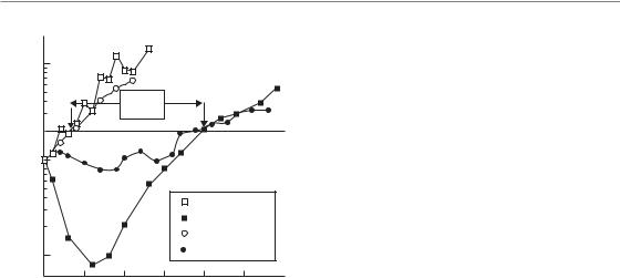
Figure 3.2 Data from Endlich et al. (2000) demonstrating early and late forms of cell death. The ST4 lymphoid cells die rapidly by apoptosis before mitosis. L5178Y-S cells also die by apoptosis following irradiation, but only after attempting to complete mitosis. In this case the initial DNA damage response is not sufficient to induce cell death and the cells die because of problems that occur during mitosis.
is the induction of apoptosis that is initiated as part of the DDR. The DDR is activated within minutes of irradiation, and this leads to p53 activation and to the upregulation of pro-apoptotic proteins. Of course, the DDR also induces pro-survival pathways at the same time, including DNA repair pathways and cell-cycle checkpoints. However, in this case these pro-survival pathways are largely irrelevant because apoptosis is initiated regardless of whether repair takes place or not. In this case, activation of apoptosis is a direct result of the initial levels of damage put into the cell. Consequently, for this early form of apoptosis, the genes that regulate this process can significantly influence radiosensitivity. Loss of p53, for example, leads to a defect in apoptosis, loss of the early form of cell death, and an increase in radioresistance.
Early activation of cell death pathways can also occur in certain cell types as a result of damage caused to cellular structures other than the DNA. In endothelial cells that make up blood vessels, relatively high radiation doses (above 15 Gy) have been reported to induce apoptosis as a result of damage to the cellular membrane and the activation of an enzyme known as ceramide synthase (Garcia-Barros et al., 2003). Endothelial cells
contain very high amounts of this enzyme, and as a result after irradiation can produce large amounts of ceramide. As is the case for apoptosis induced by the DDR, ceramide-induced apoptosis results from pathways activated in response to initial damage caused by irradiation and is not sensitive to DNA repair and checkpoint pathways. Thus, for this form of cell death, the gene products that participate in the activation of apoptosis are important determinants of cellular radiosensitivity.
Late cell death: post-mitotic
The vast majority of proliferating normal and tumour cells die at a relatively long time after irradiation, usually after attempting mitosis one or more times (Fig. 3.2). Time-lapse video microscopy has clearly demonstrated that following a transient delay (owing to activation of checkpoints) most cells resume proliferation and progress through the cell cycle one, two or more times before eventually permanently ceasing proliferation (Forrester et al., 1999). This has been known for more than 30 years and gave rise to the initial characterization of radiation-induced cell
When and why cells die after irradiation 35
Dicentric
Acentric fragment
|
|
|
|
Mitotic catastrophe |
|
|
|
|
|
50% |
|
|
|
cell death |
|
||||
|
|
|
|
|
|
|
|
|
|
DNA damage response |
50% |
|
Survival |
|
|
|
Reciprocal translocation
Aberrations per cell
Reciprocal translocation
Dicentrics
Time after irradiation
Figure 3.3 This figure, adapted from Brown and Attardi (2005), demonstrates the stochastic nature of cell death after irradiation. The DNA repair processes frequently lead to events in which chromosomes are not repaired correctly. It has been shown that irradiated cells produce approximately equal amounts of reciprocal translocations and dicentrics. The broken chromosomes in these cases are ligated to each other in a random or stochastic manner. Formation of a dicentric chromosome prevents proper mitosis and leads to cell death, whereas a reciprocal translocation that does not involve an important region of the genome is stable (sometimes for many decades). Thus, a population of irradiated cells will have approximately equal numbers of both types of aberrations and over time the cells with dicentrics will be lost owing to mitotic catastrophe-induced death. The initial amount of DNA damage and activation of the DNA damage response is the same in both types of cells but the outcome is very different. The outcome in this case is determined by the ability of the cells to avoid mitotic catastrophe. Adapted by permission from Macmillan Publishers Ltd.
death as reproductive or mitotic cell death. In this case, cell death does not occur until after the cell attempts to divide.
In cells that die at long irradiation times, the DDR activates both cell-cycle checkpoints and DNA repair systems that aid in the survival of the irradiated cells. In these cell types, the DDR is unable to induce apoptosis despite the fact that p53 or other pro-death pathways may be induced. Instead, DNA repair is allowed to take place and can have a large influence on the outcome and radiosensitivity of the cell. Consequently, most proliferating cells from animal models and patients with defects in DNA DSB repair show uniformly large increases in sensitivity to radiation-induced cell death.
Although DNA repair and checkpoint pathways play important roles in determining cell survival, cell death takes place at long times after irradiation takes place at times when the checkpoints are no longer active and when DNA repair processes have largely completed. The halftime for repair is approximately 2–4 hours for end-joining and perhaps somewhat longer from homologous recombination. Thus, only a very small fraction of
the initial DNA damage can be detected at times where cell death occurs. The signal for cell death in this case does not arise from the radiationinduced damage itself, but rather from the consequences of failure to properly complete mitosis. Mitotic catastrophe is therefore considered to be responsible for the majority of cell death in irradiated proliferating cells.
Why does irradiation cause proliferating cells to undergo mitotic catastrophe and cell death? This appears to result from the fact that, although DDR pathways remove much of the initial damage caused by irradiation, they are unable to prevent some cells with DNA breaks or DNA rearrangements from entering mitosis. The consequences of incomplete or improper DNA repair become readily visible as chromosomes condense in metaphase as a series of different types of chromosome aberrations. The fate of cells harbouring chromosome aberrations is largely determined by the nature of the chromosome aberration itself (Fig. 3.3) (Brown and Attardi, 2005). Studies have demonstrated approximately equal numbers of reciprocal translocations and non-reciprocal
36 Cell death after irradiation
Apoptotic fraction
Surviving fraction
0.8
0.6
0.4
0.2
0.0
0
100
10
10
10
10
0
 p21
p21  p21
p21
24 |
48 |
72 |
96 |
120 |
Time (hours)
 p21
p21  p21
p21
10 |
20 |
30 |
40 |
50 |
|
Etoposide (μ |
|
|
|
|
|
0.8 |
|
|
|
|
|
|
|
|
|
|
|
|
|
|
|
|
|
|
|
|
|
|
|
|
|
|
|
|
|
|
|
|
|
|
|
|
|
|
|
p21 |
|
|
|
|
|
|
|
|
|
|
|
|
|
|
|
|
|
|
|
||||||||||
|
|
|
|
|
|
|
|
|
|
|
|
|
|
|
|
|
|
|
|
|
|
|
|
|
|
||||||||||
|
fraction |
|
|
|
|
|
p21 |
|
|
|
|
|
|
|
|
|
|
|
|
|
|
|
|
|
|
|
|||||||||
|
0.6 |
|
|
|
|
|
|
|
|
|
|
|
|
|
|
|
|
|
|
|
|
|
|
|
|
|
|
|
|
|
|
|
|
|
|
|
|
|
|
|
|
|
|
|
|
|
|
|
|
|
|
|
|
|
|
|
|
|
|
|
|
|
|
|
|
|
|
|
|
||
|
|
|
|
|
|
|
|
|
|
|
|
|
|
|
|
|
|
|
|
|
|
|
|
|
|
|
|
|
|
|
|
|
|
|
|
|
Apoptotic |
0.4 |
|
|
|
|
|
|
|
|
|
|
|
|
|
|
|
|
|
|
|
|
|
|
|
|
|
|
|
|
|
|
|
|
|
|
|
|
|
|
|
|
|
|
|
|
|
|
|
|
|
|
|
|
|
|
|
|
|
|
|
|
|
|
|
|
|
|
|
||
|
|
0.2 |
|
|
|
|
|
|
|
|
|
|
|
|
|
|
|
|
|
|
|
|
|
|
|
|
|
|
|
|
|
|
|
|
|
|
|
|
|
|
|
|
|
|
|
|
|
|
|
|
|
|
|
|
|
|
|
|
|
|
|
|
|
|
|
|
|
|
|
|
|
|
|
|
|
|
|
|
|
|
|
|
|
|
|
|
|
|
|
|
|
|
|
|
|
|
|
|
|
|
|
|
|
|
|
|
|
|
|
|
|
|
|
|
|
|
|
|
|
|
|
|
|
|
|
|
|
|
|
|
|
|
|
|
|
|
|
|
|
|
|
|
|
|
|
0.0 |
|
|
|
|
|
|
|
|
|
|
|
|
|
|
|
|
|
|
|
|
|
|
|
|
|
|
|
|
|
|
|
|
|
|
|
|
|
|
|
|
|
|
|
|
|
|
|
|
|
|
|
|
|
|
|
|
|
|
|
|
|
|
|
|
|
|
|
|
|
|
|
|
|
|
|
|
|
|
|
|
|
|
|
|
|
|
|
|
|
|
|
|
|
|
|
|
|
|
|
|
|
|
|
|
|
|
|
|
|
24 |
48 |
|
72 |
96 |
|
|
|
120 |
|
144 |
|||||||||||||||||||||
|
|
0 |
|
|
|
|
|
||||||||||||||||||||||||||||
|
|
|
|
|
|
|
|
|
|
|
|
|
|
|
Time (hours) |
|
|
|
|
|
|
|
|
||||||||||||
|
|
100 |
|
|
|
|
|
|
|
|
|
|
|
|
|
|
|
|
|
|
|
|
|
|
|
|
|
|
|
|
|
|
|
|
|
|
|
|
|
|
|
|
|
|
|
|
|
|
|
|
|
|
|
|
|
|
|
|
|
|
|
|
|
|
|
|
|
|
|
|
|
|
|
|
|
|
|
|
|
|
|
|
|
|
|
|
|
|
|
|
|
|
|
|
|
|
|
|
|
|
|
|
|
|
|
||
fractionSurviving |
|
10 |
|
|
|
|
|
|
|
|
|
|
|
|
|
|
|
|
|
|
|
|
|
|
|
|
|
|
|
|
|
|
|
|
|
|
|
|
|
|
|
|
|
|
|
|
|
|
|
|
|
|
|
|
|
|
|
|
|
|
|
|
|
|
|
|
|
|
|
||
|
10 |
|
|
|
|
|
|
|
|
|
|
|
|
|
|
|
|
|
|
|
|
|
|
|
|
|
|
|
|
|
|
|
|
|
|
|
|
10 |
|
|
|
|
|
|
|
|
|
|
|
|
|
|
|
|
|
|
|
|
|
|
|
|
|
|
|
|
|
|
|
|
|
|
|
|
|
|
|
|
|
|
|
|
|
|
|
|
|
|
|
|
|
|
|
|
|
|
|
|
|
|
|
|
|
|
|
|
|
|
|
|
|
|
|
|
|
|
|
|
|
|
|
|
|
|
|
|
|
|
|
|
|
|
|
|
|
|
|
|
|
|
|
|
|
|
|
|
|
|
|
|
|
|
|
|
|
|
|
|
|
|
|
|
|
|
|
|
|
|
|
|
|
|
|
|
|
|
|
|
|
|
|
|
|
|
|
|
|
|
|
|
|
|
|
|
|
|
|
|
|
|
|
|
|
|
|
|
|
|
|
|
|
|
|
|
|
|
|
|
|
|
|
|
|
|
|
|
|
|
|
|
|
|
|
|
|
|
|
|
|
|
|
|
|||||||||
|
|
|
|
|
|
|
p21 |
|
|
|
|
|
|
|
|
|
|
|
|
|
|
|
|
|
|
|
|||||||||
|
|
10 |
|
|
|
|
p21 |
|
|
|
|
|
|
|
|
|
|
|
|
|
|
|
|
|
|
|
|||||||||
|
|
|
|
|
|
|
|
|
|
|
|
|
|
|
|
|
|
|
|
|
|
|
|
|
|
|
|
|
|
|
|
|
|
|
|
|
|
|
|
|
|
|
|
|
|
|
|
|
|
|
|
|
|
|
|
|
|
|
|
|
|
|
|
|
|
|
|
|
|
|
|
|
|
|
|
|
|
|
|
|
|
|
|
|
|
|
|
|
|
|
|
|
|
|
|
|
|
|
|
|
|
|
|
|
|
|
|
|
|
|
|
|
|
|
|
|
|
|
|
|
|
|
|
|
|
|
|
|
|
|
|
|
|
|
|
|
|
|
|
|
|
|
|
|
|
|
|
|
|
|
|
|
|
|
|
|
|
|
|
|
|
|
|
|
|
|
|
|
|
|
|
|
|
|
|
|
|
|
|
|
|
|
|
|
|
|
|
|
|
|
|
|
|
|
|
|
|
|
|
|
|
|
|
|
|
|
|
|
|
|
|
|
|
|
|
|
|
0 |
|
|
|
|
3 |
|
|
|
|
|
|
6 |
|
9 |
|
|
|
|
12 |
|
|
|
|
||||||||||
|
|
|
|
|
|
|
|
|
|
|
|
|
|
|
Dose (Gy) |
|
|
|
|
|
|
|
|
||||||||||||
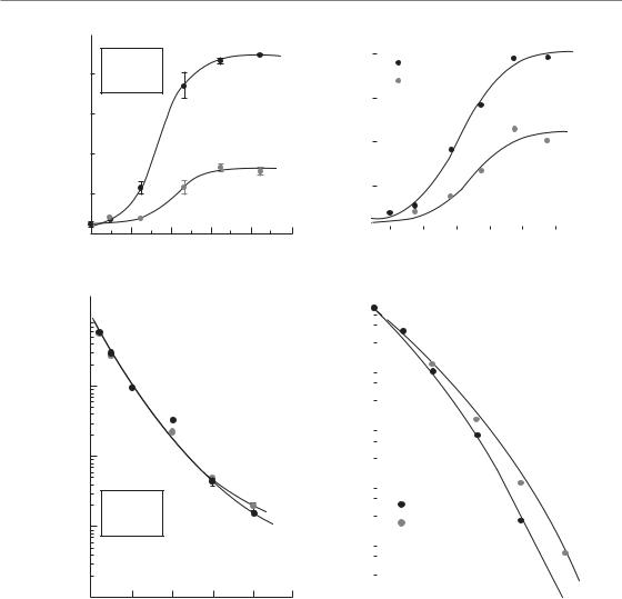
Figure 3.4 This figure, adapted from Wouters et al. (1997), demonstrates the discordance between assays of cell death and cell survival. The two cell lines differ only in the expression of the p21 cyclin-dependent kinase gene (CDKN1A). The p21 knockout cells show increased apoptosis after etoposide or irradiation (top panels) compared with the p21 wild-type cells. However, when assessed by clonogenic survival the p21 knockouts show a similar sensitivity to etoposide and a slight resistance to irradiation compared with the p21 wild-type cells. Here, apoptosis takes place after mitotic catastrophe and is just one mode of cell death that contributes to the loss of clonogenicity. Adapted with permission of American Association for Cancer Research.
translocations (a dicentric chromosome acentric fragment) are formed after irradiation. Both of these types of aberrations result from misrepair in which chromosome ends are incorrectly ligated together in a largely stochastic process. However, whereas cells with dicentric and acentric fragments all die, those with reciprocal translocations often
survive. The presence of two centromeres in dicentric chromosomes prevents their separation at metaphase, and consequently leads to mitotic catastrophe and eventually cell death. Some cells with dicentric chromosomes may manage to complete mitosis; however, loss of genetic material present in the acentric fragment (which forms a so-called
When and why cells die after irradiation 37
|
101 |
|
|
|
|
|
|
volume |
|
|
Growth |
|
|
|
|
|
|
delay |
|
|
|
|
|
|
|
|
|
|
|
|
|
tumour |
100 |
|
|
|
|
|
|
Relative |
|
|
|
|
p53 |
0 Gy |
|
|
|
|
|
|
|
||
|
|
|
|
|
p53 |
15 Gy |
|
|
|
|
|
|
p53 |
0 Gy |
|
|
10 |
|
|
|
p53 |
15 Gy |
|
|
0 |
5 |
10 |
15 |
20 |
25 |
30 |
|
|
|
Time after treatment (days) |
|
|
||
Figure 3.5 Although the mode of cell death may not affect the overall number of cells that die, it can dramatically affect the timing of their death. In this tumour regrowth experiment (from Brown and Wouters, 1999), tumours composed of p53 wild-type and knockout cells are irradiated and followed as a function of time. The unirradiated tumours grow at a similar rate. However, the p53 wild-type tumours undergo rapid apoptosis after irradiation and the tumours thus also shrink rapidly in size. The p53 knockout tumours do not undergo apoptosis and thus are considerably larger after irradiation during this first week. However, the total regrowth delay (measured when tumours reach twice their starting size) is identical for the two tumour types. This indicates that the total number of cells killed by irradiation is the same in both tumour types. In this case apoptosis alters the speed at which the cells die, but does not affect the total number of initially irradiated cells that will eventually die. Adapted with permission of American Association for Cancer Research.
micronuclei) in subsequent mitosis may lead to subsequent death later. This explains the good correlation that has been observed between the formation of dicentric chromosomes or micronuclei formation and cell survival. Reciprocal translocations do not cause problems at metaphase, and thus do not cause mitotic catastrophe or cell death. In fact, these types of aberrations can be found in cells from people exposed to irradiation many years later.
As mentioned previously, cells that experience mitotic catastrophe may ultimately undergo a
secondary form of programmed cell death such as apoptosis, autophagy, necrosis or senescence. In this case this secondary form of death is not the cause, but simply the method through which cells die. This has led to a great deal of confusion about the importance of various forms of cell death, such as apoptosis, as determinants of radiosensitivity. Whereas activation of apoptosis and other programmed cell death programmes are responsible for why cells die at early times after irradiation, they are not similarly responsible for why cells die at long times after irradiation. As a result, alteration of a particular gene may dramatically alter the levels of radiation-induced apoptosis, without altering the overall ability of the cell to survive (Wouters et al., 1997). In this case, cells are dying as the result of undergoing mitotic catastrophe and will die regardless of whether apoptosis is subsequently induced (Fig. 3.4).
Although apoptosis or other programmed cell death pathways may not affect overall survival after irradiation, they can dramatically influence the rate at which cells die and thus the early response of tumours to treatment (Brown and Wouters, 1999). Because apoptosis leads to rapid and complete destruction of the cell, tumours containing cells capable of undergoing apoptosis after mitotic catastrophe may shrink much faster than a similar tumour consisting of cells with the same overall radiosensitivity that do not similarly undergo apoptosis. For this reason, it is dangerous to draw any conclusions about tumour radiosensitivity from initial changes in tumour size after treatment (Fig. 3.5).
Time-lapse microscopy studies have demonstrated that, in cells which experience mitotic catastrophe, both the timing and nature of cell death is highly variable (Fig. 3.6) (Endlich et al., 2000). As discussed earlier, a surviving cell is considered as one that can proliferate indefinitely. In tissue culture this is quantified by the ability to form a colony of a certain size after irradiation (usually 50 cells). Conversely, cell death in this context means that eventually all progeny of an irradiated cell will die. An irradiated cell that is destined to die (not produce a colony) may, however, still proceed through mitosis many times. The resulting daughter cells can die at very different times after irradiation. For example, following the first mitosis, one of the cells may die and the other may proceed through DNA
38 Cell death after irradiation
(a) Clonogenic unirradiated cell |
|
|
|
|
|
|
|
|
|
|
|
|
|
|
|
(c) Clonogenic irradiated cell |
|
|
|
|
|
|
|
|
|
|
|
|
|
|
|
|
|
|
|
|
|
|
|
|
|
|
|
|||||||||||||||||||||||||||||
|
|
|
|
|
|
|
|
|
|
|
? |
|
|
|
|
|
|
|
|
|
|
|
|
|
|
|
|
|
|
|
|
|
|
– increased post-mitotic senescence |
|
|
|
|
|
|
|
|
|
|
|
|
|
|
|
|
|
|
|
|
||||||||||||||||||
|
|
|
|
|
|
|
|
|
? |
|
|
|
|
|
|
|
? |
|
? |
|
|
|
|
|
|
|
|
|
|
|
|
|
|
|
|
|
|
|
|
|
|
|
|
|
|
|
|
|
|
|||||||||||||||||||||||
|
|
|
|
|
|
|
|
|
|
|
|
|
|
|
|
|
|
? |
|
|
|
|
|
|
? |
|
|
|
|
|
|
|
|
|
|
|
|
|
|
|
|
|
|
|
|
|
|
|
|
|
|
|
|
|
|
|
|
|
|
|
|
|
|
|
|
|
|
|||||
|
|
|
|
|
|
|
|
|
|
|
|
|
|
|
|
|
|
|
|
|
|
|
|
? |
|
|
|
|
|
|
|
|
|
|
|
|
|
|
|
|
|
|
|
|
|
|
|
|
|
|
? |
|
|
|
|
|
|
|
|
|
|
|
|
|
|
|
|
|
|
|
|
|
|
|
|
|
|
|
|
|
|
|
|
|
|
? |
|
? |
|
|
|
|
|
|
|
|
|
|
|
|
|
|
|
|
|
|
|
|
|
|
|
|
|
|
|
|
|
|
|
|
|
|
|
|
|
|
|
|
|
|
|
|
|
|
|
|
|
|
? |
? |
|
|
|
|
|
|
|
|
|
|
|
|
|
|
|
|
|
|
|
|
|
|
|
|
|
|
|
|
|
|
|
|
|
|
|
|
|
|
|
|
|
|
|
|
|
|
|
|
|
|
|
|
|
|
|
|
|
|
|
|
|
|
|
|
|
|
|
? |
|
|
|
|||||||
|
|
|
|
|
|
|
|
|
|
|
|
|
? |
|
|
? |
|
|
|
|
|
|
|
|
|
|
|
|
|
|
|
|
|
|
|
|
|
|
|
|
|
|
|
|
|
|
|
|
|
|
|
|
|
|
|
|
|
|
|
|
|
? |
|
|
|
|
|
|||||
|
|
|
|
|
|
|
|
|
|
|
|
|
|
|
|
|
|
|
|
? |
|
|
|
|
|
|
|
|
|
|
|
|
|
|
|
|
|
|
|
|
|
|
|
|
? |
|
|
|
|
|
|
|
|
|
|
|
|
|
|
|
|
|
|
|
|
|
|
|
|
|
|
|
|
|
|
|
|
|
|
|
|
|
|
|
|
|
|
|
|
|
|
|
?? |
|
|
|
|
|
|
|
|
|
|
|
|
|
|
|
|
? |
|
|
|
|
|
|
|
|
? |
|
|
? |
|
|
|
|
|
|
|
|
|
|
|
|
|
|
|
|
|||||||
|
|
|
|
|
|
|
|
|
|
|
|
|
? |
|
|
|
|
|
|
|
|
|
|
|
|
|
|
|
|
|
|
|
|
|
|
|
|
|
|
|
|
|
|
|
|
|
|
|
|
|
|
|
|
|
|
|
|
|
|
|
|
|
|
|
|
|
|
|
? |
|||
|
|
|
|
|
? |
|
|
|
|
|
|
|
|
|
|
|
|
|
|
|
|
|
|
|
|
|
|
|
|
|
|
|
|
|
|
|
|
|
|
|
|
|
|
|
|
|
|
|
|
|
|
|
|
|
|
|
|
|
|
|
|
|
|
|
|
|
|
|
|
|
|
|
|
|
|
|
|
|
|
|
|
|
|
|
|
|
|
|
|
|
|
|
? |
? |
|
|
|
|
|
|
|
|
|
|
|
|
|
|
|
|
|
|
|
|
|
|
|
|
|
|
|
|
|
|
|
|
|
|
? |
? ? |
? |
|
|||||||||||||
|
|
|
|
|
|
|
|
|
|
|
|
|
|
|
|
|
|
? |
|
|
|
|
|
|
|
|
|
|
|
|
|
|
|
|
|
|
|
|
|
|
|
|
|
|
|
|
|
|
|
|
|
|
|
|
|
|
|
|
|
|
|
|
|
|
|
|
|
|
|
|
|
|
|
|
|
|
|
|
|
|
? ? |
|
|
|
|
|
|
|
|
|
|
|
|
|
|
|
|
|
|
|
|
|
|
|
|
|
|
|
|
|
|
|
|
|
|
|
|
|
|
|
|
|
|
|
? |
|
|
|
? |
? |
|
|
|
|
|
|
|
|
|
||||||
|
|
|
|
|
|
|
|
? |
|
|
|
|
|
|
|
|
|
|
|
|
|
|
|
|
|
|
|
|
|
|
|
|
|
|
|
|
|
|
|
|
|
|
|
|
|
|
|
|
|
|
|
|
|
|
|
|
|
? |
|
|
|
|
|
|
|
|
|
|
|
|
|
|
|
|
|
|
|
|
|
|
|
? |
|
|
|
|
|
|
|
|
|
|
|
|
|
|
|
|
? |
|
? |
|
|
|
|
|
|
|
|
|
|
|
|
|
|
|
? |
|
|
|
|
|
|
|
|
|
|
|
|
? |
|
|
|
|
|
|
|
|
|||||||
|
|
|
|
|
|
|
|
|
|
|
|
|
|
|
|
|
|
|
|
|
|
? |
|
|
|
|
|
|
|
|
|
|
|
|
|
|
|
|
|
|
|
|
|
|
|
|
|
|
|
|
|
|
|
|
|
|
|
|
|
|
|
|
|
|
||||||||
|
|
|
|
|
|
|
|
|
|
|
|
|
|
|
|
|
|
|
|
|
|
|
|
|
|
|
? |
|
|
|
? |
|
|
|
|
|
|
|
|
|
|
|
|
|
|
|
|
|
|
|
|
|
|
|
|
? |
|
|
|
|
|
|
|
? |
|
|
|
|
|
|
||
|
|
|
|
|
|
|
|
|
|
|
|
? |
|
|
|
|
|
|
|
|
|
|
|
|
|
? |
|
|
|
|
|
|
|
|
|
|
|
|
|
|
|
|
|
|
|
|
|
|
|
|
? |
|
|
|
|
|
|
|
|
|
|
|
? |
|
|
|
|
|||||
|
|
|
|
|
|
|
|
|
? |
|
|
|
|
|
|
|
|
|
|
|
|
|
|
|
|
|
|
|
|
|
|
|
|
|
|
|
|
|
|
|
|
|
|
|
|
|
|
|
|
|
|
|
|
|
|
|
|
|
|
|
|
|
|
|
|
|
|
|
|
|||
|
|
|
|
|
|
|
|
|
|
|
|
|
? |
|
|
|
? |
|
|
|
|
? |
|
|
|
|
|
|
|
|
|
|
|
|
|
|
|
|
|
|
|
|
|
|
|
|
|
|
|
|
|
|
|
|
|
|
|
|
|
|
|
|
|
|
|
|
|
|
||||
|
|
|
|
|
|
|
|
|
|
|
|
? |
|
|
|
|
|
|
|
|
|
|
|
? |
|
|
|
|
|
|
|
|
|
|
|
|
|
|
|
|
|
|
|
|
|
|
|
|
? |
|
|
|
|
? |
|
|
|
|
|
|
|
|
|
|
|
|
|
|
|
|
||
(b) Clonogenic irradiated cell |
|
|
(d) Non-clonogenic irradiated cell |
||
– increased post-mitotic apoptosis |
– pre-mitotic apoptosis |
||||
? |
? |
? |
|
|
|
|
|
|
|||
|
? |
|
|
|
|
|
|
? |
|
|
|
|
|
? |
|
|
|
|
|
? ?? |
? |
|
|
?
?

 ?
?
?
? |
|
|
? |
? |
|
? |
||
|
||
? |
? |
|
? |
||
? |
? |
|
|
? |
|
|
? |
|
|
? |
(e)Non-clonogenic irradiated cell
–post-mitotic apoptosis
(f)Non-clonogenic irradiated cell
–post-mitotic senescence
? ? ?
?
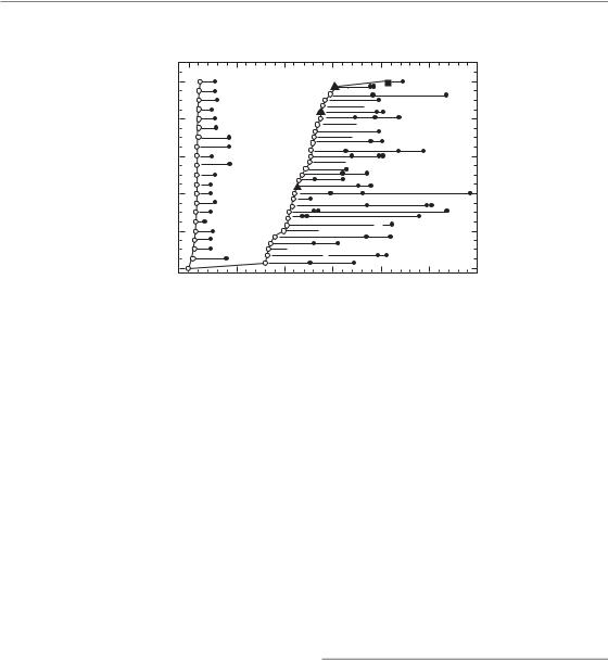
Figure 3.6 This figure, adapted from Forrester et al. (1999), tracks the fate of several irradiated cells as a function of time (left to right) following exposure to radiation. (a) An unirradiated cell is shown as an example. Each cell
division is indicated by a split of one line into two. After six or seven divisions enough cell progeny have been created to produce a colony that can be scored as a survivor. The initial cell is thus said to be clonogenic. Two cells that survive irradiation and eventually form colonies are shown in (b) and (c). In (b), the first division produces two daughter cells that both progress to mitosis and divide producing four cells. One of these four cells dies by apoptosis. Another one undergoes several more divisions but produces progeny that all eventually die. The other two cells both produce many surviving progeny that contribute to the long-term clonogenic potential of the initially irradiated cell. Note, that many of the progeny die in this case even though the initial cell has ‘survived’. In (c) the irradiated cell is also clonogenic. In this case, one of the first two daughter cells produces cells which all eventually undergo senescence. Irradiated cells that are non-clonogenic are shown in (d), (e) and (f). In (d), a cell dies by apoptosis before mitosis. In (e) cells die by apoptosis after completing two divisions and in (e) cells undergo senescence after undergoing one or more mitoses. Adapted with permission of American Association for Cancer Research.
replication and mitosis to produce two more cells. Eventually, these cells will also die, although they may or may not attempt mitosis many times. Furthermore, the type of cell death that each daughter cell undergoes can be different. Consequently, a single irradiated cell can actually die through multiple modes of cell death! A similar situation also exists for irradiated cells that are destined to survive. These cells may also produce daughter cells with different survival potential. One daughter may die,
while the other continues to proliferate and thus confers the status of ‘survived’ on the initially irradiated cell. Consequently, irradiated cells that die following cell division produce a pedigree of cells with different types that can only be tracked by timelapse microscopy (Forrester et al., 1999). Examples of cells destined for survival or death are shown in Fig. 3.6. This figure highlights the many problems associated with trying to quantify or ascribe a particular form of cell death after irradiation and the

Bibliography 39
importance of the clonogenic survival assay for determining the ultimate response of individually irradiated cells.
‘Bystander’ death
A much less understood type of cell death that has been described in response to irradiation is known as bystander-induced death (Mothersill and Seymour, 2004). A number of experiments have challenged the widely held view that radiation kills cells exclusively by direct damage. The bystander effect describes a phenomenon in which cell death can occur in cells owing to irradiation of neighbouring cells. Evidence for this effect has come from studies using high linear energy transfer (LET) α-particles in which a larger fraction of cells die than are estimated to have been traversed. Supportive data have also been generated using microbeam irradiation, in which select cells or nuclei can be irradiated with particles (both α-particles and protons have been used) or soft X-rays. In these experiments, irradiation of a select group of cells leads to increased cell death in the unirradiated cells. In addition to cell death, bystander effects have also been observed for other known biological effects of irradiation, including DNA damage, chromosomal aberrations, mutation, transformation and gene expression.
The precise mechanism or importance of bystander effects has not yet been determined. Some studies have shown that transfer of media from irradiated cells to unirradiated cells can also cause the bystander effect. This would suggest that irradiated cells secrete factors that can be damaging to unirradiated cells. Other experiments have shown that bystander effects are more easily observed when cells are physically connected to irradiated cells by gapjunctions. This allows communication (transfer or molecules) directly between the cells. For example, irradiation may result in increased levels of longlived reactive oxygen species that could be shared among irradiated and unirradiated cells. The bystander effect is probably most important at low doses of radiation which cause damage to only a small number of cells, and may thus be of most relevance to risk estimation.
Key points
1.Most cell death is controlled or programmed in some way.
2.Major death pathways include apoptosis, senescence, autophagy and necrosis.
3.Measuring one form of cell death (e.g. apoptosis) will not necessarily correlate with how many cells die.
4.The form of cell death may influence the rate at which cells die and thus tumour regression.
5.Most cell death after radiation occurs late in response to mitotic catastrophe and not from the initial response to damage.
■BIBLIOGRAPHY
Brown JM, Attardi LD (2005). The role of apoptosis in cancer development and treatment response. Nat Rev Cancer 5: 231–7.
Brown JM, Wouters BG (1999). Apoptosis, p53, and tumor cell sensitivity to anticancer agents. Cancer Res 59: 1391–9.
Campisi J, d’Adda di Fagagna F (2007). Cellular senescence: when bad things happen to good cells.
Nat Rev Mol Cell Biol 8: 729–40.
Chu K, Teele N, Dewey MW, Albright N, Dewey WC (2004). Computerized video time lapse study of cell cycle delay and arrest, mitotic catastrophe, apoptosis and clonogenic survival in irradiated 14–3-3sigma and CDKN1A (p21) knockout cell lines.
Radiat Res 162: 270–86.
Endlich B, Radford IR, Forrester HB, Dewey WC (2000). Computerized video time-lapse microscopy studies of ionizing radiation-induced rapid-interphase and mitosis-related apoptosis in lymphoid cells. Radiat Res 153: 36–48.
Forrester HB, Vidair CA, Albright N, Ling CC, Dewey WC (1999). Using computerized video time lapse for quantifying cell death of X-irradiated rat embryo cells transfected with c-myc or c-Ha-ras. Cancer Res 59: 931–9.
Garcia-Barros M, Paris F, Cordon-Cardo C et al. (2003). Tumor response to radiotherapy regulated by endothelial cell apoptosis. Science 300: 1155–9.

40 Cell death after irradiation
Hayflick L (1965). The limited in vitro lifetime of human diploid cell strains. Exp Cell Res 37: 614–36.
Klionsky DJ (2007). Autophagy: from phenomenology to molecular understanding in less than a decade. Nat Rev Mol Cell Biol 8: 931–7.
Mothersill C, Seymour CB (2004). Radiation-induced bystander effects – implications for cancer. Nat Rev Cancer 4: 158–64.
Okada H, Mak TW (2004). Pathways of apoptotic and non-apoptotic death in tumour cells. Nat Rev Cancer 4: 592–603.
Rubinsztein DC, Gestwicki JE, Murphy LO, Klionsky DJ (2007). Potential therapeutic applications of autophagy. Nat Rev Drug Discov 6: 304–12.
Taylor RC, Cullen SP, Martin SJ (2008). Apoptosis: controlled demolition at the cellular level. Nat Rev Mol Cell Biol 9: 231–41.
Wouters BG, Giaccia AJ, Denko NC, Brown JM (1997). Loss of p21Waf1/Cip1 sensitizes tumors to radiation by an apoptosis-independent mechanism. Cancer Res 57: 4703–6.
■ FURTHER READING
Steel GG (2001). The case against apoptosis. Acta Oncol 40: 968–75.