


2
Irradiation-induced damage and the DNA damage response
BRADLY G. WOUTERS AND ADRIAN C. BEGG
2.1 |
DNA damage by ionizing radiation |
|
2.6 |
Checkpoint activation |
17 |
|
(and other sources) |
11 |
2.7 |
DNA DSB repair |
20 |
2.2 |
The DNA damage response |
14 |
2.8 |
Other DNA repair pathways |
24 |
2.3 |
Sensors of damage |
14 |
Key points |
26 |
|
2.4 |
Signalling to effector pathways |
16 |
Bibliography |
26 |
|
2.5 |
Programmed cell death – apoptosis |
17 |
Further reading |
26 |
|
|
|
|
|
|
|
2.1 DNA DAMAGE BY IONIZING RADIATION (AND OTHER SOURCES)
Ionizing radiation (IR) consisting of electromagnetic radiation, or photons, is the type of radiation most commonly used for the treatment of patients with radiotherapy. Typical energies of the photons produced by 4–25 MV linear accelerators found in radiotherapy departments range from less than 100 keV to several MeV (the maximum energy of the machine being used). From its name, the principal damaging effects of this type of radiation arise from its ability to ionize, or eject electrons, from molecules within cells. Almost all the photons produced by linear accelerators have sufficient energy to cause such ionizations. Most biological damage, however, is done by the ejected electrons themselves, which go on to cause further ionizations in molecules they collide with, progressively slowing down as they go. At the end of electron tracks, interactions with other molecules become more frequent, giving rise to clusters of ionizations (Goodhead, 2006). The pattern and density of ionizations and their relationship with the size of the DNA double helix is shown in Fig. 2.1. The clusters
are such that many ionizations can occur within a few base pairs of the DNA. These clusters are a unique characteristic of IR, in contrast to other forms of radiation such as UV or DNA-damaging drugs such as topoisomerase inhibitors. Only a few per cent of the damage is clustered, but when these clusters occur in DNA, the cell has particular difficulty coping with the damage.
Ionized molecules are highly reactive and undergo a rapid cascade of chemical changes, which can lead to the breaking of chemical bonds. This can disrupt the structure of macromolecules such as DNA, leading to severe consequences if not repaired adequately or in time. Ionizing radiation deposits its energy randomly, thus causing damage to all molecules in the cell. However, there are multiple copies of most molecules (e.g. water, mRNA, proteins and others) and most undergo a continuous rapid turnover, limiting the consequences of damaging just a few molecules of one type. In contrast, DNA is present in only two copies, has very limited turnover, is the largest molecule thus providing the biggest target, and is central to all cellular functions. The consequence of permanent damage to DNA can therefore be serious and often lethal for the cell.
12 Irradiation-induced damage and the DNA damage response
There is also compelling experimental evidence that the DNA is the principal target for radiationinduced cell killing. Elegant experiments have been carried out irradiating individual cells with small polonium needles producing short-range α-particles (Warters and Hofer, 1977). High doses could be given to plasma membranes and cytoplasm without causing cell death. However, as soon as the needle was placed so that the nucleus received even one or two α-particles, cell death resulted. Other experiments used radioactively labelled compounds to irradiate principally the
3
|
|
|
|
|
|
|
|
|
|
S |
|
|
|
|
|
|
|
|
|
|
|
|||||||
|
|
|
|
|
|
|
|
|
|
|
|
|
|
|
|
|
|
C G |
|
|
|
|||||||
|
|
|
|
|
|
|
|
|
|
|
|
|
|
|
|
|
|
T A |
|
|
|
|||||||
|
|
|
|
|
|
|
|
|
|
|
|
|
|
|
|
|
|
|
|
|
||||||||
|
|
|
|
|
|
|
|
|
|
|
|
|
A T |
|
|
|
|
|
|
|
|
|||||||
|
|
|
|
|
|
|
|
C |
|
|
|
|
|
|
|
|
|
|
|
|||||||||
|
|
|
|
|
|
|
|
|
|
G |
|
|
|
|
|
|
|
|
|
|
|
|
|
|||||
|
|
|
|
|
|
|
|
|
|
|
|
|
|
|
|
|
|
|
|
|||||||||
|
|
|
|
|
|
|
C G |
|
|
|
|
|
|
|
|
|
|
|
||||||||||
|
|
|
|
|
|
|
|
C G |
|
|
|
|
|
|
|
|
|
|
|
|||||||||
|
|
|
|
|
|
|
|
T |
|
|
A |
|
|
|
|
|
|
|
|
|
|
|
|
|
||||
|
|
|
|
|
|
|
|
|
|
|
|
|
|
|
|
|
|
|
|
|||||||||
|
|
|
|
|
|
|
|
|
|
|
|
C G |
|
|
|
|
|
|||||||||||
|
|
|
|
|
|
|
|
|
|
|
|
|
|
|
|
|
||||||||||||
|
|
|
|
|
|
2.3 nm |
|
|
|
|
|
|
|
|
|
|
|
C G |
|
|
|
|
||||||
|
|
|
|
|
|
|
|
|
|
|
|
|
|
|
|
|
|
|
|
|
||||||||
|
|
|
|
|
|
|
|
|
|
|
|
|
|
|
|
|
|
|
|
|
||||||||
|
|
|
|
|
|
|
|
|
|
|
|
|
|
|
|
|
|
|
|
|
|
|
|
|
|
|
|
|
|
|
|
|
|
|
|
|
|
|
|
|
|
|
|
|
|
|
|
A T |
|
|
|
||||||
|
|
|
|
|
|
|
|
|
|
|
|
|
|
|
|
|
|
|
|
|
|
|||||||
|
|
|
|
|
|
|
|
|
|
|
|
|
|
|
|
|
|
|
|
|
|
|||||||
|
|
|
|
|
|
|
|
|
|
|
|
|
|
|
G C |
|
|
|
|
|
||||||||
|
|
|
|
|
|
|
|
|
|
|
|
|
|
|
|
|
|
|
|
|||||||||
|
|
|
|
|
|
|
|
|
|
C G |
|
|
|
|
|
|
|
|
|
|
|
|
||||||
|
|
|
|
|
|
|
|
|
|
|
|
|
|
|
|
|
|
|
|
|
|
|||||||
|
|
|
|
|
|
|
|
C G |
|
|
|
|
|
|
|
|
|
|
|
|
|
|||||||
|
|
|
|
|
|
|
|
|
|
|
|
|
|
|
|
|
|
|
|
|||||||||
|
|
|
|
|
|
|
|
G C |
|
|
|
|
|
|
|
|
|
|
|
|||||||||
|
|
|
|
|
|
|
|
|
|
|
|
|
|
|
|
|
|
|
||||||||||
|
|
|
|
|
|
|
C |
|
G |
|
|
|
|
|
|
|
|
|
|
|
|
|||||||
|
|
|
|
|
|
|
|
T |
|
|
|
|
|
|
|
|||||||||||||
|
|
|
|
|
|
|
|
|
|
|
A |
|
|
|
|
|
|
|||||||||||
|
|
|
|
|
|
|
|
|
|
|
|
|
|
|
G |
|
|
|
|
|
C |
|
|
|
||||
|
|
|
|
|
|
|
|
|
|
|
|
|
|
|
|
|
|
|
|
|
||||||||
0 |
10 |
20 |
30 |
40 |
|
|
|
|
|
|
|
|
|
|
|
CG |
|
|
|
|||||||||
|
|
|
|
S |
3 |
|
|
|
|
|
||||||||||||||||||
|
|
|
Range in water (nm) |
|
|
|
|
|
|
|
|
|
|
|||||||||||||||
(a) |
(b) |
|
|
|
|
|
|
|
|
|
|
|
|
|
|
|
|
|
|
|
||||||||
|
|
|
|
|
|
|
|
|
|
|
|
|
|
|
|
|
|
|
|
|
|
|
||||||
Figure 2.1 (a) Computer-simulated tracks of 1 keV electrons. Note the scale in relation to the 2.3 nm diameter of the DNA double helix (adapted from Chapman and Gillespie, 1981). (b) Illustrating the concept of a local multiply-damaged site produced by a cluster of ionizations impinging on DNA.
plasma membrane (125I-concanavalin), or principally the DNA (3H-labelled thymidine), and compared this with homogeneous cell irradiation with X-rays. Cell death closely correlated only with dose to the nucleus, and not with either the plasma membrane or the cytoplasm (Table 2.1).
Because of the importance of DNA, cells and organisms have developed a complex series of processes and pathways for ensuring that the DNA remains intact and unaltered in the face of continuous attack from within (e.g. oxidation and alkylation owing to metabolism) and from the outside (e.g. ingested chemicals, UV and ionizing radiation) (Harper and Elledge, 2007). These include different forms of DNA repair to cope with the different forms of DNA damage induced by different agents.
Specialized repair systems have therefore evolved for detecting and repairing damage to bases [base excision repair (BER)], single-strand breaks [singlestrand break repair (SSBR), closely related to BER], and double-strand breaks [homologous recombination (HR) and non-homologous end-joining (NHEJ)]. All these lesions are produced by ionizing radiation, and each of these repair pathways is described in more detail in Sections 2.7 and 2.8. There are also other DNA repair pathways, such as those for correcting mismatches of bases in DNA which can occur during replication, such as mismatch repair (MMR), and for repairing bulky lesions or DNA adducts such as those formed by UV light and some drugs such as cisplatin [nucleotide excision repair (NER)]. However, neither MMR nor NER appears to be important for ionizing radiation, since cells with mutations or deletions in genes on
Table 2.1 Toxicity of radioisotopes depends upon their subcellular distribution
Radiation dose to part of the cell* (Gy)
Radiation source/type |
Nucleus |
Cytoplasm |
Membranes |
X-rays |
3.3 |
3.3 |
3.3 |
3H-Thymidine |
3.8 |
0.27 |
0.01 |
125I-Concanavalin |
4.1 |
24.7 |
516.7 |
*For each of these three treatments a dose has been chosen that gives 50 per cent cell killing in Chinese hamster ovary (CHO) cells. The absorbed radiation doses to the nucleus, cytoplasm or membranes have then been calculated. 3H-Thymidine is bound to DNA and 125I-concanavalin to cell membranes. It is the nuclear dose that is constant and thus correlates with cell killing, not the cytoplasmic or membrane doses. From Warters et al. (1977) with permission.
DNA damage by ionizing radiation (and other sources) 13
these pathways are not more sensitive to IR. In contrast, mutations or deletions in BER, SSBR, HR or NHEJ genes can all, under certain circumstances, lead to increased radiosensitivity.
To give an idea of the scale of damage, 1 Gy of irradiation will cause in each cell approximately 105 ionizations, 1000 damages to DNA bases, around 1000 single-strand DNA breaks (SSBs) and around 20–40 double-strand DNA breaks (DSBs). To put this into further perspective, 1 Gy will kill only about 30 per cent of cells for a typical mammalian cell line, including human. This relatively limited cytotoxicity, despite large numbers of induced lesions per cell, is the consequence of efficient DNA repair.
Cellular DNA comprises two opposing strands linked by hydrogen bonds and forming a double helical structure. Each strand is a linear chain of the four bases – adenine (A), cytosine (C), guanine (G) and thymine (T) – connected by sugar molecules and a phosphate group, the so-called sugar–phosphate backbone (Fig. 2.2). The order
of the bases is the code determining not only which protein is made but whether a gene is active (transcribed) or not. In turn, this double helix is wound at regular intervals around a complex of a specific class of proteins (histones), forming nucleosomes, resembling beads on a string. Many other proteins are also associated with the DNA; these control DNA metabolism, including transcription, replication and repair. The DNA plus its associated proteins is called chromatin. There are further levels of folding and looping, finally making up the compact structure of the chromosomes.
This structure poses various challenges to the cell for repairing DNA damage. First, specialized proteins have to be sufficiently abundant and mobile to detect damage within seconds or minutes of it occurring. Second, the chromatin usually needs to be remodelled (e.g. the structure opened up) to allow access of repair proteins (van Attikum and Gasser, 2005). This may entail removal of nucleosomes close to the break, among other changes. The correct repair, accessory and signalling proteins
5
O
O P O CH2 O |
G |
Base |
|
||
O |
|
|
O |
H |
C |
|
|
O P O CH2 O
Deoxyribose
O
OH
|
O P O CH2 |
T |
|
|
|
O |
|
||
|
O |
|
|
|
|
3 |
O |
H |
|
|
|
|
||
Phosphodiester |
O |
P |
O CH2 |
A |
bond |
O |
|||
|
O |
|
|
|
|
5 |
|
|
|
|
|
|
|
|
|
|
|
OH |
H |
|
|
|
|
3 |
Figure 2.2 The structure of DNA, in which the four bases (G, C, T and A) are linked through sugar groups to the sugar–phosphate backbone.
14 Irradiation-induced damage and the DNA damage response
DNA damage sensors
M
NKu70
R |
ATM |
|
Ku80 DNA-PKcs |
ATRIP ATR |
|
|
|
|
|
Damage signalling |
|
|
|
|
|
P |
P |
P |
γ H2AX |
|
|
|
|
|
|||
|
|
|
|
|
MDC1 |
|
|
|
|
|
|
53BP1 |
Foci |
|
|
|
|
|
BRCA1,2 |
|
|
|
|
|
|
RAD51 |
|
|
|
|
|
|
p53 |
|
|
|
|
P |
|
p21 |
|
|
|
|
|
|
BAX |
|
|
|
|
|
|
Chk1/2 |
|
|
|
|
|
|
CDC25A/C |
|
|
|
|
Effector pathways |
|
........ |
|
Checkpoints |
Cell death |
|
DNA repair |
Figure 2.3 The DNA damage response can be divided into sensors and effectors. The sensors consist of protein complexes which recognize DNA damage and include MRN–ATM, Ku–DNA-PKcs, and ATRIP–ATR (see text). These proteins signal to many other proteins which activate three important effector pathways: checkpoints, DNA repair and cell death. Examples of some of the proteins which signal from the sensors to the effector pathways are listed.
then need to be recruited, often mediated by histone modifications, and tightly coordinated. This includes stopping various processes such as transcription and cell-cycle progression to concentrate on repair. Repair progress needs to be continually monitored so that the chromatin will be reset to its original state after completion of repair, and then normal cellular processes resumed.
2.2 THE DNA DAMAGE RESPONSE
The DNA damage response (DDR) is a highly complex and coordinated system that determines the cellular outcome of DNA damage caused by radiation. The DDR is not a single pathway, but rather a group of highly interrelated signalling pathways, each of which controls different effects on the cell. This system can be divided into two parts, the sensors of DNA damage and the effectors of damage
response (Fig. 2.3). The sensors consist of a group of proteins that actively survey the genome for the presence of damage. These proteins then signal this damage to three main effector pathways that together determine the outcome for the cell. These effector pathways include (1) programmed cell death pathways that kill damaged cells, (2) DNA repair pathways that physically repair DNA breaks and (3) pathways that cause temporary (or permanent) blocks in the progress of cells through the cell cycle – the damage checkpoints.
2.3 SENSORS OF DAMAGE
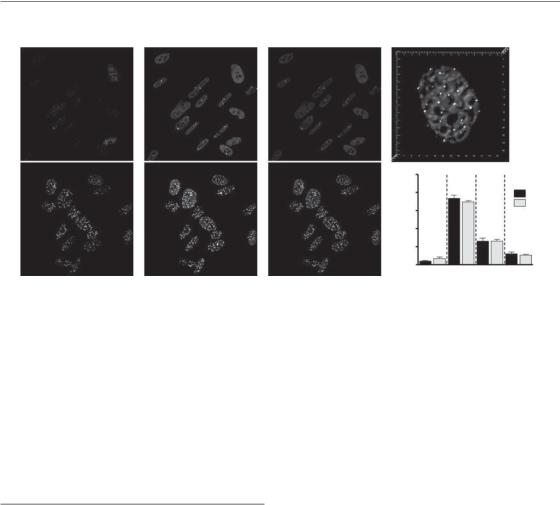
Foci formation
The initial cellular response to DSBs is characterized by the physical recruitment of a large number of different proteins to the sites of DNA damage. This clustering or recruitment of various proteins can be visualized microscopically as small regions or speckles in the nucleus after DNA damage following staining with antibodies to these proteins (Fig. 2.4). These subnuclear regions are commonly referred to as ionizing radiation induced ‘foci’ (IRIF). A large number of the proteins involved in the DDR have been shown to form IRIF at or very near to the actual sites of DSBs. Consequently, it is thought that each focus represents a platform from which DNA repair and signalling to the other effectors of the DDR occurs.
One of the earliest events known to occur in the DDR is the phosphorylation of a protein called histone H2AX (Stucki and Jackson, 2006). This is a variant of histone H2A, a component of the core nucleosome structure around which DNA is packaged. H2AX is distributed throughout the entire nucleus and makes up about 10–15 per cent of total cellular histone H2A. Starting within a few minutes of DSB formation, H2AX becomes phosphorylated in a region that extends over an extensive region around the site of the unrepaired DSBs. This phosphorylated form, known as γH2AX, is necessary for the recruitment of many of the other proteins involved in the DDR and the resulting formation of IRIF. Cells that have been engineered to lack H2AX show substantial defects in IRIF formation and are radiosensitive. In recent years,
Sensors of damage 15
γ H2AX |
53BP1 |
Merge |
NIR
2 Gy 30
Foci per nucleus
50 |
|
|
|
40 |
|
|
γH2AX |
30 |
|
|
53BP1 |
|
|
|
|
20 |
|
|
|
10 |
|
|
|
0 |
|
|
|
NIR |
0.5 |
4 |
24 |
Time after 2 Gy (hours)
Figure 2.4 Examples of ionizing radiation-induced nuclear foci. Unirradiated and irradiated (2 Gy) cells have been fixed and stained with antibodies that recognize γH2AX and 53BP1, two proteins that interact at sites of DNA damage to form foci after induction of DNA double-strand breaks. These foci form rapidly and then resolve, consistent with the kinetics of DNA double-strand break rejoining. Photographs courtesy of Farid Jallai and Rob Bristow, Princess Margaret Hospital. See colour plate section for full colour images.
the presence of γH2AX foci, which can be detected using microscopy, has become a highly sensitive method for detecting the presence and/or repair of individual DSBs in irradiated cells.
The sensors of DSBs
Phosphorylation of H2AX at and around the sites of DSBs is one of the earliest events in the DDR, occurring within 5–30 min after DSB induction. This indicates that one or more kinases, enzymes which phosphorylate other proteins, are activated at the sites of DSBs. Three related kinases have been shown to be able to phosphorylate H2AX at sites of DSBs (Falck et al., 2005).
ATM–MRN
The phosphorylation of H2AX at sites of DSBs produced by radiation occurs primarily by the ataxia telangiectasia mutated (ATM) protein. The gene that encodes this protein is mutated in the autosomal recessive syndrome ataxia telangiectasia (AT), which presents clinically as oculocutaneous telangiectasia and progressive cerebellar ataxia (O’Driscoll and Jeggo, 2006). These patients are
frequently found to be highly radiosensitive and have an increased risk of developing cancer. Cells from AT patients or normal cells in which the ATM protein is inhibited are extremely radiosensitive, and are defective in H2AX phosphorylation, IRIF formation and many other aspects of the DDR.
Although there is still some debate about the exact sequence of events that occurs immediately after a DSB is created, it is now recognized that ATM requires at least one additional protein complex in order to ‘find’ the DSB and become activated. Recruitment of ATM to the DSB requires a protein complex known as MRN. This complex contains three proteins, MRE11, RAD50 and NBS1. The NBS1 protein is the product of the gene that is mutated in Nijmegen breakage syndrome (NBS). This syndrome is highly similar to AT and patients are also radiosensitive. Cells that lack NBS1 show similar defects in H2AX phosphorylation, IRIF formation and other aspects of the DDR. One of the key functions of NBS1 is to directly bind to ATM, and bring it to the sites of damage. Thus, when NBS1 is defective, ATM is unable to relocate to DSBs and is therefore unable to phosphorylate H2AX and initiate the DDR. This provides a molecular basis for the similarities between NBS and AT clinical syndromes.

16 Irradiation-induced damage and the DNA damage response
Several lines of evidence indicate that MRN is the functional sensor of DSBs induced by radiation (Stucki and Jackson, 2006). First, the RAD50 protein has been shown to directly bind to DNA; second, NBS1 is required for recruitment of ATM to the break; third, MRN assembles at sites of DSBs faster than any other known protein; and fourth, MRN is the only known DNA repair complex that does not depend on any other protein to form foci at DSBs. MRN is a multifunctional complex. Not only does it sense the DSB, bind DNA and recruit ATM, it is also important for the ‘processing’ of the DSB. For example, it has exonuclease activity and can digest the ends of the DNA break which may not be compatible for ligation.
Thus, the earliest events in the DDR are considered to be the recruitment of MRN and ATM to DSBs. ATM is normally present in the cell, but in an inactive form. Activation of ATM occurs once it becomes associated with a DSB resulting in phosphorylation of H2AX at the site of the DSB. H2AX phosphorylation then spreads over relatively large chromatin regions (megabases) in both directions, an event that is regulated by an additional protein called MDC1. This protein acts as an adaptor/ mediator by directly binding to both ATM and phosphorylated H2AX, and in this way is able to spread ATM phosphorylation of H2AX in both directions of the break. The spreading of this signal significantly alters the chromatin structure around the DSB and is thought to be important for assisting access of other DNA repair proteins to the break. The spreading also acts to ‘amplify’ the DSB signal. The presence of large areas of H2AX phosphorylation around a single DSB explains the relatively large foci that are observed microscopically.
DNA-PKcs-KU
In cells that completely lack the ATM protein, phosphorylation of H2AX and IRIF formation can still occur through an alternative mechanism but with somewhat delayed kinetics. In these cells, H2AX phosphorylation is mediated by the catalytic subunit of the DNA-dependent protein kinase (DNA-PKcs). DNA-PKcs is a kinase that is structurally related to ATM and which is very important in the non-homologous end joining pathway of
DNA repair (see Section 2.7, NHEJ). In normal cells or in cells treated with an ATM inhibitor
(which blocks the activity of the ATM protein), DNA-PKcs is unable to phosphorylate H2AX. Thus, in addition to its early role in activating components of the DDR, the ATM protein also appears to actively exclude DNA-PKcs at sites of DSBs.
The mechanism through which DNA-PKcs finds DSBs is very similar to that of ATM. Like ATM, DNA-PKcs is unable to act as a sensor of damage itself. This sensor function is carried out by the Ku70–Ku80 complex, which directly binds to the ends of DSBs. Binding of Ku70–Ku80 to DSBs then recruits DNA-PKcs allowing phosphorylation of H2AX.
ATR–ATRIP
The third kinase capable of phosphorylating H2AX is ATR, which stands for AT-related kinase. This enzyme does not appear to play any substantial role in the initial recognition of DSBs produced by radiation. Instead, it phosphorylates H2AX in response to other types of DNA damage and abnormalities such as single-stranded DNA and stalled or broken replication forks. It is thus very important for the types of damage that occur during normal DNA replication. The recruitment of ATR to sites of DNA damage requires another protein called ATRIP (ATR interacting protein). Thus, just like ATM and DNAPKcs, ATR is recruited to sites of damage by a different protein that acts as the sensor of DNA damage.
Although ATR is not important in the initial detection of DSBs, it does play a role in this pathway after ATM is activated. As discussed above, the ATM–MRN complex leads to processing of the DNA at sites of DSBs. This processing can create stretches of single-stranded DNA, which will then activate ATR. Thus, ATR can be activated ‘downstream’ of ATM activation. Although highly related to ATM, ATR phosphorylates a distinct set of proteins that participate in the DDR. Consequently, components of the DDR effector pathways (DNA repair, checkpoints and cell death) are also dependent on ATR after radiation treatment.
2.4 SIGNALLING TO EFFECTOR PATHWAYS
Activation of ATM, DNA-PKcs and ATR leads to the phosphorylation not only of H2AX, but also of many other cellular proteins. Amazingly, recent
Checkpoint activation 17
studies have shown that as many as 700 proteins are substrates for the ATM and ATR kinases in response to DNA damage (Matsuoka et al., 2007). Phosphorylation of these other proteins act as the ‘signals’ to activate the various different downstream effectors of the DDR (apoptosis, cell-cycle checkpoints and DNA repair). The ATM protein plays perhaps the most important role in transmitting these signals in response to radiation-induced DSBs and is thus considered to be a master regulator of the DDR.
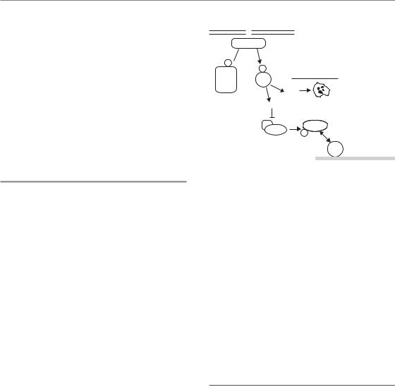
2.5 PROGRAMMED CELL DEATH – APOPTOSIS
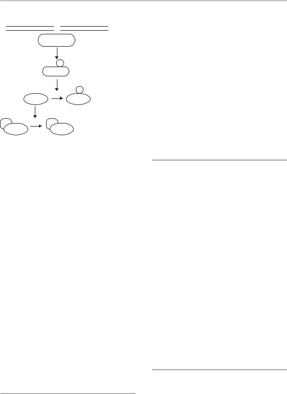
Two of the important proteins which are phosphorylated following activation of ATM are p53 and MDM2. p53 is one of the most commonly mutated tumour suppressors whose function is to regulate genes that control both cell-cycle checkpoints (see below) and programmed cell death through a programme known as apoptosis (see Chapter 3 for details). Consequently, activation of p53 after irradiation can lead either to a block in proliferation or directly to cell death (Fig. 2.5).
The p53 gene is regulated at the protein level by binding to its partner MDM2. This association leads to rapid ubiquitination and destruction of p53 through the proteasome pathway. Thus, in unstressed normal cells, p53 is made continuously but is degraded and is thus non-functional. Following DNA damage, ATM phosphorylates both p53 and MDM2. These events destabilize the p53–MDM2 interaction, and as a result the p53 protein is no longer degraded. In addition to this stabilization, direct phosphorylation of p53 by ATM leads to its activation as a transcription factor and thus the upregulation of its many target genes. These target genes include the pro-apoptotic genes BAX and PUMA, which in certain cells can be sufficient to induce cell death. Thus, in some cells, activation of the DDR itself can lead to rapid induction of cell death through apoptosis. The purpose of this DDR effector is likely to be similar to the function of p53 itself, namely tumour suppression. Because DNA damage can lead to dangerous mutations, it may be more beneficial to the organism to eliminate the cell rather than trying to repair the damage. This would be predicted to be especially important in rapidly proliferating tissues, and
DSB
ATM
P
P
Early apoptosis
MDM2 
 p53
p53
Bax
Puma
p21
G1 arrest
Rb
P
G1 cyclin/CDK
E2F
S-phase genes
Figure 2.5 Cells irradiated in the G1 phase are influenced by the action of p53. Ataxia telangiectasia mutated (ATM) protein is activated by double-strand DNA breaks (DSBs) and phosphorylates both MDM2 and p53. This leads to stabilization and activation of p53, which then induces genes that can promote apoptosis (Bax, Puma) and induce checkpoints. Induction of p21 inhibits the action of cyclin–cyclin-dependent kinase (CDK) complexes that are necessary for the entry into
S phase. Consequently cells are blocked at the G1/S border. In many cancer cells, p53 or other components of this checkpoint are mutated and so it is non-functional.
indeed these tissues tend to display radiationinduced apoptosis (see Chapter 3 for more details).
2.6 CHECKPOINT ACTIVATION
The second major effector pathway of the DDR is the activation of cell-cycle checkpoints. Treatment of cells with ionizing radiation causes delays in the movement of cells through the G1, S and G2 phases of the cell cycle (Table 2.2) (Kastan and Bartek, 2004). This occurs through the activation of DNA damage checkpoints, which are specific points in the cell cycle at which progression of the cell into the next phase can be blocked or slowed. The DDR activates four distinct checkpoints in response to irradiation that take place at different points within the cell cycle. Initially, these checkpoints were described as delays that would allow cells more time to repair DNA damage. This description is likely true to some extent, especially in terms of the importance of checkpoints in preventing mutations that might otherwise arise

18 Irradiation-induced damage and the DNA damage response
Table 2.2 |
Radiation-induced cell-cycle checkpoints and their characteristics |
|
|
|
|
|
|
|
|
Applies to cells |
|
Position |
Primary signalling proteins |
irradiated in |
Features |
|
|
|
|
G1 |
ATM, p53, p21 |
G1 |
Prevents entry into S |
S |
ATM, Chk1/Chk12, CDC25A/ |
S |
Slows progression through S |
|
CDC25C, BRCA1, BRCA2 |
|
|
G2-early |
ATM, Chk1/Chk12, CDC25A/ |
G2 |
Prevents entry into mitosis |
|
CDC25C, BRCA1, BRCA2 |
|
|
G2-late |
ATR, Chk1, CDC25A/CDC25C |
All phases |
Accumulation of cells in G2 |
|
|
|
|
because of misrepair. However, there is little evidence to support a general role for checkpoints in influencing the overall radiosensitivity (cell survival) following single doses of radiation.
All movement through the cell cycle, be it in the G1, S, G2 or M phases, is driven by cyclin-depend- ent kinases (CDKs). The CDKs phosphorylate other proteins to initiate the processes required for progression thorough the cell cycle. A CDK is active only when associated with a cyclin partner (hence their name) and different cyclin–CDK complexes are active at different points within the cell cycle. For example, cyclinD–CDK4 is active in G1 and cyclinB–CDK1 is active in mitosis. Checkpoint activation requires inhibition of the cyclin–CDK complexes and for radiation this occurs through two main mechanisms. The first is by activation of other proteins that directly inhibit the cyclin–CDK complex; these are the cyclin-dependent kinase inhibitors (CDKIs). The second is by affecting phosphorylation and activity of the CDK enzyme itself. The activity of any given CDK is frequently affected by its phosphorylation status, and may be active in either the phosphorylated or dephosphorylated state depending on the specific CDK in question.
G1 arrest
Cells contain a checkpoint at the transition between the G1 and S phases that plays an important normal role in the decision of the cell to initiate cell division. This checkpoint is thus sensitive to growth factors, nutrients and other conditions that favour proliferation. The transition from G1 to S phase is controlled by the activation of the E2F transcription factor. This factor is important for regulating many
of the genes necessary to initiate DNA replication in S phase and in G1 is kept inactive by binding to the retinoblastoma protein (Rb). As cells normally move from G1 into S, the Rb protein becomes phosphorylated by G1 cyclin–CDKs including cyclinD–CDK4 and cyclinE–CDK2. This phosphorylation causes release of Rb from E2F, allowing E2F to function as a transcription factor and initiate S phase.
As discussed above, irradiation leads to an ATM-dependent stabilization and activation of p53. One of the genes that is upregulated by p53 is the CDKI p21 (CDKN1A). p21 inhibits the G1 cyclin–CDK complexes thereby preventing phosphorylation of Rb and entry into S phase. As a result, cells that are irradiated while in the G1 phase will exhibit a delay prior to entry into S phase that is dependent on both p53 and p21.
S-phase checkpoint
The remaining radiation-induced checkpoints are controlled by two highly related proteins known as Chk1 and Chk2 (Fig. 2.6) (Bartek et al., 2004). Chk1 and Chk2 are activated by phosphorylation and are direct targets of ATR and ATM respectively. Cells that are in S phase at the time of irradiation demonstrate a dose-dependent reduction in the rate of DNA synthesis and, as a result, the overall length of time that cells need to replicate their DNA substantially increases. The target for preventing S-phase progression is the CDK2 kinase, which must be in a dephosphorylated form to be active. The dephosphorylation of CDK2 is maintained by the phosphatases CDC25A and CDC25C. When Chk1 and Chk2 are activated, they phosphorylate CDC25A
Checkpoint activation 19
|
DSB |
|
ATM/ATR |
|
P |
|
CHK1/2 |
Active |
P Inactive |
cdc25 |
cdc25 |
 Cell-cycle progression
Inactive |
Active |
cyclin/CDK |
cyclin/CDK |
Figure 2.6 The S, early G2 and late G2 checkpoints are all activated by a similar mechanism. Ataxia telangiectasia mutated protein (ATM) and/or AT-related kinase (ATR) are activated by double-strand DNA breaks (DSBs) and phosphorylate the Chk1/2 kinases (see text). These kinases then phosphorylate and inactivate CDC25A/CDC25C. CDC25A/CDC25C are required for progression through S phase and into mitosis because they activate the required cyclin–cyclin-dependent kinase (CDK) complexes in both parts of the cell cycle. Thus when Chk1/2 are phosphorylated by ATM, cellcycle checkpoints in both S and G2 are activated.
and CDC25CC leading to their destruction or inactivation. As a result, Chk1 and Chk2 activation by ATR and ATM results in an increase in the amount of phosphorylated CDK2 and thus slowed progression through S phase.
Although, ATM–Chk2 and ATR–Chk1 activation and inhibition of CDC25A/C is the main mechanism for initiation of the S-phase checkpoint, several other proteins in the DDR can also influence this response. This includes the BRCA1 and BRCA2 proteins, whose main function is in the homologous recombination branch of DNA repair (see
Section 2.7). This suggests a complex relationship between checkpoint activation and DNA repair.
G2 early checkpoint
There are two additional checkpoints in G2, both of which operate along similar lines to that
in S phase. The early G2 checkpoint is also ATM–Chk2–Cdc25A/C dependent and applies to cells that are irradiated while in G2. The checkpoint is activated by relatively low doses of radiation (1 Gy is enough) and results in a block of cell-cycle progression at the end of G2. The target of ATM–Chk2–Cdc25A/C signalling in this case is the mitotic cyclinB–CDK1 complex which, like CDK2 in S phase, must be dephosphorylated on specific sites to become active. It is called the early G2 checkpoint because it applies to cells that are irradiated while in G2 phase and rapidly blocks their movement into mitosis. As a result, there is a drop in the number cells within mitosis at short times after irradiation.
G2 late checkpoint
The late G2 checkpoint describes a long G2 delay that is observed after irradiation and is applicable to cells that have been previously irradiated while in the G1 or S phases. These cells may experience transient G1and S-phase checkpoints, but when they arrive in the G2 phase many hours later they experience a second delay before entry into mitosis. Unlike the early G2 checkpoint, this delay is strongly dose-dependent, and can last many hours after high doses of radiation. In addition, unlike all the other damage checkpoints this late G2 checkpoint is independent of ATM. Instead, the principal signalling axis occurs from ATR to Chk1 to CDC25A/CDC25C. The late G2 checkpoint is thus mechanistically similar to the S and early G2 checkpoints, but likely arises from a fundamentally different type of DNA damage. Instead of being activated directly by DSBs, this checkpoint likely reflects a type of damage that persists after other DNA repair processes have occurred and which is sufficient to activate ATR.
Checkpoints, cancer and radiosensitivity
In a large proportion of tumour cells, one or more of the G1/S, S phase and early G2 checkpoints are disabled because of genetic changes that occur during tumourigenesis. In recent years, these checkpoint responses have been linked to a

20 Irradiation-induced damage and the DNA damage response
tumour suppressor function that must be disrupted to allow oncogene-induced proliferation. This is thought to occur following activation of growth-promoting oncogenes which induce ‘inappropriate replication’ and DNA damage from replication stress. When functional, the checkpoints block further proliferation of these cells and can thus actively suppress cancer development. This idea is supported by the finding that many early cancer lesions show widespread activation of checkpoint activity.
Mutations in genes such as p53, BRCA1 or other components of the DDR that influence checkpoint activation will result in the failure to delay cell-cycle progression in response to irradiation. This may have an important consequence for genetic instability after irradiation and tumour progression, but there is little evidence to suggest that lack of these checkpoints influences overall cellular radiosensitivity. Thus, although the G1/S, S and early G2 checkpoints are often described as providing extended time for repair, this extra time seems to be more important for the quality of repair rather than the amount of repair that takes place.
The late G2 checkpoint, which unlike all others is ATR rather than ATM dependent, is the only exception since evidence exists to support it as having a role in determining radiosensitivity. For example, inhibitors of ATR that prevent this checkpoint cause radiosensitization. Thus, for reasons that are still unclear, premature entry into mitosis of cells that are in the late G2 checkpoint results in increased cell death.
Although the G1/S, S and early G2 checkpoints may not affect the radiosensitivity of cells to single doses of radiation, they may affect the response to multiple (fractionated) doses. The presence of the checkpoints in normal cells, and absence in many tumour cells, will affect the redistribution of cells at time-points after irradiation and therefore indirectly the sensitivity of cells to subsequent doses of radiation. For example, consider two populations of cells that are identical with exception of the ability to block at the G1 checkpoint. Twenty-four hours after irradiation cells that lack this checkpoint may show reduced numbers of cells in G1 phase and more cells in S phase compared with cells that have an intact checkpoint. Since radiosensitivity is different in G1 and S phase, the
two populations of cells may respond differently to subsequent irradiation at that time. Consequently, all checkpoints can potentially affect responses to radiotherapy given in multiple fractions.
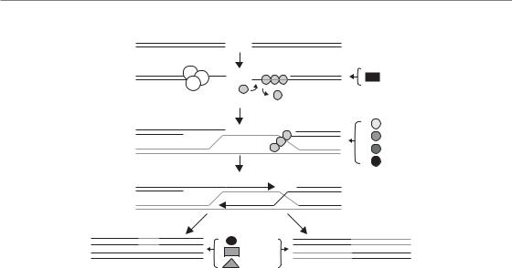
2.7 DNA DSB REPAIR
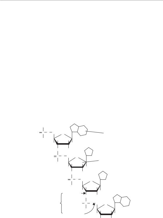
As discussed above, DSBs are detected by specialized proteins which signal to the cell that damage has occurred, thereby initiating the DNA damage response. This response effectively focuses the cell’s attention on the damage, stopping other processes such as transcription and cell-cycle progression, and, importantly, initiating repair. For DSBs, there are two main repair pathways, HR and NHEJ. These are quite different in the genes involved, the position in the cell cycle where they primarily act and in the speed and accuracy of repair. These processes are described in more detail below. Both affect the radiosensitivity of a cell.
HR
As the name implies, HR uses homologous undamaged DNA (that with an identical sequence) as the template to repair the DNA with the DSBs in it (West, 2003). By using DNA with the same sequence as a basis for repair, the process is error free. The general scheme is illustrated in Fig. 2.7. Briefly, single-strand regions are created around each side of the break, followed by their coating with specialized proteins. These single-stranded nucleoprotein filaments then invade undamaged double-stranded DNA on the neighbouring sister chromatid, forming a crossover, or bubble, structure. These bubbles are then expanded with specialized enzymes called helicases. The object of this process is to provide an undamaged DNA template of the same base sequence around the break site, so that DNA polymerases can then synthesize across the missing regions, thereby accurately repairing the break. The crossover structure then has to be reversed to reset the chromatin to its original configuration. This is done with specialized nucleases which cut, or resolve, the junctions, followed finally by connecting up, or ligating, adjacent ends with a ligase. The whole process takes several hours (up to
DNA DSB repair 21
|
DSB |
|
Resection M |
|
Coating |
R N |
|
BRCA2 |
|
|
|
MRN complex RAD51 |
RPA |
|
Strand invasion |
|
RAD51C |
|
|
|
|
|
RAD51D |
|
|
XRCC2 |
|
|
XRCC3 |
Synthesis |
DNA polymerase |
|
|
Helicase |
|
Resolution |
|
|
|
BLM |
|
|
BLAP75 |
|
Without crossover |
TOP3α |
With crossover |
Figure 2.7 Schematic of double-strand DNA break (DSB) repair by homologous recombination (HR). The principal genes known to be involved are shown, although there are others not shown which are also involved in HR. Chromatin remodelling genes are not shown. The main feature is the use of an undamaged sister chromatid sequence (light coloured lines) as template for repair. The groups of genes (right and bottom centre) are involved with the processes indicated by the horizontal arrows.
6 hours or more) to complete. Some details of the genes involved in the process are given below.
The first step is to cut back one strand on each side of the break with an exonuclease, making a 3 overhang, in order to create single-stranded regions which are necessary for subsequent strand invasion. The proteins involved in end resection include the MRN complex (described above under sensors of damage), MRE11 being the active component. The single-stranded DNA is immediately coated with RPA (replication protein A), a single-strand binding protein, which is a universal response of the cell to any single-stranded regions of DNA. This RPA is then displaced by a central protein in HR, namely RAD51. This results in the formation of a nucleoprotein filament of DNA coated with RAD51, which then undergoes the search for homologous DNA and strand invasion. Several RAD51 paralogues (genes arising from duplication of the parent gene but with modified sequence and function) help with these processes, including RAD51B, RAD51C, RAD51D, XRCC2 and XRCC3. Deletion or mutation of any of these genes can severely impair HR.
Helicases, including BLM and other members of the RecQ family, possibly with the help of RAD54,
then enlarge the subsequent ‘bubble’ structure (branch migration). The necessary DNA synthesis is carried out with DNA polymerases, the identity of which is still uncertain. One of the replicative polymerases is the likely candidate, however, since HR is mostly an error-free process, like replication, although some of the less accurate translesion synthesis polymerases have also been implicated in this process. The bubble structure then needs to be resolved by cutting the DNA at the crossover points, carried out by enzymes called resolvases. In bacteria and yeast, the identity of resolvases is known, but not in mammalian cells despite intense research. It is known that BLM exists in a complex with TopIIIa (a topoisomerase capable of untangling DNA) and a helper protein called BLAP75. There is now evidence that BLM pushes the two sides of the bubble towards each other via its helicase activity, leaving only a small crossover region, and that TopIIIa then untangles the DNA at this point, via its cutting and re-ligation activity that is common to topoisomerases, resulting in two separated sister chromatids. Finally, DNA ends are ligated probably with ligase 1, since eliminating the LIG1 gene reduces repair by HR.
22 Irradiation-induced damage and the DNA damage response
In addition to those mentioned above, two other gene families are involved, well known for causing human repair deficiency syndromes, namely BRCA genes 1 and 2 and the Fanconi anaemia family (Zhang and Powell, 2005). Mutations or deletions in one or more of these genes compromises HR. BRCA2 has perhaps the most clearly defined function in regulating the binding of RAD51 to RPAcoated single-stranded DNA, a key step in HR. The Fanconi (FANC) genes also play a significant role in HR, although knocking out these genes has a milder effect than, for example, knocking out BRCA2 or RAD51 which can lead to cell and embryonic lethality. Cells with FANC gene mutations all show increased sensitivity to DNA crosslinking agents, repair of which depends on HR. However, mild or little increased sensitivity to ionizing radiation has been found in FANC mutant cells, although increased radiosensitivity under hypoxia has been reported, which may be a consequence of the increased crosslinks formed under hypoxic irradiation, again requiring HR repair. The 13 Fanconi genes can be divided into three groups: eight in a core complex (A, B, C, E, F, G, L and M), two substrates which are ubiquitinylated by the core complex (D2 and I) and three downstream targets (D1, J and N). DNA damage leads to ubiquitinylation of the D2–I complex, which binds and regulates BRCA2 (also known as FANCD1).
The role of BRCA1 is broader, although it clearly plays a role in HR. Together with its partner BARD1 it can ubiquitinylate other proteins (it is an E3 ubiquitin ligase), thus modifying their protein interacting properties, and thus their function. BRCA1–BARD1 appear in several complexes with other proteins, each playing a different role, including with BACH1 and TOPBP1 and others in replication inhibition (part of the intra-S checkpoint), with the MRN complex in NHEJ, with RAD52– BRCA2 in homologous recombination and with RNA polymerase II in transcription. Its ubiquitinylating function is necessary for each role.
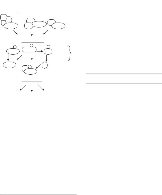
NHEJ
As its name implies, NHEJ joins two DNA DSB ends together without requiring homologous DNA sequences (Lieber, 2008). This is a more
|
|
DSB |
|
|
End binding |
Ku70 |
|
M |
End processing |
|
|
|
N |
|
|
|
|
R |
|
|
Ku80 |
|
|
|
|
RAD50 |
|
|
|
|
|
|
|
|
Gap filling by polymerase |
|
|
|
|
|
|
DNA-PKcs |
|
Holding ends, |
|
|
|
|
signalling, |
Recruitment |
processing |
|
ARTEMIS
Recruitment and ligation
XRCC4 LIG4 NHEJ1
NHEJ1
Completion
Often with small deletions or insertions
Figure 2.8 Schematic of double-strand DNA break (DSB) repair by non-homologous end-joining (NHEJ). The principal genes known to be involved are shown, although there are others not shown which are also involved in NHEJ. Chromatin remodelling genes are not shown. For clarity, processes such as end-binding have been shown on one side of the break only.
rapid process than HR but less accurate, with small deletions or insertions often resulting at the repaired break site. Although this can lead to mutations, it allows the cell to survive. An unrepaired DSB is often lethal through loss of a piece of chromosome at the next mitosis, with the potential loss of tens or hundreds of genes. In addition, only a minor fraction (a few per cent) of genomic DNA comprises gene coding or regulatory regions, so the chance of a break occurring in such regions is low, and these may also be silent (not expressed) and/or non-essential. Although NHEJ is ‘quick and dirty’, it is a good repair pathway for the cell to maximize its chance of survival.
The general scheme of NHEJ is shown in Fig. 2.8. The main steps in NHEJ, after sensing the DSB, involve nucleases to remove damaged DNA, polymerases to help repair and ligases to restore the continuity of the DNA chain. The first event in the sequence is binding of a Ku heterodimer (two different but related proteins: Ku70 and Ku80) to the DNA ends. This occurs within seconds of the break

DNA DSB repair 23
being formed because of high abundance of the Ku dimer and its high affinity for ends. The binding serves both to protect ends from degradation by exonucleases and to recruit the DNA-dependent protein kinase catalytic subunit (DNA-PKcs; officially called PRKDC), the second step in NHEJ.
Activation of PRKDC as a kinase occurs only when it is bound to the Ku complex at break sites. It has several substrates and several functions. It is a large protein and forms a physical bridge between the two ends, helping to keep them in close proximity for subsequent repair events. In addition, it phosphorylates a number of target proteins involved in checkpoints and repair. One of its targets is itself (autophosphorylation). There are a number of sites on the PRKDC protein which can be phosphorylated, some of which are autophosphorylated, while others are phosphorylated by ATM. Phosphorylation has been shown to be necessary for efficient repair by NHEJ since mutations at these sites make cells considerably more radiosensitive. Phosphorylation stimulates dissociation of the protein from DNA, or a change in conformation, allowing other repair factors access to the site, so that phosphorylation-mutant proteins remain at the site, blocking it for further repair.
PRKDC also exists in a complex with Artemis, which is recruited to DNA ends together with PRKDC. Artemis has endonuclease activity and its function is to clean up, or process, the DNA ends so they are suitable for ligation (Jeggo and Lobrich, 2005). The activation of PRKDC on endbinding leads not only to autophosphorylation but also to phosphorylation of Artemis, stimulating its nuclease activity. Break sites often show small deletions as a result. Artemis is apparently necessary for the repair of a minor fraction of DSBs. Another protein involved in end-processing in the NHEJ pathway is polynucleotide kinase (PNK), which is capable of trimming ‘dirty’ ends (e.g. those with the remnants of a sugar group instead of a ‘clean’ base with a 3 phosphate). This action renders the ends ligatable with a ligase (see below).
Radiation often produces an overhang, or nonblunt, end, either directly or after end-processing. Such gaps can be filled by a polymerase to produce blunt-ended DNA ready for ligation. The translesion synthesis polymerases λ and μ have been shown to be capable of this and have been
implicated in NHEJ. Polμ and another polymerase called terminal deoxynucleotidyl transferase (TdT) can also add a few nucleotides to a blunt end. DNA sequencing of breaks repaired by NHEJ therefore often shows small insertions. Whether replicative polymerases are also involved in these synthesis activities is not clear. The final step in repair is ligation of adjoining ends, which is carried out by ligase IV, aided by two other proteins, XRRC4 and XLF.
Making the choice between HR and NHEJ
Several factors determine which pathway is used to repair a damage-induced DSB. DNA damage detection proteins such as MRN and the Ku complex will compete for DNA ends and partially determine which pathway is subsequently used (Brugmans et al., 2007). This is called passive competition, and there is evidence supporting such a model. However, there are other factors, one of the most obvious being template availability. Homologous recombination requires the availability of an homologous stretch of DNA, which the sister chromatid provides in S and G2. Although there is no sister chromatid in G1, there is an homologous chromosome, but this is often far away in molecular terms, making it a very difficult task for the HR machinery to find and use. Therefore HR is rare or absent in G1. A further illustration of the importance and use of HR is that cells often show increased radioresistance as they progress through S phase, being most resistant in late S, a time at which almost all DNA has a paired chromatid available for HR. Knocking out or reducing HR genes eliminates this late-S resistance (Tamulevicius et al., 2007).
There are also active regulators of HR. As described above, end-resection at the break site, producing single-stranded DNA, is necessary for HR. End resection does not occur in G1, probably since it depends on specific CDKs, which are not active until late in G1. This further favours the NHEJ pathway in this phase. There are also antirecombination genes in yeast with homologues in mammalian cells, with the job of preventing unwanted recombination, which can lead to genetic instability.

24 Irradiation-induced damage and the DNA damage response
Finally, a comment on clinical relevance. Since HR is a pathway specific to S- and G2-phase cells, it occurs only in dividing cells. Conversely, NHEJ occurs in all phases of the cell cycle, and is thus neither phase specific nor cycle specific. The relevance for radiotherapy is that NHEJ is used by all cells and tissues, including those that are slowly dividing or non-dividing. This includes the doselimiting late-reacting tissues such as spinal cord and stromal tissue, which give rise to fibrosis and telangiectasia. In attempting to find DNA-repair- inhibiting drugs to improve radiotherapy, targeting NHEJ is therefore likely to be a more risky strategy than inhibiting HR.
The link between SSBs and DSBs
Single-strand breaks can lead to the formation of DSBs in two main ways. First, ionizing radiation damage often occurs in clusters, such that some SSBs will also have damage to DNA bases in their near vicinity. During repair of the base damage by BER, SSBs are formed temporarily (see Section 2.8). It has been shown that, if base damage occurs on the opposite strand to a radiation-induced SSB, the temporary nick formed during BER can combine with the radiation break on the opposite strand causing a DSB. Second, if a SSB encounters a replication fork during S phase, this leads to collapse of the fork and a single-ended DSB. The cell attempts to repair these S-phase DSBs by HR. In this case NHEJ is not an option, since there is only one double-stranded end and not two, and so there is no adjacent end for the end-joining process. Thus HR provides a backup repair pathway for unrepaired SSBs.
The latter mechanism has clinical relevance, since it has been shown that drugs which inhibit poly-(ADP-ribose) polymerase (PARP), a SSB detector protein, are particularly effective in tumours with HR deficiencies, such as breast tumours with BCRA1 or BRCA2 deficiencies. The mechanism is probably that the PARP inhibitors suppress SSB repair, resulting in greater numbers of unrepaired SSBs, which therefore have a greater chance of hitting a replication fork. Under normal circumstances the resulting DSB would be repaired by HR, so the absence or reduction of
this backup pathway leads to a substantial increase in DSBs and thus cellular lethality.
2.8 OTHER DNA REPAIR PATHWAYS
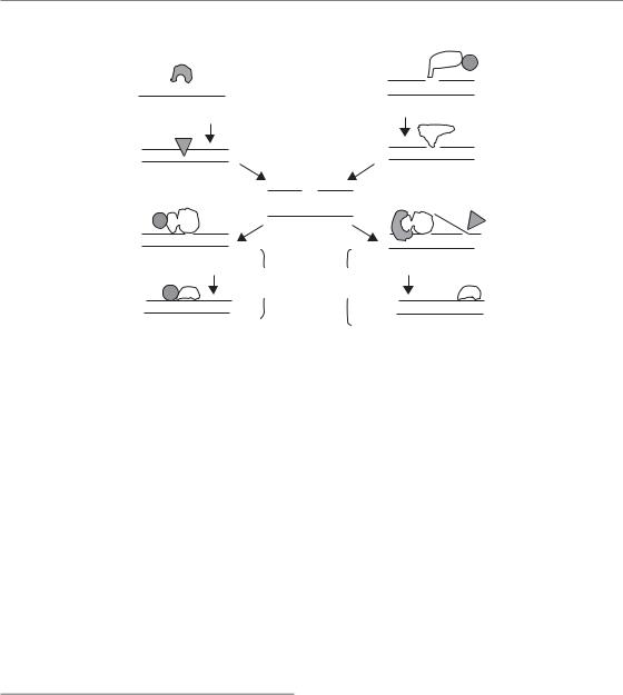
Base excision repair and single-strand break repair (SSBR)
As mentioned in Section 2.1, DSBs, although the most lethal lesion induced by ionizing radiation, are not the most common. Base damage and SSBs far outweigh DSBs in number, being up to 50 times more frequent. Base damage and SSBs also occur without irradiation as a consequence of normal metabolism. It has been estimated that 100 000 such damages occur each day in every cell in the body. The repair pathways – BER and SSBR – have therefore evolved to repair such damage efficiently and maintain genome integrity (Fortini and Dogliotti, 2007).
An outline of the related BER and SSBR pathways is shown in Fig. 2.9. Briefly, in BER, most of the damaged bases in the DNA will be detected and removed by specialized proteins called glycosylases. There are several such enzymes, each specific for a particular type of base damage. They cut out the damaged base without cutting the DNA backbone, resulting in an abasic site. This will be recognized by another class of enzyme, AP endonuclease, which will cut the DNA backbone leaving a nick, or SSB. Subsequent repair follows one of two pathways: short patch or long patch. As their names imply, short patch repair involves replacing the damaged base only, while in long patch repair up to 10 nucleotides are cut out and replaced. Each requires DNA synthesis to replace the missing bases, carried out by DNA polymerase β for short patch repair and mainly DNA polymerases δ and ε (replicative enzymes) for long patch repair. As always, ligases complete the job: ligase 3 for short patch and ligase 1 for long patch. Repair of SSB is similar, although radiation itself causes the break rather than being a repair intermediate. Since these breaks are often ‘dirty’, with ends not recognized by ligases, there is an extra end-processing step, mainly by the enzyme PNK. Once a clean nick is produced, short or long patch repair can then follow, as for BER. This short summary does not include all proteins involved in the
Other DNA repair pathways 25
|
BER |
|
|
|
SSBR |
|
|
|
Glycosylase |
|
|
|
|
PARP XRCC1 |
|
|
X |
|
X-ray-induced |
P |
OH |
||
|
|
|
|
|
|||
|
|
‘dirty’ break |
|
|
|
||
|
|
|
|
|
|
||
Glycosylase removes |
|
|
Break detected by PARP |
||||
damaged base |
|
|
|
|
|
|
|
APE1 |
|
|
|
PNK |
|
||
|
|
|
OH |
P |
|
||
|
|
|
|
|
|
||
APE1 makes nick (SSB) |
|
|
|
PNK ‘cleans’ ends |
|||
|
|
|
|
|
|
||
|
|
OH |
P |
|
|
|
|
|
|
C TAT |
T G G T |
|
|
|
FEN1 |
XRCC1 |
|
G ATA C A C C A |
PCNA |
|
|||
POLB |
|
|
|
|
|||
|
Common |
|
POLδ |
TG |
G |
||
|
|
intermediate |
|
6 |
|||
|
|
|
|
||||
|
|
|
|
|
|
||
Pol beta inserts single |
|
|
|
Replicative pols replace |
|||
correct base |
SHORT |
LONG |
|
2–10 bases |
|||
XRCC1 |
|
|
|
|
|
||
LIG3 |
PATCH |
PATCH |
|
|
|
LIG1 |
|
|
|
|
|
|
|
||
Ligation by ligase3 |
Ligation by ligase1 |
|
Figure 2.9 Schematic of the related pathways of base excision repair (BER) and single-strand break repair (SSBR). The X (top left) represents a damaged base. Different base damages are recognized and removed by different glycosylases as the first step in BER. Both pathways result in a common nicked intermediate, which is processed by one of two subpathways (short or long patch repair). APE1 apurinic/apyrimidinic endonuclease-1; PARP, poly (ADP-ribose) polymerase; PNK, polynucleotide kinase; POL, polymerase.
pathway. Two mentioned here briefly because of their importance are PARP-1, which efficiently and rapidly detects SSBs, and XRCC1, which is a helper protein for both PNK (damage processing) and ligase 3. Mutation, deletion or inhibition of either of these can lead to reduced repair and radiosensitization. Inhibition of PARP is particularly effective in HR-deficient tumours (see above), illustrating the relevance of the BER/SSBR pathways for possible clinical exploitation.
MMR and NER
The mismatch repair pathway corrects mispaired nucleotides (e.g. C with T). As with all repair pathways it comprises a recognition step, an excision and resynthesis step and ligation. Most studies with knockout cells for one or more mismatch repair genes have not found any increase in radiosensitivity. The details of the pathway will therefore not be described in detail here. However, this pathway clearly has relevance for cancer treatment, since MMR-deficient cells have altered sensitivity to some chemotherapy agents (e.g. cisplatin). In addition,
radiosensitization by thymidine analogues such as IUdR is enhanced in MMR-deficient cells because of inability to remove the modified base. The MMR status of cells is therefore of importance for outcome, not for radiotherapy alone, but for combinations of radiotherapy with some chemotherapy or radiosensitizing agents.
Nucleotide excision repair copes with bulky lesions, such as those caused by UV light (thymine dimers), and DNA adducts induced by cisplatin. However, as with MMR, knocking out NER genes has, in general, little effect on sensitivity to ionizing radiation, and so no detailed discussion of NER will be included here. There is one situation, however, where NER genes can affect the radiation response. Irradiation under hypoxia produces a greater number of DNA crosslinks than under oxic conditions. Such crosslinks require, among other factors, the excision activity of two NER genes, ERCC1 and XPF. Defects in either of these genes can lead to modest increases in radiosensitivity of hypoxic cells. The status of the NER pathway is therefore relevant to radiotherapy in combination with certain chemotherapy agents, and possibly to hypoxic tumours treated with radiotherapy alone.

26 Irradiation-induced damage and the DNA damage response
Key points
1.DNA is the critical target for radiationinduced cell killing.
2.Cells activate a DNA-damage response that consists of sensors and effectors.
3.Effector pathways include apoptosis, cellcycle checkpoints and DNA repair.
4.DNA DSBs are the most important and difficult lesion to repair.
5.DSBs are repaired by both HR and NHEJ.
■BIBLIOGRAPHY
Bartek J, Lukas C, Lukas J (2004). Checking on DNA damage in S phase. Nat Rev Mol Cell Biol 5: 792–804.
Brugmans L, Kanaar R, Essers J (2007). Analysis of DNA double-strand break repair pathways in mice. Mutat Res 614: 95–108.
Chapman JD, Gillespie CJ (1981). Radiation-induced events and their time-scale in mammalian cells. Adv Radiat Biol 9: 143–98.
Falck J, Coates J, Jackson SP (2005). Conserved modes of recruitment of ATM, ATR and DNA-PKcs to sites of DNA damage. Nature 434: 605–11.
Fortini P, Dogliotti E (2007). Base damage and singlestrand break repair: mechanisms and functional significance of shortand long-patch repair subpathways. DNA Repair (Amst) 6: 398–409.
Goodhead DT (2006). Energy deposition stochastics and track structure: what about the target? Radiat Prot Dosimetry 122: 3–15.
Harper JW, Elledge SJ (2007). The DNA damage response: ten years after. Mol Cell 28: 739–45.
Jeggo PA, Lobrich M (2005). Artemis links ATM to double strand break rejoining. Cell Cycle 4: 359–62.
Kastan MB, Bartek J (2004). Cell-cycle checkpoints and cancer. Nature 432: 316–23.
Lieber MR (2008). The mechanism of human nonhomologous DNA end joining. J Biol Chem 283: 1–5.
Matsuoka S, Ballif BA, Smogorzewska A et al. (2007). ATM and ATR substrate analysis reveals extensive protein networks responsive to DNA damage.
Science 316: 1160–6.
O’Driscoll M, Jeggo PA (2006). The role of double-strand break repair – insights from human genetics. Nat Rev Genet 7: 45–54.
Stucki M, Jackson SP (2006). gammaH2AX and MDC1: anchoring the DNA-damage-response machinery to broken chromosomes. DNA Repair (Amst) 5: 534–43.
Tamulevicius P, Wang M, Iliakis G (2007). Homologydirected repair is required for the development of radioresistance during S phase: interplay between double-strand break repair and checkpoint response.
Radiat Res 167: 1–11.
van Attikum H, Gasser SM (2005). The histone code at DNA breaks: a guide to repair? Nat Rev Mol Cell Biol 6: 757–65.
Warters RL, Hofer KG (1977). Radionuclide toxicity in cultured mammalian cells. Elucidation of the primary site for radiation-induced division delay.
Radiat Res 69: 348–58.
Warters RL, Hofer KG, Harris CR, Smith JM (1997). Radionuclide toxicity in cultured mammalian cells: elucidation of the primary site of radiation damage. Curr Top Radiat Res 12: 389–407.
West SC (2003). Molecular views of recombination proteins and their control. Nat Rev Mol Cell Biol 4: 435–45.
Zhang J, Powell SN (2005). The role of the BRCA1 tumor suppressor in DNA double-strand break repair. Mol Cancer Res 3: 531–9.
■ FURTHER READING
Caldecott KW (2007). Mammalian single-strand break repair: mechanisms and links with chromatin. DNA Repair (Amst) 6: 443–53.
Löbrich M, Jeggo PA (2007). The impact of a negligent G2/M checkpoint on genomic instability and cancer induction. Nat Rev Cancer 7: 861–9.