

- •Contents
- •Preface
- •1 Introduction: the significance of radiobiology and radiotherapy for cancer treatment
- •2 Irradiation-induced damage and the DNA damage response
- •3 Cell death after irradiation: how, when and why cells die
- •4 Quantifying cell kill and cell survival
- •5 Dose–response relationships in radiotherapy
- •6 Linear energy transfer and relative biological effectiveness
- •7 Tumour growth and response to radiation
- •8 Fractionation: the linear-quadratic approach
- •9 The linear-quadratic approach in clinical practice
- •10 Modified fractionation
- •11 Time factors in normal-tissue responses to irradiation
- •12 The dose-rate effect
- •13 Pathogenesis of normal-tissue side-effects
- •14 The volume effect in radiotherapy
- •15 The oxygen effect and fractionated radiotherapy
- •16 The tumour microenvironment and cellular hypoxia responses
- •17 Therapeutic approaches to tumour hypoxia
- •18 Combined radiotherapy and chemotherapy
- •19 Retreatment tolerance of normal tissues
- •20 Molecular image-guided radiotherapy with positron emission tomography
- •21 Molecular-targeted agents for enhancing tumour response
- •22 Biological response modifiers: normal tissues
- •23 Molecular targeting and patient individualization
- •24 Protons and other ions in radiotherapy
- •25 Second cancers after radiotherapy
- •Glossary of terms in radiation biology
- •Index

13
Pathogenesis of normal-tissue side-effects
WOLFGANG DÖRR
13.1 |
Introduction |
169 |
13.5 Radiation effects in specific tissues |
|
13.2 |
Early radiation effects |
170 |
and organs |
178 |
13.3 |
Chronic radiation effects |
174 |
Key points |
189 |
13.4 |
Documentation and assessment of |
|
Bibliography |
189 |
|
normal-tissue effects |
176 |
Further reading |
190 |
|
|
|
|
|
|
|
|
|
|
13.1 INTRODUCTION
Despite optimum conformation of the treatment fields to the tumour and precise treatment planning and application, the target volume in curative radiotherapy necessarily includes a substantial amount of normal tissue, for several reasons. First, malignant tumours infiltrate microscopically into normal structures, which hence must be included into the high-dose volume as a tumour margin. Second, normal tissues within the tumour, such as soft tissue and blood vessels, are exposed to the full tumour dose. Third, normal structures in the entrance and exit channels of the radiation beam may be exposed to clinically relevant doses. Therefore, effective curative radiotherapy is unavoidably associated with an accepted risk for early and late radiation side-effects (‘adverse events’) in order to achieve adequate tumour cure rates.
The optimum radiation dose in curative radiotherapy is defined as the dose that is associated with a certain low incidence of sequelae of a defined severity in cured patients (‘complicationfree healing’). The manifestation of side-effects is hence an indicator for optimum treatment and maximum tumour cure probability; side-effects
cannot, a priori, be considered as a consequence of incorrect treatment.
Early (acute) side-effects are observed during or shortly after a course of radiotherapy. In contrast, late (chronic) side-effects become clinically manifest after latent times of months to many years. The cut-off time to distinguish early from late effects has arbitrarily been set to 90 days after the onset of radiotherapy. This classification is based exclusively on the time-course (i.e. the time of first diagnosis of the pathological changes). However, early and late effects have specific (radio)biological features which distinguish them.
Early effects are usually found in tissues with a high proliferative activity that counteracts a permanent cell loss (turnover tissues), such as bone marrow, epidermis or mucosae of the upper and lower intestinal tract. The acute symptoms are based on radiation-induced impairment of cell production in the face of ongoing cell loss, which is usually independent of the treatment. The consequence is progressive cell depletion. This response is regularly accompanied by inflammatory changes, which either can be directly induced by the radiation exposure or secondary to the changes in the turnover compartment of the tissue. Healing, which is usually complete, is based on the proliferation

170 Pathogenesis of normal-tissue side-effects
of surviving tissue stem cells within the irradiated volume or migration of stem cells in from unirradiated tissue.
Late radiation side-effects are basically found in all organs. In contrast to the development of early side-effects, which are characterized by cell depletion as a leading mechanism, the pathogenetic pathways of chronic side-effects are more complex. The dominating processes occur in the parenchyma of the organs (i.e. in the tissue-specific compartments) but also in the connective and vascular tissues. Regularly, the immune system (macrophages, mast cells) contributes to the tissue reaction. Late radiation effects hence represent a multifaceted, orchestrated response with various components (Dörr et al., 2005b; Bentzen, 2006).
Late radiation sequelae, in contrast to early effects, with few exceptions, are irreversible and progressive, with increasing severity occurring with longer follow-up times. Therefore, the longer the survival times of the patients (i.e. the better the radiation therapy) the higher is the number of patients at risk for late reactions. A risk for the manifestation of a chronic reaction remains throughout the life of the patient (Jung et al., 2001).
Early and late radiation effects are independent with regard to their pathogenesis and, in general, conclusions from the severity of early reactions on the risk of late effects cannot be drawn. However, in particular situations, interactions between acute and chronic reactions can occur within one organ, resulting in consequential late effects (CLE). This is the case when the early-responding tissue compartments (e.g. epithelia) have a protective function against mechanical and/or chemical exposure. This barrier function is impaired during the acute radiation reaction because of cell depletion. In consequence, secondary traumata can impact on the target structures of the late sequelae (connective tissue, vasculature) in addition to the direct effects of radiation, which can then aggravate the late radiation response (Dörr and Hendry, 2001). Consequential late effects have been demonstrated for intestine, urinary tract, oral mucosa and particularly stressed skin localizations (Dörr and Hendry, 2001), as well as for lung (Dörr et al., 2005a).
In this chapter, after a description of the general pathogenesis of early and late radiation effects, specific effects in clinically relevant organs and
tissues will be described. This latter part also includes side-effects that follow an atypical pathogenesis, such as late cardiovascular changes or radiation cataract induction. This chapter does not include systemic sequelae such as fatigue or nausea and emesis. Moreover, responses to high single doses or a few large fractions, as administered in stereotactic radiotherapy, which may be based on different pathogenetic pathways, are excluded.
13.2 EARLY RADIATION EFFECTS
Radiation effects in skin and epidermis were doselimiting during the orthovoltage era, with peak doses occurring near the entrance sites of the beam. In contrast, with modern treatment techniques, distributions of high doses within the body can, in general, be achieved without severe epidermal toxicity.
However, early radiation effects are still relevant even in face of the progress in the physical application of radiotherapy. Early reactions significantly affect the quality of life of the patients. Some early responses are dose-limiting, such as oral mucositis in radiotherapy of advanced head and neck tumours, and hence reduce the chance for tumour cure. Moreover, early reactions can result in consequential late effects (Dörr and Hendry, 2001). In addition, the costs of supportive care are an important socio-economic factor.
Additional traumas can significantly aggravate early radiation responses. Chemotherapy is one prominent example. Moreover, in epithelial tissues, mechanical stress can influence early complications, such as epidermal irritation by clothing or in skin folds, or oral mucosal trauma through dental prostheses or sharp-edged food components. Similarly, chemical exposure, such as smoking, alcohol or spicy diet in oral mucosa, can intensify the response. Such exacerbating factors should be avoided during radiotherapy.
Precise knowledge of the pathogenesis and radiobiology of acute radiation effects forms the essential basis for the development of selective, biol- ogy-based interventions. One example is radiationinduced leukopenia after bone marrow irradiation, which can be treated by haematopoietic growth factors such as granulocyte colony-stimulating

Early radiation effects 171
Vascular |
? |
|
|
|
Secondary |
|
|
|
|
|
|
|
|
Healing |
|||
effects, |
Hypoplasia |
|
|
effects |
|
|
||
|
|
|
|
|
||||
inflammation |
|
|
|
|
(infection) |
|
|
|
|
|
|
|
|
|
|
||
|
|
|
|
|
|
|
|
|
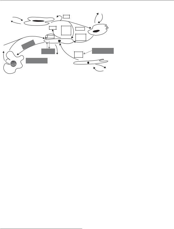
Figure 13.1 Components of early radiation effects. Early radiation effects usually start with vascular changes, clinically visible as erythema, accompanied by inflammatory changes. The depletion of functional cells, based on insufficient cellular supply in the face of ongoing differentiation and cell loss, is the dominating response to irradiation. The interaction of this phase with the vascular response is unclear. Progressive hypoplasia promotes secondary reactions (e.g. infections owing to oral mucositis, moist skin
desquamation or leukopenia). Eventually healing occurs, based on surviving stem cells within the irradiated volume or stem cells migrating in from outside the irradiated volume.
factor (G-CSF) or granulocyte/macrophage colonystimulating factor (GM-CSF) (see Chapter 22).
Phases of early radiation reactions
Different pathogenetic phases and components can be distinguished for early radiation responses in normal tissues (Sonis, 2004; Dörr et al., 2005b), as illustrated schematically in Fig. 13.1. Regularly a ‘humoral’ response is observed, based on the release of paracrine active substances, for example by vascular endothelial cells and macrophages, but also by fibroblasts or parenchymal cells such as epithelial keratinocytes. The associated changes in the functions of the target cells are accompanied by inflammatory changes. This phase usually precedes the clinically dominating reaction (i.e. the reduction in the number of functional cells). This hypoplasia is seen, for example, as epidermal or mucosal epitheliolysis or as leukopenia. Based on the breakdown of epithelial structures, which normally constitute a protective barrier function, secondary infections are frequently seen, which can even progress into septicaemia. Eventually, with the exception of very severe reactions, healing occurs, based on surviving stem cells within the irradiated volume or on migrating stem cells,
for example bone marrow stem cells from the circulation or epidermal/mucosal stem cells migrating from the margins into the irradiated area.
CHANGES IN CELLULAR FUNCTION
Early after the onset of radiotherapy, after the first or the first few fractions, increased protein expression is observed (e.g. in endothelial cells, vascular smooth muscle cells or macrophages). These proteins are predominantly pro-inflammatory, such as interleukin-1a (IL-1a) and other interleukins, tumour necrosis factor-α (TNF-α), or cyclooxy- genase-2 (COX-2). Similarly, the activity of inducible nitric oxide synthase (iNOS) is increased (e.g. Rubin et al., 1995; Sonis, 2004). These are only a few examples.
The paracrine, intercellular communication is modified by the induction of cytokines, their receptors, adhesion molecules and components of the cell–matrix interaction (Dörr et al., 2005b). For example, keratinocytes of the epidermis and oral mucosa show an increased expression of epidermal growth factor (EGF), its receptor (EGFR) or the intercellular adhesion molecule-1 (ICAM-1). The EGFR is subsequently internalized and translocated into the nucleus, and can act as a transcription factor and modulate DNA repair (Dittmann et al., 2005).
Initiation and regulation of these processes – because of their early onset – cannot exclusively be attributed to the release of mediators during the disintegration of damaged cells or to tissue hypoplasia. However, the signals underlying these very early changes in cellular function are unclear and are the focus of current research projects. Their relevance for the pathophysiology of the early radiation effects is similarly indistinct. An impact on the clinical symptoms, such as pain, is obvious, and modification of the tissue response (e.g. of the regeneration processes – repopulation, see Chapter 11) is likely. The available data, however, do not yet allow us to distinguish which of the intracellular, paracrine or humoral aspects are causally involved in the pathogenesis, and which are epiphenomena.
CELLULAR DEPLETION
The cellular depletion phase, like leukopenia after bone marrow irradiation or epitheliolysis in
172 Pathogenesis of normal-tissue side-effects
|
|
|
Transit |
|
|
|
|
cells |
|
|
|
|
|
|
Stem |
Transit |
|
n th generation |
|
cells |
cells first |
|
|
|
generation |
|
Transit |
|
|
|
|
|
||
|
|
|
cells |
|
|
|
|
|
|
|
|
|
n th generation |
|
|
|
|
|
|
Post-mitotic, functional cells
Cell loss
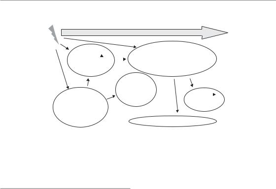
Figure 13.2 Proliferative organization of turnover tissues. Typical early reactions occur in turnover tissues. The entire cell production is based on tissue stem cells, which generate, on average, one stem cell and one transit cell in asymmetrical divisions (see also Fig. 13.3). Transit cells can undergo a limited number of divisions, which increase the cell yield per stem cell division. The cells undergo maturation and differentiation and are eventually lost. The turnover time from the initial stem cell division to cell loss is tissue specific.
epidermis and mucosa, is the clinically most relevant component of early radiation responses. As mentioned above, these changes are found in turnover tissues, with a precisely regulated equilibrium between permanent cell loss from the functional compartment and cell production in the germinal compartment. The hierarchical proliferative organization of these tissues is illustrated in Fig. 13.2.
The entire cell production takes place in the germinal components of the tissue (e.g. the basal and suprabasal layers of epithelia, intestinal crypts, or bone marrow sinuses). The basis are tissue stem cells. With very few exceptions, no cellular markers, such as surface antigens, have so far been identified that would allow for differentiation between stem cells and other proliferating cells. Therefore, the stem cell concept must be regarded as strictly functional: the stem cell population consists of cells that can completely and correctly restore the integrity and structure of a tissue after an insult. Hence, the radiation tolerance of a tissue is defined by the number and the intrinsic radiosensitivity of the stem cells.
The equilibrium between cell production and cell loss is based on the division pattern of the stem cells. On average, each stem cell division results in one cell that remains in the stem cell pool, and one cell which eventually differentiates
(Dörr, 1997). This pattern, with two different daughter cells, is called asymmetrical division.
Daughter cells which are not stem cells (transit or precursor cells) can undergo a limited number (e.g. up to 10 in bone marrow) of transit divisions, which substantially increases the yield of cells per stem cell division. The regulation of these processes remains unclear. The number of functional cells seems to feed back on the general proliferation activity. However, the number of stem cells itself seems to modulate stem cell proliferation, indicating an (additional) autoregulation within this compartment (see also Chapter 11).
In most turnover tissues, transit cells by far dominate the proliferative cell population; the relationship between the numbers of transit and stem cells depends on the number of transit divisions. Hence, studies into the proliferative activity, such as S-phase labelling with BrdUrd, Ki67labelling or mitotic counts, are dominated by transit-cell proliferation and cannot accurately assess the proliferation parameters in the stem-cell compartment. The post-mitotic cells arising from the last transit division usually undergo several steps of maturation before they reach a terminal differentiation state and are eventually lost. Their lifespan is tissue-specific, but can vary markedly between different tissues, from a few days in the epithelia of the upper and lower alimentary tract to several months in the urothelium of the urinary bladder. The overall turnover time (i.e. the time in which all cells are renewed once) defines the timecourse of the early radiation response (Fig. 13.3).
Early radiation effects 173
100% |
a |
b |
c |
d |
|
||||
number |
|
|
|
|
cell |
Time to healing, |
|
Effect 1 |
|
dose c |
|
|
||
Relative |
|
|
||
|
|
|
Effect 2 |
|
|
|
|
|
|
|
|
|
Time to healing, dose d |
|
0% |
LT1 LT2 |
TT |
|
|
|
|
|
||
Time after exposure
Figure 13.3 Radiation-induced cell depletion and clinical manifestation of early radiation effects. Radiation exposure of turnover tissues results in an impairment of cell production, while cell loss continues independent of the treatment. The rate at which cells are lost is determined by the tissue turnover time (TT). If the residual proliferation of sterilized cells (abortive divisions) is not taken into consideration, then a complete loss of cells would be observed after one turnover time. A defined clinical effect 1, which is associated with a specific reduction of the cell number, can occur dependent on the dose (dose level b, c or d), and is not observed at lower radiation doses (dose level a). The latent time to clinical manifestation, however, is independent of dose. A more severe effect level 2 is based on a higher reduction in cell numbers, and hence is observed only at higher doses (c and d). Compared with effect 1, the latent time is longer, but is also independent of dose. In contrast, the time to clinical healing is longer with higher doses (d versus c).
The radiosensitivity of the cells decreases during their differentiation process. Therefore, radiation doses administered in radiotherapy are predominantly lethal for stem cells, while transit cells are only minimally affected, and no effect is seen on post-mitotic cells. Some studies in experimental models have demonstrated with specific assays that the stem cell inactivation by radiation follows a dose–effect relationship, which corresponds in its shape to a typical cell survival curve in vitro (see Chapter 4). Such studies have been performed with microor macro-colony-forming assays in intestinal epithelium and skin (Withers and Elkind, 1970), or with spleen colony assays for surviving bone marrow stem cells (Potten and Hendry, 1983). However, despite qualitative similarities in cell
survival, there are significant quantitative differences between the in vivo and the in vitro situations.
The reduction of proliferation in the stem cell compartment results in a lack of support to the transit population, with the consequence of a decline in overall cell production. Furthermore, depending on the dose, direct effects on transit proliferation are also possible, which further affect the amplifying function of the transit compartment. Hence, increasing radiation doses result in a progressive decline in the number of precursor cells available for differentiation into post-mitotic cells. In contrast, despite the radiation exposure, differentiation and cell loss continue almost physiologically in qualitative and quantitative terms (Dörr, 1997). The radiation-induced imbalance between cell production and cell loss results in progressive hypoplasia, which becomes clinically manifest after a threshold cell depletion is reached (Fig. 13.3). Different grades of severity of an early reaction, such dry and moist desquamation in skin, are based on different degrees of cell depletion, as illustrated by different threshold levels in Fig. 13.3. As the cell loss rate depends on the turnover time of the tissue, and is independent of the treatment, the latent time until a clinical response is reached is tissue-dependent but independent of dose.
Usually, the turnover times are shorter than the latent time to complete cell depletion. For example, the turnover time in human and murine oral mucosa is in the range of 5 days (Dörr, 1997), but it takes about 10 days for ulceration to develop in mouse mucosa after single dose of irradiation (Dörr and Kummermehr, 1991) and about 9 days after a (fractionated) dose of 20 Gy in human mucosa (van der Schueren et al., 1990). This prolongation is due to the residual proliferative capacity (abortive divisions) of sterilized cells even after high doses (see also Chapter 11).
In epithelial tissues, the progressive cell depletion is associated with an exudative response, which results in the formation of a pseudomembrane consisting of fibrin and cellular remnants, which covers the ulcerative lesion. Healing of acute radiation effects is based on stem cells surviving within the irradiated volume or migrating in from outside. For the restoration of the stem cell population, symmetrical divisions, with the generation of two stem cell daughters, are
174 Pathogenesis of normal-tissue side-effects
required (see also Chapter 11). It is likely that this process is regulated via the local environment, which does not provide the signals that allow the daughter cells to differentiate. This is depicted as a differentiation block, which results in the recruitment of both daughter cells into the stem cell pool. The generation of transit cells through asymmetrical divisions recurs only when a sufficient number of stem cells have been produced.
The higher the dose, the fewer stem cells survive the treatment. Therefore, the clinically manifest response persists over a longer time with higher doses. This is illustrated by the EORTC (European Organization for Research and Treatment of Cancer) 22851 study in head and neck tumours (Horiot et al., 2006), where accelerated fractionation (and hence a biologically more effective treatment) resulted in clearly increased oral mucositis rates even at 6 weeks after the treatment (see Chapter 11). Also, complete restoration of cell numbers and tissue architecture takes longer with higher doses (Fig. 13.3), which should be considered if treatment breaks are introduced in clinical protocols for healing of early effects.
13.3 CHRONIC RADIATION EFFECTS
Tissue organization models have been developed that define late-responding tissues as flexible or F-type tissues. In these tissues, in contrast to earlyresponding hierarchical tissues (see Section 13.2), no clear separation can be made between proliferating and functional cells. Proliferating cells are recruited into the functional population on demand, and vice versa. It is assumed that the clinical manifestation of late radiation effects is based on a defined, critical depletion of functional cells (such as for early reactions). The compensatory proliferation of the surviving parenchymal cells, which were originally functional cells, results in mitotic death and hence accelerates cell loss and therefore shortens the time to the loss of organ function. The higher the initial cell depletion (i.e. the higher the dose) the more relevant this mechanism becomes. Hence, this model predicts a dose-dependent shortening of the latent time to the clinical manifestation of the effect, which is indeed a general clinical observation.
Induction |
Progression |
Manifestation |
|
Parenchyma, |
Parenchymal |
Loss of |
|
endothelium, |
|||
damage |
function |
||
fibroblasts |
|||
|
|
||
Macrophages |
|
|
Figure 13.4 Pathogenesis of late radiation effects. Chronic radiation effects are based on complex pathophysiological processes. These involve radiationinduced changes in organ-specific parenchymal cells (cell death), fibroblasts (differentiation) and vascular
endothelial cells (loss of capillaries). All these cells, as well as macrophages, interact through a variety of cytokines and growth factors. This orchestrated response results in progressive parenchymal damage and eventually in loss of function within the irradiated volume. The clinical consequences depend on the architecture of the organ and the volume irradiated (see Chapter 14).
An alternative model (Withers et al., 1988) assumes that, in late-responding tissues, structures with stem cell-like characteristics, so-called tissue rescuing units (TRU) or functional subunits (FSU) exist. Their radiation-induced inactivation results in the clinical radiation responses seen. For some tissues or organs, relatively independent structures can be defined, such as nephrons in the kidney, or bronchioli in the lung, which may represent TRU.
Undoubtedly, parenchymal cells in any organ are inactivated by radiation exposure. However, it is also accepted that, in addition to the parenchyma of the organs and the organ-specific cells, further tissue structures and cell populations are involved in the pathogenesis of late effects (Fig. 13.4). These are predominantly vascular endothelial cells, mainly in small blood vessels and capillaries, and connective tissue fibroblasts. Endothelial cell death, by apoptosis or as delayed mitotic death, is induced by radiation exposure. In contrast, mitotic fibroblasts are triggered into differentiation to post-mitotic fibrocytes, with the consequence of a drastically increased collagen synthesis and deposition (Rodemann and Bamberg, 1995). Moreover macrophages, irradiated or recruited into the tissue after irradiation, are also known to contribute to the pathogenesis of late radiation reactions. Reactive oxygen and nitrogen species,

Chronic radiation effects 175
chronically produced by various cell populations, in combination with chronic hypoxia, and a perpetual cascade of cytokines (Rubin et al., 1995; Dörr and Herrmann, 2003), seem to play an essential role in the pathogenesis of chronic radiation sequelae (Bentzen, 2006). The interactive response of the individual components of late radiation reactions results in progressive parenchymal damage and eventually in loss of function within the irradiated volume (Fig. 13.4). The clinical consequences depend on the architecture of the organ and the volume irradiated (see Chapter 14).
Each of the participating cellular components of a late effect responds to radiation exposure with a specific dose-dependence, which, in an orchestrated response, then defines the overall dose–response for the different clinical endpoints of the entire tissue. Hence, it is unlikely that the radiation sensitivity of one single cellular component can be used as a predictor of the sensitivity of the whole organ. For different organs, the relevance of the different pathogenetic components can differ (see Section 13.5). For example in the liver, the radiation response of the parenchymal cells (hepatocytes) is less important for the clinical symptoms (i.e. veno-occlusive disease; Dörr et al., 2005b). In the lung, type II pneumocytes (a slow turnover H-type tissue), endothelial cells and fibroblasts seem to contribute similarly to radia- tion-induced fibrosis (Dörr and Herrmann, 2003). In contrast, late fibrotic changes in the bladder are secondary to the functional impairment, which is predominantly based on urothelial and endothelial changes, and is not primarily radiation-induced (Dörr et al., 2005b).
Vasculature and endothelial cells
Irradiation causes changes in the function of the endothelial cells (Schultz-Hector, 1992; Dörr et al., 2005b). Endothelial cell vacuolization and foci of endothelial detachment are regularly seen. Also, transudation of serum components into the vessel wall and subendothelial oedema have been observed, and formation of thrombi and occlusion of capillaries have been reported (Fajardo et al.,
2001). Leukocyte adhesion and infiltration into the vessel wall is regularly observed. Based on all
these changes, irradiation eventually results in a progressive loss of capillaries, associated with a ‘sausage-like’ appearance of the arterioles, indicating substantial impairment of perfusion. The detailed interrelation of the individual changes described above with the eventual capillary loss remains unclear. Delayed mitotic death, based on the long turnover times of the endothelium, may contribute. The role of radiation-induced endothelial apoptosis, which occurs at early times after irradiation, is currently being discussed. As a result of the insufficient supply of oxygen and nutrients, atrophy of the downstream parenchyma develops. The morphological and functional consequences of this atrophy differ between the organs.
Telangiectasia (i.e. pathologically dilated capillaries) are observed in all irradiated tissues and organs. The pathogenesis is unclear but it is assumed that endothelial cell damage is involved. The loss of smooth muscle cells surrounding larger capillaries and veins may also contribute to the development of telangiectasia. In the intestine, the urinary system or the central nervous system (CNS), telangiectasia can be clinically relevant because of the tendency for capillary haemorrhage. In the skin, telangiectasia can be a cosmetic problem, but has also been used as a quantitative endpoint for radiobiological studies (see Section 13.5). In arterioles, progressive sclerosis of the tunica media is observed, which also results in impaired supply of the downstream parenchyma. The bases are presumably direct radiation effects on the cells in the media layer, in combination with endothelial changes.
Studies on the pathogenesis of late radiation effects have been performed in various experimental animal models, and predominantly with high single doses or few large fractions. Hence, no clear conclusions on the correlation of the individual changes described above and their time-course after fractionated radiotherapy can be drawn, although it can be assumed that similar changes occur.
Fibroblasts
In living organisms, a balance between mitotic fibroblasts and post-mitotic fibrocytes exists.
176 Pathogenesis of normal-tissue side-effects
|
|
High dose |
|
|
of response |
|
|
|
|
|
|
|
|
|
Grade |
|
Defined clinical effect |
|
|
|
|
|
|
|
|
|
Low |
|
|
|
|
dose |
|
|
|
|
|
|
|
|
|
FU1 |
FU2 |
|
(a) |
|
Time after irradiation |
|
|
100 |
|
FU2 |
FU1 |
|
|
|
Responders (%)
0 |
|
(b) |
Dose |
Figure 13.5 Time-course and dose dependence of late radiation sequelae. Late radiation effects are progressive in nature. The latent time for a specific clinical effect as well as the progression rate are dependent on dose (a). In consequence, an increasing number of individuals presenting with the effect (responders) is found in the individual dose groups with prolongation of the follow-up time, i.e. from follow-up 1 (FU1) to follow-up 2 (FU2). Therefore, a shift of the dose–effect curve to lower doses is found with increasing follow-up (b).
Irradiation results in stimulation of the differentiation of fibroblasts into fibrocytes, with the consequence of substantially increased collagen synthesis and deposition (Rodemann and Bamberg, 1995), which affects organ function. This process is modulated by the synthesis and release of transforming growth factor-β (TGF-β) from various cell populations, which further triggers fibroblast differentiation (Hakenjos et al., 2000). Increased expression of TGF-β at the mRNA and protein level can be observed over long time intervals in various cell populations (Bentzen, 2006).
Latent times
The latent time for chronic radiation effects, as well as the rate at which the severity of the clinical changes progresses, is inversely dependent on dose (Fig. 13.5). This dose-dependence results from several processes: with higher doses, more endothelial cells are damaged and the progression of the loss of capillaries is faster. Therefore, less time is required until tissue function is lost. Similarly, higher doses trigger more fibroblasts into differentiation, which results in a higher synthesis rate of collagen, and the collagen level
associated with loss of tissue function is reached earlier. Parenchymal radiation effects contribute to these processes in an organ-specific manner. As a consequence of the dose-dependence of latent time and progression rate, with increasing followup time responses are also seen at lower dose levels (Fig. 13.5). Hence, the isoeffective doses for a defined clinical response decrease with increasing follow-up time. In consequence, the definition of tolerance doses for late effects always requires information about the follow-up time on which the estimate is based.
13.4 DOCUMENTATION AND ASSESSMENT OF NORMAL-TISSUE EFFECTS
Two aspects must be considered for the documentation and quantification of normal tissue complications: the frequency of assessment and the scoring system used. Early reactions can undergo considerable changes in their clinical manifestation in short periods. For example, oral mucositis can change from a slight erythema response to confluent epithelial denudation over just a few days, particularly if additional damage is inflicted

Documentation and assessment of normal-tissue effects 177
Table 13.1 Systems for documentation of side-effects, with examples for oral mucositis |
|
|||
|
|
|
|
|
Grade |
General |
RTOG/EORTC |
CTCAE v3 |
WHO |
|
|
|
|
|
0 |
No change |
No change |
No change |
No change |
1 |
Mild |
Erythema, mild soreness, |
Erythema; normal diet |
Soreness, erythema |
|
|
painless erosions |
|
|
2 |
Moderate/clear |
Painful erythema, oedema |
Patchy ulceration; can eat |
Erythema, ulcers; can eat |
|
|
or ulcers; can eat |
and swallow modified diet |
solids |
3 |
Severe/significant |
Painful erythema, oedema |
Confluent ulcerations, |
Ulcers; requires liquid |
|
|
or ulcers; cannot eat |
bleeding with minor trauma; |
diet only |
|
|
|
unable to adequately aliment |
|
|
|
|
or hydrate orally |
|
4 |
Life-threatening |
Requires parenteral or |
Tissue necrosis; significant |
Alimentation not |
|
|
enteral support |
spontaneous bleeding |
possible |
5 |
Death owing to |
Death owing to |
Death owing to |
Death owing to |
|
side-effects |
side-effects |
side-effects |
side-effects |
RTOG/EORTC, Radiation Therapy and Oncology Group and the European Organisation for Research and Treatment of Cancer; CTCAE v3, the Common Terminology Criteria for Adverse Events, version 3; WHO, World Health Organization.
by spicy food, etc. In contrast, chronic radiation sequelae develop slowly and, more importantly, are usually irreversible. In conclusion, detailed assessment of early reactions requires scoring at least on a weekly basis during and for some weeks after radiotherapy. For studies on oral mucositis, even more frequent assessment is recommended. Late effects should be scored at intervals of several months after the end of radiotherapy, in order to assess the dynamics of their development, and may, at later time points, be documented at annual intervals. It must be emphasized that, for some chronic reactions, such as in the heart or the urinary bladder (see Section 13.5), the time to clinical manifestation of the reaction, particularly after low radiation doses (see Section 13.3), can be in the range of decades. Hence, life-long follow-up of patients is recommended.
Standardized classification systems have been established for documentation of normal-tissue reactions suitable for comparison between investigators, institutions and studies. In general, complications are graded from 0 (no response) to 5 (lethal), as shown in Table 13.1:
●Grade 1 reactions (mild) are reversible and heal spontaneously without any specific therapeutic intervention or interruption of the oncological treatment.
●Grade 2 reactions (moderate/clear) can be treated on an outpatient basis and do not require a radiation dose reduction or treatment interruption.
●Grade 3 effects (severe, pronounced) frequently require hospitalization and intense supportive care, and often necessitate interruption of the treatment and/or dose modifications.
●Grade 4 reactions are life-threatening, require immediate hospitalization and intense therapeutic interventions, plus cessation of radiotherapy.
The most widely used classification systems in radiation oncology (see the references in the Further reading section) are:
●RTOG/EORTC classification, jointly developed by the Radiation Therapy and Oncology Group and the European Organisation for Research and Treatment of Cancer;
●CTCAE v3, the Common Terminology Criteria for Adverse Events, version 3, developed by the National Cancer Institute (NCI);
●WHO (World Health Organization) classification;
●LENT/SOMA system (Late Effects in Normal Tissue/Subjective Objective Management Analytic) was developed specifically for scoring late sequelae resulting from oncological treatment.

178 Pathogenesis of normal-tissue side-effects
In principle, all these classification systems are comparable, and the scores from one system may be translated into the scores for another protocol (Table 13.1), but with exceptions. This translation is definitely precluded if sum scores are calculated, as has been suggested for LENT/SOMA, where the information on individual symptoms is lost; this method cannot, therefore, be recommended.
In addition to these scores, more detailed protocols, or systems specifically designed for side-effects in certain organs, for example OMAS (Oral Mucositis Assessment Scale), have been suggested. For clinical reports on side-effects, the scoring protocol applied must be described in detail, particularly if modified versions of the original protocols are applied.
13.5 RADIATION EFFECTS IN SPECIFIC TISSUES AND ORGANS
In this section the response and tolerance of some clinically important dose-limiting normal tissues will be summarized. In general, an overview of clinical symptoms and consequences for the various radiation sequelae can be found in the classification protocols (see Section 13.4). As a guideline for clinical treatment planning, Table 13.2 provides estimated tolerance dose levels for various endpoints, and corresponding α/βvalues. However, the numbers in this table should be used with considerable caution, as they are influenced by a number of factors, particularly the irradiated volume (see Chapter 14).
Skin
The sequence of events in skin during radiotherapy is illustrated in Fig. 13.6 (page 182). Skin erythema is closely related to radiation effects in vessels, with intermittent phases of vasodilation. In contrast, epidermal changes are based on the radi- ation-induced impairment of cell production, as described in Section 13.2. The clinical manifestation is dry desquamation (radiodermatitis sicca), followed by moist desquamation. According to the turnover time of human epidermis of 20–45 days, this phase is usually seen at 2–3 weeks after the onset of radiotherapy. The skin reaction displays a
significant area effect (see Chapter 14, Section 14.5). Any variation in overall treatment time can have a large influence on skin tolerance: as an approximation, skin tolerance doses decrease by about 3–4 Gy/week when treatment duration is shortened from the standard 6–8 weeks.
Chronic subcutaneous fibrosis, clinically manifest as induration, is based on an increase in collagen fibres and a reduction of fatty tissue (see Section 13.3). The development of skin telangiectasia (Fig. 13.7, page 182) illustrates the progression of vascular injury in the dermis. The corresponding latent time distribution and the cumulative incidence for various grades of the response are shown in Fig. 13.8 (page 182). With high-energy X-rays, in contrast to orthovoltage radiotherapy, the maximum dose is deposited below the surface and late damage may therefore occur without preceding early reactions.
Skin appendices
After a cumulative dose of 12 Gy, a loss in the function of sebaceous glands is observed, and at slightly higher doses the perspiratory glands also respond, both resulting in a typical dry skin. In hair follicles, single doses of 4 Gy or 10 Gy result in transient or permanent hair loss. In fractionated protocols, significantly higher doses up to 40 Gy still allow hair regrowth within 1 year, but regrowth is frequently associated with discoloration.
Oral mucosa and oesophagus
Oral mucositis is the most severe and frequently dose-limiting early side-effect of radio(chemo)therapy for head and neck tumours. Erythema, focal and confluent mucositis/ulceration are the lead symptoms (Table 13.1). Almost all patients with curative radiotherapy in this region develop some form of mucositis, with usually more than 50 per cent confluent reactions (depending of the definition of ‘confluency’). The latter typically develop during the third to fourth week of a conventionally fractionated protocol with 5 2 Gy/week. Oral mucosa is most sensitive to changes in dose intensity (i.e. weekly dose) and overall treatment time.
Accelerated protocols regularly result in earlier onset, an aggravation of the response, and/or an
Table 13.2 Tolerance doses and fractionation response (α/β ratio) for acute and late organ damage in humans |
|
||||
|
|
|
|
|
|
|
|
Time to manifestation |
α/β ratio |
Tolerance dose for |
|
Organ |
Endpoint |
during/after irradiation |
(Gy) |
total volume (Gy) |
Comments |
|
|
|
|
|
|
Cartilage, growing |
Growth arrest |
Next growth spurt |
6 |
20 |
|
Cartilage, adult |
Necrosis |
Months–years |
|
70 |
Associated with vascular |
|
|
|
|
|
damage |
Bone, adult |
Osteoradionecrosis |
Years–decades |
60 |
Mandible: 40–50 |
Vascular damage and trauma |
Connective tissue |
Fibrosis |
9 months–years |
2 |
60 |
Most frequent late reaction |
Capillaries |
Capillary changes/loss |
6 months–years |
3 |
60 |
Contribute to a variety of |
|
|
|
|
|
(late) radiation effects |
Large vessels |
Wall changes, stenosis |
Years |
|
70 |
Resembles atherosclerotic |
|
|
|
|
|
changes |
Heart |
ECG-changes, arrhythmia |
During RT |
|
20 |
Reversible |
|
Cardiomyopathy (pericarditis) |
Months–years |
3 |
40 |
Late myocardial infarction |
Skin |
Erythema |
During RT |
9–10 |
40 (100 cm2) |
|
|
Dry radiodermatitis |
During RT |
10 |
Varies with localization |
|
|
|
|
|
|
(additional |
|
|
|
|
60 (100 cm2) |
mechanical/chemical stress) |
|
Moist radiodermatitis |
During RT |
10 |
|
|
|
Gangrene, ulcer |
|
3 |
55 (100 cm2) |
Vasculature! |
Hair follicles |
Hair loss |
During RT (4th week) |
7 |
40 |
Discoloration! |
Sebaceous glands |
Dry skin |
During RT (2nd week) |
|
12 |
Transient loss of function |
Perspiratory glands |
Dry skin, loss of transpiration |
During RT (4th week) |
|
30–40 |
Long-lasting or permanent |
|
|
|
|
|
loss of function |
Oral mucosa |
Ulcerative mucositis |
During RT |
10 |
20 |
|
|
|
(2nd to 3rd weeks) |
|
|
|
|
Atrophy/fibrosis |
|
|
60–70 |
|
Salivary glands |
Transient loss of |
During RT (2nd week) |
|
10–20 |
|
|
function – xerostomia |
|
|
|
|
|
Permanent loss of |
Continuous development |
3 |
25 |
One-third capacity is |
|
function – xerostomia |
from the early response |
|
|
sufficient for saliva |
|
|
|
|
|
production |
Oesophagus |
Dysphagia |
|
|
40–45 |
Early mucositis |
|
Ulcer–fistula |
During RT–months |
|
55 |
|
Stomach |
Atony |
During RT |
|
20 |
‘Radiation sickness’ |
|
Ulcer |
Months |
4 |
50 |
|
Small intestine |
Malabsorption |
During RT |
8 |
30 |
Reduced tolerance due to |
|
|
|
|
|
fixation of intestinal |
|
|
|
|
|
loops, e.g. postoperative |
|
Ulcer/obstruction |
Months |
4 |
40 |
|
(Continued )
Table 13.2 (Continued) |
|
|
|
|
|
|
|
|
|
|
|
|
|
Time to manifestation |
α/β ratio |
Tolerance dose for |
|
Organ |
Endpoint |
during/after irradiation |
(Gy) |
total volume (Gy) |
Comments |
|
|
|
|
|
|
Large intestine |
Diarrhoea, pain |
During–post RT |
|
10–20 |
|
|
Ulcer/obstruction |
Months–years |
|
45 |
Ileus symptoms possible |
Rectum |
Proctitis |
During RT |
|
50 |
|
|
Chronic inflammation, |
Months–years |
5 |
60 |
Partial irradiation of the |
|
ulcer |
|
|
|
circumference increases |
|
|
|
|
|
tolerance |
Liver |
Veno-occlusive disease |
2–3 weeks |
|
30 |
Lethal after total organ |
|
(VOD) |
|
|
|
irradiation. Hence late |
|
|
|
|
|
effects only after partial |
|
|
|
|
|
organ irradiation |
|
Fibrosis |
Months–years |
1 |
|
|
Biliary tract |
Stenosis/stricture |
Months–years |
|
|
|
Pancreas |
Fibrosis |
Months–years |
|
50–60 |
No early symptoms known, |
|
|
|
|
|
included in ‘radiation |
|
|
|
|
|
sickness’? |
Kidney |
Nephropathy |
9 months–years |
2 |
20 |
|
Ureter |
Stricture |
2 years |
|
60–70 |
Vascular effects, potential |
|
|
|
|
|
interaction with surgery |
Urinary bladder |
Cystitis |
During RT |
10 |
20–35 |
Uncommon pathophysiology, |
|
|
|
|
|
no urothelial depletion |
|
Shrinkage, ulceration |
Years–decades |
5–10 |
50 |
Strong consecutive component |
Urethra |
Stricture |
Months–years |
|
60-70 |
Reduced tolerance after |
|
|
|
|
|
transurethral resection of the |
|
|
|
|
|
prostate (TURP) |
Larynx |
Oedema |
During RT |
|
45 |
|
|
Chronic oedema, necrosis |
Months |
2–4 |
70 |
Permanent changes in voice |
|
|
|
|
|
quality, necrosis after decades |
Lung |
Pneumonitis |
2–6 weeks |
5 |
12–14 |
Single-dose irradiation |
|
Pneumonitis |
4–6 weeks |
5 |
45 |
|
|
Fibrosis |
6 months–2 years |
4 |
|
|
Testis |
Permanent sterility |
Weeks–months |
|
1.5 |
Negative fractionation effect |
Ovary |
Permanent sterility |
Weeks–months |
|
2.5 |
Strong inverse age |
|
|
|
|
|
dependence |
Uterus |
Atrophy |
Months–years |
|
100 |
|
Vagina |
Mucositis |
During RT |
|
30 |
|
|
Ulcer, fibrosis |
Months–years |
|
50 |
|
Breast, child |
Growth arrest |
At puberty |
|
10 |
|

Breast, adult |
Fibrosis/atrophy |
Years |
2–3 |
60 |
|
Adrenal glands |
Loss of function |
Months–years |
|
90 |
|
Pituitary gland/ |
Growth hormone |
Months–years |
|
18–24 |
Growth retardation |
diencephalon (children) |
deficit |
|
|
|
|
Cerebrum, child |
Somnolence syndrome |
During–post RT |
|
24 |
Specific response in children |
Cerebrum, adult |
Necrosis |
Months–years |
|
55 |
|
Spinal cord |
Lhermitte syndrome |
Weeks–months |
|
35 |
Reversible |
Cervical/thoracic |
Radiation myelopathy |
6 months–2 years |
2 |
55 |
|
Thoracic/lumbar |
Radiation myelopathy |
6 months–2 years |
2 |
55 |
|
Peripheral nerves |
Functional impairment |
Months–years |
|
60 |
Frequently associated with |
Eye lens1 |
|
|
1–21 |
51 |
connective tissue fibrosis |
Cataract |
Months–years |
Surgical management |
|||
Lachrymal system |
Dry eye, ulceration |
Weeks–months |
3 |
40 |
Most critical radiation effect |
|
|
|
|
|
in the eye |
Retina |
Retinopathy |
Weeks–months |
|
45 |
|
Optic nerve |
Neuropathy |
Months–years |
2 |
55 |
|
Chiasma opticum |
Loss of vision |
Months–years |
2 |
55 |
|
Conjunctiva |
Kerato-conjunctivitis |
During–post RT |
|
50 |
Reversible |
Ear |
Serous otitis |
During–post RT |
|
30 |
|
|
Inner ear injury |
During RT |
|
30 |
Slight hearing loss (15 dB) |
|
|
plus; months |
|
|
frequently not recognized by |
|
|
|
|
|
patients; overlap with age |
|
|
|
|
|
effects |
Taste |
Taste impairment, |
During RT |
|
30 |
Reversible |
|
loss |
plus; months |
|
|
|
Lymph nodes |
Permanent atrophy |
Months–years |
|
70 |
|
Lymphatic vessels |
Sclerosis |
Months–years |
|
90 |
Frequently associated with |
|
|
|
|
|
connective tissue fibrosis |
Bone marrow |
Transient hypoplasia |
During RT |
10 |
2 |
Total body irradiation |
|
Lethal aplasia (1 year) |
|
5 |
4 |
Total body irradiation |
|
Permanent aplasia |
During–post RT |
|
|
Compensation by unirradiated |
parts; post-irradiation homing of circulating stem cells possible
1Tolerance doses for the eye lens are currently under discussion and may be significantly lower (1/10th) than usually estimated. Time to manifestation: relative to irradiation with 5 2 Gy/week. Times after the treatment relate to the last fraction.
α/β value: see also Chapters 8 and 9. Missing values indicate that no valid estimates are possible. RT, radiotherapy.
Tolerance dose total organ relates to irradiation of large volumes that include the entire organ or, for ubiquitous tissues (connective tissue, capillaries, etc.), to larger volumes. For partial organ irradiation, see Chapter 14. Early reaction/late reaction, see Section 13.1.
Adapted from Herrmann et al. (2006), with permission.
182 Pathogenesis of normal-tissue side-effects
|
Dry skin, epilation |
|
|
|
|
|
|
|
Subcutaneous fibrosis |
||||||
|
|
|
|
|
|
|
|
||||||||
|
|
|
|
|
|
|
|
|
|
|
|
||||
|
|
Erythema |
|
|
|
|
|
|
|
|
Telangiectasia |
|
|||
|
|
|
|
|
|
|
|
|
|
|
|||||
|
|
Dry desquamation |
|
|
|
|
|
Atrophy, dyskeratosis |
|||||||
|
|
|
Hyperpigmentation |
|
|
|
|
||||||||
|
|
|
|
|
|
|
|||||||||
|
|
|
|
|
|
|
|
|
|
|
|||||
|
|
|
|
Moist desquamation |
|
|
|
|
|
|
|
||||
|
|
|
|
|
|
|
Re-epithelialization |
|
|
|
|
|
|||
|
|
|
|
|
|
|
|
|
|
Hair regrowth |
|
|
|
||
|
|
|
|
|
|
|
|
|
|
|
|
|
|||
|
|
|
|
|
|
|
|
|
|
|
|
|
|
||
Radiotherapy, 5 |
|
|
|
|
|
|
|
|
|
|
|
|
|
||
|
|
|
|
|
|
|
|
|
|
|
|
|
|
|
|
1 |
2 |
3 |
4 |
5 |
6 |
7 |
8 |
9 |
10 |
1 |
3 |
5 |
7 |
9 |
|
|
|
|
Time (weeks) |
|
|
|
|
|
Time (years) |
|
|||||
Figure 13.6 Sequence of radiation effects in skin and appendices. Shown are the time-courses of early and late skin reactions induced by conventional radiotherapy with 5 2 Gy per week over 6 weeks, if the same skin area is exposed to the maximum dose of 2 Gy at each dose fraction. The duration of radiotherapy is indicated on top of the abscissa.
|
|
5 |
|
|
N |
|
|
|
5 |
|
|
|
|
|
|
score |
4 |
|
|
|
|
|
|
3 |
|
|
|
|
|
|
|
Telangiectasia |
|
|
|
|
|
|
|
2 |
|
|
|
|
|
|
|
1 |
|
|
|
|
|
|
|
|
|
|
|
|
|
|
|
|
0 |
|
|
|
|
|
|
|
0 |
20 |
40 |
60 |
80 |
100 |
120 |
|
|
|
Follow-up (months) |
|
|
||
Figure 13.7 Clinical manifestation of skin telangiectasia. Progression of skin telangiectasia in individual patients treated with five fractions of 1.8 Gy per week to a total of 35 fractions. Note the pronounced differences between patients and the continuous increase in severity even up to 8 years. From Turesson (1990), with permission.
increase in the frequency of patients with severe reactions (Fig. 13.9, page 183). Repopulation processes have been most intensely studied in this tissue (see Chapter 11).
Chronic effects of radiotherapy include mucosal atrophy and ulceration, and telangiectasia, which render the epithelium vulnerable. Any additional trauma may secondarily result in osteonecrosis. The early radiation response of the oesophagus
(/year) |
0.15 |
|
|
|
|
|
|
|
|
|
|
|
|
|
|
|
|
|
|
|
|
||
|
|
|
|
|
|
|
|
|
|
|
|
function |
0.10 |
|
|
|
|
|
|
|
|
|
|
density |
0.05 |
|
|
|
|
|
|
|
|
|
|
Prob. |
|
|
|
|
|
|
|
|
|
|
|
0 |
|
|
|
|
|
|
|
|
|
|
|
|
|
|
|
|
|
|
|
|
|
||
|
|
|
|
|
|
|
|
|
|
||
|
5 |
10 |
15 |
||||||||
|
0 |
||||||||||
(a)Observation time (years)
(%) |
80 |
|
|
|
telangiectasia |
|
|
|
|
60 |
|
Grade |
|
|
|
|
|
||
|
|
|
|
|
|
40 |
|
Grade |
|
of |
|
|
|
|
0 |
|
|
|
|
Incidence |
|
|
|
|
|
20 |
|
|
|
|
|
|
Grade 3 |
|
|
0 |
5 |
10 |
15 |
(b)Observation time (years)
Figure 13.8 Time-course of telangiectasia. (a) The latent time distribution for any grade of telangiectasia as observed in 174 treatment fields with an intermediate probability of developing any response. The probability density function may be interpreted as the fraction of patients who developed the response within a specific year after irradiation. (b) The cumulative incidence of telangiectasia as a function of time for various grades, after radiotherapy with 44.4 Gy in 25 fractions. The model calculations are based on observations in 401 treatment fields. From Bentzen
et al. (1990), with permission.
Radiation effects in specific tissues and organs 183
|
100 |
|
|
|
|
|
|
(%) |
80 |
|
|
|
|
|
|
|
|
|
|
|
|
|
|
mucositis |
60 |
|
|
|
|
|
|
|
|
|
|
|
|
|
|
Confluent |
40 |
|
|
|
|
|
|
20 |
|
|
|
|
|
|
|
|
|
|
|
|
|
|
|
|
0 |
2 |
4 |
6 |
8 |
10 |
12 |
|
0 |
Week number
Figure 13.9 Prevalence of confluent mucositis. The percentage of patients presenting with confluent mucositis within a given treatment week after onset of radiotherapy was plotted for accelerated radiotherapy: CHART (continuous hyperfractionated accelerated radiotherapy; filled circles) or conventional radiotherapy (open circles). From Bentzen et al. (2001), with permission.
mirrors that of the oral mucosa and in the chronic phase, strictures may develop.
Teeth
Radiation caries, with a very fast manifestation, is a frequent complication of radiotherapy in adults. The response is based on both direct radiation effects at the dentine–enamel border zone, and indirectly on radiation effects in the salivary glands (xerostomia, see below) and the associated changes in the oral micromilieu. Rigorous pretreatment dental restoration or extractions are of major importance, because of the risk of osteoradionecrosis, if extractions are required after radiotherapy. In order to avoid dose peaks in the mucosa around metallic dental implants, mucosal retractors should be used to displace the mucosa by about 3 mm.
Salivary glands
Salivary glands are sensitive to radiation exposure: after the first week of therapy (accumulated dose of 10–15 Gy) saliva production is significantly reduced, frequently after a transient phase of hypersalivation. After total doses in excess of
40 Gy to both parotid glands, saliva production practically stops after about 4 weeks and does not recover at all after doses over 60 Gy. Volume effects are very pronounced, and sparing of partial volumes usually leads to recovery of function.
Chronic xerostomia has a major impact on the quality of life of the patients. It depends not only on reduced serous fluid production in the parotid glands, but also on the reduced amount of mucin from the submandibular glands, and reduced function of the small salivary glands. The submandibular glands produce most of the mucinous components of the saliva; by their water-binding capacity they keep the mucous membranes hydrated and have a barrier function.
Stomach
Functional impairment, with a prolongation of the time for gastric emptying, and a reduction in HCl secretion are frequently seen. The symptoms are equivalent to those of gastritis. Ulceration, mainly based on vascular effects, can develop as a late effect at doses of 25–40 Gy.
Intestine
‘Fixed’ intestinal loops (e.g. through postoperative adhesions) are particularly at risk as these may be permanently located within a high-dose volume, in contrast to mobile loops. The same is true for the rectum.
The sequence of radiation-induced events in the intestine includes:
●initial increase in motility, followed by an atonic phase
●loss of epithelium and villi owing to proliferative impairment in the crypts, with the consequence of:
–water electrolyte and protein loss into the lumen, resulting in diarrhoea
–changed resorption (including increased resorption of some substances, which must be considered if drugs are administered orally)
●risk of sepsis.
Late effects include chronic ulcers, based on an orchestrated response of all intestinal wall

184 Pathogenesis of normal-tissue side-effects
components, plus mechanical/chemical stress from faeces, as well as infections. Fibrotic remodelling may result in stenosis and ileus. Frequently, telangiectasia are found, which may result in bleeding.
It has been demonstrated experimentally by inhibition of pancreatic secretion by somatostatin analogues that pancreatic enzymes contribute to the manifestation of the early effect. Interestingly, this treatment also reduces late fibrosis, underlining the consequential nature of chronic changes in the intestine.
Liver
The liver is radiosensitive, with a tolerance dose of around 30 Gy in 2-Gy fractions. However, liver tolerance is only dose-limiting when the whole organ is irradiated (see Chapter 14, Section 14.5). An example is total-body irradiation preceding bone marrow transplantation. In this situation, the lung is well known as a dose-limiting organ, but liver and kidney are also at risk, especially after regimes equivalent to single doses of 10 Gy or higher.
Two phases of radiation hepatopathy are recognized, with acute radiation hepatitis being the more dominant. This acute phase develops approximately 2–6 weeks after irradiation, with signs of liver enlargement and ascites. Liver function tests during this period are abnormal. Acute hepatitis usually presents as veno-occlusive disease (VOD), characterized by central vein thrombosis whereby occlusion of the centrilobular veins causes atrophy and loss of the surrounding hepatocytes. Total liver VOD is usually lethal.
Chronic hepatopathy, which obviously can develop only after partial organ irradiation, has a variable latency ranging from 6 months to more than a year post-irradiation, and shows progressive fibrotic changes in both centrilobular and periportal areas. These alterations are accompanied by blood-flow redistribution through recanalization or newly formed veins, and regenerative proliferation of hepatocytes and bile ducts.
Upper respiratory tract
The mucosa of nose, paranasal sinuses and trachea respond to irradiation similarly to oral epithelium, but appear to be slightly more radioresistant.
Early changes in the larynx are oedema and perichondritis. Doses above 50 Gy may result in a long-lasting impairment of the quality of the voice, which must be considered in patients depending on their voice in their professional life.
Lung
In the lung, two separate radiation syndromes can be distinguished clinically: early pneumonitis, usually observed at 4–6 weeks after the end of radiotherapy, and fibrosis, which develops slowly over a period of several months to years. The lung is among the most sensitive of late-responding organs, but with a pronounced volume effect (see Chapter 14). In addition to a reduction in irradiated volume, reduced doses per fraction are most effective in avoiding severe (clinically manifest) lung reactions.
Clinical signs or symptoms of radiation pneumonitis are reduced pulmonary compliance, progressive dyspnoea, decreased gas exchange and dry cough. When there is insufficient functional reserve, cardiorespiratory failure may occur within a short time. The development of chronic radiation pneumopathy (i.e. lung fibrosis) follows the general pathways described in Section 13.3. The complexity of the signalling cascades is illustrated in Fig. 13.10. Local fibrotic responses must be expected in all patients with early reactions, indicating a strong consequential component of the late reaction (Dörr et al., 2005a). Higher age and tamoxifen treatment significantly increase the incidence of early pneumopathy.
Kidney
The kidney is among the most sensitive of the late-responding critical organs. Radiation damage develops very slowly and may take years to be recognized. Radiation nephropathy usually manifests as proteinuria, hypertension and impairment in urine concentration. Anaemia is usually present, and has been attributed both to haemolysis and to a decreased production of erythropoietin. A mild form of nephritis, presenting only as a sustained proteinuria, may be observed over a period of many years. Parts of one or both kidneys can
Radiation effects in specific tissues and organs 185
|
|
|
|
|
|
|
|
|
|
Cell death |
Cell death |
Cytokines |
|
|
|
Macrophage |
TNF |
|
Cytokines |
||
|
|
|
|
|
|
|
|
|
||
|
|
|
|
|
|
|
|
|
|
|
|
|
|
|
|
|
|
IGF-2 |
TGF- |
MDGF |
|
|
|
|
|
|
|
|
|
PDGF |
|
|
|
|
|
|
|
|
|
|
|
|
|
|
|
cytokines |
|
|
|
TGF- |
TGF-α |
|
||
|
ous |
|
|
IGF-1 |
|
Alveolar |
||||
|
|
|
|
|
|
|
||||
|
Vari |
|
|
|
|
|
|
|
TGF-β |
type II cell |
|
|
|
|
|
|
|
|
|
||
|
|
|
|
1 |
|
|
|
|
|
|
|
|
|
|
|
|
|
|
|
|
|
|
|
F |
|
|
nal |
|
Progenitor |
|
||
Cell |
TG |
|
|
|
|
|
||||
|
|
m |
|
fibroblast |
|
|||||
death |
|
|
|
ter |
on |
|
Early events |
|||
|
|
ed |
|
TGF |
|
|
||||
|
|
ati |
|
|
||||||
|
Induc |
|
|
|
|
IL-1 |
||||
|
|
|
|
|
|
|
||||
|
|
fferenti |
|
|
|
|
||||
|
|
di |
|
|
|
|
Cell death |
PDGF |
|
|
|
|
|
|
|
|
|
|
|||
|
|
Later events |
|
|
|
|||||
|
|
Terminally differentiated fibrocyte |
|
Endothelial cell |
|
|||||
|
|
|
|
Cell death |
||||||
|
(PMFVI): enhanced collagen synthesis |
|
Cytokines |
|||||||
Figure 13.10 Possible cellular interactions and events after irradiation of lung tissue. IGF, insulin-like growth factor; IL-1, interleukin 1; MGDF, megakaryocyte growth and development factor; PDGF, platelet-derived growth factor; TNF, tumour necrosis factor. Modified from Rodemann and Bamberg (1995), with permission.
receive much higher doses without affecting excretory function. However, after partial kidney irradiation hypertension may develop after a latent period of up to or beyond 10 years.
The fractionation sensitivity of the kidney is high (i.e. the α/β ratio is low). The dose tolerated by the kidney does not increase with increasing time after radiotherapy, but declines because of a continuous progression of damage, after doses well below the threshold for induction of functional deficit, which usually precludes re-irradia- tion (see Chapter 19, Section 19.3).
The pathogenesis of radiation nephropathy is complex. Most studies suggest glomerular endothelial injury as the start of a cascade leading to glomerular sclerosis and later tubulo-interstitial fibrosis. Several experimental studies have shown the importance of the renin–angiotensin system in the induction of glomerular sclerosis via upregulation of plasminogen activator inhibitor 1 (PAI- 1) and enhanced fibrin deposition. Owing to loss of tubular epithelial cells, fibrin may then leak into the interstitium causing the onset of tubulointerstitial fibrosis.
Urinary bladder
In patients, two phases of radiation-induced changes in the urinary bladder are observed, with both a reduction in bladder storage capacity and a consequential increase in micturition frequency. An early phase occurs 2–6 weeks after the start of
fractionated irradiation, which is characterized morphologically by hyperaemia and mucosal oedema. Experimentally, in mice, two waves of early injury have been observed. Mechanistically, the first phase seems to be related to direct radia- tion-induced changes of the prostaglandin metabolism (which regulates the tone of the bladder wall), as suggested by the beneficial effect of aspirin when administered during this phase. The second early phase is associated with changes in urothelial barrier function, but without epithelial cell depletion (which is not expected at this time because of the very long turnover time of the urothelium). Infection may complicate this early response, which then may progress to desquamation and ulceration.
A chronic phase develops with latent times that are inversely dose-dependent and can range up to 10 years or longer. The morphological correlate in the initial late phase is a progressive mucosal breakdown, ranging from superficial denudation to ulceration and even the formation of fistulae. The urothelial changes are accompanied by urothelial areas of compensatory hyperproliferation. Vascular changes and signs of local ischaemia have been described. These processes progress into secondary fibrosis of the bladder wall. Telangiectasia can result in severe bleeding episodes.
The early changes clearly correlate with the chronic radiation sequelae, illustrating a strong consequential component. A schematic illustration of the sequence of events leading to late fibrosis, as concluded from animal studies, is given in Fig. 13.11.
186 Pathogenesis of normal-tissue side-effects
Acute phase |
|
Late phase |
|
|
|
|||
Inflammation: |
|
Vascular damage: |
||||||
|
||||||||
|
|
|
|
|
||||
ICAM-1, |
|
|
|
Late vasodilatation |
Albumin |
|||
|
|
|||||||
COX-2, |
|
|
irregular shape |
leakage |
||||
vasodilatation |
|
ICAM-1 |
|
|
|
|||
|
|
|
|
|
|
|
|
|
|
|
|
|
|
Urothelial |
Hypoxia? |
||
|
|
|
|
restoration: |
||||
|
|
|
|
|
|
|
||
Damaged |
Ki67, EGFR |
Ki67 |
|
|
||||
|
occludin |
|
|
|||||
urothelial barrier: |
|
urothelial |
||||||
|
|
|||||||
UP-III, |
|
Hypoxia? |
hyperplasia |
|||||
urothelial cell |
|
Chronic stress? |
|
|
|
|||
depletion |
|
Fibrosis |
|
|
|
|||
|
|
|
|
|
|
|
|
|
Figure 13.11 Pathogenesis of radiation effects in the urinary bladder. The individual processes have been studied in mouse urinary bladder after single dose irradiation. Morphological changes were related to functional impairment as assessed by transurethral cystometry. COX-2, cyclooxygenase-2; EFGR, epidermal growth factor receptor; ICAM-1, intercellular adhesion molecule-1; UP-III, uroplakin III. Modified with permission from Jall (2006).
Nervous system
The nervous system is less sensitive to radiation injury than some other late-responding tissues such as the lung or kidney. However, damage to this organ results in severe consequences such as paralysis: although tolerance doses are often quoted at the 5 per cent complication level (TD5) they generally are chosen to include a wide margin of safety.
A schematic outline of the development of various delayed lesions in the CNS as studied in animals is given in Fig. 13.12.
BRAIN
The most important radiation syndromes in the CNS develop a few months to several years after therapy. The often-used separation into early or late delayed injury is not very useful, as different types of lesions with overlapping time-distribu- tions occur. Some reactions occurring within the first 6 months comprise transient demyelination (‘somnolence syndrome’) or the much more severe leukoencephalopathy. The more typical radiation necrosis may also occur by 6 months, but even after as long as 2–3 years. Histopathologically, changes that occur within the first year are mostly
restricted to the white matter. For times beyond 6–12 months, the grey matter usually also shows changes along with more pronounced vascular lesions (telangiectasia and focal haemorrhages). Radionecrosis of the brain with latent times between 1 and 2 years usually shows a mixture of histological characteristics.
The brain of children is more sensitive than in adults. Functional deficits, such as a reduction in IQ, can at least partly be attributed to radiotherapy.
SPINAL CORD
Radiation-induced changes in the spinal cord are similar to those in the brain in terms of latent period, histology and tolerance dose. Among the relatively early syndromes, the Lhermitte sign is a frequently occurring, usually reversible type of demyelinating reaction, which develops several months after completion of treatment and lasts for a few months to more than a year. It may occur at doses as low as 35 Gy in 2-Gy fractions, well below tolerance for permanent radiation myelopathy, when long segments of cord are irradiated, and does not predict for later development of permanent myelopathy.
As in the brain, the later types of myelopathy include two main syndromes. The first, occurring
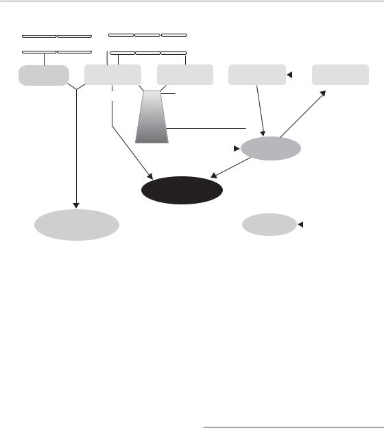
Radiation effects in specific tissues and organs 187
|
|
|
Vasculature |
|
|
|
|
|
|
|
|
Myelinated nerve fibers |
|
||||||||||||||||||||
|
|
|
|
|
|
|
|
|
|
|
|
|
|
|
|
|
|
|
|
|
|
|
|
|
|
|
|
|
|
|
|
|
|
Artery/vein |
|
|
|
|
|
Capillary |
|
|
|
|
|
|
|
|
|
|
|
|
|
|
|
|
|
|
|
|
|
||||||
|
|
|
|
|
|
|
|
|
|
|
|
|
|
|
|
|
|
|
|
|
|
|
|
|
|
||||||||
|
|
|
|
|
|
|
|
|
|
|
|
|
|
|
|
|
|
|
|
|
|
|
|
|
|
|
|
|
|||||
Smooth |
Vascular |
|
Astrocyte |
Oligodendrocyte |
|
|
|
O-2A progenitor |
|
||||||||||||||||||||||||
muscle cell |
endothelium |
|
|
|
|
|
|||||||||||||||||||||||||||
|
|
|
|
|
|
|
|
|
|
|
|
|
|
|
|
|
|
|
|
|
|||||||||||||
|
|
|
White |
cell adhesion |
|
|
Swelling/ |
|
|
|
|
|
|
|
|
|
|
|
|
|
|
|
|
||||||||||
|
|
|
|
|
|
|
|
|
|
|
|
|
|
|
|
|
|
|
|
|
|||||||||||||
|
|
|
|
|
hyperplasia |
|
|
|
Early (24h) |
|
|
||||||||||||||||||||||
|
|
|
|
|
|
|
|
|
|
|
|
|
|
|
|
|
|
|
|
||||||||||||||
|
|
|
|
Increased |
|
|
|
|
|
|
|
|
|
|
|
|
|
|
|
|
|
|
|
|
|
|
|
|
|
|
|||
|
|
|
|
|
|
|
|
|
|
|
|
|
|
|
|
|
Apoptosis |
|
|
Trigger |
|
|
|
||||||||||
|
|
|
|
permeability |
|
|
|
|
|
|
|
|
|
|
|
|
|
|
|
|
|
|
|||||||||||
|
|
|
|
|
|
Cytokine |
|
|
|
|
|
|
|
|
|
|
|
proliferation |
|
|
|
||||||||||||
|
|
|
|
|
|
|
|
|
|
|
|
|
|
|
|
|
|
|
|
|
|
|
|
||||||||||
|
|
|
|
|
|
|
|
|
|
|
(TNF-mediated?) |
|
|
late |
|
|
|||||||||||||||||
|
|
|
|
|
|
|
|
|
|
cascades |
|
|
|
|
|
|
|
|
|
|
|
|
|||||||||||
|
|
|
|
|
|
|
|
|
|
|
|
|
|
|
|
|
|
|
|
|
|||||||||||||
|
|
|
|
|
Perivascular |
|
|
|
|
|
|
|
|
|
|
|
|
|
|
|
|
|
|
|
|
|
|||||||
|
|
|
|
|
|
|
|
|
|
|
|
|
|
|
|
|
|
|
|
|
|
|
|
|
|
||||||||
|
|
|
|
|
|
oedema |
|
|
|
|
|
|
|
|
|
|
|
|
Demyelination |
|
|
||||||||||||
|
|
|
|
|
|
|
|
|
|
|
|
|
|
|
|||||||||||||||||||
|
|
|
|
|
|
|
|
|
|
|
|
|
|
|
|
|
|
|
|
|
|
|
|||||||||||
|
|
|
|
|
|
|
|
|
|
|
|
|
|
|
|
|
|
|
|
|
|
||||||||||||
|
|
|
|
|
|
|
Ischaemia/ |
|
|
|
|
|
|
|
|
|
|
|
|
|
|
|
|
|
|
|
|
|
|
|
|||
|
|
|
|
|
|
|
|
reperfusion |
|
|
|
|
|
|
|
|
|
|
1–3 months |
|
|
||||||||||||
|
|
|
|
|
|
|
|
|
|
|
|
|
|
|
|
|
|||||||||||||||||
Telangiectasia, |
|
|
|
|
|
|
|
|
|
|
|
|
|
|
|
|
|
|
|
|
|||||||||||||
|
|
|
|
|
|
|
|
|
|
|
|
|
|
|
|
|
|
|
|
|
|
|
|
|
|
|
Permanent |
|
|||||
haemorrhages |
|
|
|
|
|
|
|
|
|
|
White matter |
|
|
|
|
|
|
|
|
|
|
|
|
|
depletion |
|
|||||||
|
|
|
|
|
|
|
|
|
|
|
|
|
|
|
|
|
|
|
|
|
|
|
|
|
|
|
|
|
|||||
|
|
|
|
|
|
|
|
|
|
|
|
|
|
necrosis |
|
|
|
|
|
|
|
|
|
|
|
|
|
|
|
|
|||
|
|
Late |
6–12 months |
|
|
|
Glial |
|
|
|
|
|
|
|
|
||||||||||||||||||
|
|
|
|
|
|
|
|
|
|
|
|
|
|
|
|
|
|
||||||||||||||||
vasculopathies |
|
|
|
|
|
|
|
|
atrophy |
|
|
|
|
|
|
|
|
||||||||||||||||
|
|
|
|
|
|
|
|
|
|
|
|
|
|
|
|||||||||||||||||||
2 years
1–10 years
Figure 13.12 Schematic outline of tissue components and cell types and their potential role in the pathophysiology of radiation-induced lesions in the central nervous system. From van der Kogel (1991), with permission.
from about 6–18 months, is mostly limited to demyelination and necrosis of the white matter, whereas the second (with a latency of usually 1 to4 years) is mostly a vasculopathy. The tolerance dose of the spinal cord largely depends on the size of the dose per fraction: variations in overall treatment time up to 10–12 weeks have a negligible effect in conventional schedules using one fraction per day (see Chapter 11). For longer times or intervals, substantial recovery occurs which has important implications for retreatment (see Chapter 19, Section 19.3).
PERIPHERAL NERVES
Radiation effects in peripheral nerves, mainly plexuses and nerve roots, are probably more common than effects in the spinal cord but are less well documented. Peripheral nerves are often quoted as being more resistant to radiation than the cord or the brain, but this view is not well supported by clinical data. As is the case for all nervous tissues, a dose of 60 Gy in 2-Gy fractions is associated with a less than 5 per cent probability
of injury, but this probability rises steeply with increasing radiation dose.
The brachial plexus is often included in treatments of the axillary and supraclavicular nodes in breast cancer patients. Clinically, plexopathy is characterized by mixed sensory and motor deficits, developing after a latent period ranging from 6 months to several years. The pathogenesis involves progressive vascular degeneration, fibrosis and demyelination with loss of nerve fibres.
Heart
In the heart, low doses can result in reversible functional changes in electrocardiogram (ECG), which are not predictive for late radiation sequelae. At higher doses, morphological changes can be observed. The most common type of radiationinduced heart effect is pericarditis with a variable degree of pericardial effusion. This complication has a relatively early onset (about 50 per cent occurrence within the first 6 months, remainder within 2 years). It is asymptomatic and clears spontaneously in the majority of patients.

188 Pathogenesis of normal-tissue side-effects
Radiation-induced cardiomyopathy is another form of complication that presents either as reduced ventricular ejection or conduction blocks and develops slowly over a period of 10–20 years. Current estimates of doses giving a 50 per cent complication probability are approximately 50 Gy in 2-Gy fractions. With long-term follow-up of patients treated for Hodgkin’s disease or breast cancer, the enhanced risk of ischaemic heart disease after periods in excess of 10 years has increasingly been reported. The large variation in risk estimates reported in different studies suggests that volume effects are important, but also that sensitive substructures are present. In this respect the heart auricles and the proximal parts of the coronary arteries have been suggested to be particularly sensitive to radiation damage.
Histopathologically, late damage to the myocardium is characterized predominantly by diffuse interstitial and perivascular fibrosis, and loss of cardiomyocytes. Vascular radiation effects also contribute significantly to myocardial infarction after radiation exposure of the heart. However, the (molecular) pathophysiology of these effects at present remains unclear.
Cartilage and bone
Growing cartilage (epiphysial plate) is extremely radiosensitive; single doses of 4–7 Gy are sufficient to induce changes in chondrocytes within a few days, with a loss of the columnar structure and a reduction in cellular density. The reduced cell production translates into (bone) growth impairment, which is more pronounced at earlier age. In contrast, adult cartilage (e.g. in joints, larynx or trachea), as well as adult bone, is relatively radiation resistant. However, late effects in these structures, including osteoradionecrosis, must be considered as an interaction with vascular radiation effects.
Sense organs
EYE
Inclusion of the eye into the high-dose volume results in keratoconjunctivitis, which, however,
resolves soon after the end of radiotherapy. In the eye lens, epithelial degeneration in the equator zone, where proliferation occurs physiologically, is observed after low radiation doses. The damaged fibres develop vacuolization and partly retain their nuclei. Eventually, a usually posterior subcapsular radiation cataract develops in varying degrees. The latent times are inversely related to dose, and range from 6 months to several decades (hence frequently preventing evaluation by the radiation oncologist). The tolerance doses usually reported are in the range of 4–5 Gy for fractionated irradiation and around 1 Gy for single-dose exposure. However, more recent epidemiological studies indicate a clearly lower tolerance. A fractionation effect seems to be pronounced for the eye lens, but no long-term restoration can be expected.
As cataracts can readily be treated with modern surgical techniques (although with significant postoperative complication rates), late effects in the lachrymal glands (loss of function) and consequently in the cornea, depicted as ‘dry eye’, are becoming more important and doselimiting. After moderate radiation doses, these can result in chronic corneal ulceration and loss of the eye.
EAR
The most frequent early radiation effect in the ear is a serous inflammation (otitis media), which affects hearing function. In addition, doses30 Gy result in direct effects in the inner ear, with the consequence of permanent hearing impairment.
TASTE
Radiation effects on taste acuity are a multifactorial process, including direct changes (cell loss) in taste buds, xerostomia with reduced cleansing of the buds and changes in smelling ability. Taste impairment is usually observed after doses of around 30 Gy. Usually, the changes in the individual taste qualities resolve after radiotherapy, in intervals up to 1 year, but a general increase in threshold concentration may remain.

Bibliography 189
Key points
1.Early radiation effects, developing in turnover tissues, are dominated by tissue hypoplasia.
2.The latent time of early effects is largely independent of dose, while severity and duration are dose dependent.
3.Additional trauma aggravates early reactions.
4.Healing of early responses, based on surviving stem cells, is usually complete.
5.Late radiation sequelae, observed after months to years after therapy, are progressive and irreversible.
6.Late effects are based on an interactive response of parenchymal cells, vascular endothelium and fibroblasts, with a contribution of macrophages and other cells.
7.The latent time of chronic reactions is inversely dependent on dose.
■BIBLIOGRAPHY
Bentzen SM (2006). Preventing or reducing late side effects of radiation therapy: radiobiology meets molecular pathology. Nat Rev Cancer 6: 702–13.
Bentzen SM, Turesson I, Thames HD (1990). Fractionation sensitivity and latency of telangiectasia after postmastectomy radiotherapy: a graded-response analysis. Radiother Oncol 18: 95–106.
Bentzen SM, Saunders MI, Dische S, Bond SJ (2001). Radiotherapy-related early morbidity in head and neck cancer: quantitative clinical radiobiology as deduced from the CHART trial. Radiother Oncol 60: 123–35.
Dittmann K, Mayer C, Rodemann HP (2005). Inhibition of radiation-induced EGFR nuclear import by C225 (Cetuximab) suppresses DNA-PK activity. Radiother Oncol 76: 157–61.
Dörr W (1997). Three As of repopulation during fractionated irradiation of squamous epithelia: Asymmetry loss, Acceleration of stem-cell
divisions and Abortive divisions. Int J Radiat Biol 72: 635–43.
Dörr W, Hendry JH (2001). Consequential late effects in normal tissues. Radiother Oncol 61: 223–31.
Dörr W, Herrmann T (2003). Pathogenetic mechanisms of lung fibrosis. In: Nieder C, Milas L, Ang KK (eds)
Biological modification of radiation response. Berlin: Springer, 29–36.
Dörr W, Kummermehr J (1991). Proliferation kinetics of mouse tongue epithelium under normal conditions and following single dose irradiation. Virchows Arch B Cell Pathol Incl Mol Pathol 60: 287–94.
Dörr W, Bertmann S, Herrmann T (2005a). Radiation induced lung reactions in breast cancer therapy. Modulating factors and consequential effects.
Strahlenther Onkol 181: 567–73.
Dörr W, Herrmann T, Riesenbeck D (eds) (2005b).
Prävention und therapie von nebenwirkungen in der strahlentherapie. Bremen: UNI-MED Science.
Fajardo LF, Berthrong M, Anderson RE (2001). Radiation pathology. New York: Oxford University Press.
Hakenjos L, Bamberg M, Rodemann HP (2000). TGF- beta1-mediated alterations of rat lung fibroblast differentiation resulting in the radiation-induced fibrotic phenotype. Int J Radiat Biol 76: 503–9.
Herrmann T, Baumann M, Dörr W (2006). Klinische strahlenbiologie – kurz und bündig, 4th edn. Munich: Elsevier.
Horiot JC, Bontemps P, van den Bogaert W et al. (2006). Accelerated Fractionation (AF) compared to Conventional Fractionation (CF) improves locoregional control in the radio therapy of advanced head and neck cancers: results of the EORTC 22851 randomized trial. Radiother Oncol 44: 111–21.
Jaal J (2006). Radiation effects in the urinary bladder (mouse): histopathologic features and modification by recombinant human keratinocyte growth factor, Thesis, Technische Universität Dresden.
Jung H, Beck-Bornholdt HP, Svoboda V, Alberti W, Herrmann T (2001). Quantification of late complications after radiation therapy. Radiother Oncol 61: 233–46.
Potten CS, Hendry JH (eds) (1983). Cytotoxic insults to tissues: effects on cell lineages. Edinburgh: Churchill-Livingstone.
Rodemann HP, Bamberg M (1995). Cellular basis of radiation-induced fibrosis. Radiother Oncol 35: 83–90.
Rubin P, Johnston CJ, Williams JP, McDonald S, Finkelstein JN (1995). A perpetual cascade of cytokines postirradiation leads to pulmonary fibrosis.
Int J Radiat Oncol Biol Phys 33: 99–109.

190 Pathogenesis of normal-tissue side-effects
Schultz-Hector S (1992). Radiation-induced heart disease: review of experimental data on dose response and pathogenesis. Int J Radiat Biol 61: 149–60.
Sonis ST (2004). A biological approach to mucositis.
J Support Oncol 2: 21–32.
Turesson I (1990). Individual variation and dose dependency in the progression rate of skin telangiectasia. Int J Radiat Oncol Biol Phys 19: 1569–74.
van der Kogel AJ (1991). Central nervous system radiation injury in small animal models. In: Gutin PH, Leibel SA, Sheline GE (eds) Radiation injury to the nervous system. New York: Raven Press, 91–111.
van der Schueren E, van den Bogaert W, Vanuytsel L, van Limbergen E (1990). Radiotherapy by multiple fractions per day (MFD) in head and neck cancer: acute reactions of skin and mucosa. Int J Radiat Oncol Biol Phys 19: 301–11.
Withers HR, Elkind MM (1970). Microcolony survival assay for cells of mouse intestinal mucosa exposed to radiation. Int J Radiat Biol 17: 261–7.
Withers HR, Taylor JM, Maciejewski B (1988). Treatment volume and tissue tolerance. Int J Radiat Oncol Biol Phys 14: 751–9.
Brown JM, Mehta MP, Nieder C (eds) (2006).
Multimodal concepts for integration of cytotoxic drugs and radiation therapy. Berlin: Springer.
Dörr W (ed.) (2001). Growth factors in the pathogenesis of radiation effects in normal tissues. Munich: Urban and Vogel.
Dörr W (2006). Skin and other reactions to radiotherapy – clinical presentation and radiobiology of skin reactions. Front Radiat Ther Oncol 39: 96–101.
Grötz KA, Riesenbeck D, Brahm R et al. (2001). Chronic radiation effects on dental hard tissue (radiation caries). Classification and therapeutic strategies.
Strahlenther Onkol 177: 96–104.
National Cancer Institute, Washington DC, USA (2006). Common terminology criteria for adverse events v3.0 (CTCAE). http://ctep.cancer.gov/ forms/CTCAEv3.pdf
Pavy JJ, Denekamp J, Letschert J et al. (1995). EORTC Late Effects Working Group. Late effects toxicity scoring: the SOMA scale. Radiother Oncol 35: 11–5.
■ FURTHER READING
Bentzen SM, Dörr W, Anscher MS et al. (2003). Normal tissue effects: reporting and analysis. Semin Radiat Oncol 13: 189–202.