

- •Contents
- •Preface
- •1 Introduction: the significance of radiobiology and radiotherapy for cancer treatment
- •2 Irradiation-induced damage and the DNA damage response
- •3 Cell death after irradiation: how, when and why cells die
- •4 Quantifying cell kill and cell survival
- •5 Dose–response relationships in radiotherapy
- •6 Linear energy transfer and relative biological effectiveness
- •7 Tumour growth and response to radiation
- •8 Fractionation: the linear-quadratic approach
- •9 The linear-quadratic approach in clinical practice
- •10 Modified fractionation
- •11 Time factors in normal-tissue responses to irradiation
- •12 The dose-rate effect
- •13 Pathogenesis of normal-tissue side-effects
- •14 The volume effect in radiotherapy
- •15 The oxygen effect and fractionated radiotherapy
- •16 The tumour microenvironment and cellular hypoxia responses
- •17 Therapeutic approaches to tumour hypoxia
- •18 Combined radiotherapy and chemotherapy
- •19 Retreatment tolerance of normal tissues
- •20 Molecular image-guided radiotherapy with positron emission tomography
- •21 Molecular-targeted agents for enhancing tumour response
- •22 Biological response modifiers: normal tissues
- •23 Molecular targeting and patient individualization
- •24 Protons and other ions in radiotherapy
- •25 Second cancers after radiotherapy
- •Glossary of terms in radiation biology
- •Index

18
Combined radiotherapy and chemotherapy
VINCENT GRÉGOIRE AND MICHAEL BAUMANN
18.1 |
Introduction: clinical overview of |
|
18.4 |
Toxicity resulting from concomitant use of |
|
|
combined radiotherapy and |
|
|
chemotherapy and radiotherapy |
253 |
|
chemotherapy |
246 |
18.5 |
The therapeutic ratio |
255 |
18.2 |
Interaction between chemotherapy |
|
Key points |
256 |
|
|
and radiotherapy |
248 |
Bibliography |
256 |
|
18.3 |
Molecular mechanisms of interaction |
|
Further reading |
258 |
|
|
between chemotherapy and radiotherapy |
250 |
|
|
|
|
|
|
|
|
|
18.1 INTRODUCTION: CLINICAL OVERVIEW OF COMBINED RADIOTHERAPY AND CHEMOTHERAPY
In solid adult tumours, owing to its limited biological efficacy, chemotherapy is very seldom used as a sole curative treatment modality. It is, however, used more and more in combination with curative treatments such as surgery and radiotherapy, at least for locally advanced diseases. Chemotherapy can be delivered before a local treatment in an induction or neo-adjuvant setting, it can be delivered during a local treatment (i.e. during the course of radiotherapy) in a concomitant setting, and it can be delivered after a local treatment in an adjuvant setting. The rationale for these various schedules of administration will be discussed in Section 18.2. Table 18.1 summarizes the evidence-based data supporting the combined use of chemotherapy and radiotherapy in the most common adult tumours.
In brain glioblastoma, a recent EORTC–NCIC (European Organisation for Research and Treatment of Cancer–National Cancer Institute of
Canada) study demonstrated that the concomitant and adjuvant use of temozolomide to standard brain radiotherapy (60 Gy in 6 weeks) was associated with a significant improvement in overall survival increasing from 10.4 per cent (radiotherapy alone arm) to 26.5 per cent following radiotherapy plus temozolomide (Stupp et al.,
2005). Only minimal additional toxicity was observed in the combined modality group. The benefit of temozolomide was particularly striking in patients expressing a silencing of the MGMT (O-6-methyl-guanine DNA methyltransferase) DNA-repair gene by promoter methylation (Hegi et al., 2005).
In head and neck squamous cell carcinoma (SCC), meta-analyses have been conducted to ascertain the benefit and optimal scheduling of chemotherapy administration in relation to primary radiotherapy (Pignon et al., 2000; Budach et al., 2006). A significant benefit in survival was observed only when chemotherapy was associated concomitantly with radiotherapy. The benefit was higher in patients receiving platinum-based chemotherapy. The use of induction chemotherapy in patients with laryngeal or hypopharyngeal

Introduction: clinical overview 247
Table 18.1 Evidence-based data supporting combined chemotherapy and radiotherapy |
|
|||
|
|
|
|
|
Disease site |
Induction |
Concomitant |
Adjuvant |
References |
|
|
|
|
|
Brain glioblastoma |
– |
(level 2) |
(level 2) |
Stupp et al. (2005) |
Head and neck SCC |
(level 1) |
(level 1) |
(level 2) |
Budach et al. (2006), |
|
|
|
|
Pignon et al. (2000), |
|
|
|
|
Forastière et al. (2003), |
|
|
|
|
Cooper et al. (2004), |
|
|
|
|
Bernier et al. (2004) |
Non-small cell |
(level 1) |
(level 1) |
– |
Rowell and O’Rourke (2004) |
lung cancer |
|
|
|
|
Small cell lung cancer |
(level 1) |
(level 1) |
(level 1) |
Pignon et al. (1992) |
Cancer of uterine cervix |
(level 1) |
(level 1) |
– |
Green et al. (2001, 2005), |
|
|
|
|
NACCCMA (2004) |
Oesophageal carcinoma |
– |
(level 1) |
– |
Wong & Malthaner (2006) |
Rectal carcinoma |
– |
(level 2) |
– |
Bosset et al. (2005a, b), |
|
|
|
|
Wolmark et al. (2000) |
Anal carcinoma |
– |
(level 2) |
– |
Bartelink et al. (1997) |
|
|
|
|
|
Level of evidence: Level 1, multiple randomized studies/meta-analysis; Level 2, one or two randomized studies, requiring further confirmation.
SCC, squamous cell carcinoma.
SCC did not translate into a benefit, but was instead associated with a lower laryngectomy-free survival compared with concomitant chemoradiotherapy (Forastière et al., 2003). In patients with a high risk of loco-regional recurrence after primary surgery (R1 or R2 resection, extracapsular tumour extension), postoperative concomitant chemoradiotherapy with 3-weekly cisplatin (100 mg/m2) was also associated with a significant benefit in survival (Bernier et al., 2004; Cooper et al., 2004).
A meta-analysis has been conducted to determine the effectiveness and toxicity of concomitant chemoradiotherapy regimens compared with radiotherapy alone for non-small cell lung carcinoma (Rowell and O’Rourke, 2004). Fourteen randomized studies including 2393 patients were reviewed. At 2 years following treatment, there was a significant reduction in the death rate (relative risk of 0.93, p 0.01) and a significant improvement in loco-regional progression-free survival (relative risk of 0.84, p 0.03) and in progressionfree survival at any site (relative risk of 0.90, p 0.005) in favour of the combined treatment.
In comparison with sequential chemotherapy and
radiotherapy, concomitant treatment was associated with a 14 per cent reduction in the risk of death. The incidence of oesophagitis, neutropenia and anaemia were, however, significantly increased with concomitant chemoradiotherapy.
In limited stage small cell lung cancer, metaanalysis has also demonstrated the benefit of combining chemotherapy with thoracic radiotherapy, indicating an improved absolute overall survival of 5.4 1.4 per cent at 3 years (Pignon et al., 1992). Data are, however, conflicting regarding the optimal combination and timing between chemotherapy and radiotherapy and further clinical research is needed to resolve this issue (PijlsJohannesma et al., 2005).
In cancer of the uterine cervix, several metaanalyses have been performed to evaluate the benefit of combining radiotherapy with chemotherapy. A recent review including 24 trials totalling 4921 patients (from which data were available for 61–75 per cent of patients) has shown that concomitant chemoradiotherapy improved absolute survival by 10 per cent (95 per cent confidence interval 8–16 per cent) over radiotherapy alone (Green et al.,

248 Combined radiotherapy and chemotherapy
2005). Cisplatin was the most commonly used chemotherapy. The benefit was observed for both loco-regional control (odds ratio of 0.61, p 0.0001) and distant recurrence (odds ratio of 0.57, p 0.0001) (Green et al., 2001). However, this improvement was associated with an increased risk of haematological and gastrointestinal early toxicities. A similar analysis was performed to evaluate the benefit of induction chemotherapy (NACCCMA Collaboration, 2004). The results are much more heterogeneous and no definite conclusion can be drawn. There was a trend towards improved survival for regimens with cisplatin dose intensities greater than 25mg/m2 per week or cycle lengths shorter than 14 days. Conversely, in all other settings, a detrimental effect of induction chemotherapy on survival was found.
In localized oesophageal carcinoma, a metaanalysis of 19 randomized trials comparing radiotherapy and concomitant chemotherapy with radiotherapy alone has shown an absolute survival benefit of 9 per cent (95 per cent confidence interval 5–12 per cent) and 4 per cent (95 per cent confidence interval 3–6 per cent) at 1 year and 2 years, respectively (Wong and Malthaner, 2006). However, this benefit was associated with a significant increase in severe and life-threatening toxicities.
For patients with Dukes’ stages B and C carcinoma of the rectum, a randomized study conducted by the NSABP (National Surgical Adjuvant Breast and Bowel Project) demonstrated that postoperative concomitant chemoradiotherapy reduced the incidence of loco-regional relapse compared with postoperative chemotherapy alone (13 per cent versus 8 per cent at 5 years, p 0.02) (Wolmark et al., 2000). However, postoperative chemoradiotherapy did not have any influence on disease-free survival or overall survival. A recent EORTC randomized study demonstrated that the concomitant use of preoperative concomitant chemotherapy and radiotherapy was biologically more active than radiotherapy alone (Bosset et al., 2005a). Concomitant chemoradiotherapy was associated with a significant reduction in loco-regional relapse compared with radiotherapy alone, but did not have any effect on overall survival (Bosset et al., 2005b).
In patients with locally advanced squamous cell carcinoma of the anal canal, concomitant
5-fluorouracil, mitomycin and radiotherapy (45 Gy
plus a boost of 15–20 Gy) resulted in an 18 per cent increase in 5-year loco-regional control and a 32 per cent increase in colostomy-free survival in comparison with radiotherapy alone (Bartelink et al., 1997). No significant difference in early and late side-effects was observed between the two arms.
In summary, the combined use of chemotherapy with radiotherapy has typically translated into a significant benefit in overall survival in sites where radiotherapy plays a substantial role. This benefit is mainly a consequence of an improvement in loco-regional control rather than a decrease in the risk of distant metastasis. In all the reported studies, the therapeutic ratio (defined as the advantage in efficacy over the disadvantage in toxicity) was, however, less clearly assessed and/or reported. In general, an increase in early toxicity was observed in all the trials. For late toxicity, systematic reporting of data is lacking, but the few available reports also indicate an increase in late radiation effects.
Even if the benefits of combined modality chemoradiotherapy appear irrefutable, the reported clinical trials generally do not allow any information to be derived on the actual underlying mechanisms of interaction between chemotherapeutic drugs and ionizing radiation. Have the benefits and side-effects resulted from a simple additivity of two effective therapeutic interventions, or from a more complex molecular interplay between the two modalities? If the latter is the case, was the combination of treatments appropriately designed based on the known mechanisms of interaction and the biodistribution and pharmacokinetics of the drugs?
18.2 INTERACTION BETWEEN CHEMOTHERAPY AND RADIOTHERAPY
Spatial cooperation
Spatial cooperation is the term used to describe the use of radiotherapy and chemotherapy to target disease in different anatomical sites. The commonest situation is where radiation is used to treat the primary tumour and chemotherapy is

Interaction between chemotherapy and radiotherapy 249
added to deal with systemic spread. There is an analogous situation in leukaemia where chemotherapy is the main treatment and radiotherapy is used to deal with disease in a ‘seclusion site’ such as the brain. Another example is the treatment of breast cancer where surgery and postoperative radiotherapy deal with the loco-regional disease and adjuvant chemotherapy deals with the micrometastatic disease.
If spatial cooperation is effective, this should result in a reduction of distant failures after the combined therapy. The successful exploitation of spatial cooperation depends critically on the effectiveness of the chemotherapy used. In the common solid tumours, chemotherapy seldom achieves a surviving fraction lower than 10 6. Even small metastatic deposits of 0.1 g may contain 107–108 tumour cells and, if the majority of these are also clonogenic, standard chemotherapy may fail to control even a small amount of disseminated disease. For spatial cooperation to succeed more widely, we need more effective drugs or methods of specifically targeting existing drugs to the tumour cells, thus allowing dose escalation.
If the rationale underlying spatial cooperation between radiotherapy and chemotherapy is indeed to target different anatomical sites, the optimal way of combining these modalities is sequentially in order to avoid the likely increase in side-effects if given concomitantly.
Independent cell kill and ‘shared’ toxicity
This term describes the simple concept that if two effective therapeutic modalities can both be given at full dose then, even in the absence of interactive processes, the tumour response (total cell kill) should be greater than that achieved with either agent alone. To exploit this mechanism, the radiotherapy and chemotherapy should have nonoverlapping toxicities and the chemotherapy should not enhance normal-tissue damage within the radiation field. Such a situation may be obtained by temporal separation of the two modalities but, even if this can be achieved without a negative influence on tumour control, the patient will probably have to tolerate a wider range
of toxic reactions. This needs to be taken into account when assessing the overall benefit. If independent cell killing can be successfully exploited, it could potentially lead to both improved local control and reduced distant failure, without any interactions between the modalities.
The treatment of early-stage Hodgkin’s disease is a good illustration of this concept. Both radiation (mantle field irradiation, 40 Gy) and chemotherapy (including alkylating agents) are highly effective in providing long-term cure for these patients (Table 18.2). However, the use of both modalities is associated with a relatively high incidence of late complications (e.g. mainly induction of secondary solid tumours and cardiopathy for radiotherapy, and induction of lymphoma and leukaemia for chemotherapy). Hence, modern treatment of early-stage Hodgkin’s disease combines different chemotherapy regimens (fewer courses and different drugs) with radiotherapy delivered on the involved fields only and to a lower dose. Long-term efficacy is similar. Data are not yet mature enough to inform conclusively about any reduced incidence of late toxicity, but it is expected to be ‘shared’ between the two modalities.
When independent cell kill is the mechanism of interaction between radiotherapy and chemotherapy, obviously the optimal way of combining these modalities is sequentially to avoid the likely increase in side-effects when given concomitantly.
Cellular and molecular interaction
This term describes the situation in which radiation and chemotherapy interact with each other at the cellular or molecular level such that the net effect is greater than the simple addition of the individual effects of the two modalities. As illustrated in Fig. 18.1, this interaction is likely to translate into a modification of the shape of the cell survival curves, i.e. a steeper slope of the tangent to the initial part of the curve (increase in the α parameter of the linear-quadratic model) for the combined treatment. A classical way of expressing the benefit of a combined treatment is through the use of a dose-modifying factor (DMF), which is defined as the ratio of isoeffective radiation doses in the absence and presence of the radiosensitizer.
250 Combined radiotherapy and chemotherapy
Table 18.2 Comparative efficacy and toxicity between several treatment options for early-stage Hodgkin’s disease
|
|
|
|
|
|
|
|
|
|
|
|
|
|
|
|
|
|
Radiotherapy: |
|
|
|
|
||||||||
|
|
|
|
|
|
|
|
|
|
|
|
|
|
|
|
|
|
extended field, 40Gy |
||||||||||||
10-year overall survival |
|
|
|
|
|
80–90% |
|
|
|
|
|
|
|
|
||||||||||||||||
Complications (RR): |
|
|
|
|
|
|
|
|
|
|
|
|
|
|
|
|
|
|
|
|
|
|||||||||
Leukaemia induction |
|
|
|
|
|
|
|
|
11.0 |
|
|
|
|
|
|
|
|
|
|
|||||||||||
Lymphoma induction |
|
|
|
|
|
|
|
|
21.0 |
|
|
|
|
|
|
|
|
|
|
|||||||||||
Solid tumour induction |
|
|
|
|
|
2.8 |
|
|
|
|
|
|
|
|
|
|
||||||||||||||
Cardiopathy |
|
|
|
|
|
|
|
|
|
|
|
|
2.2–3.1 |
|
|
|
|
|
|
|
|
|||||||||
RR, relative risk. |
|
|
|
|
|
|
|
|
|
|
|
|
|
|
|
|
|
|
|
|
|
|
|
|
|
|||||
|
100 |
|
|
|
|
|
|
|
|
|
|
|
|
|
|
|
|
|
|
|
|
α |
|
|
|
β |
|
|
|
|
|
|
|
|
|
|
|
|
|
|
|
|
|
|
|
|
|
|
|
|
|
|
|
|
|
|
|
|
|||
|
|
|
|
|
|
|
|
|
|
|
|
|
|
|
|
|
|
|
|
|
|
|
|
|
|
|
|
|
||
|
|
|
|
|
|
|
|
|
|
|
|
|
|
|
|
|
|
|
|
|
|
(Gy ) (Gy ) |
||||||||
|
|
|
|
|
|
|
|
|
|
|
|
|
|
|
|
|
|
|
|
|
|
|||||||||
|
|
|
|
|
|
|
|
|
|
|
|
|
|
|
|
|
|
|
|
|
|
|
|
|
|
|
||||
|
|
|
|
|
|
|
|
|
|
|
|
|
|
|
|
Rx |
|
|
|
|
0.30 |
0.02 |
|
|
|
|
||||
|
|
|
|
|
|
|
|
|
|
|
|
|
|
|
|
|
|
|
|
|
|
|
||||||||
fraction |
10 |
|
|
|
|
|
|
|
|
|
|
|
|
|
|
Rx |
|
|
|
|
0.38 |
0.04 |
|
|
|
|
||||
|
|
|
|
|
|
|
|
|
|
|
|
|
|
|
|
|
|
|
|
|
||||||||||
|
|
|
|
|
|
|
|
|
|
|
|
|
|
|
|
|
|
|
|
|
||||||||||
10 |
|
|
|
|
|
|
|
|
|
|
|
|
|
|
|
|
|
|
|
|
|
|
|
|
|
|
|
|
|
|
|
|
|
|
|
|
|
|
|
|
|
|
|
|
|
|
|
|
|
|
|
|
|
|
|
|
|
|
|
||
|
|
|
|
|
|
|
|
|
|
|
|
|
|
|
|
|
|
|
|
|
|
|
|
|
|
|
|
|
||
|
|
|
|
|
|
|
|
|
|
|
|
|
|
|
|
|
|
|
|
|
|
|
|
|
|
|
|
|
||
|
|
|
|
|
|
|
|
|
|
|
|
|
|
|
|
|
|
|
|
|
|
|
|
|
|
|
|
|
||
|
|
|
|
|
|
|
|
|
|
|
|
|
|
|
|
|
|
|
|
|
|
|
|
|
|
|
|
|
||
Surviving |
|
|
|
|
|
|
|
|
|
|
|
|
|
|
|
|
|
|
|
|
|
|
|
|
|
|
|
|
|
|
|
|
|
|
|
|
|
|
|
|
|
|
|
|
|
|
|
|
|
|
|
|
|
|
|
|
|
|
|
||
|
|
|
|
|
|
|
|
|
|
|
|
|
|
|
|
|
|
|
|
|
|
|
|
|
|
|
|
|
|
|
|
|
|
|
|
|
|
|
|
|
|
|
|
|
|
|
|
|
|
|
|
|
|
|
|
|
|
|
|
|
|
|
|
|
|
|
|
|
|
|
|
|
|
|
|
|
|
|
|
|
|
|
|
|
|
|
|
|
|
|
|
|
|
|
|
|
|
|
|
|
|
|
|
|
|
|
|
|
|
|
|
|
|
|
|
|
|
|
|
|
|
|
|
|
|
|
|
|
|
|
|
|
|
|
|
|
|
|
|
|
|
|
|
|
|
|
|
|
|
|
|
|
|
|
|
10 |
|
|
|
|
Rx alone |
|
|
|
|
|
|
|
|
|
|
|
|
|
|
|
|
|
|
||||||
|
|
|
|
|
dFdC (5 |
|
|
|
|
|
|
|
|
|
|
|
|
|
|
|
|
|
|
|||||||
|
|
|
|
|
|
|
|
|
|
|
|
|
|
|
|
|
|
|
|
|
|
|
|
|||||||
|
10 |
|
|
|
|
3 hours prior to Rx |
|
|
|
|
|
|
|
|
|
|
|
|
|
|||||||||||
|
|
|
|
|
|
|
|
|
|
|
|
|
|
|
|
|
|
|||||||||||||
|
|
|
|
|
|
|
|
|
|
|
|
|
|
|
|
|
|
|
|
|
|
|
|
|
|
|
|
|
|
|
|
|
|
|
|
|
|
|
|
|
|
|
|
|
|
|
|
|
|
|
|
|
|
|
|
|
|
|
|
|
|
|
|
|
|
|
|
|
|
|
|
|
|
|
|
|
|
|
|
|
|
|
|
|
|
|
|
|
|
|
|
|
|
0 |
|
|
2 |
|
4 |
6 |
|
8 |
10 |
12 |
14 |
||||||||||||||||||
|
|
|
|
|
|
|
|
|
Absorbed dose (Gy) |
|
|
|
|
|
|
|
|
|||||||||||||
Chemotherapy |
Chemotherapy–radiotherapy: |
(MOPP-ABVD) |
involved field, 40Gy |
80–90% |
90% |
70.0 |
Not known yet |
22.0 |
Not known yet |
1.1 |
Not known yet |
1.0 |
Not known yet |
by only 1 per cent and 2 per cent, respectively. This example illustrates that the two modalities needed to be given within a narrow time-frame of opportunity to translate into a clinical benefit. This mechanism of interaction is likely to play a substantial role in achieving a benefit of combined chemoradiotherapy in the majority of solid tumours in adults.
18.3 MOLECULAR MECHANISMS OF INTERACTION BETWEEN CHEMOTHERAPY AND RADIOTHERAPY
Enhanced DNA/chromosome damage and repair
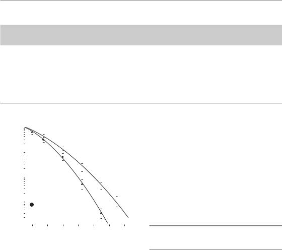
Figure 18.1 Head and neck SQD-9 cell-survival curves with or without preincubation with gemcitabine (dFdC) at a dose of 5 μM for 3 hours. In the presence of the drug, the initial slope of the cell survival curve is steeper, reflecting an interaction with ionizing radiation.
The concept of DMFs can be used in describing tumour effect or normal tissue toxicity.
A good clinical illustration of this type of interaction between chemotherapy and radiotherapy is in the treatment of locally advanced head and neck squamous cell carcinoma, in the meta-analysis of over 10 000 individual patients (Pignon et al., 2000). Patients were categorized according to whether chemotherapy had been given before (induction chemotherapy), during (concomitant chemotherapy) or after radiotherapy (adjuvant chemotherapy). A significant absolute 5-year benefit of 8 per cent was found only when concomitant chemotherapy was given. For induction or adjuvant chemotherapy, the 5-year survival was improved
Little is known about the capacity of chemotherapeutic agents to increase the efficiency with which ionizing radiation induces DNA damage. Compounds such as iododeoxyuridine (IdUrd) and bromodeoxyuridine (BrdUrd) when incorporated into DNA have been shown to enhance radiationinduced DNA damage, likely through the production of reactive uracilyl radicals and halide ions, which in turn induce DNA single-strand- breaks (SSBs) in the neighbouring DNA (Iliakis et al., 1991). However, several commonly used chemotherapy agents have been shown to inhibit the repair of radiation damage (i.e. DNA and/or chromosome damage). Examples are nucleoside analogues, cisplatin, bleomycin, doxorubicin and hydroxyurea. Some of these drugs inhibit the repair processes by interfering with the enzymatic machinery involved in the restoration of the DNA/ chromosome integrity. Fludarabine, for example, is a nucleoside analogue, which is incorporated
Molecular mechanisms of interaction 251
damage |
100 |
|
|
|
|
|
80 |
|
|
F-ara-A (100 |
|
||
|
0.5 hours prior to RT |
|||||
initial |
60 |
|
|
|
|
|
of |
|
|
RT alone |
|
|
|
Per cent |
40 |
|
|
|
||
|
|
|
|
|
||
20 |
|
|
|
|
|
|
|
|
|
|
|
|
|
|
0 |
20 |
40 |
60 |
80 |
100 |
|
|
Time after irradiation (min) |
|
|||
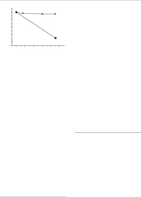
Figure 18.2 Inhibition of repair of chromosome breaks by the nucleoside analogue F-ara-A. Human
lymphocytes were irradiated with a single X-ray dose of 2 Gy and incubated at 37°C in the presence or absence of F-ara-A. At 90 min post-irradiation, 70 per cent of the chromosome breaks have repaired in the control sample; in the sample incubated with F-ara-A, only
5 per cent of breaks have repaired. RT, radiotherapy.
into DNA and blocks DNA primase, DNA polymerase α and ε and DNA ligase, and which has been shown to inhibit the repair of chromosome break repair (Fig. 18.2) (Gregoire et al., 1999).
Some of these drugs, like radiation, can directly produce DNA damage which manifests as DNA breaks, adducts and intercalation. For cisplatin, there is also an increase in the number of radiationinduced strand breaks. This might occur by conversion of radiation-induced SSBs to double strand breaks (DSBs) during the repair of DNA–platinum adducts. Inhibition of repair, or the conversion of SSBs to DSBs, has the effect of increasing the slope of the radiation survival curve and leads to an enhanced response (Fig. 18.1). Enhancement, which occurs as a result of repair inhibition, will be more pronounced in fractionated schedules than for single doses. A major problem with DNA repair inhibition as an exploitable mechanism for obtaining a therapeutic gain is the lack of evidence for a selective anti-tumour effect. For this strategy to be effective, some sort of tumour drug targeting may be required.
Cell-cycle synchronization
The vast majority of chemotherapeutic agents are inhibitors of cell division and are thus mainly active
on proliferating cells. Agents such as gemcitabine, fludarabine, methotrexate and 5-fluorouracil inhibit various enzymes involved in DNA synthesis and repair in S-phase cells; agents such as etoposide, doxorubicin, alkylating agents and platinum compounds induce DNA strand breaks and DNA strand crosslinks in any phase of the cell cycle, but will only become potentially lethal in replicating cells; agents such as taxol, taxotere and Vinca alkaloids inhibit mitotic spindle formation and thus are mainly active during mitosis.
As a consequence of this cell-cycle phase selective cytotoxicity of chemotherapeutic agents, the remaining surviving cells will be synchronized. If radiation could be delivered when these synchronized cells have reached a more radiosensitive phase of the cell cycle (e.g. G2 mitosis), a tremendous potentiation of the radiation effect could be observed. Such a mechanism of interaction between drugs and ionizing radiation has often been reported in preclinical experimental models (Gregoire et al., 1994). However, in clinics, because of the difficulty in assessing the appropriate timing between drug injection and radiotherapy delivery, it is unlikely that cell synchronization can be successfully exploited. Furthermore, considering that radiotherapy is typically delivered on a fractionated basis, it is also likely that this effect would be lost after a few fractions.
Enhanced apoptosis
Apoptosis (or interphase cell death) is a common mechanism of cell death induced by chemotherapeutic agents (Kaufmann and Earnshaw, 2000). These drugs can trigger one or more of the pathways leading to apoptosis. For the anti-metabolites, DNA incorporation is a necessary event to ensure a robust apoptotic response, hence the specific sensitivity of S-phase cells to these agents. Within this framework, it has been hypothesized that combining these drugs with ionizing radiation, which is very effective in inducing DNA SSBs or DSBs in every phase of the cell cycle, could facilitate their DNA incorporation and thus trigger an enhanced apoptotic reaction. This hypothesis was investigated in tumour models in vivo, using single doses of X-rays combined with gemcitabine (Milas et al., 1999). An increased apoptotic response was indeed observed when the two modalities were combined, but comprehensive

252 Combined radiotherapy and chemotherapy
analysis of the data did not demonstrate any synergistic enhancement, only an additive effect.
Re-oxygenation
As discussed in Chapters 15 and 16, hypoxia, a common feature of the majority of human solid tumours, is associated with a poorer response to radiotherapy. One reason for this is that the functionally insufficient tumour vascular network does not permit an adequate diffusion of oxygen throughout the whole tumour mass. It has therefore been proposed that chemotherapy, by inducing some degree of tumour shrinkage, might facilitate a more even diffusion of oxygen and increase overall tumour oxygenation, which in turn would increase
tumour radiosensitivity. In the murine mammary carcinoma MCA-4, it was indeed shown that intratumoural PO2 increased progressively in the few hours following taxol administration, from 6.2 mmHg in untreated tumours to 10.0 mmHg in treated tumours (Milas et al., 1995). This progressive tumour reoxygenation was associated with a significant parallel increase in the tumour radioresponse compared with control animals that did not receive taxol. This mechanism, which likely could play a role with any chemotherapeutic drug, has, however, never been tested with other agents.
Inhibition of cell proliferation
Table 18.3 summarizes information from preclinical experiments on the mechanisms of interaction
Table 18.3 Summary of the preclinical data regarding the mechanisms of interaction between ionizing radiation and chemotherapeutic agents
|
DNA damage |
|
Chromosome |
|
|
|
|
Induction |
Repair |
aberration |
Cell cycle |
Apoptosis |
Re-oxygenation |
|
|
|
|
|
|
|
Antimetabolites |
|
|
|
|
|
|
5-Fluorouracil |
|
|
|
|
? |
? |
Methotrexate |
? |
? |
? |
? |
? |
? |
Hydroxyurea |
? |
|
|
|
? |
? |
Gemcitabine |
|
|
|
|
|
? |
Fludarabine |
|
|
|
|
|
? |
Plant derivatives |
|
|
|
|
|
|
Vinca alkaloids |
? |
|
? |
|
? |
? |
Etoposide |
? |
? |
|
|
|
? |
Camptothecin |
? |
? |
|
|
|
? |
Taxanes |
? |
|
|
|
|
|
Antibiotics |
|
|
|
|
|
|
Doxorubicin |
|
|
|
|
? |
? |
Mitomycin-C |
? |
? |
|
? |
? |
? |
Bleomycin |
? |
|
|
|
? |
? |
Actinomycin-D |
? |
? |
? |
? |
|
|
Alkylating agents |
|
|
|
|
|
|
Cisplatin |
? |
|
? |
|
? |
? |
BCNU |
? |
|
|
? |
? |
? |
Cyclophosphamide |
? |
? |
|
? |
? |
? |
, Not demonstrated; , demonstrated; , conflicting data; ?, unknown. BCNU, β-chloro-nitrosourea.
Toxicity resulting from concomitant use 253
between chemotherapeutic agents and ionizing radiation. For the majority of these agents, the exact cellular and molecular mechanisms of interaction are not precisely known. Nevertheless, these agents are routinely used in the clinic and have been shown to be effective in combination with radiotherapy. Furthermore, even for those agents for which mechanisms of interaction with radiation have been elucidated in experimental models, it is unlikely that the clinical regimen has been designed to benefit fully from these interactions. Indeed, in the clinical setting, logistical considerations may come into play to explain ways in which drugs and radiotherapy are combined, and such considerations may not be entirely compatible with a full exploitation of the molecular and
|
|
15 |
Control |
|
|
diameter |
|
|
|
4.5 Gy q.d. |
|
|
|
|
|
||
|
13 |
|
|
|
|
|
|
|
|
|
|
tumour |
|
11 |
|
|
|
|
9 |
|
Fludarabine |
|
|
Mean |
|
7 |
|
q.d. |
|
|
|
|
|
(400 mg/kg) |
Fludarabine |
|
|
|
|
|
|
|
|
|
|
|
3 hours prior to RT |
5
0 |
5 |
10 |
15 |
20 |
25 |
Time after first fraction (days)
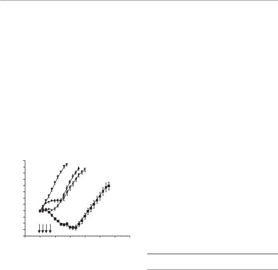
Figure 18.3 Regrowth delay experiment in a mouse sarcoma (SA-NH). Tumours (8 mm diameter) were treated with four daily i.p. administrations of fludarabine (arrows), irradiated with four daily fractions of 4.5 Gy, or given four daily doses of fludarabine 3 hours prior to four daily fractions of 4.5 Gy. Control mice were injected with saline. Each datum point represents the mean of nine or ten mice. When treated with radiation alone, tumours kept growing and only a 5.3 0.5 (SEM) day regrowth delay was seen compared with control animals. During fludarabine treatment alone, tumour proliferation was inhibited, but tumours started growing again as soon as drug administration stopped; overall, a regrowth delay of 5.5 0.7 (SEM) days was seen. When both modalities were combined, a decrease in tumour size was observed during treatment and the regrowth delay of 14.3 0.9 (SEM) days was greater than the additive effect of the two modalities alone.
cellular interactions between drugs and ionizing radiation.
It is therefore reasonable to propose that a prominent mechanism of interaction between drugs and radiotherapy would be a simple inhibition of the cellular proliferation that takes place during the radiation interfraction interval. Such a mechanism is illustrated in Fig. 18.3 (Gregoire et al., 1999). This interaction would be much less sensitive to the exact timing between drug and radiation dose delivery, provided that the drug is delivered at some point during the radiotherapy schedule. This being the case, it would be best to administer the drug towards the end of the radiation treatment course, when tumour cell repopulation had been triggered (see Chapter 10, Section 10.4). This was the rationale of a phase II trial conducted on head and neck squamous cell carcinoma, where two courses of cisplatin/5FU were given over the last 2 weeks of radiotherapy as a socalled ‘chemoboost’ (Corry et al., 2000). Results were encouraging but have not yet been tested in a randomized phase III trial.
18.4 TOXICITY RESULTING FROM CONCOMITANT USE OF CHEMOTHERAPY AND RADIOTHERAPY
Early toxicity
As discussed in Chapter 13, early toxicity after radiotherapy (e.g. oral mucositis, skin reaction, oesophagitis, proctitis and bone marrow depletion) typically results from an imbalance between physiological loss of mature cells and renewal from the stem cells or the precursor cells. As mentioned above, all chemotherapeutic agents are active on proliferative cells, and thus on their own also produce an imbalance between precursors and mature cells. It is thus anticipated that concomitant association between drugs and radiation will result in an increased early toxicity. Table 18.4 summarizes experimental data on early toxicity observed during concomitant association between drug and radiation. It shows that, for all classes of drugs, an increase in radiation-induced

254 Combined radiotherapy and chemotherapy
Table 18.4 Summary of the preclinical data regarding the toxicity of concomitant chemoradiation
|
Early effects |
Late effects |
|
|
|
Antimetabolites |
|
|
5-Fluorouracil |
(GI, skin) |
? |
Methotrexate |
(GI) |
? |
Hydroxyurea |
(GI) |
? |
Gemcitabine |
(GI) |
(lung) |
Fludarabine |
(GI) |
(CNS) |
Plant derivatives |
|
|
Vinca alkaloids |
(GI, BM) |
? |
Etoposide |
? |
? |
Taxanes |
(GI) |
? |
Antibiotics |
|
|
Doxorubicin |
(GI, skin) |
(heart, lung) |
Mitomycin-C |
(GI, BM) |
(lung) |
Bleomycin |
(GI, skin) |
(skin, lung) |
Actinomycin-D |
(GI, BM, skin) |
(lung) |
Alkylating agents |
|
|
Cisplatin |
(GI) |
(kidney) |
BCNU |
(GI) |
(lung) |
Cyclophosphamide |
(GI, skin) |
(lung, bladder, CNS) |
BCNU, β-chloro-nitrosourea; BM, bone marrow; CNS, central nervous system; GI, gastrointestinal., Not demonstrated; , demonstrated; , conflicting data; ?, unknown.
early toxicity has been reported. These findings are in agreement with the clinical trials that compared radiotherapy alone with concomitant chemoradiation, which indicated a significant increase in early toxicity in the combined modality arm.
There is a considerable body of experimental data which demonstrates that normal-tissue damage after combined modality treatment is strongly influenced by the sequence and timing of the modalities. Many commonly used drugs cause a substantial increase in normal-tissue radiation injury when the modalities are given in close sequence but not when they are separated in time (Fig. 18.4) (Gregoire et al., 1997). However, this finding conflicts with the requirement to use concomitant chemoradiation (thus with a narrow window of association) for improving loco-regional tumour control. Thus, unless pharmacokinetic
studies show a different pattern of drug biodistribution between tumour cells and normal cells, it is likely that the optimal sequence of drug administration for tumour radiosensitization is the one that will also produce the greatest increase in early radiation toxicity.
Late toxicity
In contrast to early radiation effects, which typically occur during treatment and in rapidly renewing tissues, late effects can affect all types of tissues after a latent period, which typically is expressed in months to years. Late damage also tends more to be irreversible and a radiation dosedependency has been well documented in a large number of tissues. The pathophysiology of late radiation effects is discussed at length in
The therapeutic ratio 255
circumference |
|
|
|
|
|
Gemcitabine 48 |
||
120 |
|
|
|
|
hours prior to RT |
|||
|
|
|
|
Gemcitabine 3 |
||||
|
|
|
|
|
||||
100 |
|
|
|
|
hours prior to RT |
|||
|
|
|
|
RT alone |
|
|||
|
|
|
|
|
|
|||
80 |
|
|
|
|
|
|
|
|
per |
|
|
|
|
|
|
|
|
60 |
|
|
|
|
|
|
|
|
of crypts |
|
|
|
|
|
|
|
|
40 |
|
|
|
|
|
|
|
|
20 |
|
|
|
|
|
|
|
|
Number |
|
|
|
|
|
|
|
|
0 |
|
|
|
|
|
|
|
|
|
|
|
|
|
|
|
|
|
|
6 |
8 |
10 |
12 |
14 |
16 |
18 |
20 |
Absorbed dose (Gy)
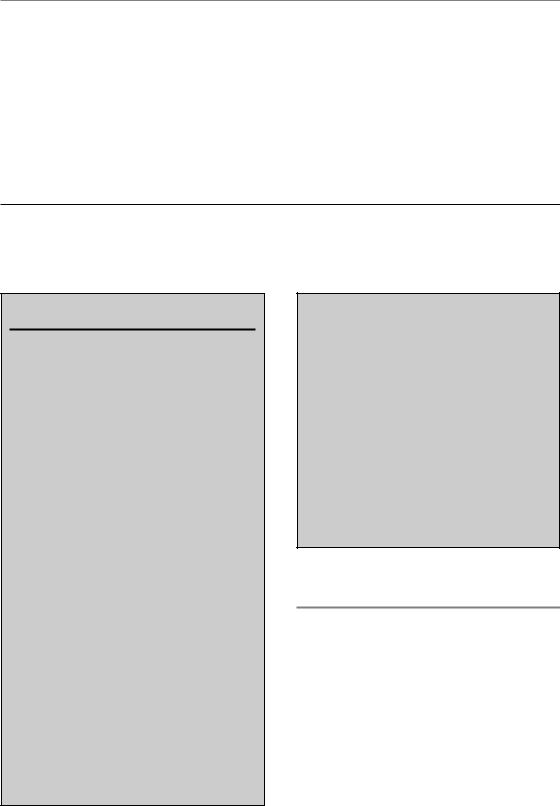
Figure 18.4 Radioenhancement of early jejunal damage after single-dose irradiation in mice. Mice were total body irradiated with single doses of 250 kV X-rays with or without prior administration (at 3 or 48 hours) of a single dose (150 mg/kg) of gemcitabine. The crypt cell regeneration assay was used. When injected 3 hours prior to irradiation, gemcitabine induced a marked radiosensitization (dose-modifying factor,
DMF 1.3). When injected 24 hours prior to irradiation a small radioprotection was observed (DMF 0.9).
Chapter 13. Although it may involve various cell types, any therapeutic intervention that may affect the repair of radiation (DNA) damage in the tumour is likely to also increase late normal-tissue radiation damage (Table 18.4). Furthermore, the risk of late effects after combined chemoradiotherapy can be further increased when the drugs have a specific toxicity for tissues within the irradiated volume, such as bleomycin for lung toxicity, doxorubicin for cardiac toxicity and cisplatin for renal toxicity. In the clinical setting, the design of protocols and the choice of the various drugs to combine with radiotherapy needs to integrate this knowledge. For example, bleomycin should be avoided together with radiotherapy for tumours of the mediastinum. For postoperative irradiation of the left breast or chest wall in women receiving doxorubicin, adequate planning should be made to avoid irradiation of the myocardium.
18.5 THE THERAPEUTIC RATIO
The therapeutic ratio (TR), or therapeutic gain, is the relative expected benefit of a combined modality treatment, integrating both the tumour and the normal-tissue effects. It is defined as the ratio of DMFs for tumour over normal tissues. A therapeutic ratio above unity indicates that, overall, the combined modality treatment is relatively more effective for tumour control than for normal tissue toxicity; conversely, a therapeutic ratio below unity indicates that the combined treatment is relatively more toxic than beneficial. The therapeutic ratio needs to be determined for both early and late normal-tissue toxicity. Table 18.5 presents an example of a concomitant association of cisplatin and 5-fluorouracil with radiotherapy for the treatment of locally advanced SCC of the cervix (Morris et al., 1999). It shows that, when early toxicity is taken into account, the therapeutic ratio is far below unity. However, although very distressful, early side-effects are usually manageable with extensive supportive care during treatment, and in this clinical example they fully resolved within a few weeks after the end of treatment. When late effects are considered, the therapeutic ratio is well above unity, illustrating the potential net benefit of the combined treatment strategy in this particular clinical setting.
When designing a new clinical trial or a new clinical strategy with concomitant chemotherapy and radiotherapy, one may need to slightly decrease the dose intensity of the standard treatment (i.e. radiotherapy) to obtain a therapeutic ratio above unity, but still have a beneficial effect at the tumour level. This was done in a trial comparing radiotherapy alone (70 Gy in 7 weeks) for locally advanced head and neck squamous cell carcinoma, with intercalated chemotherapy– radiotherapy (three cycles of 20 Gy in 2 weeks plus 1 week of cisplatin/5FU) (Merlano et al., 1992). At 3 years, the overall survival was increased from 23 per cent to 41 per cent without any increase in early toxicity.
The choice between an ‘equal toxicity’ design or an ‘equal dose’ design must therefore be considered on a site-by-site basis depending on the objective of the trial or clinical strategy.
256 Combined radiotherapy and chemotherapy
Table 18.5 Comparison of efficacy and side-effects after concomitant chemoradiotherapy for locally advanced squamous cell carcinoma of the cervix
|
Radiotherapy alone1 (%) |
Chemoradiotherapy2 (%) |
Therapeutic ratio |
Recurrence rate at 5 years |
35 |
19 |
– |
Early effects (grades 3–5) |
5 |
45 |
0.2 |
Early effects (excluding |
2 |
10 |
0.4 |
haematological toxicity) |
|
|
|
(grades 3–5) |
|
|
|
Late effects (grades 3–5) |
11 |
12 |
1.7 |
1External pelvic radiotherapy up to 45 Gy in 4.5 weeks followed by a brachytherapy implant with a total dose equal to or greater than 85 Gy; n 193.
2Cisplatin (75 mg/m2, day 1) 5-fluorouracil (1g/m2 per day, days 1–4) 3, every 3 weeks; n 195.
Key points
1.Proper design combined drug and radiotherapy treatment depends on the objective desired. Sequential association (neo-adjuvant or adjuvant) is preferred when target cell populations are different and/or when the objective is to optimize the dose intensity of chemotherapy and radiotherapy in both chemosensitive and radiosensitive disease. Concomitant association is preferred when cellular or molecular interactions are used to improve loco-regional control of the disease.
2.Although several mechanisms of interaction between drugs and radiation have been identified (modulation of DNA and chromosome damage and repair, cell-cycle synchronization, enhanced induction of apoptosis, re-oxygenation), in a clinical setting it is most likely that a key benefit is the inhibition of tumour cell proliferation by drugs during the radiation interfraction interval.
3.Concomitant administration of chemotherapy and radiation gives increased early normal tissue toxicity due to inhibition of stem cell or precursor cell proliferation. Late normal-tissue damage is likely to be enhanced through inhibition of DNA repair, and by a specific mechanism of drug toxicity in sensitive tissues (e.g. doxorubicin in the heart, bleomycin in the lung).
4.The therapeutic ratio (TR) expresses the relative benefit of a combined modality treatment, integrating both the tumour and the normal-tissue effects. For ‘equal dose’ trials, TR is typically below unity for early toxicity and above unity for late radiation damage.
5.Several randomized trials with concomitant chemoradiotherapy have been conducted in brain, head and neck, lung, oesophagus, cervix and colorectal cancers. A significant increase in loco-regional control has been found in some disease sites (e.g. brain, head and neck, cervix) with a consequent improvement in patient survival.
■BIBLIOGRAPHY
Bartelink H, Roelofsen F, Eschwege F et al. (1997). Concomitant radiotherapy and chemotherapy is superior to radiotherapy alone in the treatment of locally advanced anal cancer: results of a phase III randomized trial of the European Organization for Research and Treatment of Cancer Radiotherapy and Gastrointestinal Cooperative Groups. J Clin Oncol 15: 2040–9.
Bernier J, Domenge C, Ozsahin M et al. (2004). Postoperative irradiation with or without concomitant chemotherapy for locally advanced head and neck cancer. N Engl J Med 350: 1945–52.

Bibliography 257
Bosset JF, Calais G, Mineur L et al. (2005a). Enhanced tumoricidal effect of chemotherapy with preoperative radiotherapy for rectal cancer: preliminary results – EORTC 22921. J Clin Oncol 23: 5620–7.
Bosset JF, Calais G, Mineur L et al. (2005b). Preoperative radiation (Preop RT) in rectal cancer: effect and timing of additional chemotherapy (CT) 5-year results of the EORTC 22921 trial. J Clin Oncol (Meeting Abstracts) 23(Suppl. 16S): 3505.
Budach W, Hehr T, Budach V, Belka C, Dietz K (2006). A meta-analysis of hyperfractionated and accelerated radiotherapy and combined chemotherapy and radiotherapy regimens in unresected locally advanced squamous cell carcinoma of the head and neck. BMC Cancer
6: 28.
Cooper JS, Pajak TF, Forastiere AA et al. (2004). Postoperative concurrent radiotherapy and chemotherapy for high-risk squamous-cell carcinoma of the head and neck. N Engl J Med 350: 1937–44.
Corry J, Rischin D, Smith JG et al. (2000). Radiation with concurrent late chemotherapy intensification (‘chemoboost’) for locally advanced head and neck cancer. Radiother Oncol 54: 123–7.
Forastière AA, Goepfert H, Maor M et al. (2003). Concurrent chemotherapy and radiotherapy for organ preservation in advanced laryngeal cancer. N Engl J Med 349: 2091–8.
Green JA, Kirwan JM, Tierney JF et al. (2001). Survival and recurrence after concomitant chemotherapy and radiotherapy for cancer of the uterine cervix: a systematic review and meta-analysis. Lancet 358: 781–6.
Green J, Kirwan J, Tierney J et al. (2005). Concomitant chemotherapy and radiation therapy for cancer of the uterine cervix. Cochrane Database Syst Rev
CD002225.
Gregoire V, Van NT, Stephens LC et al. (1994). The role of fludarabine-induced apoptosis and cell cycle synchronization in enhanced murine tumor radiation response in vivo. Cancer Res 54: 6201–9.
Gregoire V, Beauduin M, Rosier JF et al. (1997). Kinetics of mouse jejunum radiosensitization by 2 ,2 - difluorodeoxycytidine (gemcitabine) and its relationship with pharmacodynamics of DNA synthesis inhibition and cell cycle redistribution in crypt cells. Br J Cancer 76: 1315–21.
Gregoire V, Hittelman WN, Rosier JF, Milas L (1999). Chemoradiotherapy: radiosensitizing nucleoside analogues (review). Oncol Rep 6: 949–57.
Hegi ME, Diserens AC, Gorlia T et al. (2005). MGMT gene silencing and benefit from temozolomide in glioblastoma. N Engl J Med 352: 997–1003.
Iliakis G, Pantelias G, Kurtzman S (1991). Mechanism of radiosensitization by halogenated pyrimidines: effect of BrdU on cell killing and interphase chromosome breakage in radiation-sensitive cells.
Radiat Res 125: 56–64.
Kaufmann SH, Earnshaw WC (2000). Induction of apoptosis by cancer chemotherapy. Exp Cell Res 256: 42–9.
Merlano M, Vitale V, Rosso R et al. (1992). Treatment of advanced squamous-cell carcinoma of the head and neck with alternating chemotherapy and radiotherapy. N Engl J Med 327: 1115–21.
Milas L, Hunter N, Mason KA, Milross C, Peters LJ (1995). Tumor reoxygenation as a mechanism of taxol-induced enhancement of tumor radioresponse.
Acta Oncol 34: 409–12.
Milas L, Fujii T, Hunter N et al. (1999). Enhancement of tumor radioresponse in vivo by gemcitabine. Cancer Res 59: 107–14.
Morris M, Eifel PJ, Lu J et al. (1999). Pelvic radiation with concurrent chemotherapy compared with pelvic and para-aortic radiation for high-risk cervical cancer. N Engl J Med 340: 1137–43. NACCCMA (Neoadjuvant Chemotherapy for Cervical Cancer Meta-Analysis) Collaboration (2004).
Neoadjuvant chemotherapy for locally advanced cervix cancer. Cochrane Database Syst Rev
CD001774.
Pignon JP, Arriagada R, Ihde DC et al. (1992). A metaanalysis of thoracic radiotherapy for small-cell lung cancer. N Engl J Med 327: 1618–24.
Pignon JP, Bourhis J, Domenge C, Designe L (2000). Chemotherapy added to locoregional treatment for head and neck squamous-cell carcinoma: three metaanalyses of updated individual data. MACH-NC Collaborative Group. Meta-Analysis of Chemotherapy on Head and Neck Cancer. Lancet 355: 949–55.
Pijls-Johannesma MC, De Ruysscher D, Lambin P, Rutten I, Vansteenkiste JF (2005). Early versus late chest radiotherapy for limited stage small cell lung cancer. Cochrane Database Syst Rev
CD004700.

258 Combined radiotherapy and chemotherapy
Rowell NP, O’Rourke NP (2004). Concurrent chemoradiotherapy in non-small cell lung cancer.
Cochrane Database Syst Rev CD002140.
Stupp R, Mason WP, van den Bent MJ et al. (2005). Radiotherapy plus concomitant and adjuvant temozolomide for glioblastoma. N Engl J Med 352: 987–96.
Wolmark N, Wieand HS, Hyams DM et al. (2000). Randomized trial of postoperative adjuvant chemotherapy with or without radiotherapy for carcinoma of the rectum: National Surgical Adjuvant Breast and Bowel Project Protocol R–02.
J Natl Cancer Inst 92: 388–96.
Wong R, Malthaner R (2006). Combined chemotherapy and radiotherapy (without surgery) compared with radiotherapy alone in localized carcinoma of the esophagus. Cochrane Database Syst Rev CD002092.
■ FURTHER READING
Browman GP, Hodson DI, Mackenzie RJ, Bestic N, Zuraw L (2001). Choosing a concomitant chemotherapy and radiotherapy regimen for squamous cell head and neck cancer: a systematic review of the published literature with subgroup analysis. Head Neck 23: 579–89.
Steel GG (1988). The search for therapeutic gain in the combination of radiotherapy and chemotherapy.
Radiother Oncol 11: 31–53.
Steel GG, Peckham MJ (1979). Exploitable mechanisms in combined radiotherapy–chemotherapy: the concept of additivity. Int J Radiat Oncol Biol Phys 5: 85–91.
Wouters A, Pauwels B, Lardon F, Vermorken JB (2007). Review: implications of in vitro research on the effect of radiotherapy and chemotherapy under hypoxic conditions. Oncologist 12: 690–712.