

- •Contents
- •Preface
- •1 Introduction: the significance of radiobiology and radiotherapy for cancer treatment
- •2 Irradiation-induced damage and the DNA damage response
- •3 Cell death after irradiation: how, when and why cells die
- •4 Quantifying cell kill and cell survival
- •5 Dose–response relationships in radiotherapy
- •6 Linear energy transfer and relative biological effectiveness
- •7 Tumour growth and response to radiation
- •8 Fractionation: the linear-quadratic approach
- •9 The linear-quadratic approach in clinical practice
- •10 Modified fractionation
- •11 Time factors in normal-tissue responses to irradiation
- •12 The dose-rate effect
- •13 Pathogenesis of normal-tissue side-effects
- •14 The volume effect in radiotherapy
- •15 The oxygen effect and fractionated radiotherapy
- •16 The tumour microenvironment and cellular hypoxia responses
- •17 Therapeutic approaches to tumour hypoxia
- •18 Combined radiotherapy and chemotherapy
- •19 Retreatment tolerance of normal tissues
- •20 Molecular image-guided radiotherapy with positron emission tomography
- •21 Molecular-targeted agents for enhancing tumour response
- •22 Biological response modifiers: normal tissues
- •23 Molecular targeting and patient individualization
- •24 Protons and other ions in radiotherapy
- •25 Second cancers after radiotherapy
- •Glossary of terms in radiation biology
- •Index

23
Molecular targeting and patient individualization
ADRIAN C. BEGG
23.1 |
Introduction |
316 |
23.8 |
Prediction of normal-tissue tolerance |
326 |
23.2 |
Molecular targeting |
317 |
23.9 |
How should clinicians respond to |
|
23.3 |
The question of tumour specificity |
320 |
|
prediction results? |
328 |
23.4 |
Patient individualization |
321 |
23.10 |
Summary |
328 |
23.5 |
What determines tumour response |
|
Key points |
328 |
|
|
after radiotherapy? |
322 |
Bibliography |
329 |
|
23.6 |
Measuring single parameters |
322 |
Further reading |
331 |
|
23.7 |
Measuring multiple parameters |
|
|
|
|
|
(genome wide) |
324 |
|
|
|
|
|
|
|
|
|
23.1 INTRODUCTION
Radiotherapy is effective as a single modality. It is prescribed either alone or as an adjuvant therapy in more than half of all cancer patients. Improvements in radiotherapy have been considerable over the last two decades, in large measure owing to improvements in instrumentation, machine software and diagnostic techniques, enabling more accurate dose delivery to the tumour while minimizing the dose to surrounding healthy tissue. Despite these technical improvements, some patients still fail after radiotherapy and some still develop severe side-effects. Improvements are thus still needed. These improvements can, in principle, be achieved from the biological rather than the technical side.
One approach is to try and make tumour cells more sensitive to radiation. This can be done using drugs, usually small molecules, since these often have some pharmacological advantages, but also with antibodies to surface receptors linked to
radioresistance pathways (e.g. epidermal growth factor receptor, EGFR). In addition, the expression of specific genes can be altered using short antisense RNA, short interference RNA (siRNA) or aptamers, all aimed at genes involved in radioresistance.
Some commonly used anti-cancer drugs are already known to radiosensitize cells, in addition to being cytotoxic. Cisplatin is a good example. However, future improvements must come from applying more tumour-specific drugs, and cisplatin and most other cytotoxic agents are not tumour specific, often causing serious side-effects in normal tissues. What is therefore needed is sufficient knowledge of the molecular biology of individual tumours, elucidating which pathways are deregulated. These pathways can then be attacked, providing a measure of tumour specificity. In the future, the combination of more accurate and complete molecular diagnostic methods, together with development of a wider range of radiosensitizing treatment options

Molecular targeting 317
(drugs, antibodies or genetic manipulation, targeted to a range of pathways affecting the radiation response), will allow treatments tailored to the individual, maximizing tumour cell kill and minimizing normal-tissue damage.
This chapter describes which pathways can be targeted to increase cell killing by radiation. In addition, it will review current molecular diagnostic methods for finding the deregulated pathways in individual tumours, ultimately aiding the choice of tumour-specific treatments. It is important to keep in mind at the outset that making all cells more radiosensitive is relatively easy, while making tumour cells more radiosensitive than normal-tissue cells is considerably more difficult, but is the goal that must be achieved to improve cancer therapy.
23.2 MOLECULAR TARGETING
DNA repair
The ability to repair DNA damage is probably the most important determinant of resistance to ionizing radiation. The most important pathways are those for repairing base damages, single-strand breaks (SSBs) and double-strand breaks (DSBs); for details, see Chapter 2. They include base excision repair (BER), the related single-strand break repair pathway (SSBR), and two pathways for repairing double-strand breaks, namely homologous recombination (HR) and non-homologous end-joining (NHEJ). To a lesser extent, radiation also produces DNA–DNA and DNA–protein crosslinks, particularly under hypoxic irradiation conditions. Crosslink repair involves yet other genes. There are experimental data on cell lines showing that interfering in any of these pathways can make cells more sensitive to radiation, although interference with DSB repair usually produces the greatest increases in sensitivity.
DNA polymerase beta (polβ) is a central enzyme in both BER and SSBR. Cells deficient in polβ, or expressing a dominant negative construct to polβ, thereby inhibiting its function, have been shown to be more sensitive to ionizing radiation in vitro under certain conditions (Vermeulen et al.,
2007). In addition, cells deficient in XRCC1, a
central helper protein in BER and SSBR, have also been shown to be more radiosensitive than wildtype cells (Thompson et al., 1990). A number of drugs have also been developed that are capable of inhibiting polβ. Some of these have been shown to modify the response of cells in vitro to alkylating agents and radiosensitizing thymidine analogues, which produce a type of damage repaired primarily by BER. At least one of these (methoxyamine) has also been tested in animal tumour models and shown to potentiate the action of alkylating agents, although it has not been tested with radiation (Liu and Gerson, 2004). Inhibitors also exist for APEX1, another central protein in BER, and can potentiate the response to some drugs (Luo and Kelley, 2004), although they have not been tested with radiation. Finally, small-molecule drugs have also been developed which inhibit poly-ADP-ribose polymerase 1 (PARP1), a critical break detector protein in SSBR. The PARP inhibitors and methoxyamine are the only drugs designed to target BER and SSBR which are now entering clinical trials.
Targeting double-strand break repair (DSBR) has received considerable attention, since this can substantially increase radiosensitivity, as shown from studies with knockout cells deficient in one of these essential genes. DNA-PKcs (DNAdependent protein kinase) is a key enzyme in the major pathway of NHEJ. Several drugs have been developed against this enzyme and been shown to inhibit DNA repair and radiosensitize cells. These agents can also sensitize cells to DSB-inducing drugs such as etoposide (direct) and cisplatin (indirect). Tumour specificity is an issue here, since DNA-PKcs is also central to DNA repair in normal tissues. This is exemplified by severe combined immunodeficiency (SCID) mice, which are deficient in DNA-PKcs and which show hypersensitivity to radiation in all normal tissues examined.
MRN and ATM
Sensing DNA breaks and signalling their presence in order to halt the cell cycle and recruit repair proteins is an important aspect of the damage response after radiation. Inhibition of sensing and signalling can thus also lead to radiosensitization.
The MRN complex of three proteins acting

318 Molecular targeting and patient individualization
together, MRE11, RAD50 and NBS1, is central to damage detection and signalling (see Chapter 2), and knocking out one or more of these genes leads to a marked increase in radiosensitivity. One of their functions is to activate ATM (gene is mutated in ataxia telangiectasia), which is a crucial damage signalling molecule. Several natural compounds such as caffeine and newly developed more-specific drugs (Hickson et al., 2004) can inhibit this enzyme. These drugs can abrogate the checkpoint response after irradiation, can reduce DNA repair and can lead to radiosensitization. Some of these drugs are undergoing clinical development. Tumour specificity is an issue here, since normal tissues also rely on MRN and ATM for an efficient damage response.
The PI3K/AKT pathway
One of the most important signal transduction pathways affecting the response to radiation is that involving phosphoinositide-3-kinase (PI3K) and protein kinase B (AKT) (see Chapter 21). Activation by phosphorylation of AKT, a key molecule in the pathway, is associated with resistance to radiation in both preclinical and clinical studies. Activation can be caused by overexpression of EGFR, deletion or mutation of the tumour suppressor PTEN, mutational activation of the RAS oncogene, and others (Valerie et al., 2007). Many drugs have been developed that are targeted to different members of this pathway, and several of these drugs have been shown to radiosensitize cells. Some of these have already shown promise in clinical trials (see Chapter 21). Tumours often overexpress EGFR, or express oncogenic RAS or contain inactivating PTEN mutations, leading to a degree of tumour specificity for further attacks on this pathway.
Nuclear factor kappa B (NFκB)
NFκB is a transcription factor associated with a protective survival response after DNA damage. Radiation activates the NFκB pathway in a number of cell types, and inhibiting this activation by both genetic and chemical methods has been shown to lead to an increase in apoptosis. This
pathway has therefore also been proposed as a target for radiosensitizing tumour cells (Magne et al., 2006). Some proteasome inhibitors (see below) also affect the NFκB pathway, and part of their effect may result from inhibition of NFκB. Inhibitors have also been developed to the upstream activator IKK, which increase the apoptotic response.
Proteasomes
Protein turnover, and its precise control, is essential for many cellular processes and for cell survival. The most common way the cell degrades proteins is to tag them with ubiquitin (requiring special enzymes) which then results in the protein being fed to a degradation organelle, the proteasome. This is a proteinase complex responsible for degrading most intracellular proteins, including those important for cell-cycle regulation and apoptosis. Several proteasome inhibitors have been found, including peptidyl aldehydes, lactacystin and a dipeptide boronic acid analogue (bortezomib). Proteasome inhibitors have been shown to induce apoptosis and to sensitize malignant cells in culture, and tumours in vivo, to radiation (Weber et al., 2007) and to some chemotherapy agents. The exact mechanisms are unknown, although effects on the cell cycle, on the NFκB signalling pathway and on apoptosis probably play a role, since all these can under some circumstances affect radiosensitivity. It has also been reported that transformed (malignant) cells are more sensitive to such inhibitors than normal cells, implying a degree of tumour specificity. Why this should be so is not clear, although, if confirmed, it would make proteasome inhibitors promising agents for improving radiotherapy. At least one of these agents, bortezomib, is approved for clinical use.
Chromatin structure
Histone acetylation is another potential target for radiosensitization. Efficient DNA repair is usually associated with modifications of the chromatin structure in order to recruit repair proteins and facilitate their access to damage; these changes then have to be reversed on completion of repair. One important type of chromatin

Molecular targeting 319
modification is the acetylation of histones, the proteins that form nucleosomes. Acetylation of histones removes a positive charge, reducing the affinity between histones and DNA and allowing easier access of transcription and other factors. The degree of histone acetylation is determined by competition between histone acetylases (HATs; putting on acetyl groups) and histone deacetylases (HDACs; taking them off). A number of HDAC inhibitors have been developed and found to have anti-tumour activity in preclinical cancer models. In addition, they can also significantly enhance tumour cell radiosensitivity (Cerna et al., 2006). The mechanisms are not entirely understood, but presumably relate to the recruitment of repair factors and/or resetting the chromatin after repair. The HDAC inhibitors are undergoing clinical trials as single agents, and in combination with radiotherapy.
Checkpoints
Blocks at various stages of the cell cycle are a universal feature of the response of mammalian cells to irradiation (see Chapter 2). However, the link between the presence or length of these blocks and cell kill after irradiation is often weak. Thus for the G1/S and intra-S checkpoints, abrogation of the blocks can be achieved by genetic manipulation of one or more checkpoint genes without altering radiosensitivity. However, two distinct G2 checkpoints exist, one rapid and dose independent and the other slow and dose dependent (see Chapter 2), and the dose-dependent block appears to be associated with radiosensitivity (Xu et al., 2002). Abrogation of this block increases radiosensitivity and it therefore represents a target for improving radiotherapy. In addition, abrogation of two or more blocks can be more effective than removing one block. Inhibition of the G2 checkpoint thus appears to more effectively sensitize cells to DNA damage if the cells also lack the G1 checkpoint, as in cells with mutant p53. This, in principle, provides some tumour specificity, since a large proportion of human cancers have p53 pathway mutations while the surrounding normal tissues will not. Drugs such as caffeine and pentoxyfylline can abrogate the G2 block and increase radiosensitivity. These drugs also inhibit
ATM, and so whether the effects seen are solely because of reducing damage-induced cell-cycle delays or effects on repair is not clear. At the time of writing, one drug, 7-hydroxystaurosporine (UCN-01), which also abrogates the G2 block and radiosensitizes cells, is undergoing clinical testing.
Apoptosis
Cells often die from apoptosis after irradiation or chemotherapy (although other forms of death can also dominate in solid tumours, see Chapter 3). It has therefore been proposed that increasing apoptosis would increase the effects of radiotherapy (Belka et al., 2004). Several apoptosis inducers have shown synergy in combination with radiation in preclinical models, such as perifosine (an alkylphospholipid compound with structural similarity to phospholipids that are the main constituents of cellular membranes), TRAIL [tumour necrosis factor (TNF)-related apoptosis inducing ligand], gossypol (a natural compound from cotton seeds), and others. It has not been proven that the synergy seen between radiation and these compounds is caused by the increases observed in apoptosis, since apoptosis does not always correlate with clonogenic cell kill (cells not dying of apoptosis can die in other ways, see Chapter 3). There is also no obvious reason why this approach should be tumour specific other than the observation that tumours often arise by evading apoptosis (Hanahan and Weinberg, 2000), leading to the conjecture that restoring apoptosis sensitivity might restore treatment sensitivity.
Hypoxia
Targeting hypoxia to increase tumour radiosensitivity is an attractive approach since the occurrence of hypoxia is almost exclusively restricted to tumours. The aim here is to make only the hypoxic cells more radiosensitive, or to selectively kill hypoxic cells. Many preclinical and clinical studies have shown the feasibility and effectiveness of this approach. Chapters 16 and 17 discuss the biology of hypoxia and ways to combat it.

320 Molecular targeting and patient individualization
23.3 THE QUESTION OF TUMOUR SPECIFICITY
Most DNA repair pathways operate universally and equally well in normal tissues and tumours (Table 23.1a). There is no known consistent tumour specificity concerning expression or function of DNA repair genes, or ATM and other damage detectors, or for chromatin modifications associated with repair or for the NFκB pathway. A partial exception is the HR pathway for repairing DSBs. This operates only in proliferating cells (S/G2 phase), and is therefore proliferation specific. Tumours tend to exhibit higher proliferation rates than normal tissues, especially compared with late-reacting normal tissues after radiotherapy. This provides partial tumour specificity. Targeting homologous recombination may also reduce hypoxic radioresistance, thereby contributing to tumour specificity (Sprong et al., 2006). However, no drugs have yet been described that specifically inhibit HR proteins.
Proteasome inhibitors have been reported to work better on transformed cells, but it is not yet known how general this observation is.
The attraction of targeting DNA repair, including damage sensing and signalling, is that it is an excellent way of making cells more radiation sensitive (Choudhury et al., 2006). However, as stated above, in most cases it suffers from a lack of tumour specificity. There are two possible ways in which to increase tumour specificity. The first is to deliver the sensitizing drug or other sensitizing agent (e.g. siRNA, antibody) specifically to the tumour. This remains an elusive goal, and has yet to succeed sufficiently well to improve clinical cancer treatment using either cytotoxic agents alone or as radiosensitizers. Progress in this area would certainly help the application of the approaches discussed here.
The second approach is to attack tumours where they are already weakened (Table 23.1b). Since almost all malignant tumours are genetically
Table 23.1 DNA repair as a target for improving radiotherapy
Tumour
|
Pathway |
Role |
specificity |
Reason |
(a) |
BER/SSBR |
Base damage/ |
No |
Important for repairing damage in normal tissue and tumour |
|
|
SSBR |
|
|
|
HR |
DSB repair |
Partial |
Lower hypoxic radioresistance if mutated; proliferation |
|
|
|
|
dependent |
|
NHEJ |
DSB repair |
No |
Important for repairing damage in normal tissue and tumour |
|
ATM |
Sensing DSB |
No |
Important for repairing damage in normal tissue and tumour |
|
MRN |
Sensing DSB |
No |
Important for repairing damage in normal tissue and tumour |
|
Pathway |
Role |
Mutations |
Possible ways to exploit |
|
|
|
in cancer |
|
(b) |
BER/SSBR |
Base damage/ |
Yes |
Anti-HR drug (reduce backup repair pathway) |
|
|
SSBR |
|
|
|
HR |
DSB repair |
Yes |
PARP inhibitor (anti-SSBR; reduce backup repair pathway) |
|
NHEJ |
DSB repair |
Yes |
Anti-NHEJ drug (greater effect on tumour weakened by |
|
|
|
|
pathway mutation) |
|
ATM |
Sensing DSB |
Yes |
Anti-ATM drug (greater effect on tumour weakened by |
|
|
|
|
pathway mutation) |
|
MRN |
Sensing DSB |
Yes |
Anti-MRN drug (greater effect on tumour weakened by |
|
|
|
|
pathway mutation) |
BER, base excision repair; SSBR, single-strand break repair; HR, homologous recombination; NHEJ, non-homologous end-joining; ATM, gene mutated in ataxia telangiectasia; MRN, MRN complex (MRE11, RAD50, NBS1); DSB, double-strand break; PARP: poly-ADP-ribose polymerase.

|
|
Patient individualization 321 |
|
||
Table 23.2 Drugs for improving radiotherapy: tumour specificity of targets |
||
|
|
|
|
Tumour specificity |
Remarks |
|
|
|
Intrinsic radiosensitivity |
|
|
DNA repair |
Partial |
Only some pathways (e.g. HR); or if mutations in pathway |
|
|
(see Table 23.1) |
Cell-cycle checkpoints |
Partial |
Only some checkpoints affect radiosensitivity (see Chapter |
|
|
2); will also affect proliferating normal tissues |
Cell death |
Partial? |
Tumours are less apoptosis prone? |
(e.g. pro-apoptotic) |
|
|
Signal transduction |
Partial |
If overexpression of target, and/or activation of pathway |
(e.g. PI3K) |
|
|
Hypoxia |
Yes |
Tumours are usually hypoxic, normal tissues rarely; acute |
|
|
and chronic hypoxia may require different approaches |
Repopulation |
|
|
Anti-cell-cycle drugs |
Partial |
Tumours proliferate more rapidly than late-reacting normal |
|
|
tissues |
Signal transduction |
Partial |
Tumours proliferate more rapidly than late-reacting normal |
(e.g. EGFR) |
|
tissues; if overexpression of target, and/or activation of pathway |
EGFR, epidermal growth factor receptor; HR, homologous recombination.
unstable, mutations or deletions of many genes are often found in a tumour, which result in losses of gene or pathway function. This includes DNA repair genes. Therefore agents that reduce expression or inhibit function of a gene or pathway which is already compromised by genetic mutation in the tumour are likely to have more effect on such a tumour than on surrounding normal tissues with fully functional repair pathways. This idea remains speculative, and requires a detailed knowledge of deregulated pathways in individual tumours. An example from the chemotherapy world is the dramatically increased effect of PARP inhibitors in tumours with reduced homologous recombination, such as are found in BRCA1/2 heterozygous individuals because BRCA genes are important for homologous recombination (Helleday et al., 2007). Similar combinations with radiation are therefore being sought, but, without such tumour specificity, the therapeutic gain from using general radiosensitizers will remain limited.
Of the three main factors affecting the response to fractionated irradiation, hypoxia remains the most obvious tumour-specific target (Table 23.2), since tumours almost always contain hypoxic
areas whereas normal tissues rarely do. Tumour proliferation, or repopulation, can be attacked with anti-proliferative drugs. These drugs will also adversely affect proliferating normal tissues leading to enhanced early reactions, but should have much less effect on the dose-limiting late-reacting normal tissues.
23.4 PATIENT INDIVIDUALIZATION
The goal of much current research is to develop rapid and robust methods enabling us to understand enough about each patient and their tumour to be able to choose the best treatment for that individual. At present, treatment choice is usually based on parameters such as tumour site, histological type, tumour stage and performance status. Within these broad categories, some tumours show less response to radiotherapy than others. If these tumours could be identified before treatment, alternative therapies might be selected that may give a better chance of cure than the standard therapy. This may involve one of the targeted therapies discussed above.

322 Molecular targeting and patient individualization
Individual patients also differ in their tolerance of radiation therapy. Among a group of patients given the same treatment protocol, some suffer more severe normal-tissue reactions than others. It is these severe reactors that limit the dose of radiation that can be prescribed to a group of patients. If severe reactors could be identified prior to therapy it might be possible to improve their management (e.g. by reducing their treatment dose, applying anti-fibrosis or anti-thrombotic therapies, etc.) as well as that of the rest of the patient group (e.g. by increasing their dose).
The following sections will deal with these two aspects of the individualization of radiation therapy: predicting tumour response and predicting normal-tissue response.
23.5 WHAT DETERMINES TUMOUR RESPONSE AFTER RADIOTHERAPY?
Determinants of tumour response to radiotherapy can be put into three broad categories: intrinsic radiosensitivity, proliferation rate and the extent of hypoxia. These are largely independent, such that a group of intrinsically radiosensitive tumours could have a range of proliferation rates and degrees of hypoxia; a group of tumours with high proliferation rates could have a range of radiosensitivities and degrees of hypoxia, etc. These three factors should thus be considered separately, and the goal is to measure them all to maximize the chance of accurately predicting response. In addition to these radiobiological parameters, other factors that can determine success or failure are tumour size at the time of treatment and the metastatic potential of the tumour. Large tumours are harder to control than small tumours simply because there are more cells to kill, which will require higher doses. This will be true even if intrinsic radiosensitivity, hypoxia and repopulation rates are equal at small and large tumour sizes. Tumour size should therefore be taken into account when assessing the performance of a predictive assay. Metastatic potential is clearly important for survival but should be considered separately from factors affecting local tumour control.
23.6 MEASURING SINGLE PARAMETERS
Intrinsic radiosensitivity
Malignant tumours are intrinsically genetically unstable, and there is ample evidence from cell lines, animal tumour models and in the clinic that this leads to a wide variation in intrinsic radiosensitivity, even between tumours of similar origin and histological type. Attempts have been made to assess the radiosensitivity of human tumours by explanting cells directly from biopsies, irradiating them in culture and measuring colony-forming ability, usually specifying surviving cell fraction after 2 Gy (SF2). Such studies have shown that tumour cell radiosensitivity is a significant and independent prognostic factor for radiotherapy outcome in carcinoma of both the cervix (West et al., 1997) and head and neck (Bjork-Eriksson et al., 2000). The disadvantages of the colony assay are its poor success rate for human tumours ( 70 per cent) and the time needed to produce data (often up to 4 weeks). Subsequent studies have evaluated alternative assays that generate results in less than a week. Examples include chromosome damage, DNA damage, glutathione levels and apoptosis. However, comparing these assays with the ‘gold standard’ of clonogenic cell death in cell lines has shown variable results. Similarly, some clinical studies with such assays have shown correlations with radiotherapy outcome while others have not. It can be concluded that these functional, usually cell-based, assays have limited clinical utility as predictive assays but have been useful in confirming one mechanism underlying differences in response of tumours to radiotherapy.
Hypoxia
Tumour hypoxia is a key factor involved in determining not only resistance to treatment but also malignant progression. Evidence for an association between measurements of hypoxia in individual human tumours and response to radiation therapy has been summarized in Chapters 15–17.

Measuring single parameters 323
Hypoxia has also been shown to be a negative prognostic factor after treatment with chemotherapy or surgery. The latter is consistent with data showing that hypoxia plays a key role in tumour progression by promoting both angiogenesis and metastasis (see Chapter 16).
One method to measure tumour hypoxia includes using thin polarographic glass electrodes (Eppendorf) which are inserted into tumours, producing several measurements along each track, thus providing multiple oxygen tension measurements per tumour. This direct method of measuring hypoxia has limitations in that it is only suitable for accessible tumours. Hypoxia-specific chemical probes such as pimonidazole (Raleigh et al., 2000) and EF5 (Evans et al., 2000) have been developed and are in clinical use. These and similar compounds are usually nitroimidazoles, which undergo bioreduction only under hypoxic conditions, followed by binding of reduced products onto macromolecules. Bound adducts can be detected with specific antibodies, allowing measurement by immunohistochemistry, immunofluorescence or flow cytometry. Some studies have shown a good correlation of pimonidazole binding with outcome after radiotherapy, as in head and neck cancer (Kaanders et al., 2002), while others have not, as in cervix cancer (Nordsmark et al., 2006).
The use of fluorinated derivatives of such bioreductive drugs also allows their detection by noninvasive positron emission tomography (PET) (Krause et al., 2006). This approach has the additional advantage of sampling the whole tumour and not just a small part of it. These drugs depend upon hypoxia for their reduction, although there are other factors that can influence their quantification and make them a less direct measure of hypoxia than is the case with electrodes. However, such agents are useful for quantifying hypoxia in human tumours, although they require administration of a drug. Other methods that are being evaluated to measure tumour hypoxia in the clinic include cross-sectional imaging using computed tomography (CT), magnetic resonance spectroscopic (MRS) imaging and magnetic resonance imaging (MRI) (Padhani et al., 2007).
Another possible surrogate marker of hypoxia is tumour vascularity, because of the known association between hypoxia and angiogenesis, and
the fact that oxygen is delivered via a tumour blood supply that varies from ordered to chaotic. A variety of methods have been used to score vascularity, including intercapillary distance, vascular density, vascular ‘hot-spots’ and the proportion of tumour areas greater than a fixed distance from a vessel. Some of these methods have shown a positive correlation with outcome, while others have been negative (West et al., 2001a).
Repopulation
The importance of tumour proliferation is most clearly shown by the higher doses required to control a tumour when overall treatment time is increased (see Chapters 8 and 10). Further evidence comes from studies showing loss of local tumour control as a result of gaps in treatment, whether planned or unplanned. There is also increasing evidence from randomized trials that accelerated regimes can improve outcome (Chapter 10).
Methods for measuring tumour proliferation include counting the mitotic index (proportion of mitoses in tissue sections), determining the proportion of cells in the S phase of the cell cycle by DNA flow cytometry, measuring the tumour potential doubling time (Tpot) with thymidine analogues such as iododeoxyuridine (IdUrd) and bromodeoxyuridine (BrdUrd), and using antibodies to detect proliferation-associated proteins. A multicentre analysis of over 470 head and neck cancer patients treated with radiotherapy alone showed a lack of significance of Tpot as a predictor (Begg et al., 1999). A number of other studies have shown a significant although usually weak correlation between labelling index and radiotherapy outcome. Therefore pretreatment labelling index (LI) or Tpot measurements are apparently not sufficiently robust for determining tumour cell proliferation during radiotherapy. Proliferation, or repopulation, during fractionated radiotherapy is clearly an important factor determining outcome, but reliable ways to measure it are not yet available. A greater understanding at the cell and molecular levels is needed of why, in some tumours, radiation damage leads to an accelerated repopulation response, but in others it does not. This may
324 Molecular targeting and patient individualization
be related to differences in the cytokine and growth-factor pathways.
23.7 MEASURING MULTIPLE PARAMETERS (GENOME WIDE)
The response of a tumour to radiotherapy is complex, determined broadly by the three factors mentioned above (intrinsic radiosensitivity, hypoxia and repopulation), each in turn controlled by many genes and pathways, some of which may be dysfunctional or overactive in a particular tumour. It is therefore unlikely that measuring a single factor, or expression of single genes, will provide a reliable predictor of how the tumour will respond to treatment. To maximize the chance of reliably predicting the success of a treatment in an individual, multiple factors, or multiple genes, need to be measured, both in the tumour and in the patient.
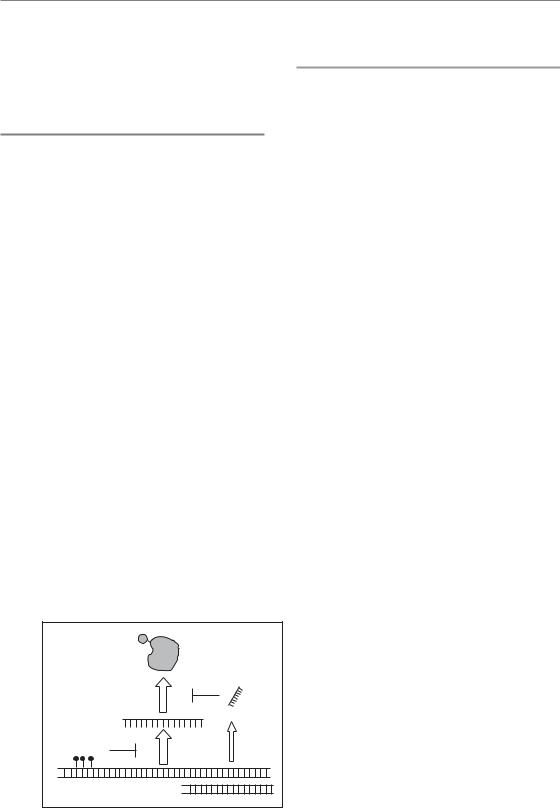
Methods for measuring genetic alterations on a genome-wide scale have made enormous progress in the last few years, including those for studying DNA changes, mRNA expression and protein expression. Application of these methods has already shown great promise for tumour diagnosis and prognosis, in addition to providing powerful new methods for fundamental studies of tumour (and other) biology. These genome-wide assays are now being applied to improve prediction in cancer treatments (see Fig. 23.1 for summary scheme).
The DNA level (comparative genomic hybridization, methylation)
Comparative genomic hybridization (CGH) allows tumour and normal-tissue DNA to be compared, producing a map of the loci in the chromosomes which are either deleted or amplified in the tumour (Pinkel and Albertson, 2005). This was originally achieved by hybridizing fluorescently labelled DNAs (e.g. tumour DNA green, normal DNA red) to metaphase spreads of normal cells. This technique has been superseded by hybridizing the labelled DNAs to a glass slide on which thousands of DNA probes, each representing a part of the genomic DNA, are spotted in an array (called array CGH, or aCGH). The advantage is reproducibility of the arrays, greater resolution because of the use of small DNA probes spaced closely on the array, and greater flexibility (widely spaced probes covering the whole genome, or closely spaced probes for part of a chromosome). Each of the observed genetic alterations covers megabases of DNA, but this is often sufficient to indicate genes of interest. Patterns of genomic changes have been correlated with outcome in several studies, although few after radiation (van den Broek et al., 2007). Matching such genomic DNA changes with gene expression data (microarrays, see below) can help pinpoint relevant genomic regions and relevant genes (Adler et al., 2006).
Methylation is a further factor at the DNA level which affects gene expression. Methylation of cytosines in promoter regions of genes can switch
LEVEL |
|
PROCESS |
ASSAY |
|
|
|
Post-translational |
|
|
Proteomics |
Figure 23.1 Genetic screening methods. |
Protein |
modification |
|
|
||
|
|
The genome can be screened at three levels |
|||
|
|
|
|
|
|
|
|
translation |
MicroRNA |
MicroRNA |
(left) using different assays (right). The |
|
|
|
scheme (middle) shows the relationship |
||
|
|
|
profiling |
||
|
|
|
|
||
|
|
|
|
between the biological processes. |
|
RNA |
|
|
|
Expression |
|
mRNA |
|
|
Combining results from more than one |
||
|
|
|
|||
|
|
|
profiling |
||
|
|
transcription |
SNP |
deregulation of important pathways, which |
|
|
|
Methylation |
|||
|
Methylation |
|
|
|
assay will give a better indication of the |
|
|
|
|
|
|
DNA |
|
|
|
profiling |
could then be targets of therapy. CGH, |
|
|
|
|
comparative genomic hybridization; SNP, |
|
|
Amplification/deletion |
|
SNP, CGH |
||
|
|
|
|
|
single nucleotide polymorphism. |
Measuring multiple parameters (Genome wide) 325
them off by altering the binding of specific proteins (transcription factors) necessary to initiate transcription. A number of methods have been designed to measure methylation status, and although these are not quite yet genome wide, they are likely to become so in the near future. Methods are based on affinity purification of methylated DNA, or fractionation of (un)methylated DNA using methyl-sensitive restriction enzymes, or chemical modification of unmethylated cytosines; in each case, microarray detection can be used for monitoring many loci. Such measurements, representing the epigenetic status of the genome, are also likely to help monitor which pathways are deregulated in tumours.
The RNA level (microarrays)
Expression microarrays are small chips (often glass slides) containing many thousands of DNA sequences in spots, one DNA sequence per spot. The DNA can be cDNA (messenger RNA backtranslated into more stable complementary DNA) but more usually oligomers (short DNA chains) representing the partial sequence of a gene. Messenger RNA, representing all expressed genes, is extracted from the tumour or other sample of interest, then fluorescently labelled and added to the chip, where each labelled RNA hybridizes only to the spot for that gene having the complementary DNA sequence. Genes which are highly expressed in the tumour then result in bright spots on the array, while lowly expressed genes show low signals. After hybridization, the array is scanned automatically and rapidly to measure the expression of all genes. Current arrays can hold almost all the genes in the human genome, estimated to be around 30 000.
Several studies have reported finding gene sets, or signatures, which predict the response to drugs or to radiation. In these studies, expression profiles of each sample are correlated with treatment sensitivity using specific statistical (bioinformatics) methods, such as supervised hierarchical clustering. Some of these studies have employed a range of cell lines with different treatment sensitivities, while others have studied a series of clinical tumours in which mRNA has been extracted
from pretreatment biopsies and the results correlated with subsequent treatment outcome. The technique thus shows promise as a predictor of outcome. However, at the present time, there are almost no examples of predictive signatures specific for a particular treatment such as radiation, although signatures have been found for combined radiation and drug treatments (Pramana et al., 2007).
Before such expression signatures can be used in the clinic to select treatment options, they require rigorous testing and validation. Usually this first involves a single institute trial in which a signature is found on a group of patient samples and tested on a second independent group. This is usually done on frozen samples from patients for whom treatment outcome is already known. If sensitivity and specificity are high for distinguishing good and poor outcomes, the signature should then be tested prospectively on a larger group of patients, preferably in a multicentre trial. At the time of writing, only a few signatures, mainly for breast cancer, and mainly for predicting the probability of metastasis, are in the prospective stage (Bogaerts et al., 2006). Most studies have been underpowered (low sample numbers) and/or have not included appropriate independent validation.
The statistical methods that are employed to extract a set of genes distinguishing good and bad response consider each gene independently, and are also independent of any biological knowledge. The hope is that the genes appearing in the signature are causal for the response, and that they, or their molecular pathways, also represent drug targets for intervention. In this way, not only can response be predicted, but appropriate treatments could be indicated. A second approach starts from the biology. A set of genes on known pathways relevant to the treatment can be tested, such as DNA repair genes, or hypoxia-inducible genes or proliferation genes. The rationale is that, while individual genes may not provide a strong enough predictor, several genes on the same pathway might do so. Such gene sets can be tested on clinical microarray data to see if they can split tumours into good and bad responders. This approach has shown good success with hypoxia (Chi et al., 2006), a serum response signature (Chang et al., 2005) and others. Signatures have also been derived from cell lines with different radiosensitivities, although such radiosensitivity

326 Molecular targeting and patient individualization
signatures have not yet been adequately tested in clinical series.
The RNA level (microRNA)
MicroRNAs (miRNA) are small non-coding RNAs of 19–24 nucleotides that generally downregulate gene expression by inhibiting protein translation. It is estimated that miRNAs affect expression of up to 30 per cent of the genes in the mammalian genome. They have been shown to play a role in development, differentiation and apoptosis, and to be involved in initiation and progression of human cancers. More recently, miRNA profiling has shown the potential to predict treatment outcome (Garzon et al., 2006). The use of miRNA profiling rather than, or together with, mRNA profiling and other genome-wide techniques holds promise for more accurate prediction in the future.
The protein level (proteomics)
Since proteins (rather than mRNA) carry out the actual cellular functions, expression profiling at the protein level should, in principle, be better than at the mRNA level. However, proteins are an order of magnitude more diverse in structure, making such profiling more difficult. Despite this, rapid progress is being made, including antibody chips and mass spectrometry in various forms (Domon and Aebersold, 2006). Proteomics using powerful mass spectrometry methods as clinical predictors has so far been restricted mainly to the study of serum proteins, and usually for early detection and treatment monitoring.
In addition, simple immunohistochemistry has been made more ‘high throughput’ by the use of tissue microarrays, in which hundreds of small paraffin-embedded tumour samples from different patients are placed on one microscope slide (Simon and Sauter, 2002). Staining with a particular antibody can then be done simultaneously for all tumours in a clinical series. Scoring and registration of the data can also be automated.
This is far from genome wide, since candidate protein targets (and the antibodies which detect
them) must be chosen based on prior knowledge, and are usually restricted to up to 30 markers. However, this method is ideal for testing potential predictive markers in retrospective series.
23.8 PREDICTION OF NORMAL-TISSUE TOLERANCE
The existence of individuals with extreme sensitivity to ionizing radiation was first realized with the publication of a study showing the hypersensitivity of fibroblasts cultured from a patient with ataxia telangiectasia (Taylor et al., 1975). Then, in the 1980s, increasing evidence was found for a range of radiosensitivities within the population even without known genetic syndromes. Further evidence for differences in cellular radiosensitivity as a determinant of normal-tissue response to radiotherapy came from studies showing that inherent differences between individuals dominated normal-tissue reactions more than other contributing factors (Turesson et al., 1996). It was realized that the 5 per cent most sensitive individuals within a patient population limit the dose that can be safely applied in radiotherapy, but as it is not known beforehand who these sensitive patients are, radiation doses to most patients may be too low, jeopardizing the overall chance of cure.
In the 1990s, studies were carried out to test whether the in vitro radiosensitivity of normal cells could predict the severity of normal-tissue damage. In general, no correlations were seen with acute radiation effects. Several small studies showed correlations between fibroblast radiosensitivity and the severity of late effects (e.g. Johansen et al., 1994) but were not confirmed in subsequent larger studies (Russell et al., 1998; Peacock et al., 2000). There have been two large studies using peripheral blood lymphocytes and both showed that their radiosensitivity correlated with the severity of late effects (West et al., 2001b).
More rapid assays have also been evaluated, including the measurement of chromosome damage (Russell et al., 1995) and DNA damage (Kiltie et al., 1999). Although some significant correlations with late effects have been reported, other studies have shown no relationship. A general problem has been that experimental (assay) variability has been

Prediction of normal-tissue tolerance 327
relatively large compared with inter-individual differences in radiosensitivity.
Although clonogenic cell survival is a crucial concept for local tumour control, it may be less significant for late normal-tissue damage. For example, the radiosensitivity of fibroblasts from a fibrosis-prone mouse strain was found to be identical to that from a fibrosis-resistant strain (Dileto and Travis, 1996). Therefore, other factors clearly influence the response of normal tissues to radiation. Cytokines are known to play an important role (Rodemann and Bamberg, 1995; Bentzen, 2006). For example, transforming growth factor-β (TGF-β) is a key cytokine in fibrosis development (see Chapter 13.3), influencing fibroblast proliferation and differentiation. This is a growing and important area of research, and increased understanding of the molecular pathogenesis of radiation
injury may lead to better predictions of the radiation response of normal tissues.
Genome-wide screens are beginning to be used for predicting adverse reactions to radiotherapy. Single nucleotide polymorphisms (SNPs) in candidate genes thought to influence normal-tissue radiation injury have shown some promising results (Chang-Claude et al., 2005; Andreassen et al., 2006). Expression profiling of patient lymphocytes irradiated ex vivo has also produced signatures correlating with the severity of normal-tissue injury (Svensson et al., 2006). The results of all these studies need validating in larger independent patient series. The advantage of the SNP approach is that easily accessible lymphocytes are ideal for the measurements. In contrast, expression profiles in lymphocytes may not be a relevant surrogate for the target tissue (usually non-lymphoid) or
Cancer |
Genetic screening |
|
Analysis |
|
Therapy choice |
patient |
|
|
|
|
|
Tumour |
|
Tumour |
|
Tumour |
|
|
|
|
|||
|
SNP |
|
Deregulated pathways |
|
Choose appropriate |
|
CNV |
|
Repair weakness |
|
radiomodifying drug |
Tumour |
mRNA |
|
Radiosensitivity |
|
– attacking hypoxia |
biopsy |
microRNA |
|
Hypoxia |
|
– attacking weakness |
|
methylation |
|
Repopulation capacity |
|
– attacking |
|
proteomics |
|
...... |
|
overexpressed |
|
...... |
|
|
|
protein/activated |
|
|
|
|
|
pathway |
|
Lymphocytes |
|
Lymphocytes |
|
Normal tissue |
|
SNP |
|
Radiosensitive? |
|
Adjust radiation dose |
Blood |
CNV |
|
Chemosensitive? |
|
Avoid ineffective drug |
|
mRNA |
|
|
|
Response modulators |
|
microRNA |
|
|
|
(anti-inflammatory, |
|
methylation |
|
|
|
anti-fibrosis, stem |
|
...... |
|
|
|
cells, etc.) |
|
|
|
|
|
...... |
|
|
|
|
|
|
Figure 23.2 A schematic representation of how genetic screening can be combined with new molecular-targeted agents for individualizing therapy in the future. Samples are taken from both tumour and normal tissue and subjected to genome-wide screening methods. Analyses related to sensitivity, resistance and pathway deregulation will indicate where to attack each tumour, while protecting or sparing normal tissues. Examples: phosphoinositide-3-kinase (PI3K) pathway activation in the tumour together with high hypoxia would indicate attacking both these problems (e.g. anti- AKT/EGFR/RAS drug, plus hypoxic cytotoxin or hypoxic radiosensitizer). If normal-tissue screening indicated the patient is fibrosis-prone, anti-fibrosis therapies [related to the transforming growth factor-β (TGF-β) pathway] could be applied during follow-up. CNV, copy number variation; SNP, single nucleotide polymorphism.

328 Molecular targeting and patient individualization
response type, for example fibrosis or telangiectasia. In addition to SNPs, variations in DNA copy number and in DNA methylation patterns are now also being investigated and may further increase predictive power in the future.
23.9 HOW SHOULD CLINICIANS RESPOND TO PREDICTION RESULTS?
If and when reliable predictive assays are developed, their use will depend on the availability of alternative treatments. For example, patients with very hypoxic tumours could be assigned to treatments that include hypoxia-modifying or hypoxiaexploiting agents (e.g. ARCON, tirapazamine, etc; see Chapter 17). The type of hypoxia (chronic or acute) may also determine which modifying strategy to use. Tumours with fast repopulation potential would be candidates for accelerated fractionation, or radiotherapy combined with drugs designed to combat proliferation (e.g. EGFR inhibitors). In the short term, patients with radioresistant tumours may benefit from switching to an alternative therapeutic modality such as surgery or chemotherapy, combined chemoradiotherapy or the radiation dose to the tumour could be increased using some form of conformal radiotherapy where possible. In the long term, the goal is to be able to obtain a complete genetic picture of each tumour, thereby understanding why a tumour is radioresistant, allowing a rational choice of tumour-specific radiosensitizing drugs of the types described in the first section of this chapter. Rapid progress is being made in developing techniques for monitoring tumour genetics and this, coupled with the increasing pace of development of molecular-tar- geted drugs, should mean that more tumour-spe- cific therapies with lower toxicity should emerge in the not too distant future. Finally, if reliable information were available for predicting the risk of severe normal-tissue effects, possible strategies would be to reduce the radiation dose for radiosensitive individuals, to offer a radioprotective agent (assuming the agent does not also protect tumours), or to use a post-radiotherapy strategy designed to reduce vascular and parenchymal consequences of irradiation, such as anti-
TGF-β and anti-inflammatory approaches.
23.10 SUMMARY
Designing treatments which are tailored to the individual patient requires, first, extensive knowledge of the genetics of that individual and of their tumour. Second, the availability of a considerable array of agents that attack specific genes or pathways is essential. An agent (drug, antibody, peptide, siRNA, etc.) can then be chosen for that individual with the greatest chance of a therapeutic effect when combined with radiation (Fig. 23.2). With the rapid development of genomewide screening approaches, providing massive new information on tumour genetics, there has been considerable progress in the area of outcome prediction. It is hoped and anticipated that this will soon lead to more rational therapies.
Key points
1.Impeding DNA repair pathways can significantly increase cellular radiosensitivity. However, there is no known consistent tumour specificity concerning expression or function of DNA repair genes, or ATM and other damage detectors, or for chro-
matin modifications associated with repair or for the NFκB pathway. Hypoxia is the most tumour-specific therapeutic target.
2.Intrinsic tumour cell radiosensitivity is a significant and independent prognostic factor for radiotherapy outcome in carcinomas both of the cervix and of the head and neck, but functional, usually cell-based, measurements have limited clinical utility as predictive assays.
3.Tumour hypoxia has been shown to be a negative prognostic factor after treatment with radiotherapy, chemotherapy or surgery and the predictive capability of several pathology-based and PET/CT/MRI markers is being evaluated clinically.
4.Tumour proliferation, or repopulation, during fractionated radiotherapy, is an important factor determining outcome, but no reliable ways to measure it are yet available.

Bibliography 329
5.Methods for studying DNA changes, mRNA expression and protein expression have already shown great promise in tumour diagnosis and prognosis. These genomewide assays are now being applied to improve prediction in cancer treatments.
6.Reliable and rapid tests are not yet available for predicting the risk of severe normal-tissue effects.
■BIBLIOGRAPHY
Adler AS, Lin M, Horlings H, Nuyten DS, van de Vijver MJ, Chang HY (2006). Genetic regulators of large-scale transcriptional signatures in cancer. Nat Genet 38: 421–30.
Andreassen CN, Alsner J, Overgaard M, Sorensen FB, Overgaard J (2006). Risk of radiation-induced subcutaneous fibrosis in relation to single nucleotide polymorphisms in TGFB1, SOD2, XRCC1, XRCC3, APEX and ATM – a study based on DNA from formalin fixed paraffin embedded tissue samples. Int J Radiat Biol 82: 577–86.
Begg AC, Haustermans K, Hart AA et al. (1999). The value of pretreatment cell kinetic parameters as predictors for radiotherapy outcome in head and neck cancer: a multicenter analysis. Radiother Oncol 50: 13–23.
Belka C, Jendrossek V, Pruschy M, Vink S, Verheij M, Budach W (2004). Apoptosis-modulating agents in combination with radiotherapy-current status and outlook. Int J Radiat Oncol Biol Phys 58: 542–54.
Bentzen SM (2006). Preventing or reducing late side effects of radiation therapy: radiobiology meets molecular pathology. Nat Rev Cancer 6: 702–13.
Bjork-Eriksson T, West C, Karlsson E, Mercke C (2000). Tumor radiosensitivity (SF2) is a prognostic factor for local control in head and neck cancers. Int J Radiat Oncol Biol Phys 46: 13–19.
Bogaerts J, Cardoso F, Buyse M et al. (2006). Gene signature evaluation as a prognostic tool: challenges in the design of the MINDACT trial. Nat Clin Pract Oncol 3: 540–51.
Cerna D, Camphausen K, Tofilon PJ (2006). Histone deacetylation as a target for radiosensitization. Curr Top Dev Biol 73: 173–204.
Chang HY, Nuyten DS, Sneddon JB et al. (2005). Robustness, scalability, and integration of a woundresponse gene expression signature in predicting breast cancer survival. Proc Natl Acad Sci USA 102: 3738–43.
Chang-Claude J, Popanda O, Tan XL et al. (2005). Association between polymorphisms in the DNA repair genes, XRCC1, APE1, and XPD and acute side effects of radiotherapy in breast cancer patients.
Clin Cancer Res 11: 4802–9.
Chi JT, Wang Z, Nuyten DS et al. (2006). Gene expression programs in response to hypoxia: cell type specificity and prognostic significance in human cancers. PLoS Med 3: e47.
Choudhury A, Cuddihy A, Bristow RG (2006). Radiation and new molecular agents part I: targeting ATM-ATR checkpoints, DNA repair, and the proteasome. Semin Radiat Oncol 16: 51–8.
Dileto CL, Travis EL (1996). Fibroblast radiosensitivity in vitro and lung fibrosis in vivo: comparison between a fibrosis-prone and fibrosis-resistant mouse strain.
Radiat Res 146: 61–7.
Domon B, Aebersold R (2006). Mass spectrometry and protein analysis. Science 312: 212–17.
Evans SM, Hahn S, Pook DR et al. (2000). Detection of hypoxia in human squamous cell carcinoma by EF5 binding. Cancer Res 60: 2018–24.
Garzon R, Fabbri M, Cimmino A, Calin GA, Croce CM (2006). MicroRNA expression and function in cancer.
Trends Mol Med 12: 580–7.
Hanahan D, Weinberg RA (2000). The hallmarks of cancer. Cell 100: 57–70.
Helleday T, Lo J, van Gent DC, Engelward BP (2007). DNA double-strand break repair: from mechanistic understanding to cancer treatment. DNA Repair (Amst) 6: 923–35.
Hickson I, Zhao Y, Richardson CJ et al. (2004). Identification and characterization of a novel and specific inhibitor of the ataxia-telangiectasia mutated kinase ATM. Cancer Res 64:
9152–9.
Johansen J, Bentzen SM, Overgaard J, Overgaard M (1994). Evidence for a positive correlation between in vitro radiosensitivity of normal human skin fibroblasts and the occurrence of subcutaneous fibrosis after radiotherapy. Int J Radiat Biol 66: 407–12.
Kaanders JH, Wijffels KI, Marres HA et al. (2002). Pimonidazole binding and tumor vascularity predict

330 Molecular targeting and patient individualization
for treatment outcome in head and neck cancer.
Cancer Res 62: 7066–74.
Kiltie AE, Ryan AJ, Swindell R et al. (1999). A correlation between residual radiation-induced DNA doublestrand breaks in cultured fibroblasts and late radiotherapy reactions in breast cancer patients.
Radiother Oncol 51: 55–65.
Krause BJ, Beck R, Souvatzoglou M, Piert M (2006). PET and PET/CT studies of tumor tissue oxygenation. Q J Nucl Med Mol Imaging 50: 28–43.
Liu L, Gerson SL (2004). Therapeutic impact of methoxyamine: blocking repair of abasic sites in the base excision repair pathway. Curr Opin Investig Drugs 5: 623–7.
Luo M, Kelley MR (2004). Inhibition of the human apurinic/apyrimidinic endonuclease (APE1) repair activity and sensitization of breast cancer cells to DNA alkylating agents with lucanthone. Anticancer Res 24: 2127–34.
Magne N, Toillon RA, Bottero V et al. (2006). NF-kappaB modulation and ionizing radiation: mechanisms and future directions for cancer treatment. Cancer Lett 231: 158–68.
Nordsmark M, Loncaster J, Aquino-Parsons C et al. (2006). The prognostic value of pimonidazole and tumour pO2 in human cervix carcinomas after radiation therapy: a prospective international multi-center study. Radiother Oncol 80:
123–31.
Padhani AR, Krohn KA, Lewis JS, Alber M (2007). Imaging oxygenation of human tumours. Eur Radiol 17: 861–72.
Peacock J, Ashton A, Bliss J et al. (2000). Cellular radiosensitivity and complication risk after curative radiotherapy. Radiother Oncol 55: 173–8.
Pinkel D, Albertson DG (2005). Array comparative genomic hybridization and its applications in cancer. Nat Genet 37(Suppl): S11–7.
Pramana J, van den Brekel MW, van Velthuysen ML et al. (2007). Gene expression profiling to predict outcome after chemoradiation in head and neck cancer. Int J Radiat Oncol Biol Phys 69: 1544–52.
Raleigh JA, Chou SC, Calkins-Adams DP, Ballenger CA, Novotny DB, Varia MA (2000). A clinical study of hypoxia and metallothionein protein expression in squamous cell carcinomas. Clin Cancer Res 6: 855–62.
Rodemann HP, Bamberg M (1995). Cellular basis of radiation-induced fibrosis. Radiother Oncol 35: 83–90.
Russell NS, Arlett CF, Bartelink H, Begg AC (1995). Use of fluorescence in situ hybridization to determine the relationship between chromosome aberrations and cell survival in eight human fibroblast strains.
Int J Radiat Biol 68: 185–96.
Russell NS, Grummels A, Hart AA et al. (1998). Low predictive value of intrinsic fibroblast radiosensitivity for fibrosis development following radiotherapy for breast cancer. Int J Radiat Biol 73: 661–70.
Simon R, Sauter G (2002). Tissue microarrays for miniaturized high-throughput molecular profiling of tumors. Exp Hematol 30: 1365–72.
Sprong D, Janssen HL, Vens C, Begg AC (2006). Resistance of hypoxic cells to ionizing radiation is influenced by homologous recombination status. Int J Radiat Oncol Biol Phys 64: 562–72.
Svensson JP, Stalpers LJ, Esveldt-van Lange RE et al. (2006). Analysis of gene expression using gene sets discriminates cancer patients with and without late radiation toxicity. PLoS Med 3: e422.
Taylor AM, Harnden DG, Arlett CF et al. (1975). Ataxia telangiectasia: a human mutation with abnormal radiation sensitivity. Nature 258: 427–9.
Thompson LH, Brookman KW, Jones NJ, Allen SA, Carrano AV (1990). Molecular cloning of the human XRCC1 gene, which corrects defective DNA strand break repair and sister chromatid exchange. Mol Cell Biol 10: 6160–71.
Turesson I, Nyman J, Holmberg E, Oden A (1996). Prognostic factors for acute and late skin reactions in radiotherapy patients. Int J Radiat Oncol Biol Phys 36: 1065–75.
Valerie K, Yacoub A, Hagan MP et al. (2007). Radiationinduced cell signaling: inside-out and outside-in.
Mol Cancer Ther 6: 789–801.
van den Broek GB, Wreesmann VB, van den Brekel MW, Rasch CR, Balm AJ, Rao PH (2007). Genetic abnormalities associated with chemoradiation resistance of head and neck squamous cell carcinoma. Clin Cancer Res 13: 4386–91.
Vermeulen C, Verwijs-Janssen M, Cramers P, Begg AC, Vens C (2007). Role for DNA polymerase beta in response to ionizing radiation. DNA Repair (Amst) 6: 202–12.

Further reading 331
Weber CN, Cerniglia GJ, Maity A, Gupta AK (2007). Bortezomib sensitizes human head and neck carcinoma cells SQ20B to radiation. Cancer Biol Ther 6: 156–9.
West CM, Cooper RA, Loncaster JA, Wilks DP, Bromley M (2001a). Tumor vascularity: a histological measure of angiogenesis and hypoxia. Cancer Res 61: 2907–10.
West CM, Davidson SE, Elyan SA et al. (2001b). Lymphocyte radiosensitivity is a significant prognostic factor for morbidity in carcinoma of the cervix. Int J Radiat Oncol Biol Phys 51: 10–5.
West CM, Davidson SE, Roberts SA, Hunter RD (1997). The independence of intrinsic radiosensitivity as a prognostic factor for patient response to
radiotherapy of carcinoma of the cervix. Br J Cancer 76: 1184–90.
Xu B, Kim ST, Lim DS, Kastan MB (2002). Two molecularly distinct G(2)/M checkpoints are induced by ionizing irradiation. Mol Cell Biol 22: 1049–59.
■ FURTHER READING
West CM, McKay MJ, Holscher T et al. (2005). Molecular markers predicting radiotherapy response: report and recommendations from an International Atomic Energy Agency technical meeting. Int J Radiat Oncol Biol Phys 62: 1264–73.