


Preface
This is the fourth edition of Basic Clinical Radiobiology, which was first published in 1993. It is a teaching book which is directed at an international audience but has arisen and evolved largely from courses organized by the European Society for Therapeutic Radiology and Oncology (ESTRO) for students of radiotherapy, radiation physics and radiobiology. In this new edition, as previously, we have included as contributors many of the radiation oncologists and biologists from both Europe and North America who currently teach this material for those ESTRO courses that continue to take place now typically twice a year and attract students from all over the world.
The first three editions of this book were under the editorship of Gordon Steel, but in this new edition Gordon has passed the editing pen to his two senior co-teachers, who have both been involved in these international courses since their inception in 1990. We acknowledge and thank Gordon for his tremendous effort and expert stewardship over the first three editions, and we hope very much that, in this new edition, we have managed to maintain the high standard of content, presentation and accessibility that has always been an integral part of this project.
This new edition is the most extensive revision to Basic Clinical Radiobiology yet. New chapters have been added which review image-guided radiotherapy, biological response modifiers, the tumour microenvironment, and radiationinduced second cancers. Substantial additions have been made to the description of the pathogenesis of normal tissue side-effects, the molecular description of the DNA damage response, cell
death, and molecular targeting and individualization. With clinical trials demonstrating that tumour-targeted molecules can improve the therapeutic ratio, these topics have become important in teaching radiation biology and questions on these subject areas are appearing in board examinations for radiation oncology and medical physics.
At the same time, we continue to provide in-depth coverage of the more established subjects of dose responses and fractionation including the linear-quadratic framework, time factors and dose-rate effects, volume effects and retreatment tolerance, tumour radiobiology, combined radiotherapy and chemotherapy, and the oxygen effect. Also well-covered are high-linear energy transfer (LET) effects, but now with additional presentation of the status of clinical usage of light ions and protons, which centres are starting to adopt in their radiotherapy practices.
Thus, with including the essential core material while adequately covering the rapidly expanding field of molecular radiobiology, both necessary for a full understanding of clinical radiotherapy, this new edition of the book is larger than the previous editions. Yet, we believe we have achieved the same high level of accessibility and ease of reading that have always been the hallmarks of this book and which we hope will once again make Basic Clinical Radiobiology a valuable companion to all people involved in radiation oncology, whatever their contribution and level of expertise.
Michael Joiner
Albert van der Kogel
This page intentionally left blank

1
Introduction: the significance of radiobiology and radiotherapy for cancer treatment
MICHAEL C. JOINER, ALBERT J. VAN DER KOGEL AND G. GORDON STEEL
1.1 |
The role of radiotherapy in the |
|
1.6 |
The concept of therapeutic index |
7 |
|
management of cancer |
1 |
1.7 |
The importance of radiation biology for the |
|
1.2 |
The role of radiation biology |
4 |
|
future development of radiotherapy |
8 |
1.3 |
The time-scale of effects in radiation biology |
4 |
Key points |
10 |
|
1.4 |
Response of normal and malignant tissues |
|
Bibliography |
10 |
|
|
to radiation exposure |
5 |
Further reading |
10 |
|
1.5 |
Response curves, dose–response curves and |
|
|
|
|
|
isoeffect relationships |
6 |
|
|
|
|
|
|
|
|
|
1.1 THE ROLE OF RADIOTHERAPY IN THE MANAGEMENT OF CANCER
Radiotherapy has consistently remained one of the two most effective treatments for cancer, with more than half of all patients estimated to receive radiotherapy at some point during their management (Tobias, 1996; Delaney et al., 2005). Surgery, which has the longer history, is also the primary form of treatment in many tumour types and leads to good therapeutic results in a range of early non-metastatic tumours. Radiotherapy is a good alternative to surgery for the long-term control of many tumours of the head and neck, lung, cervix, bladder, prostate and skin, in which it often achieves a reasonable probability of tumour control with good cosmetic results. In addition to these examples of the curative role of radiation therapy, many patients gain valuable palliation by radiation. Chemotherapy is the third most important treatment modality at the present time. Following the early use of nitrogen mustard during the 1920s it has emerged to the point where a large choice of drugs is available for the management of cancer, although no more than 10–20 agents are in common use. Many patients
receive chemotherapy at some point in their management and useful symptom relief and disease arrest are often obtained. Last, targeted agents (also called small or smart molecules) are being introduced into clinical practice, and some [e.g. epithelial growth factor receptor (EGFR) inhibitors] have been associated with radiotherapy and shown promising clinical results.
Table 1.1, adapted from Delaney et al. (2005), illustrates the proportions of patients who should optimally receive radiotherapy for cancers in different sites, derived from evidence-based guidelines. The following is a brief outline exampling the role of radiotherapy in different disease sites:
●Breast – early breast cancers, not known to have metastasized, are usually treated by surgery (e.g. lumpectomy or tumourectomy) and this has a tumour control rate in the region of 50–70 per cent. Postoperative radiotherapy given to the breast and regional lymph nodes increases control by up to 20 per cent and improves long-term survival. Hormonal therapy and chemotherapy also have significant impact on patient survival. In patients who have evidence
2Introduction: the significance for cancer treatment
Table 1.1 Optimal radiotherapy utilization rate by cancer type* |
|
||
|
|
|
|
|
Proportion of all |
Proportion of patients receiving |
Patients receiving radiotherapy |
Tumour type |
cancers (%) |
radiotherapy (%) |
(% of all cancers) |
|
|
|
|
Breast |
13 |
83 |
10.8 |
Lung |
10 |
76 |
7.6 |
Melanoma |
11 |
23 |
2.5 |
Prostate |
12 |
60 |
7.2 |
Gynaecological |
5 |
35 |
1.8 |
Colon |
9 |
14 |
1.3 |
Rectum |
5 |
61 |
3.1 |
Head and neck |
4 |
78 |
3.1 |
Gall bladder |
1 |
13 |
0.1 |
Liver |
1 |
0 |
0.0 |
Oesophageal |
1 |
80 |
0.8 |
Stomach |
2 |
68 |
1.4 |
Pancreas |
2 |
57 |
1.1 |
Lymphoma |
4 |
65 |
2.6 |
Leukaemia |
3 |
4 |
0.1 |
Myeloma |
1 |
38 |
0.4 |
Central nervous system |
2 |
92 |
1.8 |
Renal |
3 |
27 |
0.8 |
Bladder |
3 |
58 |
1.7 |
Testis |
1 |
49 |
0.5 |
Thyroid |
1 |
10 |
0.1 |
Unknown primary |
4 |
61 |
2.4 |
Other |
2 |
50 |
1.0 |
Total |
100 |
– |
52.3 |
*From Delaney et al. (2005), with permission.
of metastatic spread at the time of diagnosis the outlook is poor.
●Lung – most locally advanced lung tumours are inoperable and, in these, the 5-year survival rate for radiotherapy combined with chemotherapy is in the region of 5 per cent. However, studies have shown high local tumour control after high-dose radiotherapy for early disease in patients not suitable for surgery.
●Prostate – surgery and radiotherapy have a similar level of effectiveness, with excellent longterm outcome. Early-stage disease is often treated with radiotherapy alone, either by external beam or by brachytherapy, with 5-year disease-specific control rates more than 95 per cent. Locally, more advanced tumours may require an association between anti-hormonal
treatment and external radiotherapy. Chemotherapy makes a limited contribution to local tumour control.
●Cervix – disease that has developed beyond the in situ stage is often treated by a combination of intracavitary and external-beam radiotherapy; in more advanced stages radiotherapy is frequently combined with chemotherapy. The control rate varies widely with the stage of the disease, from around 70 per cent in stage I to perhaps 7 per cent in stage IV.
●Head and neck – early-stage disease can be cured with either surgery or radiotherapy (externalbeam and/or brachytherapy). For more advanced disease, radiotherapy is typically delivered with alternative fractionation (e.g. accelerated treatment or hyperfractionation), or with concomitant

Radiotherapy in the management of cancer 3
chemoradiotherapy. More recently concomitant association of EGFR inhibitors (e.g. cetuximab) and radiotherapy has also been validated. Postoperative radiotherapy or concomitant chemoradiotherapy is also often used after primary surgery for locally advanced diseases.
●Lymphoma – in early-disease Hodgkin’s lymphoma, radiotherapy alone achieves a control rate of around 80–90 per cent, but now is more often associated with chemotherapy allowing for smaller irradiated volumes and lower doses of radiation.
●Bladder – the success of surgery or radiotherapy varies widely with stage of the disease; both approaches give 5-year survival rates in excess of 50 per cent. For early-stage bladder cancer, organ-preserving (partial) bladder irradiation is a good alternative to surgery with comparable local control rates.
●Other tumour sites – radiotherapy alone or combined with chemotherapy is also frequently used as a postoperative modality in brain tumours, pancreatic tumours or sarcomas, or as a preoperative modality in oesophageal, rectal or gastric tumours.
Substantial numbers of patients with common cancers achieve long-term tumour control largely by the use of radiation therapy. Informed debate on the funding of national cancer programmes requires data on the relative roles of the main treatment modalities. Broad estimates by DeVita et al. (1979) and Souhami and Tobias (1986) suggested that local treatment, which includes surgery and/or radiotherapy, could be expected to be successful in approximately 40 per cent of these cases; in perhaps 15 per cent of all cancers radiotherapy would be the principal form of treatment. In contrast, many patients do receive chemotherapy but their contribution to the overall cure rate of cancer may be only around 2 per cent, with some prolongation of life in perhaps another 10 per cent. This is because the diseases in which chemotherapy alone does well are rare. If these figures are correct, it may be that around seven times as many patients currently are cured by radiotherapy as by chemotherapy. This is not to undervalue the important benefits of chemotherapy in a number of chemosensitive diseases and as an adjuvant treatment, but to stress the
greater role of radiotherapy as a curative agent (Tubiana, 1992).
Considerable efforts are being devoted at the present time to the improvement of radiotherapy and chemotherapy. Wide publicity is given to the newer areas of drug development such as lymphokines, growth factors, anti-oncogenes and gene therapy. But if we were to imagine aiming to increase the cure rate of cancer by, say, 2 per cent, it would seem on a realistic estimation that this would be more likely to be achieved by increasing the results of radiotherapy from say 15 per cent to 17 per cent than by doubling the results achieved by chemotherapy.
There are four main ways in which such an improvement in radiotherapy might be obtained:
1.by raising the standards of radiation dose prescription and delivery to those currently in use in the best radiotherapy centres;
2.by improving radiation dose distributions beyond those that have been conventionally achieved, either using techniques of conformal radiotherapy and intensity modulation with photons or by the use of proton or carbon-ion beams;
3.by integrating image-guidance into daily treatment delivery;
4.by exploiting radiobiological initiatives.
The proportion of radiotherapists world-wide who work in academic centres is probably less than 5 per cent. They are the clinicians who may have access to new treatment technology, for example ion beam therapy and image guidance, or to new radiosensitizers or to new agents for targeted therapy. Chapters of this book allude to these exciting developments which may well have a significant impact on treatment success in the future. However, it should not be thought that the improvement of radiation therapy lies exclusively with clinical research in the specialist academic centres. It has widely been recognized that by far the most effective way of improving cure rates on a national or international scale is by quality assurance in the prescription and delivery of radiation treatment. Chapters 8–10 of this book deal with the principles on which fractionation schedules should be optimized, including how to respond to unavoidable gaps in treatment. For many radiotherapists this
4Introduction: the significance for cancer treatment
will be the most important part of this book, for even in the smallest department it is possible, even without access to greatly increased funding, to move closer to optimum fractionation practices.
1.2 THE ROLE OF RADIATION BIOLOGY
Experimental and theoretical studies in radiation biology contribute to the development of radiotherapy at three different levels, moving in turn from the most general to the more specific:
●Ideas – providing a conceptual basis for radiotherapy, identifying mechanisms and processes that underlie the response of tumours and normal tissues to irradiation and which help to explain observed phenomena. Examples are knowledge about hypoxia, reoxygenation, tumour cell repopulation or mechanisms of repair of DNA damage.
●Treatment strategy – development of specific new approaches in radiotherapy. Examples are hypoxic cell sensitizers, targeted agents, highlinear energy transfer (LET) radiotherapy, accelerated radiotherapy and hyperfractionation.
●Protocols – advice on the choice of schedules for clinical radiotherapy. For example, conversion formulae for changes in fractionation or dose rate, or advice on whether to use chemotherapy concurrently or sequentially with radiation. We may also include under this heading methods for predicting the best treatment for the individual patient (individualized radiotherapy).
There is no doubt that radiobiology has been very fruitful in the generation of new ideas and in the identification of potentially exploitable mechanisms. A variety of new treatment strategies have been produced but, unfortunately, few of these have so far led to demonstrable clinical gains. In regard to the third of the levels listed above, the newer conversion formulae based on the linearquadratic (LQ) equation seem to be successful. However, beyond this, the ability of laboratory science to guide the radiotherapist in the choice of specific protocols is limited by the inadequacy of the theoretical and experimental models: it will always be necessary to rely on clinical trials for the final choice of a protocol.
1.3 THE TIME-SCALE OF EFFECTS IN RADIATION BIOLOGY
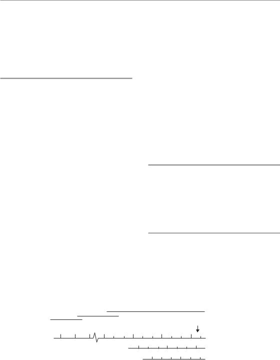
Irradiation of any biological system generates a succession of processes that differ enormously in time-scale. This is illustrated in Fig. 1.1 in which these processes are divided into three phases (Boag, 1975), described below.
Physical phase
The physical phase consists of interactions between charged particles and the atoms of which the tissue is composed. A high-speed electron takes about 10 18 s to traverse the DNA molecule and about 10 14 s to pass across a mammalian cell. As it does so it interacts mainly with orbital electrons, ejecting
Biological
Chemical |
|
|
|
||
Physical |
|
|
|
Human lifespan |
|
|
|
|
|
||
10 10 12 10 |
1 |
103 |
106 |
109 |
|
|
|
|
103 |
(seconds) |
|
|
|
1 |
106 |
||
|
|
|
|
(hours) |
|
Free-radical |
|
|
1 |
103 |
|
reactions |
|
|
|
(days) |
|
|
|
Early effects |
|||
|
Enzyme reactions |
||||
|
Late effects |
||||
Excitation |
Repair processes |
||||
|
|
||||
Ionization |
Carcinogenesis |
|
Cell proliferation |
||
|
Figure 1.1 Time-scale of the effects of radiation exposure on biological systems.

Response of tissues to radiation exposure 5
some of them from atoms (ionization) and raising others to higher energy levels within an atom or molecule (excitation). If sufficiently energetic, these secondary electrons may excite or ionize other atoms near which they pass, giving rise to a cascade of ionization events. For 1 Gy of absorbed radiation dose, there are in excess of 105 ionizations within the volume of every cell of diameter 10 μm.
Chemical phase
The chemical phase describes the period in which these damaged atoms and molecules react with other cellular components in rapid chemical reactions. Ionization and excitation lead to the breakage of chemical bonds and the formation of broken molecules known as ‘free radicals’. These are highly reactive and they engage in a succession of reactions that lead eventually to the restoration of electronic charge equilibrium. Free-radical reactions are complete within approximately 1 ms of radiation exposure. An important characteristic of the chemical phase is the competition between scavenging reactions, for example with sulphydryl compounds that inactivate the free radicals, and fixation reactions that lead to stable chemical changes in biologically important molecules.
Biological phase
The biological phase includes all subsequent processes. These begin with enzymatic reactions that act on the residual chemical damage. The vast majority of lesions, for example in DNA, are successfully repaired. Some rare lesions fail to repair and it is these that lead eventually to cell death. Cells take time to die; indeed after small doses of radiation they may undergo a number of mitotic divisions before dying. It is the killing of stem cells and the subsequent loss of the cells that they would have given rise to that causes the early manifestations of normal-tissue damage during the first weeks and months after radiation exposure. Examples are breakdown of the skin or mucosa, denudation of the intestine and haemopoietic damage (see Chapter 13, Section 13.2). A secondary effect of cell killing is compensatory cell proliferation, which occurs both in normal tissues and in tumours. At later times after
the irradiation of normal tissues the so-called ‘late reactions’ appear. These include fibrosis and telangiectasia of the skin, spinal cord damage and blood vessel damage. An even later manifestation of radiation damage is the appearance of second tumours (i.e. radiation carcinogenesis). The time-scale of the observable effects of ionizing radiation may thus extend up to many years after exposure.
1.4 RESPONSE OF NORMAL AND MALIGNANT TISSUES TO RADIATION EXPOSURE
Much of the text of this book will focus on the effects of radiation exposure that become apparent to the clinician or the patient during the weeks, months and years after radiotherapy. These effects are seen both in the tumour and in the normal tissues that are unavoidably included within the treatment plan and exposed to radiation. The primary tasks of radiation biology, as applied to radiotherapy, are to explain observed phenomena, and to suggest improvements to existing therapies (as outlined in Section 1.2).
The response of a tumour is seen by regression, often followed by regrowth (or recurrence), but perhaps with failure to regrow during the normal lifespan of the patient (which we term cure or, more correctly, local control). These italicized terms describe the tumour responses that we seek to understand. The cellular basis of tumour response, including tumour control, is dealt with in Chapter 7.
The responses of normal tissues to therapeutic radiation exposure range from those that cause mild discomfort to others that are life-threatening. The speed at which a response develops varies widely from one tissue to another and often depends on the dose of radiation that the tissue receives. Generally speaking, the haemopoietic and epithelial tissues manifest radiation damage within weeks of radiation exposure, while damage to connective tissues becomes important at later times. A major development in the radiobiology of normal tissues during the 1980s was the realization that early and late normal-tissue responses are differently modified by a change in dose fractionation and this gave rise to the current interest in hyperfractionation (see Chapter 10, Section 10.3).
6Introduction: the significance for cancer treatment
The first task of a radiobiologist is to measure a tissue response accurately and reliably. The term assay is used to describe such a system of measurement. Assays for tumour response are described in Chapter 7, Section 7.2. For normal tissues, the following three general types of assay are available:
●Scoring of gross tissue effects – it is possible to grade the severity of damage to a tissue using an arbitrary scale as is done, for example, in Figs 13.7 and 13.9. In superficial tissues this approach has been remarkably successful in allowing isoeffect relationships to be determined.
●Assays of tissue function – for certain tissues, functional assays are available that allow radiation effects to be documented. Examples are the use of breathing rate as a measure of lung function in mice, ethylenediaminetetraacetic acid (EDTA) clearance as a measure of kidney damage (Fig. 8.4), or blood counts as an indicator of bone marrow function.
●Clonogenic assays – in some tumours and some normal tissues it has been possible to develop methods by which the colony of cells that derive
from a single irradiated cell can be observed. In tumours this is particularly important because of the fact that regrowth of a tumour after subcurative treatment is caused by the proliferation of a small number of tumour cells that retain colony-forming ability. This important area of radiation biology is introduced in Chapter 4.
1.5 RESPONSE CURVES, DOSE–RESPONSE CURVES AND ISOEFFECT RELATIONSHIPS
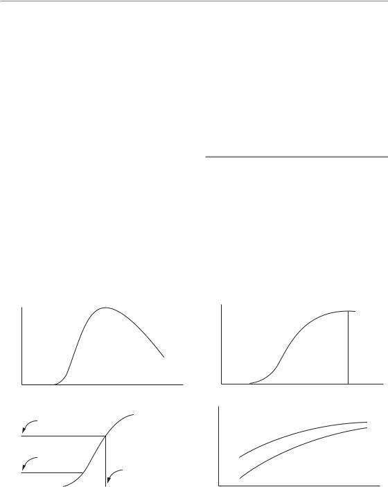
The damage that is observed in an irradiated tissue increases, reaches a peak, and then may decline (Fig. 1.2a). How should we quantify the magnitude of this response? We could use the measured response at some chosen time after irradiation, such as the time of maximum response, but the timing of the peak may change with radiation dose and this would lead to some uncertainty in the interpretation of the results. A common method is to calculate the cumulative response by integrating this curve from left to right (Fig. 1.2b). Some normal tissue
Response |
|
Response |
(a) |
Time after irradiation |
(b) |
|
R |
|
|
|
|
Response |
T |
Totaldose |
|
|
|
|
D |
|
|
|
(d) |
(c) |
Radiation dose |
Cumulative
response
R
Time after irradiation
Tumour response
Normal-tissue damage
Number of fractions
Figure 1.2 Four types of chart leading to the construction of an isoeffect plot. (a) Time-course of development of radiation damage in a normal tissue. (b) The cumulative response. (c) A dose–response relationship, constructed by measuring the response (R) for various radiation doses (D); acceptable clinical tolerance (T ) of most tissues is towards the low end of the dose–response relationship. (d) Isoeffect plot for a fixed level of normal-tissue damage (also a similar plot for tumour response).

Concept of therapeutic index 7
responses give a cumulative curve that rises to a plateau, and the height of the plateau is a good measure of the total effect of that dose of radiation on the tissue. Other normal-tissue responses, in particular the late responses seen in connective and vascular tissues, are progressive and the cumulative response curve will continue to rise (Figs 13.7 and 13.8). The quantification of clinical normal-tissue reactions is dealt with in Chapter 13, Section 13.4.
The next stage in a study of the radiation response of a tissue will be to vary the radiation dose and thus to investigate the dose–response relationship (Fig. 1.2c). Many examples of such curves are given throughout this book, for example Figs 5.6, 14.3 and 19.8. Cell survival curves (see Chapter 4, Section 4.3) are further examples of dose–response curves that are widely used in radiobiology. The position of the curve on the dose scale indicates the sensitivity of the tissue to radiation; its steepness also gives a direct indication of the change in response that will accompany an increase or decrease in radiation dose. These aspects of dose– response curves are dealt with in detail in Chapter 5.
The foregoing paragraphs have, for simplicity, referred to ‘dose’ as though we are concerned only with single radiation exposures. It is a wellestablished fact in radiation oncology that multiple radiation doses given over a period of a few weeks give a better curative response than can be achieved with a single dose. Diagrams similar to Fig. 1.2a–c can also be constructed for fractionated radiation treatment, although the results are easiest to interpret when the fractions are given over a time that is short compared with the time-scale of development of the response. If we change the schedule of dose fractionation, for example by giving a different number of fractions, changing the fraction size or changing radiation dose rate, we can then investigate the therapeutic effect in terms of an isoeffect plot (Fig. 1.2d). Experimentally this is done by performing multiple studies at different doses for each chosen schedule and calculating a dose–response curve. We then select some particular level of effect (R in Fig. 1.2c) and read off the total radiation dose that gives this effect. For effects on normal tissues the isoeffect will often be some upper limit of tolerance of the tissue, perhaps expressed as a probability of tissue failure (see Chapter 5, Section 5.1, and
Chapter 14, Section 14.2) and perhaps choosing a
lower level of effect (T in Fig. 1.2c) will be more appropriate. The isoeffect plot shows how the total radiation dose for the chosen level of effect varies with dose schedule: examples are given in Figs 8.2 and 10.3, and recommendations for tolerance calculations are set out in Chapters 8 and 9. The dashed line in Fig. 1.2d illustrates how therapeutic conclusions may be drawn from isoeffect curves. If the curve for tumour response is flatter than for normal-tissue tolerance, then there is a therapeutic advantage in using a large number of fractions: a tolerance dose given using a small number of fractions will be far short of the tumour-effective dose, whereas for large fraction numbers it may be closer to an effective dose.
1.6 THE CONCEPT OF THERAPEUTIC INDEX
Any discussion of the possible benefit of a change in treatment strategy must always consider simultaneously the effects on tumour response and on normal-tissue damage. A wide range of factors enter into this assessment. In the clinic, in addition to quantifiable aspects of tumour response and toxicity, there may be a range of poorly quantifiable factors such as new forms of toxicity or risks to the patient, or practicality and convenience to hospital staff; there are also cost implications. These must all be balanced in the clinical setting. The role of radiation biology is to address the quantifiable biological aspects of a change in treatment.
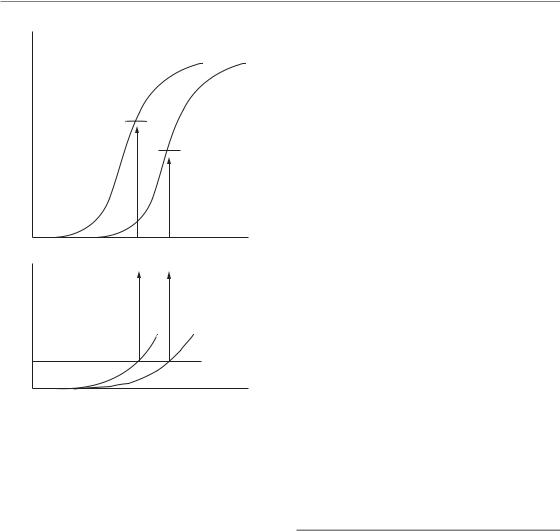
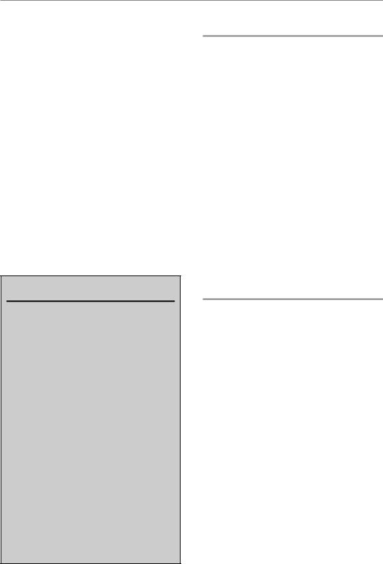
In the research setting, this can be done by considering dose–response curves. As radiation dose is increased, there will be a tendency for tumour response to increase, and the same is also true of damage to normal tissues. If, for example, we measure tumour response by determining the proportion of tumours that are controlled, then we expect a sigmoid relationship to dose (for fractionated radiation treatment we could consider the total dose or any other measure of treatment intensity). This is illustrated in the upper part of Fig. 1.3. If we quantify damage to normal tissues in some way for the same treatment schedule, there will also be a rising curve of toxicity (lower panel). The shape of this curve is unlikely to be the same as that for tumour response and we probably will not wish to determine more
8Introduction: the significance for cancer treatment
control |
With drug |
|
|
Radiation |
|
|
|
|
tumour |
B |
alone |
|
||
|
A |
|
Local |
|
|
-tissue damage |
|
Normal |
Max. tolerated |
|
level
Radiation dose
Figure 1.3 The procedure by which an improvement in therapeutic index might be identified, as a result of adding chemotherapy to radiotherapy (see text, Section 1.6).
than the initial part of this curve since a high frequency of severe damage is unacceptable. By analogy with what must be done in the clinic, we can then fix a notional upper limit of tolerance (see Chapter 14, Section 14.2). This fixes, for that treatment schedule, the upper limit of radiation dose that can be tolerated, for which the tumour response is indicated by the point in Fig. 1.3 labelled A.
Consider now the effect of adding treatment with a cytotoxic drug. We plan that this will increase the tumour response for any radiation dose and this will be seen as a movement to the left of the curve for tumour control (Fig. 1.3). However, there will probably also be an increase in damage to normal tissues which again will consist of a leftward movement of the toxicity curve. The relative displacement of the curves for the tumour and normal tissues will usually be different and this fact makes the amount of benefit from the chemotherapy very difficult to assess. How do we know whether there has been a real therapeutic
gain? For studies on laboratory animals there is a straightforward way of asking whether the combined treatment is better than radiation alone: for the same tolerance level of normal-tissue damage (the broken line) the maximum radiation dose (with drug) will be lower and the corresponding level of tumour control is indicated by point B in Fig. 1.3. If B is higher than A then the combination is better than radiation alone and represents a therapeutic gain, because it gives a greater level of tumour control for the same level of morbidity.
This example illustrates the radiobiological concept of therapeutic index: it is the tumour response for a fixed level of normal-tissue damage (see Chapter 5, Section 5.6). The term therapeutic window describes the (possible) difference between the tumour control dose and the tolerance dose. The concept can, in principle, be applied to any therapeutic situation or to any appropriate measures of tumour response or toxicity. Its application in the clinic is, however, not a straightforward matter, as indicated in Chapter 18, Section 18.1. Therapeutic index also carries the notion of ‘cost–benefit’ analysis. It is impossible to reliably discuss the potential benefit of a new treatment without reference to its effect on therapeutic index.
1.7 THE IMPORTANCE OF RADIATION BIOLOGY FOR THE FUTURE DEVELOPMENT OF RADIOTHERAPY
Radiation oncology, more than any of the other modalities for cancer treatment, is, to a large extent, a technical discipline. Improvements in the treatment of cancer with radiotherapy over recent decades have resulted mainly from improvements in technology, combining new methods of precision in dose delivery with new imaging tools. A major recent development has been the introduction of intensity modulation in combination with various functional imaging modalities such as functional magnetic resonance imaging (fMRI) and positron emission tomography(PET)/computed tomography (CT). This has led to new concepts such as ‘biological target volume’, ‘dose painting’ and ‘theragnostic imaging’ (see Chapter
20). These developments will undoubtedly lead to further improvements in tumour control rates and reductions in morbidity.

Importance of radiation biology for future development 9
In parallel with these technological advances, new developments have taken place in radiobiology, encompassing the understanding of cancer biology in general, and the radiation response in particular. These fundamental and preclinical research efforts in biology hold great promise, just as the technical innovations, for improving the radiotherapy of cancer. It is even likely that the expected improvements from technical innovations will reach a limit, and the next breakthroughs will come from biological innovations, such as the application of molecularly targeted drugs (see Chapters 21 and 22) in combination with high-precision methods to deliver radiation.
It is interesting to note that the recent rapid progress in knowledge of the biology of cancer is itself also partly due to technological innovations, especially in high-throughput methods to study the genetics of the whole cell. There are now several methods to look at the genes (DNA) and expression of those genes (RNA and protein) in high numbers (tens of thousands) all at once. The trend here is away from the study of single genes or parameters towards genome-wide studies. The many different potential causes of failure, or of severe normal-tissue reactions, necessitates such multiparameter/multigene studies. Methods to selectively manipulate gene expression represent another revolution in biology, allowing one to quickly assess the importance of any given gene by reducing or eliminating its expression (RNA interference and microRNA methods). Radiation biologists are now exploiting these techniques to better understand the molecular pathways that determine how cells respond to damage. This should lead to identification not only of new targets, but of targets that are specifically deregulated in tumours, providing the all-important tumour specificity of therapy. This should also lead to the development of more robust and accurate predictors of which tumours or normal tissues will respond well to standard radiotherapy and which will not, which could significantly improve individualized radiotherapy (see Chapter 23).
Over the last decade we have seen a change from ‘classic radiobiology’, which has often focused on fractionation, the LQ model, and the phenomenology of repair in terms of ‘sublethal’ and ‘potentially lethal’ damage. However, fractionation remains an important core subject for the application of
radiation therapy, and the development of the LQ model, together with elucidation of the importance of repopulation, has been central in understanding fractionation, leading to new and better clinical fractionation schemes and the ability to predict the response of normal tissue and tumours to nonstandard schedules (see Chapters 8–12). It is of great interest to see a change developing in the established concept of high α/βvalues for tumours and acutely responding tissues, and low α/β values for lateresponding tissues. This ‘dogma’ has now evolved into a more differentiated view, indicating that some tumours may have a lower α/βratio than surrounding normal tissues, requiring a very different approach to the design of treatment schedules. This new knowledge is now being applied to the design of hypofractionated schedules, such as for the treatment of prostate tumours, which is a dramatic deviation from clinical practice in the last decades.
In a similar manner, simple descriptions of repair and recovery have been supplemented by increasing knowledge and understanding of the molecular pathways involved in various types of repair, including those for base damage, singlestrand DNA breaks and double-strand breaks. This is leading to new ways to target deregulated repair pathways, with the promise of improving radiotherapy (see Chapter 23). In recent years, a link has been established between the EGFR pathway and DNA double-strand break repair. This is highly relevant to radiotherapy as blocking the EGFR has been shown to improve the effect of radiotherapy in head and neck cancer (see Chapter 21).
Hypoxia has always been a focus in radiation research, given its large influence on radiosensitivity (see Chapter 15). However, here again, phenomenology has now been replaced by a huge plethora of molecular studies illuminating how cells respond to hypoxia of different degrees and fluctuating over time (see Chapter 16). Hypoxia is also an important issue for other disciplines apart from cancer, and so an enormous amount of fundamental information has been contributed by these different areas, which radiation biologists can also exploit. This has led to several novel ways to either attack or exploit tumour hypoxia clinically (see Chapter 17).
Indirectly related to hypoxia is the tumour vasculature and blood supply, and this component of the tumour microenvironment has been a target for therapy for many years now. One approach is to
10 Introduction: the significance for cancer treatment
block one of the most important growth factors involved in new vessel formation and the maintenance of blood vessels, vascular endothelial growth factor (VEGF). Another approach is to modify the function of mature blood vessels. Since radiation therapy is a balancing act between damage to tumours and normal tissues, sparing the latter has always attracted the attention of radiation scientists. The trends in radiation studies of normal tissues, as above, are to elucidate the molecular pathways determining response, and by an increased understanding to both predict and ameliorate severe sideeffects (see Chapter 22).
Radiation oncology has always been at the interface of physics, biology and medicine, and, with new developments in the technology of high-precision beam delivery with functional and molecular imaging, these are exciting times. Clearly, today’s radiation oncologists and clinical physicists need to obtain a solid understanding of both radiation biology and the new developments in molecular radiation oncology: that is the purpose of this book.
■ BIBLIOGRAPHY
Boag JW (1975). The time scale in radiobiology. 12th Failla memorial lecture. In: Nygaard OF, Adler HI, Sinclair WK (eds) Radiation research. Proceedings of the 5th International Congress of Radiation Research. New York: Academic Press, 9–29.
Delaney G, Jacob S, Featherstone C, Barton M (2005). The role of radiotherapy in cancer treatment: estimating optimal utilization from a review of evidence-based clinical guidelines. Cancer 104: 1129–37.
DeVita VT, Oliverio VT, Muggia FM et al. (1979). The drug development and clinical trials programs of the division of cancer treatment, National Cancer Institute. Cancer Clin Trials 2: 195–216.
Souhami RL, Tobias JS (1986). Cancer and its management. Oxford: Blackwell Scientific.
Tobias JS (1996). The role of radiotherapy in the management of cancer – an overview. Ann Acad Med Singapore 25: 371–9.
Tubiana M (1992). The role of local treatment in the cure of cancer. Eur J Cancer 28A: 2061–9.
Key points
1.Radiotherapy is a very important curative and palliative modality in the treatment of cancer, with more than half of all patients estimated to receive radiotherapy at some point during their management.
2.The effects of radiation on mammalian tissues should be viewed as a succession of processes extending from microseconds to months and years after exposure. In choosing one endpoint of effect, it is important not to overlook the rest of this process.
3.Therapeutic index is always ‘the name of the game’ in curative cancer therapy.
4.Significant gains are still to be made by the optimization of biological and physical factors, particularly in the domain of ‘biologically based treatment planning’ and image-guided therapy.
5.Further gains will also accrue from the increasing knowledge of the molecular mechanisms underlying radiation responses, enabling tumour-specific targeting of radiosensitization.
■ FURTHER READING
Bentzen SM, Thames HD (1995). A 100-year Nordic perspective on the dose-time problem in radiobiology. Acta Oncol 34: 1031–40.
Feinendegen L, Hahnfeldt P, Schadt EE, Stumpf M, Voit EO (2008). Systems biology and its potential role in radiobiology. Radiat Environ Biophys 47: 5–23.
Salminen E, Izewska J, Andreo P (2005). IAEA’s role in the global management of cancer – focus on upgrading radiotherapy services. Acta Oncol 44: 816–24.
Willers H, Beck-Bornholdt HP (1996). Origins of radiotherapy and radiobiology: separation of the influence of dose per fraction and overall treatment time on normal tissue damage by Reisner and Miescher in the 1930s. Radiother Oncol 38: 171–3.