
10
Modified fractionation
MICHAEL BAUMANN AND VINCENT GRÉGOIRE
|
|
|
|
|
|
10.1 |
Introduction |
135 |
10.7 |
Split-course radiotherapy |
144 |
10.2 |
Conventional fractionation |
135 |
10.8 |
Reasons for increased late normal-tissue |
|
10.3 |
Modification of dose per fraction |
136 |
|
damage after modified fractionation |
145 |
10.4 |
The time factor for fractionated |
|
10.9 |
Is the same modified fractionation |
|
|
radiotherapy in tumours |
138 |
|
schedule optimal for all patients? |
145 |
10.5 |
Clinical evaluation of accelerated |
|
Key points |
146 |
|
|
radiotherapy |
140 |
Bibliography |
147 |
|
10.6 |
Comparison between hyperfractionation |
|
Further reading |
148 |
|
|
and accelerated fractionation |
144 |
|
|
|
|
|
|
|
|
|
10.1 INTRODUCTION
The clinical evaluation and implementation of modified fractionation schedules based on biological rationales is an important focus of ‘translational research’ in radiation oncology. Throughout the history of radiotherapy, the optimal distribution of dose over time has been a major issue but important progress has been made in this area over the past two decades. The relationships uncovered between total dose and fraction number for late-responding normal tissues, early-responding normal tissues and tumours provide the basic information required to optimize the dose per fraction in radiotherapy. Work still needs to be done to determine the exact time of onset, the rate and the mechanisms of repopulation in tumours and normal tissues during radiotherapy, but enough is now known about time factors to support the important conclusions that: (1) the overall duration of fractionated radiotherapy should not be allowed to extend beyond the originally prescribed time; (2) a
reduced overall treatment time should be considered in a number of clinical situations; and (3) inter-fraction time intervals should be made as long as possible in order to gain the full benefit from fractionation schedules employing multiple fractions per day.
This chapter summarizes the current status of modified fractionation in clinical radiotherapy.
10.2 CONVENTIONAL FRACTIONATION
Conventional fractionation is the application of daily doses of 1.8–2 Gy and five fractions per week (Monday to Friday) with a dose per week of 9–10 Gy. Depending on tumour histology, tumour size and localization, total doses ranging from 40 Gy to 70 Gy are given for macroscopic disease and lower doses when treating microscopic disease. These conventional fractionation schedules were developed on an empirical basis (Fletcher, 1988) and have been the mainstay of
136 Modified fractionation
curative radiotherapy over the last decades in most institutions in Europe and the USA.
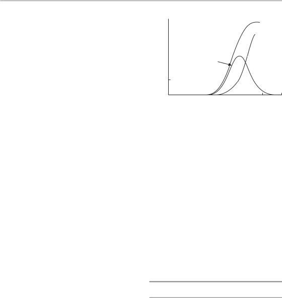
Radiosensitive tumours such as lymphomas and seminomas can be controlled with low doses of 45 Gy or even less and in this situation there is a low incidence of collateral normal-tissue damage. By contrast, glioblastoma multiforme is a very resistant tumour that is not controlled even after doses as high as 70 Gy. Most tumour types, including squamous cell carcinomas and adenocarcinomas, are of intermediate sensitivity. Small tumours, for example T1 or T2 carcinomas of the head and neck, are well controlled with acceptable normal-tissue damage using conventional fractionation and total doses between 60 Gy and 70 Gy. However, local tumour control rates rapidly decline for larger and more advanced tumours. As local tumour control increases with the total dose of radiotherapy, the question may be asked whether improved management of larger tumours could be gained by increasing the total dose of conventional fractionation above 70 Gy, say to doses between 80 Gy and 100 Gy. Such dose escalation is currently being tested for non-small cell lung cancer and for carcinoma of the prostate. One constraint is that not only tumour control rates but also the incidence and severity of nor- mal-tissue damage increase with increasing total doses (see Chapter 5). This was recognized as early as 1936 when Holthusen pointed out that the uncomplicated local tumour control rate initially increases with increasing dose but then falls again because of the steep increase in the incidence of normal-tissue damage. Figure 10.1 is redrawn from one of Holthusen’s papers: the frequency of ‘uncomplicated tumour control’ follows a bellshaped curve. Once the optimum dose is established, further improvements in uncomplicated tumour control can only be achieved by either moving the dose–effect curve for local tumour control to lower doses or the curve for normal-tis- sue damage to higher doses; the latter is the objective of hyperfractionated schedules (see Section 10.3). Conformal radiotherapy is another option currently used in dose escalation protocols, reducing the volume of normal tissue irradiated to high dose and therefore also the probability of late nor- mal-tissue damage (see Chapters 14 and 20). As dose escalation using conventional fractionation
|
1.0 |
|
Tumour control |
|
|
|
|
||
rate |
0.8 |
|
|
|
|
|
|||
0.6 |
|
|
|
|
Response |
|
Complication-free |
Normal-tissue |
|
|
|
|||
|
|
damage |
||
|
|
|
||
|
|
|
control of disease |
|
|
0.4 |
|
|
|
|
|
|
|
0.2
0.0 


 Radiation dose (Gy)
Figure 10.1 Dose–response curves for tumour control and normal-tissue damage. The uncomplicated local tumour control rate initially increases with increasing dose after which it falls again because of a steep increase in the incidence of damage to normal tissue. Adapted from Holthusen (1936).
is always associated with the prolongation of overall treatment time, some of the potential gain may be lost as a consequence of tumour-cell repopulation and this is considered in Section 10.4.
Radiotherapy is highly effective in dealing with a small number of cancer cells, as in the treatment of microscopic disease. Thus, lower doses of irradiation are also used postoperatively after complete resection of breast or head and neck cancer.
10.3 MODIFICATION OF DOSE PER FRACTION
Hyperfractionation
Hyperfractionation is the term used to describe radiotherapy with doses per fraction less than the 1.8–2.0 Gy given in conventional fractionation. The total number of fractions must be increased, hence the prefix ‘hyper-’; usually two fractions are administered per day. The biological rationale of hyperfractionation is to exploit the difference between the small effect of dose per fraction on tumour control versus the larger effect of dose per fraction on the incidence and severity of late nor- mal-tissue damage. As pointed out in Chapter 8, this differential between tumours and lateresponding normal tissues is thought to be caused
Modification of dose per fraction 137
|
100 |
|
|
Local tumour control |
|
|
|
|
100 |
|
Absence of late effects |
|
|
|
|||||
|
90 |
|
|
|
|
|
|
|
|
|
90 |
|
|
|
|
|
|
|
|
|
80 |
|
|
|
|
|
|
|
|
|
80 |
|
|
|
|
|
|
|
|
(%) |
70 |
|
|
|
|
|
|
|
|
(%) |
70 |
|
|
|
|
|
|
|
|
60 |
|
|
|
|
Hyperfractionation |
60 |
|
|
|
|
|
|
|
|
|||||
Probability |
|
|
|
|
Probability |
|
|
|
|
|
|
|
|
||||||
50 |
|
|
|
|
|
Conventional |
|
50 |
|
|
|
|
|
|
|
|
|||
40 |
|
|
|
|
|
|
40 |
|
|
|
|
|
Hyperfractionation |
||||||
|
|
|
|
|
|
|
|
|
|
|
|
|
|||||||
30 |
|
|
|
|
|
|
|
|
30 |
|
|
|
|
|
|
Conventional |
|||
|
|
|
|
|
|
|
|
|
|
|
|
|
|
|
|
||||
|
|
|
|
|
|
|
|
|
|
|
|
|
|
|
|
|
|
||
|
20 |
|
|
|
|
|
|
|
|
|
20 |
|
|
|
|
|
|
|
|
|
10 |
|
|
|
|
|
|
|
|
|
10 |
|
|
|
|
|
|
|
|
|
0 |
1 |
2 |
3 |
4 |
5 |
6 |
7 |
8 |
|
0 |
1 |
2 |
3 |
4 |
5 |
6 |
7 |
8 |
(a) |
|
Duration of trial (years) |
|
|
|
(b) |
|
Duration of trial (years) |
|
|
|
||||||||
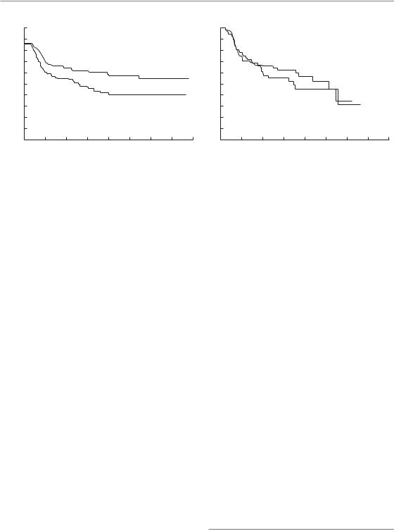
Figure 10.2 Results of the EORTC (22791) trial of dose-escalated hyperfractionation. (a) Loco-regional tumour control (log-rank p 0.02); (b) patients free of late radiation effects, grade 2 or worse (log-rank p 0.72). From Horiot et al. (1992), with permission.
by a different capacity of target cells in these tissues to recover from sublethal radiation damage between fractions.
In clinical practice hyperfractionation is usually applied to escalate the total dose compared with conventional fractionation, thereby aiming at improved tumour control rates without increasing the risk of late complications (see Fig. 8.7). Dose-escalated hyperfractionation has been tested in two large multicentre randomized clinical trials on head and neck squamous cell carcinoma [European Organisation for Cancer Research (EORTC) No. 22791 and Radiation Therapy Oncology Group (RTOG) No. 9003]. The results of the EORTC trial are shown in Fig. 10.2. In Fig. 10.2b it can be seen that hyperfractionated treatment with 70 fractions of 1.15 Gy (two fractions per day with a 4- to 6-hour interval; total dose 80.5 Gy) produced a similar incidence of pooled grades 2 and 3 late tissue damage to a conventional schedule of 35 fractions of 2 Gy (70 Gy given in the same overall time of 7 weeks). However, the larger total dose in the hyperfractionated treatment produced an increase of about 19 per cent in long-term local tumour control (Fig. 10.2a). Survival appeared higher after hyperfractionation but this difference did not reach statistical significance. In the RTOG trial (Fu et al.,
2000), local tumour control was increased by 8 per cent after hyperfractionation (68 fractions of
1.2 Gy, two fractions per day, 6 hours apart, total dose 81.6 Gy) compared with conventional fractionation using 2-Gy fractions to 70 Gy in the same overall time of 7 weeks. Overall survival was not significantly improved but the prevalence of grade 3 late effects was significantly increased after hyperfractionation. Both these clinical trials thus confirm the radiobiological expectation that local tumour control can be increased by doseescalated hyperfractionation, thereby supporting a high average α/β ratio for squamous cell carcinoma of the head and neck (Table 9.1). However, while the EORTC trial supports the view that hyperfractionation allows the total dose to be increased without a simultaneous increase in late complications, the RTOG trial indicates that this is not always the case. The potential therapeutic gain from hyperfractionation is debated by BeckBornholdt et al. (1997), Baumann et al. (1998) and Bentzen et al. (1999). Factors that contribute to an increased risk of late normal-tissue damage when multiple fractions are applied per day are discussed in Section 10.5.
Hypofractionation
This is the use of doses per fraction higher than 2.0 Gy; the total number of fractions is reduced, hence the prefix ‘hypo-’. As explained in Chapter 8,

138 Modified fractionation
Section 8.6, the radiobiological expectation is that hypofractionation will lower the therapeutic ratio between tumours and late-responding normal tissues, compared with conventional fractionation given in the same overall time. This expectation depends on the α/β ratio for the tumour being considerably higher than for late-responding normal tissues; exceptions could therefore occur for tumours that have low α/β ratios, for example some melanomas, liposarcomas and potentially early-stage prostate and breast cancer (see Table 9.1). In these cases hypofractionation may be as good or even better than conventional fractionation. For example a randomized clinical trial (Owen et al., 2006), including 1410 women with invasive breast cancer who had local tumour excision, compared 50 Gy in 25 fractions, 39 Gy in 13 fractions and 42.9 Gy in 13 fractions, all given over 5 weeks. The risk of ipsilateral tumour relapse after 10 years was 12.1 per cent in the 50 Gy group, 14.8 per cent in the 39 Gy group, and 9.6 per cent in the 429 Gy group (difference between 39 Gy and 42.9 Gy groups, p 0.027). The α/βratio of breast cancer was estimated from these data to be 4.0 Gy [95 per cent confidence interval (CI) 1.0–7.8], similar to that estimated for the late adverse effects in healthy tissue from breast radiotherapy.
Single-dose irradiation or hypofractionation with only few large fractions are widely applied in palliative radiotherapy. These schedules use lower total doses than those applied in curative radiotherapy. For this reason, and because the patients have a limited life expectancy, late normal-tissue damage is of only minor concern. A number of randomized clinical trials have shown that symptom control after palliative hypofractionated schedules is comparable to that achieved with more highly fractionated schedules. Hypofractionated schedules have the advantage of being more convenient for the patient and they help spare resources.
For stereotactic radiotherapy of small tumours (e.g. in lung, where very steep dose gradients can be achieved and hence only very small volumes of surrounding normal tissue are at risk of radiation damage) single doses or hypofractionation with few large fractions are also frequently applied in clinical practice.
Moderate hypofractionation with doses per fraction up to approximately 3.5 Gy is routinely
used for curative radiation therapy in many centres worldwide. To reduce the risk of late normaltissue damage in these schedules, slightly lower total doses are applied than for conventional fractionation. For tumours with a high α/β ratio this decrease in total dose may well lead to a reduction in tumour control probability. However, some or all of this negative effect may be compensated by the shorter overall treatment times often used for this moderate hypofractionation (see Section 10.4). There is growing interest in the use of moderate hypofractionation to escalate total doses in the context of clinical trials of conformal radiation therapy. For example, using a field-in-field technique or intensity-modulated radiotherapy (IMRT), a higher dose per fraction can be applied for boosting the macroscopic tumour while potential microscopic tumour extensions are treated at conventional doses per fraction. Such hypofractionated approaches for dose escalation avoid the necessity to prolong the overall treatment time and conserve treatment resources. However, these advantages have to be carefully weighed against the increased risk of late normaltissue injury, and clinical trials are therefore necessary to fully evaluate the therapeutic gain compared with standard approaches.
10.4 THE TIME FACTOR FOR FRACTIONATED RADIOTHERAPY IN TUMOURS
In the 1960s and 1970s the prevailing view among radiation oncologists had been that prolonged overall treatment times of fractionated irradiation did not impair local tumour control. Contrary to this general view, studies on experimental tumours were beginning to find that clonogenic cells proliferate rapidly after irradiation (see Chapter 7) so predicting that the ability of clinical radiotherapy to achieve local tumour control would actually decrease with increasing overall treatment time. Several experimental studies indicate that repopulation of clonogenic tumour cells accelerates at some point during fractionated radiotherapy (see Chapters 8 and 9). It should also be noted that other mechanisms such as long repair halftimes or increasing radiobiological
The time factor for fractionated radiotherapy 139
TCD50 (Gy)
(a)
80 |
|
|
|
|
|
|
|
|
90 |
|
|
|
|
|
|
|
|
|
|
|
|
|
|
|
|
|
80 |
|
|
|
|
|
|
|
|
70 |
|
|
|
|
|
|
|
|
70 |
|
|
|
|
|
|
|
|
|
|
|
|
|
|
|
|
|
|
|
|
|
|
|
|
|
|
60 |
|
|
|
|
|
|
|
(Gy) |
60 |
|
|
|
|
|
|
|
|
|
|
|
|
|
|
|
50 |
|
|
|
|
|
|
|
|
|
|
|
|
|
|
|
|
|
|
TCD |
50 |
|
|
|
|
|
|
|
|
|
|
|
|
|
|
|
|
|
|
|
|
|
|
|
|
|
|
50 |
|
|
Miso |
|
|
|
|
|
40 |
|
|
|
|
|
|
|
|
|
|
|
|
|
|
|
|
|
|
|
|
|
300 |
|
|
||
|
|
|
|
|
|
|
|
|
|
|
|
|
|
|
|
||
|
|
HBO |
|
|
|
|
|
|
|
|
|
|
|
|
|
|
|
|
|
|
|
|
|
|
|
30 |
|
|
|
|
|
100 |
|
|
|
|
|
|
HBO |
|
|
|
|
|
|
|
|
|
|
|
|
||
|
|
|
|
|
|
|
|
|
|
|
|
|
50 |
|
|
||
|
|
|
|
|
|
|
|
|
|
|
|
|
|
|
10 |
|
|
40 |
10 |
20 |
30 |
40 |
50 |
60 |
70 |
|
20 |
10 |
20 |
30 |
40 |
50 |
60 |
70 |
80 |
0 |
|
0 |
|||||||||||||||
|
|
Overall treatment time (days) |
|
|
(b) |
|
Overall treatment time (days) |
|
|||||||||
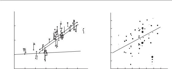
Figure 10.3 Tumour control dose (TCD50 – dose required to control 50 per cent of the tumours) in head and neck cancer as a function of overall treatment time, normalized to a dose per fraction of 2 Gy. The same large set of clinical studies has been retrospectively summarized by (a) Withers et al. (1988) and (b) Bentzen et al. (1991). In (b) each point indicates the result of a particular trial, the size of the symbol indicating the size of the trial. There is a trend for the curative radiation dose to increase with overall treatment time, although the details of this association differ between the two studies.
hypoxia during treatment might contribute to a detrimental effect of long overall treatment times on tumour control.
The possible existence of a tumour time-factor in clinical radiotherapy became more widely acknowledged following a publication by Withers et al. (1988) entitled ‘The hazard of accelerated tumour clonogen repopulation during radiotherapy’. This review examined the correlation between tumour control and overall treatment time for squamous cell carcinomas of the head and neck and led to the diagram shown in Fig. 10.3a: this shows the dose required to achieve tumour control in 50 per cent of cases (i.e. TCD50 values) plotted against the overall treatment time. Since a variety of doses per fraction were used in the various original studies summarized in this plot, the linear-quadratic (LQ) model was used with an α/β ratio of 25 Gy to convert from the actual doses per fraction used into equivalent doses using 2 Gy per fraction (EQD2, see Chapter 8, Section 8.7). The various studies also achieved different tumour control rates and it was therefore necessary to interpolate or extrapolate to the 50 per cent control level. This required an assumed value for the steepness of the dose–response relationship for the
tumours; it was assumed that the dose to increase control from 40 per cent to 60 per cent was 2.9 Gy. As can be seen from Fig. 10.3a, this retrospective review of head and neck cancer data found a clear trend: as overall time increased, a greater total radiation dose had been required to control these tumours. The other important conclusion was that there seemed to be an initial flat portion to this relationship (the so-called ‘dog-leg’). This implied that, for treatment times shorter than 3–4 weeks, tumour proliferation had little effect and that, as shown for experimental tumours, it also takes time for accelerated repopulation to be ‘switched on’ in human tumours. Withers et al. (1988) concluded that for treatment times longer than 4 weeks, the effect of proliferation was equivalent to a loss of radiation dose of about 0.6 Gy/day.
This publication gave rise to considerable debate. Subsequent analyses of the same clinical data carried out by Bentzen and Thames (1991) and by Dubben (1994) are shown in Figs 10.3b and 10.4. The analysis of Bentzen and Thames made a different assumption about the steepness of dose–response curves for tumour control: the dose to increase control from 40 per cent to 60 per cent was taken to be 10.5 rather than 2.9 Gy, which
140 Modified fractionation
|
80 |
|
|
|
|
|
70 |
|
|
|
|
(Gy) |
60 |
|
|
|
|
50 |
|
|
|
|
|
TCD |
|
|
|
|
|
|
50 |
|
|
|
|
|
40 |
50 |
60 |
70 |
80 |
|
40 |
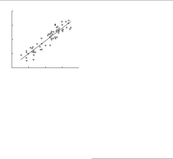
Normalized total dose (Gy)
Figure 10.4 The clinical data shown in (a) and (b) of Fig. 10.3 as reanalysed by Dubben (1994). The tumour control dose (TCD) is plotted against the prescribed total dose, normalized to a dose per fraction of 2 Gy. The positive trend in these data indicates a possible prescription bias. Reprinted with permission from Elsevier.
was thought to be more clinically realistic. In addition, the data points in Fig. 10.3b were drawn to indicate the size of the patient sample from which the estimate of TCD50 had been made. Figure 10.3b suggests that the ‘lag’ period before commencement of repopulation may have been somewhat exaggerated by the plot shown in Fig. 10.3a, although this issue is still actively discussed. Furthermore, the analysis by Dubben used the same radiobiological assumptions as the review by Withers et al. (1988) and showed that the actual local tumour control rates could be replaced by random numbers without changing the conclusion of Fig. 10.3a. As shown in Fig. 10.4, the reason for this completely unexpected finding was a highly significant correlation between TCD50 and the prescribed total dose (normalized to 2-Gy fractions). Dubben’s conclusion was that the increase of TCD50 with increasing treatment duration in Fig. 10.3a reflects only dose–time prescriptions and that these data neither confirm nor exclude a time factor of fractionated radiotherapy. The comparison among these three analyses of the same dataset is a good example of how difficult it is to draw reliable conclusions based on retrospective analyses of clinical data.
If we accept the data summarized in Fig. 10.3b, the slope of the line indicates that 0.48 Gy per day is recovered during fractionated radiotherapy of head and neck squamous cell carcinoma. If we further accept that this effect is caused by repopulation and assume reasonable estimates of tumour cell radiosensitivity, we can deduce a clonogen doubling time of less than 1 week, similar to the values of pretreatment potential doubling times measured in human tumours (see Tables 7.4 and 7.5). The potential doubling time (Tpot) is a cell kinetic parameter that indicates the rate at which cells are proliferating in an untreated tumour. Although there is much uncertainty about this, it has been suggested that during treatment the rate at which clonogenic cells within the tumour repopulate may also resemble the Tpot value. Thus, accelerated fractionation, which uses a reduced overall treatment time below the conventional 6–7 weeks, should increase tumour cure rates by restricting the time available for tumour cell proliferation. From Fig. 10.3b, for example, the dose in a 5-week schedule would be effectively larger than that in a 7-week schedule by a factor 0.48 (7 5) 6.7 Gy, or nearly 10 per cent of a 70-Gy treatment.
10.5 CLINICAL EVALUATION OF ACCELERATED RADIOTHERAPY
Through the joint activities of radiation biologists and clinicians, accelerated fractionation schedules have been developed that aim to counteract the rapid repopulation of clonogenic cells during therapy, as deduced in Section 10.4. Accelerated fractionation is defined as a shortening of the overall treatment time or, more precisely, as an increase of the average dose per week above the 10 Gy given in conventional fractionation. Early normal-tissue reactions are expected to increase using accelerated radiotherapy (see Chapter 11). In contrast, if recovery from sublethal radiation damage between fractions is complete (see Chapter 8), late normaltissue damage is expected to remain constant for accelerated fractionation schedules using 1.8- to 2-Gy fractions and total doses comparable to conventional fractionation. If the total dose and/or the dose per fraction is reduced (sometimes termed
|
|
|
|
|
|
Clinical evaluation of accelerated radiotherapy |
141 |
||||
1.0 |
|
|
|
Events Total |
1.0 |
|
|
|
Events Total |
||
0.9 |
|
|
|
0.9 |
|
|
|
||||
|
|
CHART |
286 |
552 |
|
|
CHART |
294 |
552 |
||
0.8 |
|
|
Conv. |
194 |
366 |
0.8 |
|
|
Conv. |
192 |
366 |
|
|
|
|
|
|
|
|
|
|
||
0.7 |
|
|
|
|
|
0.7 |
|
|
|
|
|
0.6 |
|
|
|
|
|
0.6 |
|
|
|
|
|
0.5 |
|
|
|
|
|
0.5 |
|
|
|
|
|
0.4 |
|
|
|
|
|
0.4 |
|
|
|
|
|
0.3 |
|
|
|
|
|
0.3 |
|
|
|
|
|
0.2 |
|
|
|
|
|
0.2 |
|
|
|
|
|
0.1 |
|
|
|
|
|
0.1 |
|
|
|
|
|
0.0 |
|
|
|
|
|
0.0 |
|
|
|
|
|
0 |
12 |
24 |
36 |
48 |
60 |
0 |
12 |
24 |
36 |
48 |
60 |
Patients at risk |
|
|
Months |
|
|
Patients at risk |
|
|
Months |
|
|
CHART 552 |
315 |
223 |
162 |
114 |
57 |
CHART 552 |
420 |
297 |
220 |
151 |
74 |
Conv. 366 |
205 |
144 |
97 |
70 |
38 |
Conv. 366 |
276 |
214 |
142 |
102 |
57 |
(a) |
|
|
|
|
|
(b) |
|
|
|
|
|
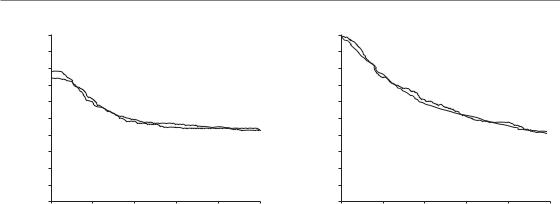
Figure 10.5 Results of a phase III randomized trial of CHART (continuous hyperfractionated accelerated radiotherapy) in squamous cell carcinoma of the head and neck. (a) Probability of loco-regional tumour control; (b) probability of overall survival of patients treated by CHART (bold line) and by conventional radiotherapy (solid line). From Dische
et al. (1997), with permission.
accelerated hyperfractionation) late normal-tissue damage could even decrease.
To test these ideas, many clinical trials of accelerated radiotherapy have been set up. As of 2008, 15 trials have been reported, some of these only as abstracts. Twelve of these trials (9/11 in head and neck squamous cell carcinoma, 2/3 in non-small cell lung cancer, 1/1 in small-cell lung cancer) indicate that, for a given total dose, accelerated fractionation schedules are more effective in obtaining local tumour control than conventional fractionation schedules, or that the results are at least identical to conventional fractionation despite administering a reduced total dose in the accelerated arm. Overall, these studies on more than 7000 patients provide strong support for the existence of a significant time factor for tumours. The following paragraphs describe some of these trials of accelerated radiotherapy in more detail.
The strategy known as CHART (continuous hyperfractionated accelerated radiotherapy) is an example of strongly accelerated fractionation. This protocol applies 36 fractions over 12 consecutive days (starting on a Monday and including the following weekend), using three fractions per day with an interval of 6 hours between the fractions within each day. Dose per fraction is 1.5 Gy to a
total of 54 Gy. Total dose is therefore reduced compared with conventional therapy, in order to remain within the tolerance of acutely responding epithelial tissues. Dische et al. (1997) have reported the results of a phase III clinical trial of CHART in 918 patients with head and neck cancer. Patients were randomized between CHART and conventional fractionation in 2-Gy fractions to 66 Gy. Figure 10.5a shows that loco-regional tumour control was identical in both treatment arms. CHART used 12 Gy less than conventional therapy in an overall time reduced by 33 days. If this dose reduction is thought to just offset repopulation, this would correspond to 0.36 Gy per day lost through tumour cell proliferation, which is somewhat lower than the 0.48 Gy per day from the data in Fig. 10.3b. This could be interpreted to mean that the lag period before onset of accelerated repopulation in head and neck carcinoma is longer than the 12-day duration of a course of CHART, but further data are required to confirm this. Overall patient survival was identical in both treatment arms (Fig. 10.5b). As expected for accelerated radiotherapy, radiation mucositis was more severe with CHART; it occurred earlier but settled sooner and was in nearly all cases healed by 8 weeks in both arms. Unexpectedly, skin reactions were less
142 Modified fractionation
|
1.0 |
HR |
|
P |
|
0.9 |
|
Events |
Total |
|
|
|
||
|
0.8 |
Chart |
269 |
338 |
|
0.7 |
Conv |
204 |
225 |
(%) |
|
|
|
|
0.6 |
|
|
|
|
Survival |
|
|
|
|
0.4 |
|
|
|
|
|
0.5 |
|
|
|
|
0.3 |
|
|
|
|
0.2 |
|
|
|
|
0.1 |
|
|
|
|
0.0 |
|
|
|
|
0 |
12 |
24 |
36 |
48 |
Patients at risk |
|
Months |
|
|
|
CHART |
338 |
201 |
104 |
57 |
32 |
Conv |
225 |
123 |
44 |
23 |
10 |
(a)
|
1.0 |
|
HR |
|
|
P |
|
0.9 |
|
|
|
||
|
|
|
|
|
|
|
|
0.8 |
|
|
|
Events |
Total |
(%) |
|
|
Chart |
285 |
338 |
|
|
|
|
||||
0.7 |
|
|
Conv |
196 |
225 |
|
control |
|
|
||||
0.6 |
|
|
|
|
|
|
tumour |
0.5 |
|
|
|
|
|
0.4 |
|
|
|
|
|
|
Local |
|
|
|
|
|
|
0.3 |
|
|
|
|
|
|
|
|
|
|
|
|
|
|
0.2 |
|
|
|
|
|
|
0.1 |
|
|
|
|
|
|
0.0 |
|
|
|
|
|
|
0 |
12 |
24 |
36 |
|
48 |
Patients at risk |
|
Months |
|
|
|
|
|
|
|
|
|
||
CHART |
338 |
144 |
67 |
41 |
|
22 |
Conv |
225 |
78 |
28 |
15 |
|
9 |
(b)
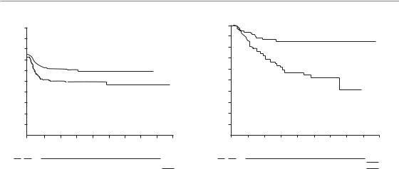
Figure 10.6 Results of a phase III randomized trial of CHART (continuous hyperfractionated accelerated radiotherapy) in non-small cell lung cancer. (a) Overall survival; (b) local tumour control of patients treated by CHART or by conventional radiotherapy (CHART results are indicated by the heavier line). HR, hazard ratio. From Saunders et al. (1999), with permission.
severe and settled more quickly in the CHARTtreated patients. Life-table analysis showed evidence of reduced severity in a number of late morbidities in favour of CHART but the magnitude of these differences in late reactions would not allow a substantial increase in the total dose of CHART without increasing the risk of late damage over the rate observed for conventional fractionation. The overall conclusion is that CHART improved the therapeutic ratio in head and neck cancer by a small margin.
Saunders et al. (1997, 1999) reported the results of the CHART–Bronchus trial. A total of 563 patients with non-small cell lung cancer were randomized between CHART (as described above) and conventional fractionation to 60 Gy in 2-Gy fractions. Despite the lower total dose, survival after 2 years was significantly increased by 9 per cent, from 21 per cent in the conventional arm to 30 per cent in the CHART arm (Fig. 10.6a). Exploratory analysis revealed that this was a consequence of improved local tumour control (Fig. 10.6b) and, in squamous cell carcinoma, a reduced incidence of distant metastases. Oesophagitis occurred earlier and reached higher scores in CHART patients, but symptoms also settled earlier and were of no major concern on longer follow-up.
Pneumonitis was not decreased in the CHART
arm. The overall conclusion of this study was that, compared with conventional fractionation with 60 Gy, CHART offers a significant therapeutic benefit for patients with non-small cell lung cancer.
The Danish Head and Neck Cancer Study Group (DAHANCA) trial 6 and 7 (Overgaard et al., 2003) provides a typical example of weakly accelerated radiotherapy. This study was aimed at finding out whether shortening of treatment time by only 1 week by using six instead of five radiotherapy fractions per week improved the tumour response in squamous cell carcinoma of the head and neck. A total of 1476 eligible patients were randomly assigned to five (n 726) or six (n 750) fractions per week at the same total dose and fraction number (66–68 Gy in 33–34 fractions to all tumour sites except well-differentiated T1 glottic tumours, which were treated with 62 Gy). All patients, except those with glottic cancers, also received the hypoxic radiosensitizer nimorazole (see Chapter 17). Overall 5-year loco-regional control rates were improved by 10 per cent from 60 per cent to 70 per cent by accelerated fractionation (p 0.0005). It is interesting to note that the whole benefit of shortening of treatment time was seen for primary tumour control but was non-significant for neck-node control. Diseasespecific survival improved (73 vs 66 per cent for
Clinical evaluation of accelerated radiotherapy 143
100 |
|
|
|
|
|
|
|
|
|
90 |
|
|
|
|
|
|
|
|
|
80 |
|
|
|
|
|
|
|
|
|
70 |
|
|
|
|
|
|
|
|
|
60 |
|
|
|
|
|
|
|
AF |
|
|
|
|
|
|
|
|
|
|
|
50 |
|
|
|
|
|
|
|
|
CF |
40 |
|
|
|
|
|
|
|
|
|
30 |
|
|
|
|
|
|
|
|
|
20 |
|
|
|
|
|
|
|
|
|
10 |
|
|
|
|
|
|
|
(Years) |
|
|
|
|
|
|
|
|
|
||
0 |
1 |
2 |
3 |
4 |
5 |
6 |
7 |
8 |
9 |
ON Number of patients at risk:
119 |
255 |
91 |
67 |
46 |
31 |
18 |
16 |
10 |
4 |
|
CF |
|
|||||||||||
96 |
257 |
115 |
84 |
60 |
43 |
27 |
19 |
11 |
0 |
|
AF |
(a)
100 |
|
|
|
|
|
|
|
|
|
|
90 |
|
|
|
|
|
|
|
|
|
CF |
80 |
|
|
|
|
|
|
|
|
|
|
70 |
|
|
|
|
|
|
|
|
|
|
60 |
|
|
|
|
|
|
|
|
|
|
50 |
|
|
|
|
|
|
|
|
AF |
|
|
|
|
|
|
|
|
|
|
|
|
40 |
|
|
|
|
|
|
|
|
|
|
30 |
|
|
|
|
|
|
|
|
|
|
20 |
|
|
|
|
|
|
|
|
|
|
10 |
|
|
|
|
|
|
|
|
(Years) |
|
|
|
|
|
|
|
|
|
|
||
|
0 |
1 |
2 |
3 |
4 |
5 |
6 |
7 |
8 |
9 |
O |
N |
Number of patients at risk: |
|
|
|
|
||||
17 |
182 |
104 |
63 |
44 |
29 |
16 |
13 |
8 |
4 |
CF |
51 |
197 |
111 |
61 |
41 |
29 |
19 |
14 |
4 |
0 |
AF |
(b) |
|
|
|
|
|
|
|
|
|
|
Figure 10.7 Results of the European Organisation for Cancer Research (EORTC No. 22851) trial of accelerated fractionation. (a) Loco-regional tumour control (log-rank p 0.02); (b) patients free of severe radiation effects, grade 3 and 4 (log-rank p 0.001). From Horiot et al. (1997), with permission.
six and five fractions; p 0.01) but not overall survival. Acute morbidity was significantly more frequent with six than with five fractions, but was transient. The overall conclusion of this study is that shortening of overall treatment time by increasing the weekly number of fractions is beneficial in patients with head and neck cancer. Very importantly, the DAHANCA trial as well as some other clinical studies clearly shows that not only large differences in overall treatment time but also a comparably small difference of only 1 week influences substantially the probability of achieving local tumour control.
In the EORTC trial No. 22851 (Horiot et al., 1997) 512 patients with head and neck cancer were randomized to receive their treatment either conventionally in a median overall time of 54 days (using 1.8–2 Gy per fraction each day, total 35–40 fractions, treatment on 5 days per week) or accelerated in a median of 33 days (using 1.6 Gy per fraction three times per day with 4 hours minimum inter-fraction interval, total 45 fractions, treatment on 5 days per week, overall time allocated to 8 days radiotherapy, 12–14 days gap and 17 days radiotherapy). The report of this EORTC trial indicates that patients who received the accelerated treatment showed a 13 per cent increase in loco-regional tumour control from 46 per cent to
59 per cent at five years (Fig. 10.7a); there was no increase in survival compared with patients receiving conventional treatment. Early radiation effects, particularly mucositis, were much more pronounced in the accelerated arm. Thirty-eight per cent of patients had to be hospitalized for acute toxicity compared with only 7 per cent of the patients in the conventional arm. Fig. 10.7b shows that grades 3 and 4 late damage (according to the EORTC/RTOG scale) also occurred significantly more frequently after the accelerated fractionation than after conventional fractionation (p 0.001). The probability of being free of severe late damage at 3 years was 85 per cent in the conventional arm but only 63 per cent in the accelerated arm. With increasing follow-up this difference is even greater. Most of the difference in late effects has been attributable to late damage to connective tissues and mucosal sequelae.
In summary, accelerated fractionation has been shown in randomized clinical trials to counteract the time factor in head and neck and lung cancer. Some of the trials indicate an improved therapeutic ratio compared with conventional fractionation. However, one of the most intriguing biological observations from the clinical trials on accelerated fractionation is that sparing of late normal-tissue morbidity compared with

144 Modified fractionation
conventional fractionation was much less than anticipated. In fact, late damage in the EORTC trial was even higher than after conventional fractionation. Possible reasons for these unexpected findings are discussed below.
10.6 COMPARISON BETWEEN HYPERFRACTIONATION AND ACCELERATED FRACTIONATION
Bourhis et al. (2006) have reported a meta-analysis of hyperfractionation and accelerated radiotherapy in patients with head and neck tumours. This analysis included 15 randomized trials with a total of 6515 patients. In all of these trials, patients had been randomized between conventional radiotherapy and hyperfractionated or accelerated radiotherapy. The majority of patients had stage III or IV oropharyngeal or laryngeal squamous cell carcinoma and were monitored with a median fol- low-up of 6 years. When all the trials were put together, a 3–4 per cent significant increase in overall survival (hazard ratio 0.92, 95 per cent CI 0.86–0.97; p 0.003) was found. There was a significant benefit for loco-regional control in favour of altered fractionation (6.4 per cent at 5 years, p 0.0001), which was particularly important for local control (Table 10.1). No effect was observed with altered fractionation on distant metastasis. The survival advantage was more pronounced with hyperfractionation (8 per cent at 5 years) than with accelerated fractionation (2 per cent for
regimens without dose reduction and 1.7 per cent for regimens with dose reduction). Interestingly, the survival advantage observed with altered fractionation regimens is of the same order of magnitude seen with concomitant chemo-radiotherapy regimens (see Chapter 18), indicating that the debate on the relative merits of these two strategies is likely to continue.
10.7 SPLIT-COURSE RADIOTHERAPY
Intentional gaps in radiation therapy have sometimes been introduced in order to allow recovery of early-responding normal tissues. However if such gaps prolong the overall treatment time then local tumour control rates are expected to decrease. As discussed for the DAHANCA 6 and 7 trial (see Section 10.5) not only differences in the overall treatment time by several weeks but also for only 1 week may significantly change the chance for local tumour control. These data imply that even small prolongations of overall treatment times in the order of one or few days should be avoided. If this is not possible, appropriate compensation has to be applied. Studies on experimental tumours suggest that, for a given overall treatment time, the magnitude of the time factor is the same for continuous fractionation or for fractionation protocols including gaps (Baumann et al., 2001). This supports the current clinical guidelines for the compensation of unscheduled treatment gaps that are discussed in Chapter 9.
Table 10.1 Hazard ratio of altered fractionated radiotherapy versus conventional radiotherapy on overall population and by type of radiotherapy for loco-regional, local, regional and metastatic control
|
|
Accelerated |
Accelerated |
|
|
|
|
|
|
radiotherapy |
radiotherapy |
|
|
|
|
|
Hyperfractionation |
(no dose reduction) |
(dose reduction) |
Overall |
p |
||
|
|
|
|
|
|
||
Loco-regional |
0.76 (0.66–0.89)* |
0.79 (0.72–0.87) |
0.90 (0.80–1.02) |
0.82 (0.77–0.88) |
0.0001 |
||
Local |
0.75 (0.63–0.89) |
0.74 (0.67–0.83) |
0.83 |
(0.71–0.96) |
0.77 |
(0.71–0.83) |
0.0001 |
Regional |
0.83 (0.66–1.03) |
0.90 (0.77–1.04) |
0.87 |
(0.72–1.06) |
0.87 |
(0.79–0.97) |
0.01 |
Metastatic |
1.09 (0.76–1.58) |
0.93 (0.74–1.19) |
0.95 (0.68–1.32) |
0.97 (0.82–1.15) |
0.75 |
||
Number of patients, n 7073.
*The 95 per cent confidence intervals are given in parenthesis. Reproduced from Bourhis et al. (2006), with permission.

Is the same modified fractionation schedule optimal for all? 145
10.8 REASONS FOR INCREASED LATE NORMAL-TISSUE DAMAGE AFTER MODIFIED FRACTIONATION
Hyperfractionation and accelerated radiotherapy both require multiple radiation treatments per day. In the case of hyperfractionation this is because in order to give an adequate number of small fractions, one per day, the overall treatment time would have to be very long. In the EORTC and RTOG trials mentioned above the fraction number was increased to 68–70, which would require once-daily treatment over 14 weeks; this would be unacceptable in view of the time factor for tumour-cell repopulation. In the case of accelerated radiotherapy, shortening the overall time but still giving one fraction per day would require an increase in dose per fraction; this would be expected to lead to an increase in late effects although hypofractionation is clinically acceptable in some situations (see Section 10.3).
A radiobiological constraint in giving multiple fractions per day is that these should not be given too close together because of incomplete repair (see Chapter 8). As indicated in Chapter 7, Section 7.3, the damage inflicted by radiation is very largely repaired in most cell types, both normal and malignant. The repair of normal-tissue stem cells is vital for their tolerance to radiation therapy and if fractions are given so close together that repair is incomplete, tissue tolerance will be reduced. Repair halftimes for many tumours and normal tissues are in the region of 0.5–2 hours (Table 9.2). Assuming exponential decay of radiation damage, it takes six halftimes for the damage to decay to 1/64 (i.e. 1–2 per cent of its initial value). The problem is that, as shown in Table 9.2, some late-responding normal tissues appear to have long repair halftimes. These tissues will therefore be especially disadvantaged by radiotherapy given with multiple fractions at a short inter-frac- tion interval: the therapeutic index will be impaired and the total radiation dose will have to be lowered in order to remain within tolerance. Most current schedules with multiple fractions per day employ inter-fraction times of at least 6 hours; in some situations even this gap may be too short.
Evidence on the clinical impact of incomplete repair comes from an analysis by Bentzen et al.
(1999) of data from the CHART head and neck trial. As noted above, several late-damage endpoints were significantly reduced after CHART, compared with conventional treatment. The reduction was much less than expected on the basis of LQ calculations and analysis of these data yielded repair halftimes in the range of 4–5 hours for the three late morbidities investigated: laryngeal oedema, skin telangiectasia and subcutaneous fibrosis. Even the lower ends of the confidence intervals were around 3 hours. This suggests that even 6 hours between dose fractions in multiple- fractions-per-day-schedules may be too short for complete repair in some situations. Long repair halftimes for late effects in human normal tissues therefore pose a significant problem for the development of novel fractionation schedules.
Consequential late effects (see Chapter 13) may also contribute to greater than expected late morbidity after modified fractionation. Compared with conventional fractionation the dose per week is increased, both in accelerated radiotherapy and in hyperfractionation. This is expected to produce an increase in early normal-tissue damage such as mucositis and more severe or more prolonged early damage may then lead to more pronounced consequential late effects.
10.9 IS THE SAME MODIFIED FRACTIONATION SCHEDULE OPTIMAL FOR ALL PATIENTS?
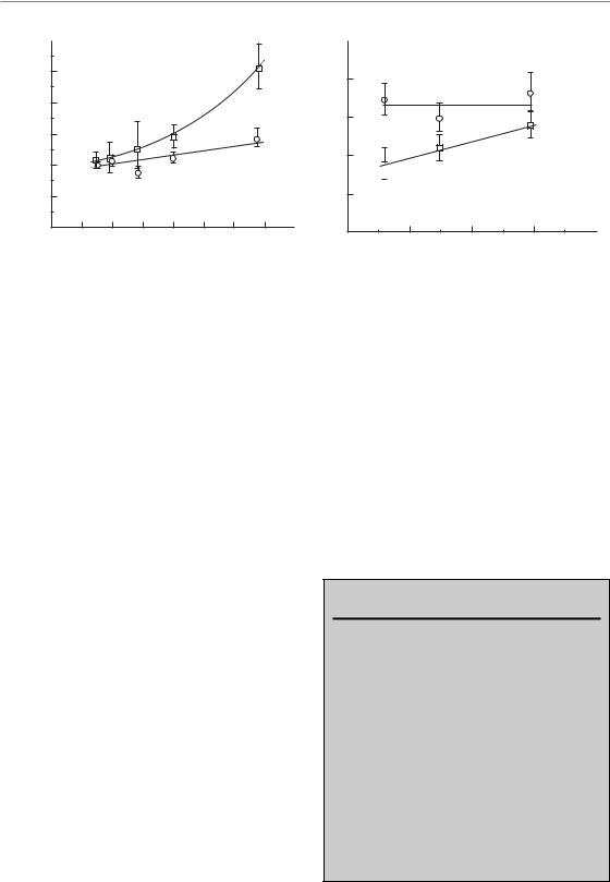
Figure 10.8 summarizes the results of experiments testing different modified fractionation schedules in two experimental squamous cell carcinoma xenografts (FaDu and GL) in mice. For a constant number of 30 fractions in FaDu tumours, the TCD50 increased by roughly 0.6 Gy for each day of prolongation for overall treatment times up to 40 days but more steeply for longer times (Fig. 10.8a); this effect was much less pronounced (0.28 Gy/day) in GL squamous cell carcinomas. In contrast, when the number of fractions was increased from 12 to 60 in a constant overall treatment time of 6 weeks (Fig. 10.8b), the TCD50 in FaDu tumours appeared to be constant while in GL tumours the TCD50 increased with increasing fraction number. Both dose-escalated hyperfractionation and accelerated
146 Modified fractionation
TCD50 (Gy)
(a)
120
100
80
60
40
20
0
0