

- •Contents
- •Preface
- •1 Introduction: the significance of radiobiology and radiotherapy for cancer treatment
- •2 Irradiation-induced damage and the DNA damage response
- •3 Cell death after irradiation: how, when and why cells die
- •4 Quantifying cell kill and cell survival
- •5 Dose–response relationships in radiotherapy
- •6 Linear energy transfer and relative biological effectiveness
- •7 Tumour growth and response to radiation
- •8 Fractionation: the linear-quadratic approach
- •9 The linear-quadratic approach in clinical practice
- •10 Modified fractionation
- •11 Time factors in normal-tissue responses to irradiation
- •12 The dose-rate effect
- •13 Pathogenesis of normal-tissue side-effects
- •14 The volume effect in radiotherapy
- •15 The oxygen effect and fractionated radiotherapy
- •16 The tumour microenvironment and cellular hypoxia responses
- •17 Therapeutic approaches to tumour hypoxia
- •18 Combined radiotherapy and chemotherapy
- •19 Retreatment tolerance of normal tissues
- •20 Molecular image-guided radiotherapy with positron emission tomography
- •21 Molecular-targeted agents for enhancing tumour response
- •22 Biological response modifiers: normal tissues
- •23 Molecular targeting and patient individualization
- •24 Protons and other ions in radiotherapy
- •25 Second cancers after radiotherapy
- •Glossary of terms in radiation biology
- •Index
25
Second cancers after radiotherapy
KLAUS RÜDIGER TROTT
25.1 |
Introduction |
339 |
25.4 |
Radiation-induced second cancers after |
|
25.2 |
Estimating the risk of radiation-induced |
|
|
treatment of paediatric malignancies |
347 |
|
second cancers after curative radiotherapy |
|
25.5 |
Conclusions |
348 |
|
of cancers in adult patients |
341 |
Key points |
350 |
|
25.3 |
Radiation-induced second cancers after |
|
Bibliography |
351 |
|
|
combined radiochemotherapy treatment of |
|
Further reading |
352 |
|
|
malignancies in young adults |
346 |
|
|
|
|
|
|
|
|
|
25.1 INTRODUCTION
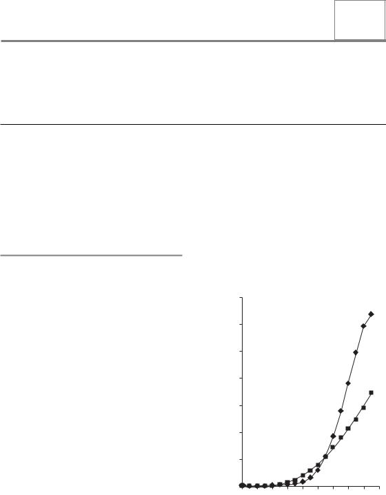
Age is the most important risk factor for developing cancer and for dying from cancer. The risk of developing cancer within the following year changes little between the end of childhood and the age of 40 years. In women the risk increases earlier than in men, yet in both sexes the most dramatic increase is after the age of 60 years, as demonstrated in Fig. 25.1. Table 25.1 shows the risk of developing cancer within 5 years after treatment (i.e. the commonly practised follow-up time after radiotherapy for patients treated at the age of 50, 55, 60, 65, 70 or 75 years), assuming that cancer rates follow those found in the general population, as shown in Fig. 25.1. Since there is no convincing evidence that the development of one cancer protects against the development of another, we may conclude that, during the typical follow-up period of a patient treated for cancer with curative intent, a relatively large proportion of patients will present with a second cancer. This frequency will vary between 1 per cent and more than 10 per cent, depending on age and sex. The results of epidemiological studies described below indicate that in cancer patients, after curative radiotherapy, the increased lifespan of cured
patients is by far the most important risk factor leading to second cancers. This risk, determined from cancer registry data which cover entire populations, may be further increased by individual
|
3500 |
|
|
|
|
|
|
|
|
|
|
3000 |
|
|
|
|
|
|
|
|
|
000 people |
2500 |
|
|
|
|
|
|
|
|
|
2000 |
|
|
|
|
|
|
|
|
|
|
per 100 |
|
|
|
|
|
|
|
|
|
|
1500 |
|
|
|
|
|
|
|
|
|
|
Incidence |
|
|
|
|
|
|
|
|
|
|
1000 |
|
|
|
|
|
|
|
|
|
|
|
|
|
|
|
|
|
|
|
|
|
|
500 |
|
|
|
|
|
|
|
|
|
|
0 |
|
|
|
|
|
|
|
|
|
|
0 |
10 |
20 |
30 |
40 |
50 |
60 |
70 |
80 |
90 |
Age group (years)
Figure 25.1 Average annual cancer incidence in the UK by gender and age attained. Diamonds, male; squares, female.

340 Second cancers after radiotherapy
Table 25.1 The spontaneous cancer incidence risk within a follow-up period of 5 years, in patients treated at different ages
|
Cancer risk within the next |
||
Age at treatment |
5 years (%) |
|
|
(years) |
Males |
Females |
|
|
|
|
|
50 |
1.5 |
2.0 |
|
55 |
2.5 |
2.7 |
|
60 |
5.0 |
3.6 |
|
65 |
7.0 |
4.6 |
|
70 |
10.0 |
5.4 |
|
75 |
12.5 |
6.3 |
|
Data from UK, England and Wales 1983–1987.
factors such as specific carcinogen exposure or by genetic predisposition.
It has been generally accepted that the majority of cancers are causally related to exposure of the individual to common carcinogens, the most important being dietary factors and smoking which, together, cause more than 50 per cent of all cancers (Doll and Peto, 1981). Most carcinogens are known to be related to more than one type of cancer. Smoking, for example, is causally related to cancer of the lung, the bladder and the head and neck. This means that a patient who has been cured of bladder cancer, for example, independent of the treatment modality, will have a greater risk than other members of the general population to develop, for example, lung cancer. The size of this increased individual risk is difficult to determine. However, in the quantification of treatment-related second cancer risks in epidemiological studies, methods have to be used which eliminate or reduce the potential bias related to specific carcinogen exposure. The best approaches are the determination of radiation dose dependence of risk or the comparison of different curative treatment modalities for the same type and stage of cancer.
The risk for the development of specific cancers is also influenced by genetic predisposition. The strength of this predisposition varies between gene mutations. Well-known examples of strong genetic predisposition are mutations of the Rb gene (predisposing for retinoblastoma and osteosarcoma) and of the BRCA1 gene (predisposing for early
breast cancer and ovarian cancer). The fact that these, and probably most, predisposing cancer gene mutations are associated with more than one type of cancer also means that people cured of one of those cancers have a higher than average probability of developing the other cancers associated with the respective mutation. However, the impact of genetic predisposition on the risk of developing a second cancer after cure from the first cancer is difficult to assess at the present state of knowledge. The most obvious example is the high risk of children treated with radiotherapy for retinoblastoma to develop osteosarcomas in the irradiated volume.
Finally, some of the modalities used to treat cancer have proven to have carcinogenic potential. A large number of studies have been published exploring the impact of various chemotherapy and radiotherapy schedules as well as their combination on the incidence of second cancers. The most detailed and comprehensive analysis of studies carried out up to 2003 has been published by van Leeuwen and Travis (2005).
It is particularly in those cancer patients who were treated as children or young adults for cancers such as Hodgkin’s and non-Hodgkin’s lymphoma, testicular cancer and paediatric malignancies that it is becoming increasingly apparent that the most important cause of death in longterm survivors is second cancers causally related to the methods of treatment of the first cancer. Radiotherapy and chemotherapy are both implicated in the induction of those second malignancies. In general, chemotherapy is mostly related to the induction of leukaemia, in particular acute myeloid leukaemia (AML), most of which occur within 10 years after primary treatment. Radiotherapy is more related to the induction of solid cancers, the vast majority having longer latency with risk persisting for several decades or maybe even life-long. There are strong indications that radiotherapy also co-increases the risk of second leukaemia and that chemotherapeutic drugs also co-increase the risk of solid cancers after radiotherapy.
The risk of treatment-related second cancers is commonly determined by the comparison of the frequency of second cancers after different treatments such as surgery versus radiotherapy or with the general population. From this, a ratio of

Estimating the risk of radiation-induced second cancers 341
frequencies is calculated that indicates relative risk (RR) of cancers caused by a specific treatment. These RR values tend to vary greatly between different second cancers for the same primary cancer and, in many cases such as leukaemia after treatment of Hodgkin’s lymphoma, may assume very high values. Although RR values form the basis of epidemiological and statistical analyses, they may be misleading in the evaluation of the clinical problem. Since many treatment-related cancers (particularly those with high RR values) are rare in the general population, a high RR may still translate into a low absolute risk. Therefore, absolute excess risk, which estimates the excess number of second malignancies per 10 000 patients per year, better reflects the second malignancy burden of treated patients than RR values (van Leeuwen and Travis, 2005).
25.2 ESTIMATING THE RISK OF RADIATION-INDUCED SECOND CANCERS AFTER CURATIVE RADIOTHERAPY OF CANCERS IN ADULT PATIENTS
Radiation is a well-established carcinogen. Therefore, it has to be assumed that successful curative radiotherapy of cancer may, in some cases, also cause a new, second cancer in addition to age-related cancer risks. In radiation protection, estimation of radiation-induced cancer follows a method which has been developed by the International Commission on Radiological Protection (ICRP) for preventive purposes in radiation worker populations. It is largely based on data derived from epidemiological studies in populations exposed to whole-body low-dose irradiation, in particular the lifespan study of the Japanese atomic bomb survivors. The method of risk estimation involves three steps of calculation:
1.Calculate the mean organ dose for the different organs at risk such as lung, stomach, colon, bone marrow and others.
2.Multiply the mean organ dose by the relevant organ weighting factor which ranges from
0.01 for skin to 0.12 for the four organs listed above.
3.Add up the weighted mean organ doses for all organs at risk. This weighted total body dose is called ‘effective dose’. This effective dose is then multiplied by the appropriate risk factor that varies between 4 and 10 per cent per Gy, depending on age and exposure rate, to calculate the lifetime cancer risk from the respective radiation exposure.
Several studies have been published recently using this approach to determine the risks from radical radiotherapy and to compare different treatment plans in radiotherapy. The risk estimates derived using this method yield very high values which may be up to two orders of magnitude higher than the risk derived directly from the epidemiological studies described below. The reasons for this discrepancy are the extraordinary dose inhomogeneities within individual organs and between organs. Epidemiological and experimental evidence actually shows that the probability of cancer induction decreases dramatically as the dose inhomogeneity in an irradiated organ increases. It is mainly for this reason that the ICRP strongly advises not to use the effective dose method to estimate the risks of radiation-induced cancer in situations of very large dose inhomogeneities with peak doses well above doses that would cause acute or chronic radiation effects (i.e. doses 5 Gy); see ICRP 60 (1991) and ICRP 103 (2007). The ICRP also recommends that data derived directly from epidemiological investigations on radiotherapy patients are better suited to estimate the risk of induction of second cancers by the radiation treatment of the first cancer.
Such data can best be collected by the comparison of the rate of second cancers in large patient cohorts who have been cured from the first cancer either by radiotherapy or by surgery. Conditions of suitability are:
●the first cancer has to be common
●the first cancer must have a good chance of cure ( 50 per cent)
●the chance of cure has to be similar from surgery and radical radiotherapy and the decision of treating by radiotherapy or surgery should be largely independent of factors affecting cancer risks
●the life expectancy of a large proportion of the cured patients must be 10 years.

342 Second cancers after radiotherapy
Two types of cancer fulfil these conditions particularly well: they are cancer of the cervix and cancer of the prostate. For both these cancers, large epidemiological studies have been conducted which provide the main source of data on second cancer risks after radiotherapy in patients of advanced age.
In addition, important information can also be derived from studies on the topographical relationship of primary cancer and second cancer in symmetrical organs, in particular in patients with a primary breast cancer and a secondary lung cancer. Moreover, studies on second cancers after radiotherapy of young people and their comparison with age-matched healthy populations also provide important information since they permit very long follow-up, yet their interpretation is difficult and may be misleading because of strong genetic susceptibility factors influencing risks and because other carcinogenic treatment modalities such as chemotherapy may also be given, which makes any identification of radiation risks difficult. However, in some cancers, in particular Hodgkin’s lymphoma, testicular cancer and paediatric malignancies, it has been possible to separate out the contributions of radiotherapy and chemotherapy and their interaction.
Suit et al. (2007) have made the most comprehensive analysis of the problem, putting the question of second cancers after radical radiotherapy into the context of radiobiological studies on cell transformation in vitro, radiation-induced cancers in experimental animals from mice to monkeys, the Japanese atomic bomb survivors, radiation workers, patients treated for benign diseases and those treated with radiotherapy for cancer. Estimating RR values in comparison with the general population and with patients treated with non-radiation modalities, they demonstrated that some (but not all) cured cancer patients have a higher risk of developing cancer than the general population. They concluded that ‘radiation can induce malignant transformation of mammalian tissue. . . . The relationship between radiation dose and risk of cancer is clearly complex and is not amenable to a simple definition applicable to all mammalian species, all members of a species, or even all organs in one inbred strain of a species. . . . Due to quite large and undefined heterogeneity in the patient populations studied, no
precise quantification of the risk of radiationinduced secondary cancer is available at present. However, the clear implication of this review . . . is that the basic concept for planning radiation treatment should be that the risk of radiationinduced secondary cancer would be reduced by any dose decrement to uninvolved normal tissues, at least down to around 0.05 Gy. The factorial decrease in risk would be greatest for reduction in dose levels below 2 Gy.’
By pooling data from 14 published radiotherapy series and by concentrating on RR, inevitably some details of the presented information which might be important for the assessment of the clinical relevance of the data was lost in the Suit et al. review. This is particularly so if one considers the dose response of individual organs at risk for second cancer induction since the spectrum of critical organs differed between the various studies. In the following, we have adopted a different approach in which the largest and most detailed studies of second cancers after radical radiotherapy have been selected to derive information that could be used not so much for risk estimation, but in the critical evaluation of treatment decisions and of dose–volume histograms in radiotherapy treatment planning.
Carcinoma of the cervix
The first analysis on the risk of second cancers after radical radiotherapy of primary cancers was a multi-institutional study on long-term survivors of cancer of the cervix. The study of Kleinerman et al. (1995) was a cohort study of the incidence of second cancers in 66 541 patients with cervical cancer reported to 13 population-based cancer registries in five countries. Out of this patient group, 49 828 (75 per cent) were treated with radiotherapy and 16 713 (25 per cent) were treated surgically. The average follow-up was 10.4 years. More than 2000 second cancers were recorded and analysed. The results are tabulated in Table 25.2 and are consistent with the results of a case–control study on the same patient population by the same investigators (Boice et al., 1988).
The results of this study are remarkable in several aspects. In contrast to all studies which form

Estimating the risk of radiation-induced second cancers 343
Table 25.2 Results of the cohort study on second cancers after radiotherapy of cervical cancer
|
|
Number of second |
|
|
Radiation |
cancers after |
|
Site of second cancer |
dose (Gy) |
radiotherapy/surgery |
Relative risk after 10 years |
|
|
|
|
Rectum |
30–60 |
274/33 |
2 after 10 years; 4 after 30 years |
Colon |
24 |
296/56 |
No increase |
Bladder |
30–60 |
265/23 |
2 after 10 years; 6 after 30 years |
Stomach |
2 |
143/19 |
1.2 |
Lung |
0.3 |
276/91 |
No increase |
Breast |
0.3 |
366/114 |
Decrease 20–40% after 10 years |
|
|
|
and 30 years |
Leukaemia |
4.5 |
82/15 |
2 |
Data from Kleinerman et al. (1995).
the basis of radiation protection regulations such as the atomic bomb survivor studies or the ankylosing spondylitis studies (United Nations Scientific Committee on the Effects of Atomic Radiation: UNSCEAR, 2000, 2006), the greatest risk occurs in the bladder (which, although significantly related to radiation exposure in the atomic bomb survivors, has a small organ weighting factor) and in the rectum (which has not been shown to be sensitive to the induction of cancer by radiation). The colon, which also receives considerable doses in radiotherapy of cervical cancer, did not show increased second cancer rates. In addition to the bladder and rectum located in the high-dose volume, only two organs found in the low-dose volumes show significantly increased cancer rates. These are the stomach, receiving a mean dose of 2 Gy, and the bone marrow, receiving a mean dose of 4.5 Gy. The leukaemia risk per Gy derived from these data is less than 10 per cent of the risk per Gy estimated from the atomic bomb survivor study, demonstrating the overriding importance of dose inhomogeneity on second malignancy risks.
Carcinoma of the prostate
Several small studies on the risks of second cancers after radical radiotherapy of prostate cancer have yielded inconclusive results. Yet the results of the very large cohort study on more than 120 000 prostate cancer patients (Brenner et al., 2000)
Table 25.3 Results of the study of second cancers after radiotherapy of prostate cancer
|
Surgery only |
Radiotherapy |
|
|
|
Number of patients |
70 539 |
51 584 |
Person-years at risk |
312 499 |
218 341 |
Mean survival time |
4.4 |
4.2 |
(years) |
|
|
Mean age at therapy |
71.4 |
70.3 |
(years) |
|
|
Mean age at second |
77 |
75.3 |
cancer (years) |
|
|
Percentage of persons at risk after: |
|
|
5–10 years |
35.8 |
33.5 |
10 years |
10.8 |
9.8 |
Number of second malignancies: |
|
|
At all times after |
5055 |
3549 |
treatment |
|
|
After 5 years |
1646 |
1185 |
After 10 years |
393 |
305 |
Data from Brenner et al. (2000).
clearly demonstrate the extent of the problem for clinical radiotherapy. The study was a cohort study on 122 123 patients with prostate cancer registered in the SEER (the National Cancer Institute’s Surveillance, Epidemiology and End Results) programme who had either surgery or radiotherapy. The results of this study are summarized in Table 25.3.

344 Second cancers after radiotherapy
Table 25.4 Risk of radiation-induced second cancer after radiotherapy of prostate cancer
Relative risk
|
After 5 years |
After 10 years |
||
All second cancers |
1.11 (p 0.007) |
1.27 |
(p 0.002) |
|
Bladder |
1.55 |
(p 0.0001) |
1.77 |
(p 0.01) |
Rectum |
1.35 |
(p 0.06) |
2.05 (p 0.03) |
|
Lung |
1.22 (p 0.01) |
1.42 (p 0.02) |
||
Leukaemia in first 10 years: |
|
|
|
|
Surgery patients |
Irradiated patients |
Relative risk in 10 years |
||
39 in 343 690 person-years |
25 in 112 422 person-years |
2 (p 0.05) |
||
Data from Brenner et al. (2000).
Comparing second cancer rates of patients treated with either radiotherapy or surgery at different follow-up times, the risk of radiation-induced second cancer and its dependence on follow-up can be calculated. Results are shown in Table 25.4. Out of the approximately 17 000 prostate cancer patients who survived more than 5 years after radical radiotherapy, 1185 (7 per cent) developed a second cancer. More than 1000 of those second cancers ( 85 per cent) result from the increased lifespan after cure from the first cancer. Just about 120–150 of those second cancers among 51 584 prostate cancer patients (0.3 per cent) are related to radiotherapy:
●approximately 50 cases of bladder cancer
●approximately 15 cases of cancer of the rectum
●approximately 50 cases of lung cancer
●approximately 12 cases of leukaemia.
As was observed in the cervix studies, bladder and rectum cancers found in the high-dose volume are most frequent. The unexpected large number of radiotherapy-associated lung cancers is probably related to the older treatment techniques using large fields delivered mostly with 60Co and a mean lung dose of 0.5 Gy has been estimated that is related to scattered radiation. This is in agreement with the risk of radiation-induced lung cancer from the atomic bomb survivor studies. Modern conformal treatment protocols lead to much lower lung exposure of about 10 per cent of the doses estimated in the Brenner et al. (2000) study. However, lung doses from some techniques such as intensity-modulated radiotherapy (IMRT) of prostate cancer may be close to those doses and may possibly be associated with similar risks of
second lung cancer. This is due to the use of more treatment fields and higher monitor units greatly increasing both collimator and phantom scatter, as well as leakage from the linear accelerator head.
The most important result of the prostate cancer study is that half of all radiation-induced second cancers occur in the high-dose volumes and the other half in the volumes exposed to those radiation doses commonly associated with radiation carcinogenesis. It is very likely that two entirely different mechanisms are involved in the highand low-dose volumes. In the low-dose volumes, we may assume the same molecular and cellular mechanisms as in other situations of low-dose radiation carcinogenesis which have been extensively explored in radiation protection research (UNSCEAR, 2006). However, radiation doses given to the bladder and the rectum very often lead to chronic radiation injury, which is characterized by progressive microvascular damage, parenchymal atrophy and chronic inflammation. This condition has been recognized for more than 100 years as a precancerous lesion. Therefore, one may classify the radiation-induced second cancers in the high-dose organs as secondary to chronic radiation injury. This attribution would have pronounced impact on the dose–risk relationship and on the optimization of treatment plans, as discussed in the concluding paragraph.
Breast cancer
Patients treated with postoperative radiotherapy for breast cancer receive significant radiation doses of more than 5 per cent of the target dose to
Estimating the risk of radiation-induced second cancers 345
Table 25.5 Ipsilateral and contralateral second lung cancers in patients treated with postoperative radiotherapy of breast cancer
Duration of |
Number of second cancers |
Lung cancer |
||
follow-up (years) |
Ipsilateral |
Contralateral |
|
mortality ratio |
|
|
|
|
|
10 |
161 |
134 |
|
1.2 |
10–15 |
65 |
44 |
|
1.5 |
15 |
57 |
21 |
|
2.7 |
Data from Darby et al. (2005).
the contralateral breast. New trial protocols are addressing this problem and studies such as the IMPORT High trial, using partial breast irradiation for high-risk patients, are suggesting a mean dose of 1 Gy as a dose constraint to the contralateral breast. Since second cancers in the contralateral breast occur more frequently than expected and constitute nearly half of all second cancers in women with breast cancer, a causal relationship with the radiation exposure from the treatment of the first cancer has been suggested. However, a nested case–control study by Storm et al. (1992) on 529 patients who developed contralateral breast cancer 8 or more years after diagnosis of the first cancer (being part of a cohort of more than 50 000 breast cancer patients in Denmark) provided evidence that there is little, if any, risk of radiationinduced breast cancer associated with exposure of the contralateral breast in postoperative radiotherapy. However, most patients in this study were over 45 years old and therefore at an age when the radiosensitivity of the breast with regard to cancer induction has been shown to be very low. In contrast, in a study on women under the age of 45 years, Boice et al. (1992) estimated that one in 10 of second breast cancers could be attributed to prior radiotherapy. Modern treatment planning permits a much lower dose to the contralateral breast than was the case in both these studies and their findings indicate that it is particularly in young breast cancer patients that the dose to the contralateral breast should be carefully controlled.
Patients treated with postoperative radiotherapy for breast cancer receive very different doses to the ipsilateral compared with the contralateral lungs. Darby et al. (2005) have reported a cohort
study on 308 861 women, included in the SEER programme, who were treated for breast cancer between 1973 and 2001 and of whom 115 165 (37 per cent) received radiotherapy as part of their primary treatment. Of these treated women, 482 (0.4 per cent) later died from lung cancer for which the affected side was clearly defined in the records. The main endpoint was which side of the lung developed a second cancer in relation to which breast was originally treated. More than 1000 cases of lung cancer (0.5 per cent) occurred in women who did not receive radiotherapy, and there was no difference between the rates of ipsilateral and contralateral lung cancers. Conversely, of the 482 cases of lung cancer that occurred in women who received radiotherapy, 283 cases (59 per cent) were ipsilateral and 199 (41 per cent) were contralateral. From these findings the risk of radiation-induced lung cancer can be estimated.
The proportion of ipsilateral second lung cancers in women who had received radiotherapy increased with increasing follow-up time from a ratio of 1.2 less than 10 years after treatment to 2.7 more than 15 years after treatment (Table 25.5). Among women diagnosed with breast cancer during 1973–1982 and receiving postoperative radiotherapy, there were 112 deaths from ipsilateral lung cancer and 51 from contralateral lung cancer, indicating a mortality ratio of about 2. Taking into account also that the contralateral lung received considerable radiation doses from scatter, the RR of second cancer in the lung from postoperative radiotherapy of breast cancer would increase even further. The estimated RR is about 3, which would translate into an absolute risk of lung cancer from postoperative radiotherapy of 0.6 per cent. There is a non-significant suggestion that, in recent years,

346 Second cancers after radiotherapy
using more advanced radiotherapy techniques, the risk of second lung cancer after postoperative radiotherapy of breast cancer after 10 years is reduced compared with the older cohort.
25.3 RADIATION-INDUCED
SECOND CANCERS AFTER COMBINED RADIOCHEMOTHERAPY TREATMENT OF MALIGNANCIES IN YOUNG ADULTS
Hodgkin’s lymphoma
The treatment results of Hodgkin’s lymphoma have improved significantly since the introduction of intensive radiotherapy, which was mainly based on the work of Kaplan at Stanford and of Musshoff in Freiburg in the 1950s. As a result, there are now thousands of long-term survivors of Hodgkin’s lymphoma who are at risk for late effects of therapy including second cancers. Wolden et al. (1998) described the incidence of second cancers in 697 patients who were less than 21 years old at the time of treatment in Stanford – some were followed up for more than 35 years. Eighty patients (11 per cent) developed 85 new malignant tumours. Twenty-five (31 per cent) were non-melanoma skin cancers. The second most frequent second cancer was breast cancer (16 patients), followed by sarcomas (13 patients). Eight second leukaemias occurred, all but one within 10 years and all eight patients had received chemotherapy with alkylating agents. The actuarial risk of second cancer at 20 years after treatment for Hodgkin’s lymphoma, at a mean attained age of 36 years, was 9.7 per cent for males and 16.8 per cent for females with more than half of their risk being breast cancer (9.2 per cent). The incidence of breast cancer was similar for patients who received both radiotherapy and chemotherapy versus radiation only as initial therapy. The most remarkable finding of this important singleinstitution study was that, among the 48 solid second cancers, 43 (90 per cent) occurred within the radiotherapy treatment field or in the penumbra region, and 40 (83 per cent) developed in volumes that had received at least 35 Gy. The authors stress
that treatment policies for Hodgkin’s lymphoma have changed dramatically over the past 30 years, putting more emphasis on multiagent chemotherapy and reduced radiation doses and treatment volumes to involved sites. Thus, the second malignancy rates seen after long follow-up in this study may not represent the risk for patients treated in the modern era. It should be noted that patients were more than twice as likely to die from their primary Hodgkin’s lymphoma than from a second cancer (11 per cent versus 4 per cent).
Dores et al. (2002) reported results of a large international study on 32 591 Hodgkin’s lymphoma patients with 2861 patients followed up for more than 20 years and 1111 patients for more than 25 years; mean age at treatment was 37 years. Second malignancies developed in 2153 patients (7 per cent) which, compared with the ageand sex-adjusted general population, was an increase of more than a factor of 2. The risk of latedeveloping solid cancers was particularly increased after radiotherapy while second leukaemias were mostly related to chemotherapy. After more than 25 years’ follow-up, there was evidence of a decrease in RR for all second cancers from a RR of 4.4 in the 20to 24-year period to a RR of 2.4. The highest absolute excess second cancer risk was for cancers of the lung and breast. Whereas the RR of all second cancers decreased with increasing age at diagnosis of Hodgkin’s lymphoma, the absolute excess risk of second cancers increased with increasing age from 30 cases per 100 000 person-years for under 21-year-old patients to 107 cases per 100 000 person-years in the 51to 60-year-old patients. This was not seen for second breast cancer, where the risk was highest in patients treated when they were under 30 years old. The authors calculated a 25-year cumulative risk of treatment-induced second cancers of 11.7 per cent, most of which was related to radiotherapy.
In a case–control study of British patients, Swerdlow et al. (2000) demonstrated that MOPP chemotherapy also leads to a dose-dependent elevated risk of lung cancer and that this risk was not further increased if radiotherapy was given together with MOPP.
In their review of late effects after treatment for Hodgkin’s lymphoma, Swerdlow and van
Leeuwen (2005) concluded that the substantial

Radiation-induced cancers after treatment of paediatric maligancies 347
increase in solid tumour risk with time since diagnosis necessitated careful, lifelong medical surveillance of all patients. Since the absolute excess risk of lung cancer was much greater among smokers than non-smokers, physicians should make a special effort to dissuade Hodgkin’s disease patients from smoking. Women treated with mantle field irradiation before the age of 30 years are at greatly increased risk of breast cancer. In many centres, from 8 years after irradiation onwards, the follow-up programme of these women includes yearly breast palpation and mammography; however, the efficacy of these measures in this specific population has not yet been demonstrated.
Testicular cancer
In a large international study on nearly 29 000 patients with testicular cancer who survived more than 1 year, Travis et al. (1997) analysed the dependence of 1406 observed second cancers (which was an overall excess of 43 per cent) on time since treatment and on treatment modality with special emphasis on the histology of first and of second tumour. The 25-year cumulative risk after treatment of seminoma was 18 per cent compared with that of an age-matched normal population and greater than that of non-seminomatous testicular cancer which was 11 per cent compared with 6 per cent in an age-matched normal population. Compared with the general population, the excess cancer risk increased steadily for at least 30 years. The most pronounced significantly increased second cancer rates among the 3306 patients surviving more than 20 years were related to cancer of the bladder, which receives the highest radiation dose of all organs at risk. Seventy bladder cancers were diagnosed among the total of 276 cancer cases in this group, mostly related to radiotherapy with a RR of 3. In a later study, Travis et al. (2000) related the risk of treatment-induced leukaemia to the type of treatment. Both radiotherapy and chemotherapy with cisplatin increased leukaemia risk in a dose-dependent way. After cisplatin chemotherapy, leukaemia risk was nearly twice that of radiotherapy; however, the absolute risk was small after both treatment modalities (15 years
cumulative risk about 0.1 per cent) compared with the risk of treatment-induced solid second cancers, most of which resulted from radiotherapy.
25.4 RADIATION-INDUCED SECOND CANCERS AFTER TREATMENT OF PAEDIATRIC MALIGNANCIES
The chances of children with cancer being cured and having a near-normal life expectancy have reached a level unimaginable 30 years ago. But the price for this progress is high. Several large studies have demonstrated that both radiotherapy and chemotherapy, and in particular the combination of both, cause a significant risk of developing a second malignancy. Leukaemia predominates in the first 10 years whereas various solid cancers develop later in life. The latter risk increases steadily with increasing survival; therefore, a major effort is needed to identify those factors that determine the size of this risk. For chemotherapy it is mainly the type of drug; for radiotherapy it is, in the first instance, the dose and the dose distribution as well as the volume irradiated.
Neglia et al. (2001) investigated a cohort of 13 581 children from the Childhood Cancer Survivor Study register in the USA who survived at least 5 years with a median follow-up of 15 years. A total of 298 second malignancies were observed after a mean latency of 12 years. Whereas the risk of secondary leukaemia (24 cases) increased to a peak after 5–9 years, the risk of solid second cancers, in particular breast (60 cases), thyroid (43 cases) and central nervous system (CNS; 36 cases), was significantly elevated during the entire followup period of up to 30 years. The authors concluded that second malignant neoplasms are infrequent but extremely serious events following therapy for primary cancers. In particular, female survivors of childhood cancer are at a significantly increased high risk of developing secondary breast cancer. Yet the authors also warned not to compromise the effectiveness of treatment of the first cancer as 2 excess malignancies were recorded per 1000 years of patient follow-up (0.2 per cent).
The study of de Vathaire et al. (1999) is the only one which has looked at the impact of radiotherapy for childhood solid malignancies on the risk

348 Second cancers after radiotherapy
of second cancers. They analysed the second cancer risk in 4400 3-year survivors treated in eight centres in France and the UK, 3109 (71 per cent) of whom received radiotherapy. For 2831 (91 per cent) of these children, individual radiation doses at 151 points of the body were determined, based on the individual treatment plans using a computer phantom. A total of 113 patients (4 per cent) developed a solid second malignant tumour (non-melanoma skin cancers excluded). The cumulative incidence of treatment-associated second solid tumours increased dramatically as the patients progressed into their thirties. Twenty-five years after treatment of the primary malignancy, the cumulative risk was about 5 per cent, 5 years later it approached 8 per cent. In 543 patients who had already attained an age 30 years, 16 second cancers were diagnosed while only 3.3 were expected – a five-fold increase. The most critical organs for radiation-induced second cancers in paediatric radiotherapy are breast, brain, bone, soft tissues and thyroid. More than 80 per cent of all second solid tumours occurred in those organs and tissues, yet there were great differences in sensitivity with age and in dose dependence: while sarcomas and brain tumours tended to develop in the high-dose volumes, carcinomas tended to occur in the intermediate to low-dose volumes.
Neglia et al. (2006) reported a case–control study in 40 children from the Childhood Cancer Survivor Study who developed a secondary glioma after a mean interval of 9 years from primary radiotherapy and in 66 meningiomas diagnosed after a mean interval of 17 years. Local radiation dose at the site of the second brain tumour was the most important risk factor. No cases were observed at 10 Gy, and the maximal risk (RR 10) was related to a mean brain dose of30 Gy. The risk of secondary glioma was particularly high in children given radiotherapy at age5 years, which may be attributed to greater susceptibility of the developing brain to radiation. In a cohort study on 14 372 participants in the Childhood Cancer Survivor Study, 108 children were diagnosed with second sarcoma at a median of 11 years after the diagnosis of childhood cancer. In a multivariate model, increased risk of secondary sarcoma was significantly associated with radiotherapy (RR 3.1) but also with treatment
with higher doses of anthracyclines (RR 2.3) or alkylating agents (RR 2.2).
The evolving progress of treatment in paediatric oncology is associated with rapidly changing treatment schedules, regarding both chemotherapy (drugs, their combination and dosage) and radiotherapy (with a tendency to decrease target volumes and doses). The epidemiological data on the risk of second cancers are therefore, inevitably, those resulting from outdated treatment techniques. Whether the methods used today are associated with a lower or possibly even higher risk cannot be directly answered and any conclusions appear premature and speculative. The short latency of leukaemias may permit the investigation of this problem for relatively recent chemotherapy schedules since chemotherapy is mainly associated with secondary leukaemia arising within10 years. Identifying the criteria determining risk in radiotherapy may be more difficult because of the long latency periods of solid cancers. Location of these solid cancers, matched to the patient’s planned dose–volume distributions, may be used in the future.
As a caveat it should be mentioned that all studies on treatment-induced second cancers after radiotherapy of paediatric malignancies, so far, show only the tip of the iceberg. The vast majority of study members are still under the age of 50 years; yet from the atomic bomb survivor studies we may conclude that, even though RR may decrease with time, most radiation-induced cancers will occur only when the cured patients reach an age 60 years. For this reason, it is of utmost importance for paediatric radiation oncology that these studies be continued for at least another 20 years.
25.5 CONCLUSIONS
Considering the evidence presented in this chapter, there can be no doubt that radical radiotherapy of malignant diseases may cause second cancers many years later. The risk of radiother- apy-induced second cancers varies considerably with the type of primary cancers (paediatric malignancies posing the highest risk), and between different treatment techniques. However, by necessity, all data on second cancer risks after

Conclusions 349
radiotherapy of first cancers relate to techniques which are more than 20 years old, and most are outdated. This poses two important questions:
1.Is the risk of radiotherapy-induced second cancers too high a price to be acceptable in the decision-making process for treating the first cancer?
2.Can the risk of radiotherapy-induced second cancer be reduced by optimizing the treatment techniques and the dose–volume distributions?
In answer to the first question, the risk of radio- therapy-induced second cancers is well below 1 per cent after radical radiotherapy of most adult cancers, such as cancers of the cervix and prostate. The risk of dying from uncontrolled local recurrences within a few years after radiotherapy is much higher than the risk of developing a second cancer 10 or 20 years later. This conclusion also applies to postoperative radiotherapy of breast cancer. For cases of juvenile and childhood malignancies, the answer has to be more guarded. Certainly, radiotherapy has made a great impact on long-term survival in these patients. After a 20-year follow-up, the risk of recurrence of the primary cancer is higher than the risk of developing a radiotherapyinduced second cancer. However, if this RR persisted throughout the remaining lifespan that those patients have been granted by the success of the radiation treatment in the first place, the risk of radiotherapy-induced second cancers would rise to levels that would cause serious concern. These considerations are based entirely on speculations on what the results of ongoing epidemiological studies may show 10 or 20 years from now and the true picture remains to be uncovered.
In answer to the second question, it is very likely, and it can be deduced from the evidence presented, that different treatment techniques are associated with different risks of radiationinduced second cancers. These variations in risk would be primarily caused by differences in dose–volume relationships. Many studies have been published in recent years which have determined the radiation doses within and outside the target volume as part of the treatment planning optimization process. The findings of the epidemiological studies in patients treated for cervix and prostate cancers suggest that two different
mechanisms, leading to radiation-induced second cancers, may exist which show very different relationships with radiation dose. One mechanism is related to chronic radiation damage in organs, such as rectum, bladder and skin, that develop acute, chronic and consequential radiation damage after high and very high radiation doses. Atrophy as a hyperproliferative disorder is known to be a precancerous lesion, in particular if associated with chronic inflammation. In the epidemiological studies on second cancers after radiotherapy for cervix and prostate cancer, about half of all radiation-induced second cancers are probably caused by this mechanism. Therefore, treatment optimization which aims at reducing the risk of severe chronic radiation damage might also reduce or minimize the risk of radiation-induced second cancer effected by this mechanism.
The epidemiological studies on second malignancies after radiotherapy of children and adolescents demonstrate that the vast majority of second, radiotherapy-induced cancers occur in tissues and organs not commonly associated in radiation protection with a great risk of radiation carcinogenesis such as brain and connective tissue. Moreover, nearly all radiotherapy-induced second cancers developed after high doses of just over 30 Gy and these doses are not sufficiently high to cause atrophy and chronic inflammation, which appears to be the mechanism of second cancer after high radiation doses in adults. In contrast, radiation exposure of organs outside the target volume is usually more homogeneous, giving mean organ doses of only a few Gy. It is in these organs that the characteristic mechanisms of radiation carcinogenesis at low radiation doses may become critical.
We conclude that there are at least three different mechanisms of radiation carcinogenesis after radiotherapy, each of which is critical at different dose levels, in different organs and in different age groups. It seems inconceivable that a single dose–volume risk relationship would be suitable to describe the treatment-related cancer risk in all clinical situations.
Owing to the relatively short latency and its distribution across the whole body, the mean radiation dose to the bone marrow appears to be the most critical factor in the majority of radiotherapy treatment plans; however, the results of the

350 Second cancers after radiotherapy
clinical studies suggest that the data from the atomic bomb survivors should not be used unchecked. The inhomogeneous dose distribution of bone marrow doses in most radiotherapy treatment plans leads to a reduction of risk by approximately a factor of 10 compared with atomic bomb survivor data.
The most critical organs in the low-dose volume with regard to radiation-induced second cancers are the lung and the stomach. In contrast to the organ-weighting factors proposed in the ICRP model, the organs shown in the post-radiotherapy studies to be at highest risk are very different: in none of the studies has the colon been found to be critical, and in many studies the breast was by far the most critical organ. The results of the various epidemiological studies of second cancers after curative radiotherapy demonstrate clearly that any optimization process has to be based on these results rather than on the mathematical models designed for radiation protection purposes for the general population or radiation workers.
Mean organ doses, or, even worse, effective doses, are not a valid predictor of second cancer risk. Individual dose–volume distributions in the different organs, both in the target volume and in the low-dose volumes, have to be critically assessed, taking into account age, sex, the specific anatomy and biology of the organ and any other factor such as chronic inflammation, all of which will potentially influence second cancer risk. So far, no criteria for optimization of dose–volume distributions have been published which would permit an evi- dence-based application in modern treatment planning of cancer. It is obvious that the most critical step in this process is the evaluation of the heterogeneity of doses in the various critical organs such as bone marrow, lung, bowel, breast and, particularly in children, brain and soft tissues. The assumptions made in many published evaluations of second cancer risks from different treatment plans, such as the comparison of conformal versus intensity-modulated radiotherapy, have to be regarded with caution as they are not consistent with the results of the clinical data presented above.
The development of evidence-based criteria for the optimization of treatment plans that include the risk of induction of second cancers in long-term survivors must await the results of
comprehensive case–control studies on the relationship between second cancer risks in specific organs and treatment parameters, in particular dose–volume distributions in the respective critical organs. Few such studies have been performed to date, yet such studies are most likely to yield clinically useful information, particularly if they are embedded in large, single-institution cohort studies or in suitable randomized clinical studies with life-long follow-up of those cancer patients who have been cured by radiotherapy.
Key points
1.In radical radiotherapy, the radiation exposure to non-involved organs and tissues may cause second cancers several decades later.
2.In adult cancer patients, the risk of radiationinduced second cancers is much smaller than the risk of recurrent primary cancer.
3.In adult cancer patients, more than 90 per cent of second cancers occurring after radiotherapy are the consequence of increased life expectancy because of cure from the first cancer.
4.The risk of radiation-induced second cancers is much greater in young and very young cancer patients. Increased cancer rates may persist life-long.
5.Most radiation-induced second cancers occur in organs and tissues in the high-dose volume but some may also appear in the low dose ( 2 Gy) volume. There are pronounced differences in the types of radia- tion-induced second cancers between children, young adults and elderly patients treated with radiotherapy. Moreover, the types of second cancers after radiotherapy are different from those induced by lowdose total body irradiation (e.g. in Japanese atomic bomb survivors).
6.There are at least three different biological mechanisms leading to second cancers after radiotherapy, depending on dose distribution and age of the irradiated patient. The

Bibliography 351
dose–risk relationship, therefore, is unlikely to follow a simple mathematical function.
7.The risk of radiation-induced second cancers from radiotherapy should not be estimated using the effective dose method proposed by the ICRP for radiation protection purposes.
■BIBLIOGRAPHY
Boice JD, Engholm G, Kleinerman RA et al. (1988). Radiation dose and second cancer risk in patients treated for cancer of the cervix. Radiat Res 116: 3–55.
Boice JD, Harvey EB, Blettner M, Stovall M, Flannery JT (1992). Cancer in the contralateral breast after radiotherapy for breast cancer. N Engl J Med 326: 781–5.
Brenner DJ, Curtis RE, Hall EJ, Ron E (2000). Second malignancies in prostate carcinoma patients after radiotherapy compared with surgery. Cancer 88: 398–406.
Darby SC, McGale P, Taylor CW, Peto R (2005). Longterm mortality from heart disease and lung cancer after radiotherapy for early breast cancer: prospective cohort study of about 300,000 women in US SEER cancer registries. Lancet Oncol 6: 557–65.
de Vathaire F, Hawkins M, Campbell S et al. (1999). Second malignant neoplasms after a first cancer in childhood: temporal pattern of risk according to type of treatment. Br J Cancer 79: 1884–93.
Doll R, Peto R (1981). The causes of cancer: quantitative estimates of avoidable risks of cancer in the United States today. J Natl Cancer Inst 66: 1191–308.
Dores GM, Metayer C, Curtis RE et al. (2002). Second malignant neoplasms among long-term survivors of Hodgkin’s disease: a population-based evaluation over 25 years. J Clin Oncol 20: 3484–94.
ICRP (1991). 1990 Recommendations of the International Commission on Radiological Protection. ICRP publication 60. Ann ICRP 21: 1–201.
ICRP (2007). The 2007 Recommendations of the International Commission on Radiological Protection. ICRP publication 103. Ann ICRP 37: 1–332.
Kleinerman RA, Boice JD Jr, Storm HH et al. (1995). Second primary cancer after treatment for cervical cancer. An international cancer registries study. Cancer 76: 442–52.
Neglia JP, Friedman DL, Yasui Y et al. (2001). Second malignant neoplasms in five-year survivors of childhood cancer: childhood cancer survivor study.
J Natl Cancer Inst 93: 618–29.
Neglia JP, Robison LL, Stovall M et al. (2006). New primary neoplasms of the central nervous system in survivors of childhood cancer: a report from the Childhood Cancer Survivor Study. J Natl Cancer Inst 98: 1528–37.
Storm HH, Andersson M, Boice JD Jr et al. (1992). Adjuvant radiotherapy and risk of
contralateral breast cancer. J Natl Cancer Inst 84: 1245–50.
Suit H, Goldberg S, Niemierko A et al. (2007). Secondary carcinogenesis in patients treated with radiation: a review of data on radiation-induced cancers in human, non-human primate, canine and rodent subjects. Radiat Res 167: 12–42.
Swerdlow AJ, Barber JA, Hudson GV et al. (2000). Risk of second malignancy after Hodgkin’s disease in a collaborative British cohort: the relation to age at treatment. J Clin Oncol 18: 498–509.
Swerdlow AJ, van Leeuwen FE (2005). Late effects after treatment for Hodgkin’s lymphoma. In: Dembo AJ, Linch DC, Lowenberg B (eds) Textbook of malignant hematology. Abingdon: Taylor & Francis, 758–68.
Travis LB, Curtis RE, Storm H et al. (1997). Risk of second malignant neoplasms among long-term survivors of testicular cancer. J Natl Cancer Inst 89: 1429–39.
Travis LB, Andersson M, Gospodarowicz M et al. (2000). Treatment-associated leukemia following testicular cancer. J Natl Cancer Inst 92: 1165–71.
UNSCEAR (2000). Annex I: Epidemiological evaluation of radiation-induced cancer. Sources and Effects of Ionizing Radiation 2: 297–450.
UNSCEAR (2006). Annex A: Epidemiological studies of radiation and cancer. Effects of Ionizing Radiation 2: (in press).
van Leeuwen FE, Travis LB (2005). Second cancers. In: DeVita VT, Hellman S, Rosenberg SA (eds) Cancer: principles and practice of oncology, 7th edn.
Philadelphia: Lippincott Williams & Wilkins, 2575–602.

352 Second cancers after radiotherapy
Wolden SL, Lamborn KR, Cleary SF, Tate DJ, Donaldson SS (1998). Second cancers following pediatric Hodgkin’s disease. J Clin Oncol 16: 536–44.
■ FURTHER READING
Henderson TO, Whitton J, Stovall M et al. (2007). Secondary sarcomas in childhood cancer survivors: a report from the Childhood Cancer Survivor Study.
J Natl Cancer Inst 99: 300–8.
Mody R, Li S, Dover DC et al. (2008). Twenty-five year follow-up among survivors of childhood acute lymphoblastic leukemia: a report from the Childhood Cancer Survivor Study. Blood 111: 5515–23.
Mudie NY, Swerdlow AJ, Higgins CD et al. (2006). Risk of second malignancy after non-Hodgkin’s lymphoma: a British Cohort Study. J Clin Oncol 24: 1568–74.
Parkin DM, Whelan SL, Ferlay J, Teppo L, Thomas DB (eds) (2002). Cancer incidence in five continents. Volume VIII. IARC Sci Publ 155: 1–781.