

- •Contents
- •Preface
- •1 Introduction: the significance of radiobiology and radiotherapy for cancer treatment
- •2 Irradiation-induced damage and the DNA damage response
- •3 Cell death after irradiation: how, when and why cells die
- •4 Quantifying cell kill and cell survival
- •5 Dose–response relationships in radiotherapy
- •6 Linear energy transfer and relative biological effectiveness
- •7 Tumour growth and response to radiation
- •8 Fractionation: the linear-quadratic approach
- •9 The linear-quadratic approach in clinical practice
- •10 Modified fractionation
- •11 Time factors in normal-tissue responses to irradiation
- •12 The dose-rate effect
- •13 Pathogenesis of normal-tissue side-effects
- •14 The volume effect in radiotherapy
- •15 The oxygen effect and fractionated radiotherapy
- •16 The tumour microenvironment and cellular hypoxia responses
- •17 Therapeutic approaches to tumour hypoxia
- •18 Combined radiotherapy and chemotherapy
- •19 Retreatment tolerance of normal tissues
- •20 Molecular image-guided radiotherapy with positron emission tomography
- •21 Molecular-targeted agents for enhancing tumour response
- •22 Biological response modifiers: normal tissues
- •23 Molecular targeting and patient individualization
- •24 Protons and other ions in radiotherapy
- •25 Second cancers after radiotherapy
- •Glossary of terms in radiation biology
- •Index

14
The volume effect in radiotherapy
WOLFGANG DÖRR AND ALBERT J. VAN DER KOGEL
14.1 |
Introduction |
191 |
14.5 Experimental and clinical data for volume |
|
14.2 |
Tolerance dose in relation to tissue |
|
effects in individual organs |
195 |
|
architecture |
192 |
Key points |
204 |
14.3 |
Mathematical modelling of volume |
|
Bibliography |
204 |
|
effects |
193 |
Further reading |
206 |
14.4 |
Effect of tissue stem cells outside the |
|
|
|
|
treatment volume |
195 |
|
|
|
|
|
|
|
14.1 INTRODUCTION
Volume specifications in radiotherapy (Fig. 14.1) are described in publications 50 and 62 of the International Commission on Radiation Units and Measurements (ICRU, 1999). Even the smallest volume, the gross tumour volume (GTV), contains normal tissue elements (e.g. blood vessels and normal connective tissue) within the tumour. In addition, the clinical target volume (CTV) encompasses a relevant number of normal parenchymal cells of the respective organ, intermingled between the suspected tumour cells. The volume difference between the CTV and the treated volume (TV) – the volume enclosed by a surface of the clinically effective isodose – is exclusively composed of normal tissue. In all these normal cells and structures, radiation side-effects may be induced. However, all the normal tissues within the TV are unavoidably exposed to the entire tumour dose, which therefore may be limited by the normal tissue volume, depending on the size of the TV.
In contrast, the irradiated volume (IV), which receives a dose that is considered significant with
regard to normal tissue tolerance, is dependent on the physical parameters of the radiation delivery, for example the type and quality of the radiation (photons, electrons, protons, energy), mode of radiotherapy (brachytherapy, conformal teletherapy, intensity-modulated radiotherapy) and treatment planning (number of fields, etc.). Technological improvements in the physical administration of radiotherapy have led to increasing conformation of the TV with the planning target volume (PTV) and of the IV with the TV. In this process of improvement of the quality of radiotherapy, the volumes of organs at risk exposed to significant doses has significantly decreased, resulting in increased inhomogeneities in the dose distributions within these organs. This has increased the importance of identifying volume effects in normal tissues.
The volume of tissue irradiated can be an important determinant of clinical tolerance without having any influence on tissue sensitivity per unit volume. An example of this is skin or mucosal ulceration. If this occurs over a large area, the ulcer will lead to pain and will heal only slowly. A small area of ulceration, by contrast, may lead only to minor discomfort and will heal more rapidly. In
192 The volume effect in radiotherapy
GTV
CTV
PTV TV
IV
Figure 14.1 Volume definitions in radiotherapy according to the International Commission on Radiation Units and Measurements (ICRU, 1999) Report 62 (ICRU, 1999). GTV, gross tumour volume (detectable tumour volume); CTV, clinical target volume (GTV plus volumes with expected subclinical spread); PTV, planning target volume (CTV plus safety margin for movements or deformations of CTV, technical uncertainties, etc.); TV, treatment volume (receiving the prescribed dose); IV, irradiated volume (that is exposed to significant doses with regard to normal tissue tolerance).
this situation the clinical tolerance is strongly dependent on irradiated volume although structural tolerance is not. There is very little evidence for any increase in cellular radiosensitivity when the irradiated volume is increased, either in skin or in other tissues. However, the pathology underlying the same clinical changes may change with dose, as has been demonstrated for lung (Novakova-Jiresova et al., 2007).
14.2 TOLERANCE DOSE IN RELATION TO TISSUE ARCHITECTURE
Withers et al. (1988) originally introduced the concept of tissue radiation tolerance based on functional subunits (FSUs), which may be considered as anatomical structures such as bronchioli, or simply as tissue stem cells. Per definition, a FSU is the largest tissue volume, or unit of cells, that can be regenerated from a single surviving clonogenic cell. Functional subunits are sterilized independently by irradiation. This results in structural
|
|
|
|
|
|
|
|
|
|
|
|
|
|
|
|
|
|
|
|
|
|
|
|
|
|
|
|
|
|
|
|
|
|
|
|
|
|
|
|
|
|
|
|
|
|
|
|
|
|
|
|
|
|
|
|
|
|
|
|
|
|
|
|
|
|
|
|
|
|
(a) |
|
|
|
|
|
(b) |
|
||
Figure 14.2 (a) Parallel and (b) serial organization of functional subunits (FSUs) in normal tissues. In parallel organized tissues (a), a critical number of functional subunits must be damaged before a clinical response (i.e. loss of function) becomes manifest (threshold volume), although structural damage may be diagnosed in individual FSUs. In contrast, in serial organs (b), failure of only one FSU results in a loss of function of the entire organ. Adapted from Withers et al. (1988), with permission from Elsevier.
damage within the exposed volume. The number of FSUs that are sterilized, and hence the severity of the damage, depends on their intrinsic radiosensitivity, and on dose and other radiobiological parameters, such as fractionation (see Chapter 5) or overall treatment time (see Chapter 11). With suitable procedures (e.g. radiological imaging) the changes can be diagnosed.
However, the clinical consequences are dependent on the arrangement of the FSU within the exposed organ (Fig. 14.2). Similar to the connection of elements in an electrical circuit, the FSU can be arranged either in parallel or in series. In organs with a parallel structure (Fig. 14.2a), FSUs function independently. Hence, a clinical radiation effect is observed only if the number of surviving FSUs is too low to sustain the physiological organ function. Hence, a threshold volume must be considered in treatment planning, which must not be exceeded but within which large doses may be administered. The risk of complications depends on the distribution of the total dose within the organ rather than on individual ‘hot spots’. Examples of organs with a (predominantly) parallel architecture are lung, kidney, and liver.

Mathematical modelling of volume effects 193
In contrast, in organs with a serial (or ‘tubular’) architecture (Fig. 14.2b), the function of the entire organ depends on the function of each individual FSU; inactivation of only one FSU results in clinical side-effects in a binary response. In these organs, the risk of complications is highly dependent on ‘hot spots’, while the dose distribution within the entire organ is less relevant. Examples of (mainly) serially structured organs are spinal cord, intestine and oesophagus.
The purely parallel or serial organization of an organ, however, represents the extreme cases. In reality, no organ is organized simply as a chain of FSUs. Moreover, as described in Chapter 13, Section 13.3, one component of late radiation effects is the response of the (micro)vasculature, and individual small vessels may be considered as serially arranged, which introduces a serial factor into parallel arranged tissues. This has led to the concept of a variable seriality factor of organs, which has been incorporated into the mathematical modelling of volume effects (Källman et al., 1992). The modelling of volume effects on the basis of their serial or parallel organization is useful and explains the apparent paradox that relatively radiosensitive organs, such as kidney and lung, can sustain the loss of more than half their total mass without significant loss of function, whereas relatively radioresistant tissues such as spinal cord can be functionally inactivated by the irradiation of only a small volume.
The relative seriality model, as well as other models for the definition of normal-tissue complication probabilities (NTCP), does not take into account the influence of cellular migration and regeneration from outside the irradiated area (Section 14.3) or other factors such as regional differences in radiation sensitivity within one organ, which have been demonstrated in lung, urinary bladder or parotid gland. Therefore, these models should be used with caution.
Many organs, such as the brain, are better described by an intermediate type of organizational structure which is neither serial/tubular nor parallel. Specific areas of the brain perform specific functions. The clinical tolerance of brain tissue is therefore much more dependent on which area of brain is irradiated than the total volume irradiated. Damage to even a small area may lead
to permanent loss of the particular function controlled by that area, since the undamaged parts of the brain are unable to take over these functions, although other brain functions may be unaffected. Similarly, the eye is an organ with many very different tissues and structures, and hence displays volume characteristics similar to those of the brain.
14.3 MATHEMATICAL MODELLING OF VOLUME EFFECTS
Theoretical models have been developed to estimate NTCP for partial-volume irradiations and inhomogeneous dose distributions. Models using power-law functions were the earliest and were followed by models with a more biophysical basis.
In the model of Lyman (1985), a power-law relationship was assumed between the tolerance dose for uniform wholeor partial-organ irradiation, where the parameter n (the exponent of the partial volume) describes the volume dependence of the tolerance dose. When n : 1.0 then the volume effect is large and the tolerance dose increases steeply with decreasing volume, and when n : 0 then the volume effect is small. The Lyman model has been extended to inhomogeneous irradiation by converting the original dose–volume histogram (DVH) into an equivalent DVH for uniform irradiation, usually by the effective-volume method (Kutcher and Burman, 1989). The resulting so-called Lyman–Kutcher–Burman (LKB) model is currently one of the most commonly used models for predicting normal-tissue complication probability.
Intermediate between the purely empirical and more biophysically based models is the relative seriality model of Källman et al. (1992). In this model, an extra parameter, s (the ‘degree of seriality’), is introduced to describe the functional organization of a tissue (Section 14.2). A nearzero value of s represents a parallel structure and an s value close to unity represents an organ with a serial organization.
The first model, which assumed that an organ can be divided into physiologically discrete compartments or FSUs, was the integral response model (Withers et al., 1988). This model allows

194 The volume effect in radiotherapy
for the spatial distribution of FSUs in the tissue to be non-uniform. The radiation response of each independent FSU is determined by Poisson statistics and the functional architecture of FSUs determines the organ’s response to partial volume irradiation (see Section 14.2).
In the concept of the equivalent uniform dose (EUD), inhomogeneous dose distributions within one organ are converted to a homogeneous dose, which would result in the same cell survival. Hence, the concept is dependent on survival parameters of tissue stem cells, clonogenic cells, or tissue-rescuing units. This might be applicable for early radiation effects. However, late radiation effects are based on a variety of target cells (see Chapter 13, Section 13.3), and the estimates of EUD must hence be considered to be empirical rather than biology-based.
Clinical application of dose–volume models
Modern developments in the high-precision delivery of radiation, particularly intensitymodulated radiotherapy (IMRT) and tomotherapy, or administration of protons or heavy ions, have stimulated clinical trials on dose escalation, notably in lung and prostate. Data are now available for tolerance to partial-organ irradiation at doses well above the levels that were previously established. The new data obtained from prospective dose-escalation studies, combined with precise knowledge of dose-distributions and dose–volume histograms, should in the future allow the derivation of more realistic parameters and a validation of mathematical models used for describing volume effects. However, there are many limitations and uncertainties in current multiparameter models and it seems unlikely that the biologically based models will quickly replace the relatively simple empirical models such as the LKB probability model.
With the rapid implementation of conformal three-dimensional radiation therapy (3D-RT), dose–volume histograms have proved useful as a tool for the evaluation and comparison of treatment plans. However, despite ample clinical data for some organs, it remains unclear which of the
DVH-derived parameters is optimal for the prediction of NTCP, as has recently been demonstrated for lung by Rodrigues et al. (2004). Moreover, the loss of information on the spatial dose-distribution in a DVH is a serious constraint in determining the relationship between local tissue damage and overall morbidity. A high-dose region in the histogram may represent a single hot-spot in the volume of interest or a number of smaller hot-spots from contiguous regions or from different regions. These could have quite different implications for tissue tolerance. Dose– volume histograms also do not differentiate between functionally or anatomically different subregions or compartments within an organ, such as in brain or eye (Section 14.2). This becomes particularly relevant if variations in radiosensitivity and/or functional consequences within the organ are evident, such as in lung, kidney, urinary bladder, parotid gland, or particularly in the eye and the brain.
The models are based on tissue-specific tolerance doses and fractionation parameters, such as the α/β ratio or even halftimes for recovery of sublethal damage. For tolerance doses the compilation by Emami et al. (1991) is frequently used; However, the numbers in this paper are not wholly based on clinical or even experimental data, but on ‘opinions and experience of the clinicians from four universities’, and hence even now require validation in clinical studies. The fractionation parameters for various endpoints in human tissues, as well as for experimental endpoints (see Chapters 8 and 9), usually have been defined with broad confidence intervals (i.e. with a significant uncertainty), which is not considered in the models. If a parameter for the overall treatment time is included, this does not take into account the complexity of the underlying processes and their consequences (see Chapter 11), for example the dose-dependence of dose compensation, and the time to onset of repopulation and its dynamics. Moreover, none of the models yet considers out-of-field effects.
Importantly, the NTCP models also do not account for the functional status of the unirradiated organ volume. For example, lung function in heavy smokers can be substantially impaired, and the usually accepted tolerance limits should thus

Experimental and clinical data for volume effects 195
be significantly lowered. Similar effects can occur with impaired liver function, or with haemorrhoids that may influence the radiation tolerance of the rectum. In general, previous or additional chemotherapy also has a potential to impair the function of the unirradiated organ volume. Consideration of the impact of functional impairment of unirradiated tissue on the tolerance limits for radiotherapy must be subject to the experience and expert knowledge of the responsible radiation oncologist. Similar to functionally inoperable patients, ‘functionally unirradiatable’ patients can be identified.
The available models, which are even being integrated into some treatment-planning systems, should therefore be used with great caution and with a clear understanding of all their pitfalls and drawbacks. They should be regarded as an aid to the evaluation and comparison of clinical data using different treatment set-ups, rather than giving accurate predictions of clinical outcome.
14.4 EFFECT OF TISSUE STEM CELLS OUTSIDE THE TREATMENT VOLUME
An important factor in the influence of irradiated volume on radiation effects is the migration of unirradiated clonogenic cells. The most prominent example is the bone marrow. After complete sterilization of the stem and precursor cells in a fraction of active marrow, this volume – provided that the supportive structures are regenerated – can be repopulated by haematopoietic stem cells that are present in the circulation, or recruited from unirradiated bone marrow. Other examples are epithelial tissues with a high cellular migratory capacity, such as skin, oral mucosa or intestinal epithelium, which exhibit a steep increase in tolerance as the irradiated field-size decreases to small areas (see Section 14.5). This type of volume effect is also seen in some other tissues with a relatively linear or tubular structural organization (e.g. spinal cord). However, none of the existing NTCP models incorporates such effects and hence is capable of adequately describing the volume effect data, the spinal cord being a good example.
Repopulation from neighbouring subunits is much less likely in tissues with a parallel type of
arrangement of the FSUs with long migratory distances between FSUs, and has not been demonstrated in kidney or lung.
14.5 EXPERIMENTAL AND CLINICAL DATA FOR VOLUME EFFECTS IN INDIVIDUAL ORGANS
In this section, volume effects will be described for selected organs: lung, spinal cord, kidney, liver, intestinal tract, parotid gland and skin. Preclinical studies of the volume effect, if available, will be summarized for these organs. The literature on clinical dose–volume effects is expanding rapidly and examples of relevant clinical data will be cited where possible.
Lung
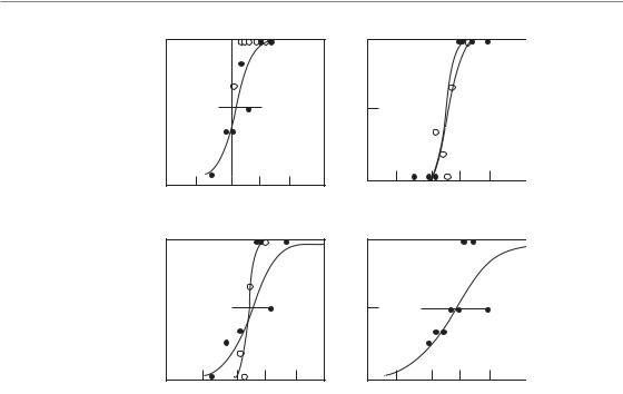
The influence of irradiated lung volume on structural and functional damage has been investigated experimentally in mice, rats, dogs and pigs. Most of these studies have focused on pneumonitis as the endpoint, and hence the data may not be wholly applicable to lung fibrosis. All studies demonstrate a pronounced volume effect for total lung function, with little or no symptomatic pneumonitis for partial irradiated volumes below 50 per cent. These volume effects depend on the compensatory capacity of the unirradiated tissue, which enables overall function to be maintained despite destruction of a substantial part of one lung (Herrmann et al., 1997). In contrast, local structural lung damage, assessed by radiography, histology or collagen content, is independent of the volume irradiated (Fig. 14.3). This indicates that cellular radiosensitivity is not influenced by the size of the irradiated volume, although the consequence of cell death for lung function is strongly dependent on volume.
Several prospective clinical studies have described the influence of a change in irradiated lung volume on local lung damage and on NTCP (reviewed in Mehta, 2005). Local structural and functional lung damage can be quantified using computed tomography (CT)-based lungdensity distributions and single-photon emission
196 The volume effect in radiotherapy
100
(%) |
50 |
|
|
||
|
|
|
responders |
(a) |
|
|
0 |
|
|
0 |
|
of |
100 |
|
Incidence |
|
|
|
|
|
|
50 |
|
|
|
|
0
0
(c)
100
50
|
|
0 |
20 |
40 |
20 |
40 |
0 |
||
|
|
|
(b)
100
50

 0
0 


 20 40 0 20 40
(d) Total dose (Gy)
Figure 14.3 Dose–response curves for radiation-induced lung damage in pigs after irradiation with five fractions, given to half of the right lung (open circles) or to the whole right lung (filled circles). Damage was assessed from radiographic (X-ray) changes (a), histological evidence of fibrosis (b), elevated hydroxyproline (collagen) levels (c), or increased breathing rate (d). Only the functional endpoint demonstrated a volume effect. From Herrmann et al. (1997), with permission.
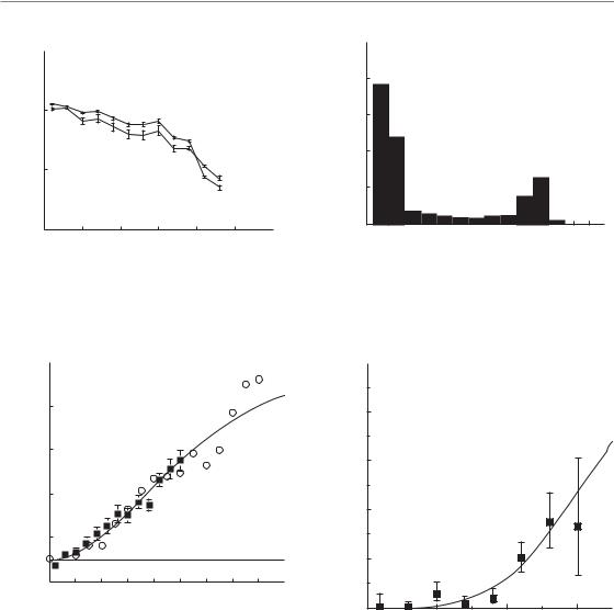
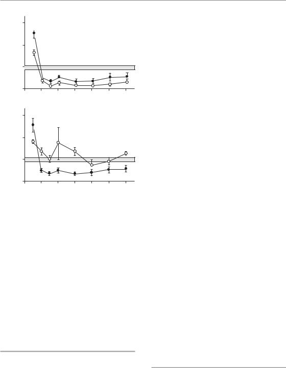
computed tomography (SPECT) ventilation and perfusion distributions. By matching preand post-treatment SPECT scans to the 3D dose distributions in the lung, changes in perfusion and ventilation can be quantified in relation to the locally delivered radiation dose (Fig. 14.4). For patients with malignant lymphoma and breast cancer, well-defined dose–response relationships have been determined for local changes in lung function per SPECT voxel (6 mm3 volume) in the irradiated areas, relative to the low-dose or unirradiated regions (Fig. 14.5). These and other studies demonstrate that the magnitude of local, structural pulmonary changes is independent of the irradiated volume but does depend on patient-related factors such as concurrent chemotherapy, smoking and age.
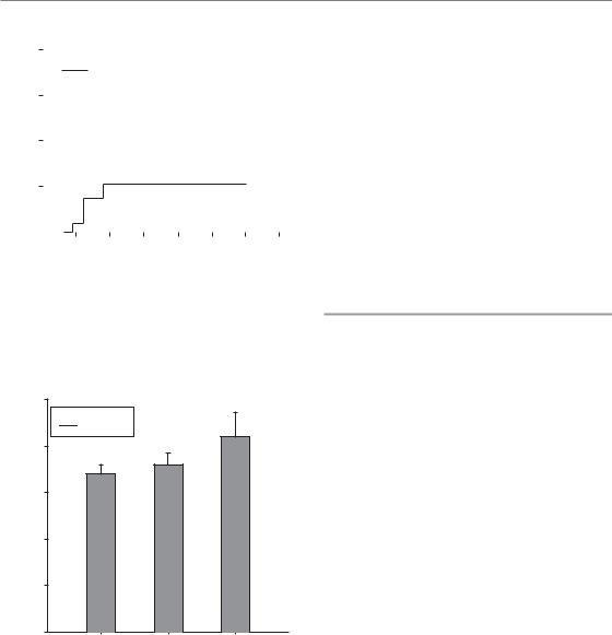
In an extension of these studies, the observed incidence of radiation-induced pneumonitis can be related to the DVH for the irradiated lung. For this analysis, the complex 3D physical dose distribution is converted into a mean biological dose to the whole lung, after a normalization procedure using the linear-quadratic (LQ) model with α/β ratios of 2.5–3 Gy. This parameter, the ‘mean normalized total lung dose’, which does not include any critical-volume parameter, correlates well with the incidence of pneumonitis, as for example shown in a large series of 540 patients treated for malignant lymphoma, lung or breast cancer. Results on pneumonitis in 264 patients treated for lymphoma, lung and breast cancer from one institute are displayed in Fig. 14.6. Further studies have focused on other, simple parameters such as the
Experimental and clinical data for volume effects 197
|
150 |
|
|
|
|
|
|
(%) |
|
|
|
|
|
|
|
SPECT values |
100 |
|
|
|
|
|
|
50 |
|
|
|
|
|
|
|
Local |
|
|
|
|
|
|
|
|
|
|
|
|
|
|
|
|
0 |
10 |
20 |
30 |
40 |
50 |
60 |
(a) |
0 |
||||||
|
|
|
|
|
|
|
|
50 |
|
|
|
|
|
|
|
40 |
|
|
|
|
|
|
(%) |
|
|
|
|
|
|
|
volume |
30 |
|
|
|
|
|
|
20 |
|
|
|
|
|
|
|
Lung |
|
|
|
|
|
|
|
|
|
|
|
|
|
|
|
|
10 |
|
|
|
|
|
|
|
0 |
|
|
|
|
|
|
|
6 |
14 |
22 |
30 |
38 |
46 |
|
(b) |
|
|
Dose (Gy) |
|
|
|
|
Figure 14.4 Dose–effect relationship for local lung perfusion (solid line) and ventilation (dashed line) at 3–4 months after irradiation, as a percentage of the pretreatment value, in a patient treated for malignant lymphoma (a). The corresponding dose–volume histogram of the three-dimensional dose distribution to the lung of this patient is shown in (b). From Boersma et al. (1994), with permission.
|
90 |
|
|
|
|
|
|
Local perfusion |
|
|
|
(%) |
70 |
|
|
|
|
|
|
|
|
|
|
per voxel |
50 |
|
|
|
|
Reduction |
30 |
|
|
|
|
|
|
|
|
|
|
|
10 |
|
|
|
|
|
|
|
|
|
|
|
0 |
20 |
40 |
60 |
80 |
Normalized total dose (Gy)
Figure 14.5 Dose–effect relationships for the average change in local lung function (i.e. perfusion) as a function of the normalized total dose. Data are shown from functional studies carried out at Duke University (circles) and The Netherlands Cancer Institute (squares). From Theuws et al. (1998) and including data from Marks et al. (1997), with permission from Elsevier.
percentage of total lung volume irradiated with defined doses (i.e. 20 Gy or 30 Gy), which hence incorporate a critical-volume component. These parameters can be used to predict the probability of radiation pneumonitis. In a comprehensive
|
100 |
(%) |
80 |
|
|
incidence |
60 |
Pneumonitis |
40 |
|
20
0
0 |
10 |
20 |
30 |
Mean normalized total dose (Gy)
Figure 14.6 Incidence of radiation pneumonitis as a function of mean normalized total dose to the whole lung. Pooled data are shown from a group of 264 patients with lung cancer, breast cancer or malignant lymphoma, treated in five different centres. The absolute number of patients contributing to each dose level is indicated. From Kwa et al. (1998), with permission from Elsevier.
review, Rodrigues et al. (2004) demonstrated that the ideal parameter for estimation of the NTCP for pneumonitis from DVH, despite ample data and studies, has not yet been identified. This raises
198 The volume effect in radiotherapy
serious doubts of whether further studies on patients treated with a range of different dose distributions will ever be sufficient to determine the best approach.
A few studies have investigated regional differences in radiation sensitivity of the lung. Experiments in mice demonstrate that functional damage is more prevalent after irradiation of the base of the lung than an equivalent volume irradiation of the apex (Travis et al., 1997). This may indicate that functional subunits are not homogeneously distributed, but are concentrated in basal areas. Cytokine release from partial liver irradiation, included in the basal lung field, may also contribute to these field localization effects. However, studies in pigs have not demonstrated differences in cellular damage in apical versus basal parts of the lung. Data from clinical studies are controversial.
In summarizing the clinical data available (Bentzen et al., 2000), tolerance limits for unaffected lung may be defined by the ratio of total vol-
ume of both lungs (Vtot) to the volume exposed to doses >25 Gy (V25). If this ratio, V25:Vtot is greater than 50 per cent, then pneumonitis can be dose
limiting, or pneumonitis is not relevant. The mean lung dose for a 5 per cent incidence of pneumonitis is in the order of 15 Gy. No conclusive data are available for fibrotic reactions.
Spinal cord
LENGTH OF IRRADIATED SPINAL CORD
A marked volume effect for irradiation of very short lengths of spinal cord ( 1 cm), and less pronounced or no volume effects for cord lengths2 cm, have been demonstrated in rats, pigs, monkeys and dogs. This suggests migration of tissue-restoring cells, with only a limited migration distance, from outside the irradiated volume (Section 14.4).
In rat spinal cord, a very steep rise in ED50 (i.e. the radiation dose at which white-matter necrosis and myelopathy are expected in 50 per cent of treated animals) was observed when the irradiated cord length was reduced below 10 mm (Fig. 14.7).
For irradiation of cord lengths between 10 and
|
100 |
|
|
80 |
|
(Gy) |
60 |
|
|
|
|
50 |
|
Split field |
ED |
|
|
40 |
2 |
|
|
|
20
0
0 |
5 |
10 |
15 |
20 |
Cord length (mm)
Figure 14.7 The influence of field size on biological response in rat spinal cord after single-dose irradiation with small fields. A steep rise in ED50 occurs as field size is reduced below 10 mm, with very little change in ED50 for larger field-sizes. The single fields were centred around C4, the two concomitant 4-mm fields were at C1/C2 and C7/T1, separated by approximately 10 mm. Data show the ED50 for induction of white-matter necrosis. From Hopewell and Trott (2000) and Bijl et al. (2002, 2006), with permission.
30 mm, little change in ED50 was found. This observation has been confirmed by more detailed studies, exploiting a high-precision proton beam for irradiation (Bijl et al., 2003): rats were irradiated with either a single field of 8 mm or two fields of 4 mm, separated by an unirradiated length of
cord of 8 mm or 12 mm (Fig. 14.8). The ED50 for myelopathy with 2 4 mm fields was 42–45 Gy,
which is less than 54 Gy for a single field of 4 mm, but considerably greater than the ED50 for 1 8 mm (25 Gy).
Single radiation doses given to 2.5-, 5- and 10-cm lengths of pig spinal cord showed only a small (c. 1 Gy) decrease in ED50 for induction of white-matter necrosis with increasing field size. At low probabilities of injury, which are clinically relevant, this difference was no longer significant (van den Aardweg et al., 1995), similar to the results in the kidney (see below). The irradiated cord length
Experimental and clinical data for volume effects 199
|
100 |
|
|
|
|
|
|
|
No bath |
|
80 |
|
|
4 Gy bath |
|
|
|
|
18 Gy bath |
(Gy) |
60 |
|
|
|
|
|
|
|
|
50 |
|
|
|
|
ED |
40 |
|
|
|
|
|
|
|
|
|
20 |
|
|
|
|
0 |
4 |
8 |
20 |
|
2 |
Shower volume (mm)
Figure 14.8 The influence of a surrounding low dose on the tolerance of a small high-dose volume (‘bath and shower’ irradiations) in the rat cervical spinal cord. The high-dose volumes of 2, 4 or 8 mm (‘shower’) were centred at C5, while the remainder of the 20-mm cervical cord was irradiated with a ‘bath’ dose of 4 Gy or 18 Gy. From Bijl et al. (2003, 2006), with permission from Elsevier.
also influenced the incidence of myelopathy in monkeys given fractionated irradiation (Schultheiss et al., 1994). The incidence of myelopathy after a total dose of 70.2 Gy (2.2 Gy per fraction) increased from 15 per cent, to 20 per cent to 37.5 per cent for field sizes of 4, 8 and 16 cm, respectively.
In an extensive study in dogs (Powers et al., 1998), irradiation of 4- and 20-cm lengths of spinal cord were compared using a fractionated schedule of 4 Gy per fraction. For functional, neurological symptoms, such as thoracic pain or paresis, a large increase in ED50 from 54 Gy for the large field to 78 Gy for the small field was found. In contrast, a much less pronounced increase was observed for morphological, necrotic lesions (Fig. 14.9). This again indicates the relevance of the endpoint studied for volume effects.
INFLUENCE OF DOSE SURROUNDING THE HIGH-DOSE VOLUME
The marked volume effect for irradiation of only very short lengths of spinal cord is clearly
|
100 |
|
|
|
|
(%) |
75 |
|
|
|
|
myelopathyof |
|
|
|
|
|
50 |
|
|
|
|
|
Incidence |
|
|
|
|
|
25 |
|
|
|
|
|
|
|
|
|
|
|
|
0 |
40 |
60 |
80 |
100 |
|
20 |
Total dose (Gy)
Figure 14.9 Influence of change in field size on spinal cord damage in dogs. Increasing the field size from
4 cm (circles) to 20 cm (squares) had a more marked influence on the development of neurological signs of injury (dotted lines) than on the occurrence of severe pathological lesions (solid lines). Redrawn from Powers et al. (1998), with permission.
compromised when a small dose is given to the surrounding tissue. This has been demonstrated for rat spinal cord in ‘bath-and-shower’ experiments (Bijl et al., 2003), where graded subtolerance doses (‘bath’) were given to a large segment (20 mm) of spinal cord and a high dose (‘shower’) was given to small segments of 2–8 mm in the centre of the low-dose volume. The ED50 for a 4-mm field given 53 Gy alone was reduced to 39 Gy with a bath dose of only 4 Gy. For a 2-mm high-dose segment, the ED50 was reduced from 88 Gy to 61 Gy. Paralysis was based on necrotic lesions in the high-dose region whereas no histological changes were seen in the bath volume. The hypothesis to describe these observations was that migration of presumptive stem cells into the highdose region was compromised by the bath dose.
In further experiments with the same model (Bijl et al., 2003), the shower dose was placed at the edge rather than the centre of the low-dose segment. Assuming migration phenomena, the tolerance in this setup should be similar to that with high-dose irradiation alone (88 Gy) for a 2-mm shower segment. However, the observed ED50 was intermediate, at 69 Gy, indicating additional

200 The volume effect in radiotherapy
mechanisms underlying the volume effect for small cord lengths. It is important to note that none of the existing NTCP models take these nonlocal effects into account.
LATERAL DOSE DISTRIBUTION
With conformal radiotherapy, variations not only in the length of spinal cord irradiated but also in the lateral distribution of the dose can occur. These effects have also been studied in rats irradiated with high-precision proton beams (Bijl et al., 2005). The left lateral half of the spinal cord was irradiated with a penumbra (20–80 per cent isodose) of 1.1 mm or 0.8 mm, or the midline of the cord with a penumbra of 0.8 mm. The irradiated length of spinal cord was 20 mm in all experiments. The resulting ED50 values for paralysis were 29 Gy and 33 Gy for lateral irradiation, respectively, and 72 Gy for midline irradiation; the corresponding homogeneous irradiation of a 20-mm cord segment resulted in an ED50 of 20 Gy. Hence, the grey matter is highly resistant to radiation as no lesions observable by light microscopy were induced, even after a single dose as high as 80 Gy; all lesions were restricted to white matter structures.
In a recent analysis of clinical data from singledose radiosurgery of 230 vertebral column metastases in 177 patients (Ryu et al., 2007), an average dose of 9.8 1.5 Gy to 10 per cent of the spinal cord volume, given in single-dose radiosurgery, was associated with only one spinal cord injury in 86 patients surviving more than 1 year.
In summary, there is a clear volume effect for severe lesions in the spinal cord which lead to irreversible signs of myelopathy; this is most pronounced at high levels of injury. At low probabilities of injury (< 5 per cent), which usually define clinical tolerance doses, a volume effect may not be detectable and should have minimal impact on the clinical practice of maintaining spinal cord dose below 55 Gy. However, when clinical conditions require the choice of higher dose levels closer to tolerance, such as in a re-irradiation situation, the existence of a volume effect might be taken into consideration. The volume effect in the spinal cord is complex, with an impact of the surrounding dose as well as of the lateral dose distribution across the tissue. It must be emphasized
that none of the existing NTCP models take this complexity into account.
Kidney
Volume effects in the irradiated kidney are strongly influenced by the duality of this organ and by its parallel organization, with a large reserve capacity. Unirradiated parts of one kidney and the contralateral kidney are able to undergo drastic postirradiation hypertrophy and increase their performance to compensate for functional impairment within another part of the organ and thus maintain renal function.
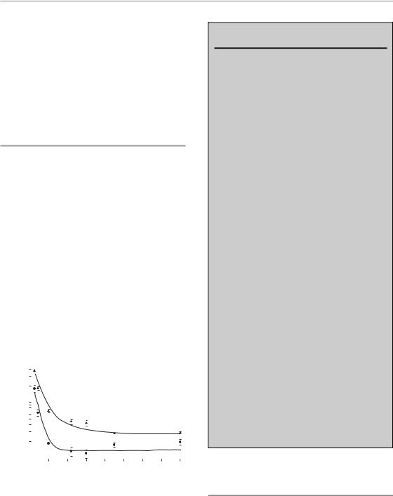
Experiments in pigs (Robbins and Hopewell, 1988) have demonstrated that the individual function of a kidney after unilateral irradiation can actually be poorer than after irradiation of both kidneys with the same dose. Total renal function is, however, much less reduced after irradiation of only one kidney (Fig. 14.10). Interestingly, if the unirradiated kidney is removed after unilateral irradiation, the previously non-functional, irradiated kidney may be capable of partial restoration of glomerular filtration rate and effective renal plasma flow to maintain a viable level of total renal function. These experiments demonstrate that the functional response of a unilaterally irradiated kidney depends on the compensatory response in the unirradiated, contralateral organ. The presence and increased function of the unirradiated kidney may actually promote functional impairment, or inhibit functional recovery, in the irradiated kidney.
A pronounced volume effect was also observed in scintigraphic studies in 91 patients who received abdominal irradiation with various doses and to varying volumes (Köst et al., 2002), but only at higher incidence levels for the loss of kidney function. The dose for a 5 per cent incidence was in the range of 3–6 Gy, independent of volume, owing to uncertainties in the dose–effect analysis. However, the doses estimated for a 50 per cent incidence clearly increased with decreasing kidney volume, from approximately 8 Gy for 100 per cent to 27 Gy for 10 per cent of the volume.
In a study in 44 patients receiving radiochemotherapy for gastric cancer (Jansen et al.,
2007), where the left kidney was included in the
Experimental and clinical data for volume effects 201
function |
300 |
|
|
kidney |
200 |
Individual |
100 |
|
|
0 |
|
|
|
(a) |
0 |
8 |
16 |
24 |
|
300 |
|
|
|
function |
200 |
|
|
|
kidney |
100 |
|
|
|
Total |
|
|
|
|
|
|
|
|
|
|
0 |
|
|
|
0 |
8 |
16 |
24 |
(b) |
Time after irradiation (weeks) |
|
|
Figure 14.10 Time-related changes in glomerular filtration rate in pigs in which one (open circles) or both (filled circles) kidneys were irradiated with a single dose of 12.6 Gy. (a) The change in individual kidney function, as a percentage of control values; (b) the total renal function in the same pigs. Redrawn from Robbins and Hopewell (1988), with permission from Elsevier.
high-dose radiotherapy volume, the V20 (left kidney) and mean left kidney dose were identified as parameters associated with decreased kidney function.
Liver
The liver is usually regarded as a prime example of a parallel-type organ, with liver acini as FSUs. Similar to lung, structural damage (i.e. loss of function within the irradiated volume) can be demonstrated with scintigraphy and other methods. Whole-liver irradiation with doses around
30 Gy (2 Gy per fraction) is generally associated
with the induction of 5–10 per cent hepatitis. Until the introduction of 3D treatment planning, the relationship between tolerance dose and partial volume irradiation was very conservatively interpreted. However, extensive clinical data on liver tolerance to partial organ irradiation has now been accumulated at the University of Michigan for more than 200 patients (Dawson and Ten Haken, 2005). LKB–NTCP analyses resulted in a large volume–effect parameter (the n-exponent in the power law function) of 0.97, demonstrating that the liver indeed behaves as a parallel-type organ with a large reserve capacity. No cases of radiationinduced liver disease grade 3 according to RTOG/EORTC (Radiation Therapy and Oncology Group and the European Organisation for Research and Treatment of Cancer) were observed when the mean liver dose was 31 Gy. Estimates of tolerance doses associated with 5 per cent risk of liver disease after uniform irradiation to partial volumes were 47 Gy for two-thirds and 90 Gy for one-third of the total volume. A negligible complication risk, regardless of dose, is associated with irradiation of a partial volume of 25 per cent. A recent analysis by Kim et al. (2007) in 105 patients with hepatocellular carcinoma revealed that the total liver volume receiving 30 Gy also appears to be a useful dose–volumetric parameter for predicting the risk of hepatic toxicity and should be limited to 60 per cent.
The tolerance of patients with primary liver cancer is lower than that of patients with liver metastases or without liver tumours (Dawson and Ten Haken, 2005). However, the functionality of the unirradiated liver volume, which may be impaired by chemotherapy, alcohol consumption or other trauma, has to be taken into consideration. The presence of liver cirrhosis is known to be the predominant risk factor for hepatic toxicity following radiotherapy.
Intestinal tract
The results of several trials have been published for treatment of prostate, rectal or cervical cancer, relating the bowel volume irradiated, i.e. various
DVH parameters, to the incidence of complications. Restricting dose conformation to the PTV
202 The volume effect in radiotherapy
can substantially decrease the normal tissue volumes exposed to significant doses. For example, a study from the Royal Marsden Hospital in London showed that the volume of small bowel irradiated to 90 per cent of the prescribed dose could be reduced from 24 per cent using conventional fields to 18 per cent for 3D conformal therapy and 5–8 per cent for IMRT, depending on the number of fields used. For the rectum, the high-dose irradiated volumes could be reduced from 89 per cent to 51 per cent and 6–16 per cent, respectively (Nutting et al., 2000). It should be noted that early complications in the intestine result in an increased incidence of late effects, thus representing a consequential component (see Chapter 13). Therefore, volume–effect studies on late complications may be affected by a varying influence of early effects.
ORAL MUCOSA
The clinical consequences of oral mucositis are closely related to the localization where the reaction occurs. For example, complications can be significantly reduced if the lips are excluded from the irradiated volume. Also, changes in taste acuity can be prevented by a reduction of the tongue volume included in the high-dose volume.
OESOPHAGUS
A study on 215 patients treated for non-small cell lung cancer (Wei et al., 2006) revealed that acute oesophageal symptoms were dominated by DVH parameters, such as the mean dose, or the relative volume treated to doses above 20 Gy (rV20), but were independent of clinical factors.
SMALL BOWEL
The mobility of the small intestine largely prevents irradiation of the same segment or loop during subsequent fractions. Hence, the dose to individual loops can vary over wide ranges, and usually is lower than the dose to the target volume. However, previous surgery, related or independent of the oncological disease, as well as inflammatory changes in the abdominal cavity, can compromise mobility and hence significantly
increase the dose to bowel segments fixed in the high-dose volume. These uncertainties appear to be one of the reasons for the conflicting data for a correlation of dose and volume with the incidence of small bowel symptoms.
Volume effects for small bowel obstruction have been demonstrated in patients treated with extended field radiotherapy (bowel volumes800 cm3), as reviewed by Letschert et al. (1994). No volume effects for bowel obstruction were observed in patients receiving postoperative radiotherapy for rectal carcinoma. In these patients, the incidence of chronic diarrhoea was 31 per cent for volumes 77 cm3, compared with 42 per cent for volumes 328 cm3. The type of surgery was a strong influence on the incidence of chronic diarrhoea and malabsorption.
RECTUM
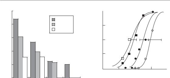
The incidence of late rectal bleeding and other chronic changes has generally been found to correlate with irradiated volume exposed to high doses, as quantified by DVH parameters. Some studies have demonstrated a significant dose–volume relationship for late rectal bleeding using a single cutoff value for the rectal volume irradiated to certain doses (Fig. 14.11). Other studies describe a more complex relationship with several cut-off levels which significantly discriminated between a high or low risk of severe rectal bleeding, or a continuous relationship between rectal bleeding and dose– volume parameters. A study on rectal mucosal changes assessed by rectoscopy using a specific score in 35 patients receiving external–beam radiotherapy and high dose-rate brachytherapy for carcinoma of the cervix revealed ED50 values for irradiated volumes of 2 cm3, 1 cm3 or 0.1 cm3 of 68 Gy, 73 Gy and 84 Gy (Fig. 14.12). The corresponding doses for changes according to LENT/SOMA were 73 Gy, 78 Gy and 97 Gy.
One aspect to be considered for highly conformal radiotherapy, particularly for the rectum, is the dose distribution through the cross-section of the organ. Gross clinical symptoms are observed only if a larger part or the entire circumference is exposed to significant doses. In rats, it was demonstrated that shielding of half the circumference completely prevented late bowel obstruction, which occurred
Experimental and clinical data for volume effects 203
(%) |
40 |
|
|
|
|
|
|
V90 57% |
|
|
|
|
|
|
|
|
|
|
|
|
|
|
||||||
|
|
|
|
|
|
|
|
|
|
|
|
|
|
|
|
|
|
|
|
|||||||||
bleedingrectal |
|
|
|
|
|
|
|
V90 57 Gy |
|
|
|
|
|
|
|
|
|
|
|
|
|
|
||||||
|
|
|
|
|
|
|
|
|
|
|
|
|
|
|
|
|
|
|
|
|
|
|||||||
|
30 |
|
|
|
|
|
|
|
|
|
|
|
|
|
|
|
|
|
|
|
|
|
|
|
|
|
|
|
|
|
|
|
|
|
|
|
|
|
|
|
|
|
|
|
|
|
|
|
|
|
|
|
|
|
|
|
|
of late |
20 |
|
|
|
|
|
|
|
|
|
|
|
|
|
|
|
P |
0.03 |
|
|
|
|
|
|
|
|||
|
|
|
|
|
|
|
|
|
|
|
|
|
|
|
|
|
|
|
|
|
|
|||||||
|
|
|
|
|
|
|
|
|
|
|
|
|
|
|
|
|
|
|
|
|
|
|||||||
|
|
|
|
|
|
|
|
|
|
|
|
|
|
|
|
|
|
|
|
|
|
|
|
|
|
|
|
|
Incidence |
10 |
|
|
|
|
|
|
|
|
|
|
|
|
|
|
|
|
|
|
|
|
|
|
|
|
|
|
|
|
|
|
|
|
|
|
|
|
|
|
|
|
|
|
|
|
|
|
|
|
|
|
|
|
|
|
|
|
|
0 |
|
|
|
|
|
|
|
|
|
|
|
|
|
|
|
|
|
|
|
|
|
|
|
|
|
|
|
|
|
|
|
|
|
|
|
|
|
|
|
|
|
|
|
|
|
|
|
|
|
|
|
|
|
|
|
|
|
|
|
|
|
|
|
|
|
|
|
|
|
|
|
|
|
|
|
|
|
|
|
|
|
|
|
|
|
|
0 |
10 |
20 |
|
30 |
|
40 |
50 |
60 |
70 |
||||||||||||||||||
|
|
|
|
|
|
|
Time after treatment (months) |
|
|
|
|
|
|
|
||||||||||||||
Figure 14.11 Actuarial incidence of rectal bleeding in patients with rectal volumes (V90) irradiated to at least 90 per cent of the isodose (approximately 60 Gy) of greater than 57 per cent or less than 57 per cent. From Wachter et al. (2001), with permission from Elsevier.
100

 VRS
VRS
|
80 |
|
|
(Gy) |
60 |
|
|
|
|
|
|
50 |
|
|
|
ED |
40 |
|
|
|
|
|
|
|
20 |
|
|
|
0 |
|
|
|
2.0 |
1.0 |
0.1 |
|
Rectal volume irradiated (cm3) |
||
Figure 14.12 The ED50 values for rectoscopic mucosal changes [Vienna Rectoscopy Score (VRS) 3] in patients receiving teletherapy and brachytherapy for cervical cancer in rectal volumes of 2 cm3, 1 cm3 or 0.1 cm3. Data from Georg et al. (unpublished), with permission of the authors.
with increasing dose in animals with irradiation of the entire rectal wall. The incidence (but not the area) of mucosal ulceration was similar in both groups. Similarly, an intrarectal balloon can be
applied for displacement of the posterior rectal wall and hence for a reduction in dose to this part, which results in a decrease in side-effects. In contrast, emptying of the rectum before irradiation increases dose homogeneity and reduces variations of DVH parameters for the PTV, but may increase the risk of rectal complications.
As with other organs, such as the lung (see above), the functional status of the rectum can influence the volume tolerance. Huang et al. (2002) demonstrated that an individual history of haemorrhoids clearly correlated with the incidence of rectal bleeding at a given volume irradiated.
Parotid gland
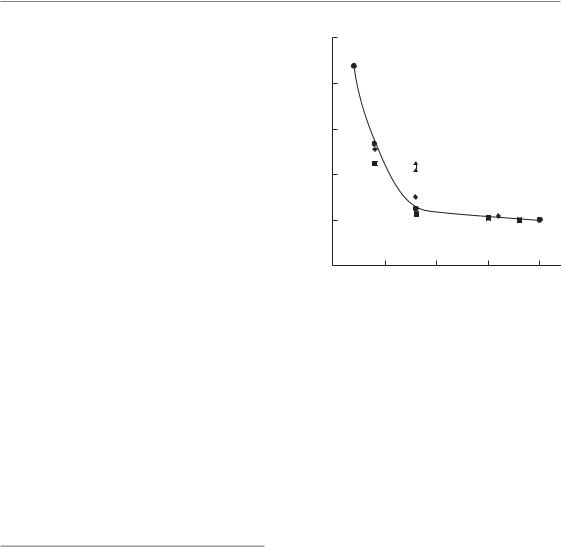
The parotid glands are important dose-limiting organs in treatment of the head and neck with conventional radiation techniques, as they often cannot be spared. Doses above 40–50 Gy lead to permanent loss of function contributing to xerostomia (parotid glands produce c. 60 per cent of the saliva) and impairment of quality of life. One of the major advantages of the introduction of 3D-conformal techniques in the head and neck area is the possibility of limiting the irradiated volume to parts of the parotids and this has resulted in a reduction of permanent xerostomia. A DVH-based prospective trial showed complete recovery of salivary flow at 1 year after mean doses to the parotid of around 26 Gy (Eisbruch et al., 1999). The anatomical organization in acini as FSUs was clearly reflected in the outcome of modelling the partial-volume data, as derived from the DVH. The LKB model showed the volumeeffect parameter n to be close to 1.0, which indicates a nearly parallel behaviour, whereby the mean dose determines NTCP. In these studies partialvolume thresholds were (approximately) 45 Gy for 25 per cent, 30 Gy for 50 per cent and 15 Gy for 67 per cent of the total volume.
Rat studies (Konings et al., 2006) have demonstrated significant regional variations in radiation sensitivity in the parotid gland. The dose effect for late changes in saliva production, but not for early changes, was different for the cranial and caudal part of the glands, with a substantially higher radiosensitivity of the cranial part. The reduction
204 The volume effect in radiotherapy
in saliva output after a single dose of 30 Gy was 65 per cent for irradiation of the cranial and 25 per cent for irradiation of the caudal 50 per cent volume of the gland, and 100 per cent after irradiation of the entire organ. Histological studies showed that irradiation of the cranial 50 per cent volume also caused late development of secondary damage in the shielded caudal part at late time-points.
Skin
The skin shows an ‘area’ effect similar to oral mucosa. In studies in pig skin (Hopewell et al., 1986), no effect of the irradiated area was observed for acute epidermal changes when the field diameter was larger than 20 mm, and for late effects when the diameter was larger than 10 mm. At smaller diameters, a steep rise in isoeffective doses was found (Fig. 14.13). In the orthovoltage era, this area effect of small fields used to be exploited by using a ‘sieve technique’, where part of the skin was shielded with the bridges of a lead sieve, and hence tolerable reactions occurred only in the small irradiated fields, but did not achieve confluency. This allowed for curative tumour doses despite the unfavourable depth-dose distribution of these low-energy X-rays.
(Gy) |
400 |
|
|
|
|
|
|
|
|
|
|
|
|
|
|
|
|
|
|
|
|
||
200 |
|
|
|
|
|
|
|
|
|
|
|
|
|
|
|
|
|
|
|
|
|
||
damage |
100 |
|
|
|
|
|
|
|
|
|
|
|
|
|
|
|
|
|
|
|
|
|
|
skin |
50 |
|
|
|
|
|
|
|
Acute |
|
|
|
|
|
|
|
|
|
|
|
|||
|
|
|
|
|
|
|
|
|
|||
|
|
|
|
|
|
|
|
|
|||
|
|
|
|
|
|
|
|
|
|||
50 |
|
|
|
|
|
|
|
|
|
|
|
|
|
|
|
|
|
|
|
Late |
|
|
|
|
|
|
|
|
|
|
|
|
|
||
ED |
10 |
|
|
|
|
|
|
|
|
|
|
|
|
|
|
|
|
|
|
|
|
||
|
|
|
|
|
|
|
|
|
|
|
|
|
|
|
|
|
|
|
|
|
|
|
|
|
|
|
|
10 |
20 |
30 |
40 |
||||
|
0 |
|
|||||||||
Source diameter (mm)
Figure 14.13 The influence of field-diameter on the dose required to induce acute and late skin reactions in 50 per cent of pigs after single-dose irradiation with small fields. A steep rise in ED50 is seen below a diameter of 20 mm for early reactions and 10 mm for late sequelae, respectively. At larger field-sizes, the ED50 is independent of the area exposed. From Hopewell and Trott (2000), with permission.
Key points
1.Structural tissue tolerance depends on cellular radiation sensitivity and is independent of volume irradiated. Functional tolerance depends on tissue organization and functional reserve capacity.
2.Tissues with a parallel organization (e.g. lung) have a large reserve capacity and show a pronounced volume effect, with a threshold volume below which functional damage does not occur. The risk of developing a complication depends on dose distribution throughout the whole organ rather than the maximum dose to a small area.
3.Tissues with a serial organization (e.g. spinal cord) have little or no functional reserve and the risk of developing a complication is less dependent on volume irradiated than for tissues with a parallel organization. The risk of complication is strongly influenced by high-dose regions and hot-spots.
4.Migration of surviving clonogenic cells into the edge of irradiated fields can lead to a steep increase in tissue tolerance for field diameters up to 20 mm in some tissues (e.g. spinal cord, intestine, skin).
5.Theoretical models have been developed to estimate NTCP for partial-volume irradiations and inhomogeneous dose distributions. Simple power-law and probability models have been expanded to incorporate parameters relating to tissue architecture and reserve capacity. These models need to be validated against clinical data emerging from conformal treatment schedules.
■BIBLIOGRAPHY
Bentzen SM, Skoczylas JZ, Bernier J (2000). Quantitative clinical radiobiology of early and late lung reactions. Int J Radiat Biol 76: 453–62.
Bijl HP, van Luijk P, Coppes RP, Schippers JM, Konings AW, van der Kogel AJ (2002). Dose-volume effects in the rat cervical spinal cord after proton irradiation.
Int J Radiat Oncol Biol Phys 52: 205–11.

Bibliography 205
Bijl HP, van Luijk P, Coppes RP, Schippers JM, Konings AW, van der Kogel AJ (2003). Unexpected changes of rat cervical spinal cord tolerance caused by inhomogeneous dose distributions. Int J Radiat Oncol Biol Phys 57: 274–81.
Bijl HP, van Luijk P, Coppes RP, Schippers JM, Konings AW, van Der Kogel AJ (2005). Regional differences in radiosensitivity across the rat cervical spinal cord.
Int J Radiat Oncol Biol Phys 61: 543–51.
Bijl HP, van Luijk P, Coppes RP, Schippers JM, Konings AW, van der Kogel AJ (2006). Influence of adjacent low-dose fields on tolerance to high doses of protons in rat cervical spinal cord. Int J Radiat Oncol Biol Phys 64: 1204–10.
Boersma LJ, Damen EM, de Boer RW et al. (1994). Doseeffect relations for local functional and structural changes of the lung after irradiation for malignant lymphoma. Radiother Oncol 32: 201–9.
Dawson LA, Ten Haken RK (2005). Partial volume tolerance of the liver to radiation. Semin Radiat Oncol 15: 279–83.
Eisbruch A, Ten Haken RK, Kim HM, Marsh LH, Ship JA (1999). Dose, volume, and function relationships in parotid salivary glands following conformal and intensity-modulated irradiation of head and neck cancer. Int J Radiat Oncol Biol Phys 45: 577–87.
Emami B, Lyman J, Brown A et al. (1991). Tolerance of normal tissue to therapeutic irradiation. Int J Radiat Oncol Biol Phys 21: 109–22.
Herrmann T, Baumann M, Voigtmann L, Knorr A (1997). Effect of irradiated volume on lung damage in pigs.
Radiother Oncol 44: 35–40.
Hopewell JW, Trott KR (2000). Volume effects in radiobiology as applied to radiotherapy. Radiother Oncol 56: 283–8.
Hopewell JW, Coggle JE, Wells J, Hamlet R, Williams JP, Charles MW (1986). The acute effects of different energy beta-emitters on pig and mouse skin. Br J Radiol Suppl 19: 47–51.
Huang EH, Pollack A, Levy L et al. (2002). Late rectal toxicity: dose-volume effects of conformal radiotherapy for prostate cancer. Int J Radiat Oncol Biol Phys 54: 1314–21.
ICRU (1999). Prescribing, recording and reporting photon beam therapy. ICRU report 62. Oxford: Oxford University Press.
Jansen EP, Saunders MP, Boot H et al. (2007). Prospective study on late renal toxicity following postoperative chemoradiotherapy in gastric cancer.
Int J Radiat Oncol Biol Phys 67: 781–5.
Källman P, Agren A, Brahme A (1992). Tumour and normal tissue responses to fractionated non-uniform dose delivery. Int J Radiat Biol 62: 249–62.
Kim TH, Kim DY, Park JW et al. (2007). Dose-volumetric parameters predicting radiation-induced hepatic toxicity in unresectable hepatocellular carcinoma patients treated with three-dimensional conformal radiotherapy. Int J Radiat Oncol Biol Phys 67: 225–31.
Konings AW, Faber H, Cotteleer F, Vissink A, Coppes RP (2006). Secondary radiation damage as the main cause for unexpected volume effects: a histopathologic study of the parotid gland. Int J Radiat Oncol Biol Phys 64: 98–105.
Köst S, Dörr W, Keinert K, Glaser FH, Endert G, Herrmann T (2002). Effect of dose and dosedistribution in damage to the kidney following abdominal radiotherapy. Int J Radiat Biol 78: 695–702.
Kutcher GJ, Burman C (1989). Calculation of complication probability factors for non-uniform normal tissue irradiation: the effective volume method. Int J Radiat Oncol Biol Phys 16: 1623–30.
Kwa SL, Lebesque JV, Theuws JC et al. (1998). Radiation pneumonitis as a function of mean lung dose: an analysis of pooled data of 540 patients. Int J Radiat Oncol Biol Phys 42: 1–9.
Letschert JG, Lebesque JV, Aleman BM et al. (1994). The volume effect in radiation-related late small bowel complications: results of a clinical study of the EORTC Radiotherapy Cooperative Group in patients treated for rectal carcinoma. Radiother Oncol 32: 116–23.
Lyman JT (1985). Complication probability as assessed from dose-volume histograms. Radiat Res Suppl 8: S13–9.
Marks LB, Munley MT, Spencer DP et al. (1997). Quantification of radiation-induced regional lung injury with perfusion imaging. Int J Radiat Oncol Biol Phys 38: 399–409.
Mehta V (2005). Radiation pneumonitis and pulmonary fibrosis in non-small-cell lung cancer: pulmonary function, prediction, and prevention. Int J Radiat Oncol Biol Phys 63: 5–24.
Novakova-Jiresova A, van Luijk P, van Goor H, Kampinga HH, Coppes RP (2007). Changes in expression of injury after irradiation of increasing volumes in rat lung. Int J Radiat Oncol Biol Phys 67: 1510–8.
Nutting CM, Convery DJ, Cosgrove VP et al. (2000). Reduction of small and large bowel irradiation using

206 The volume effect in radiotherapy
an optimized intensity-modulated pelvic radiotherapy technique in patients with prostate cancer. Int J Radiat Oncol Biol Phys 48: 649–56.
Powers BE, Thames HD, Gillette SM, Smith C, Beck ER, Gillette EL (1998). Volume effects in the irradiated canine spinal cord: do they exist when the probability of injury is low? Radiother Oncol 46: 297–306.
Robbins ME, Hopewell JW (1988). Effects of single doses of X-rays on renal function in the pig after the irradiation of both kidneys. Radiother Oncol 11: 253–62.
Rodrigues G, Lock M, D’Souza D, Yu E, van Dyk J (2004). Prediction of radiation pneumonitis by dose-volume histogram parameters in lung cancer – a systematic review. Radiother Oncol 71: 127–38.
Ryu S, Jin JY, Jin R et al. (2007). Partial volume tolerance of the spinal cord and complications of single-dose radiosurgery. Cancer 109: 628–36.
Schultheiss TE, Stephens LC, Ang KK, Price RE, Peters LJ (1994). Volume effects in rhesus monkey spinal cord.
Int J Radiat Oncol Biol Phys 29: 67–72. Theuws JC, Kwa SL, Wagenaar AC et al. (1998).
Prediction of overall pulmonary function loss in relation to the 3-D dose distribution for patients with breast cancer and malignant lymphoma.
Radiother Oncol 49: 233–43.
Travis EL, Liao ZX, Tucker SL (1997). Spatial heterogeneity of the volume effect for radiation pneumonitis in mouse lung. Int J Radiat Oncol Biol Phys 38: 1045–54.
van den Aardweg GJ, Hopewell JW, Whitehouse EM (1995). The radiation response of the cervical spinal
cord of the pig: effects of changing the irradiated volume. Int J Radiat Oncol Biol Phys 31: 51–5.
Wachter S, Gerstner N, Goldner G, Potzi R, Wambersie A, Potter R (2001). Rectal sequelae after conformal radiotherapy of prostate cancer: dose-volume histograms as predictive factors. Radiother Oncol 59: 65–70.
Wei X, Liu HH, Tucker SL et al. (2006). Risk factors for acute esophagitis in non-small-cell lung cancer patients treated with concurrent chemotherapy and three-dimensional conformal radiotherapy. Int J Radiat Oncol Biol Phys 66: 100–7.
Withers HR, Taylor JM, Maciejewski B (1988). Treatment volume and tissue tolerance. Int J Radiat Oncol Biol Phys 14: 751–9.
■ FURTHER READING
Deasy JO, Niemierko A, Herbert D et al. (2002). Methodological issues in radiation dose-volume outcome analyses: summary of a joint AAPM/NIH workshop. Med Phys 29: 2109–27.
Kong FM, Pan C, Eisbruch A, Ten Haken RK (2007). Physical models and simpler dosimetric descriptors of radiation late toxicity. Semin Radiat Oncol 17: 108–20.
Sundar S, Symonds P, Deehan C (2003). Tolerance of pelvic organs to radiation treatment for carcinoma of cervix. Clin Oncol 15: 240–7.