

Therapeutic Micro-Nano Technology BioMEMs - Tejlal Desai & Sangeeta Bhatia
.pdf316 |
JAY T. GROVES |
to a particular protein of interest triggered a phase transition only when the appropriate cognate ligand was incorporated into the colloid membrane.
Quantitative analysis of the colloidal phases is performed by extracting the pair distribution function, g(r ). Bead positions are measured from wide-field ( 1 mm2) images by an object locating algorithm to a precision of 15 nm. Experiments are typically carried out at bead area fractions of φ ≈ 0.2, corresponding to 104 individual beads per image; g(r ) is then generated from > 108 measured pair distances. Higher area fractions allow determination of g(r ) with higher precision, however the system is more likely to become trapped in non-equilibrium configurations. Plots of g(r ) for different time points during a phase transition triggered by protein binding are illustrated in Figure 17.6B. The condensed phase g(r ) is characterized by a large peak at the nearest neighbor separation distance of one bead diameter (r0) and secondary peaks occurring at r = √3r0 and 2r0, corresponding to next nearest neighbors in the hexagonal crystallites. Independent measurements of g(r ) were highly consistent. Standard deviations in the magnitude of the r0 peak determined from separate colloidal preparations were generally less than 5%. Dispersed phases, consisting of random distributions and correspondingly flat g(r ) functions, are visibly distinguishable from condensed phases. Quantitative determination of g(r ) additionally distinguishes a range of intermediate distributions. These can be transient, such as the dispersing crystallites in Figure 17.6, t = 30 s. Intermediate degrees of order can also observed in near equilibrium distributions, corresponding with differing amounts of protein binding on the membrane surface. The colloid detection strategy can thus be used to extract binding affinity constants for membrane associated molecules, without the use of labels or sophisticated instrumentation. A low-magnification imaging system and some image analysis is all that is required.
17.4.3. Electrical Manipulation
Ion channels are a very important class of membrane proteins, and measurements of electrical conductance through these channels has been an major area of study for many years. Conventional techniques such as patch clamping [105], in which a micropipette is carefully brought into contact with a membrane, and the black lipid membrane (BLM) method [106], which consists of a reconstituted free standing bilayer membrane spanning an aperture between two aqueous chambers, have yielded a wealth of information. However, these are challenging techniques, which require a high degree of craftsmanship to perform successfully. Consequently, there has been much interest in developing supported membrane systems on conductive substrates to produce chip-based versions of the patch clamp or BLM techniques.
For electrical measurements, it is critical that the membrane be highly insulating. Indeed the success of patch clamp and BLM techniques is based largely on the extremely high electrical resistance achievable in these configurations (> 10M cm2). Whereas freestanding bilayers are self-healing and virtually defect free, the proximity of a solid substrate can stabilize defects in supported membranes. This is particularly true for highly hydrated polymeric substrates. Nonetheless, working systems have been achieved with indium-tin- oxide (ITO), doped silicon, and gold (when a tethering strategy is also employed) serving as conductive substrates for supported membranes [55, 107, 108]. Typical values of the electrical resistance in these supported membrane systems range from 10−3–1 M cm2.
SUPPORTED LIPID BILAYERS AS MIMICS FOR CELL SURFACES |
317 |
Electrical measurements are generally made by impedance spectroscopy in the 10−1–105 Hz range with 50 mV applied voltage using conventional impedance analyzers.
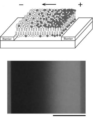
The fluid nature of the supported membrane permits dynamic rearrangement of membrane components over macroscopic length scales. Electrophoretic forces, resulting from application of electric fields tangent to the membrane, can induce bulk transport of charged and uncharged membrane components [21]. When this supported membrane microelectrophoresis is combined with patterns of diffusion barriers, several novel types of membrane manipulation are enabled [12, 22, 74].
The electrophoretic drift velocities of molecules in a membrane can be used as a basis for molecular separation. Subtle differences in drift velocity can be amplified by driving the membrane through a geometrical Brownian ratchet, consisting of a two-dimensional labyrinth of asymmetrical diffusion barriers [26]. Percolation of molecules through the array of barriers results in a net drift perpendicular to the field, which depends on the ratio of the parallel electrophoretic drift velocity to the diffusion coefficient. Attractive features of this type of separation are that it is continuous process and that it can be performed in a membrane environment.
Electrophoretic rearrangement membranes entirely confined within a boundary of diffusion barriers can lead to equilibrium concentration profiles [12, 22, 24, 25, 109]. Electric field strengths in the 10–100 V/cm range typically produce concentration profiles on the 100 μm size scale. These can be readily imaged by fluorescence microscopy when membranes are doped with fluorescent probes (Figure 17.7). The shape of the concentration profile results from a balance between the applied electrophoretic forces (including both electrical and electroosmotic) and entropy. Concentration profile imaging has proven to be a highly informative analytical probe for weak interactions among molecules in membranes. It also provides a rapid assay for membrane lateral fluidity. The ability to locally concentrate membrane components by electrophoretic rearrangement can be of utility as well [23].
17.4.4. Live Cell Interactions
One of the most enabling applications of supported membrane technology is its usage to create phantom cell surfaces for interactions with living cells. Some of the earliest implementations of supported membranes were for purposes of live cell studies, and this continues to be a promising growth area [7, 110]. Supported membrane systems are uniquely poised to recapture the rich biological functionality that emerges when cell surface proteins are permitted to move freely about. A hallmark example of this functionality is illustrated by the formation of a T cell immunological synapse between a living T cell and a supported membrane, which displays the relevant proteins (Figure 17.8) [10]. In addition to achieving antigen specificity, proteins in both the T cell and the supported membrane undergo large-scale spatial rearrangements in their respective membranes to form highly organized patterns. Supported membrane technology has been instrumental in the study of these immune recognition processes and continues to offer new insights [11]. The ability to fabricate patterns of diffusion barriers with sub-cellular (sub-micron) spatial dimensions provides a method of imposing constraints on protein motion and transport in the supported membrane. Through engagements with cell surface proteins on the living cells, these freedom-of-motion constraints can be transmitted to living signal transduction networks. Patterned supported membrane systems thus offer a method of imposing spatial constraints on biochemical
318 |
JAY T. GROVES |
E
A
Barrier |
|
Barrier |
Substrate
B
100 mm
FIGURE 17.7. A. Schematic of a electric field—induced concentration gradient in a confined patch of supported membrane. B. Image of a field-induced concentration gradient of the negatively-charged fluorescent probe (Texas Red DPPE) in an otherwise neutral supported membrane. Adapted from [12].
signaling networks in living cells. Application of these concepts can be expected to yield new information on the function of live cell signaling systems, and may further offer new ways of transmitting specific signals to cells from non-living materials.
At a more industrial level, live cell assays are used extensively within the pharmaceutical industry. Hybrid live cell-supported membrane assays, such as the T cell immune synapse formation mentioned above, offer many unique capabilities, which may be of significant utility in drug discovery applications. As one example, consider the ability to precisely control the specific protein composition of each membrane. In addition, hybrid live cellsupported membrane assays can be executed in array format.
An interesting aspect of performing live cell experiments on membrane arrays is that different results can be observed compared with cells assayed on substrates consisting of only a single composition [13, 84]. For example, phosphatidylserine (PS) is known to promote the pathological adhesion of erythrocytes (abnormally expressing PS in the outer leaflet of their membrane) to endothelial cells in diseases such as malaria and diabetes. Membrane arrays displaying different mixtures of lipids have been used as cell culture

SUPPORTED LIPID BILAYERS AS MIMICS FOR CELL SURFACES |
319 |
T-Cell
Silica Substrate
FIGURE 17.8. Schematic of a hybrid live cell—supported membrane synapse, modeled after actual synapses formed between living T cells and supported membranes displaying majorhistocompatibility protein (MHC) and intercellular adhesion molecule (ICAM). Adapted from [11].
substrates to characterize PS-mediated cell adhesion. The cells clearly distinguish between lipid composition, selectively adhering to and growing on PS containing membrane corrals. Less adhesion to PS-free membrane was observed when the cells had the option of growing on PS-containing membrane. Although the reasons for this are not clear, it suggests that side- by-side cell discrimination experiments are likely to provide more insightful results than can be obtained form the same experiments performed in isolated wells of a cell culture plate. A key feature of the assay is that the spacing of the different membrane corrals is sufficiently small that cells can sample multiple membranes before settling down; 200-micron square corrals were used in the experiments mentioned above.
17.5. CONCLUSION
Supported membrane technology has received increasing interest in recent years. This interest is fueled by prospects of creating cell mimetic surfaces for the study of biological membranes and membrane processes as well as the construction of biologically inspired devices. Substantial progress has been made in areas of membrane patterning, electrical integration, support modification, and biological functionalization. Further advancement can certainly be expected. Supported membrane systems combine elements of surface chemistry, soft condensed matter materials science, and biology. This is a complex intersection of scientific disciplines, which at the same time presents promising and intriguing prospects for creating new interfaces between living and non-living systems.
320 |
JAY T. GROVES |
REFERENCES
[1]S.J. Singer and G.L. Nicolson. The fluid mosaic model of cell membranes. Science, 175:720–731, 1972.
[2]K. Jacobson, E.D. Sheets, and R. Simson. Revisiting the fluid mosaic model of membranes. Science, 268:1441–1442, 1995.
[3]R.G.W. Anderson and K. Jacobson. A role for lipid shells in targeting proteins to caveolae, rafts, and other lipid domains. Science, 296:1821–1825, 2002.
[4]K. Simmons and E. Ikonen. Functional rafts in cell membranes. Nature, 387:569–572, 1997.
[5]H.M. McConnell and M. Vrljic. Liquid-liquid immiscibility in membranes. Annu. Rev. Biophys. Biomol. Struct., 32:469–492, 2003.
[6]S.L. Veatch and S.L. Keller. Organization in lipid membranes containing cholesterol. Phys. Rev. Lett., 89(26):268101, 2002.
[7]A.A. Brian and H.M. McConnell. Allogenic stimulation of cytoxic T cells by supported planar membranes.
Proc. Natl. Acad. Sci. U.S.A., 81:6159–6163, 1984.
[8]E. Sackmann Supported membranes: Scientific and practical applications. Science, 271:43–48, 1996.
[9]E. Sackmann and M. Tanaka. Supported membranes on soft polymer cusions: fabrication, characterization, and applications. TIBTECH, 18:58–64, 2000.
[10]A. Grakoui et al. The immunological synapse: A molecular machine controlling T cell activation. Science, 285:221–227, 1999.
[11]J.T. Groves and M.L. Dustin. Supported planar bilayers in studies of immune cell adhesion and communication. J. Immunol. Methods, 278:19–32, 2003.
[12]J.T. Groves and S.G. Boxer. Micropattern formation in supported lipid membranes. Acc. Chem. Res., 35:149–157, 2002.
[13]J.T. Groves. Membrane array technology for drug discovery. Curr. Op. Drug Disc. Dev., 5(4):606–612, 2002.
[14]Bray, M.D. Levin, and C.J. Morton-Firth. Receptor clustering as a cellular mechanism to control sensitivity. Nature, 393(7):85–88, 1998.
[15]G. Maheshwari et al. Cell adhesion and motility depend on nanoscale RGD clustering. J. Cell Sci., 113:1677– 1686, 2000.
[16]J.E. Gestwicki and L.L. Kiessling. Inter-receptor communication through arrays of bacterial chemoreceptors. Nature, 415(3):81–84, 2002.
[17]P.A. vanderMerwe and S.J. Davis. The immunological synapse—a multitasking system. Science, 295:1479– 1480, 2002.
[18]C. Dietrich et al. Lipid rafts reconstituted into model membranes. Biophys. J., 80:1417–1428, 2001.
[19]Y. Kaizuka and J.T. Groves. Structure and dynamics of supported intermembrane junctions. Biophys. J., 86:905–912, 2004.
[20]C. Dietrich et al. Partitioning of Thy-1, GM1, and cross-linked phsopholipid analogs into lipid rafts reconstituted in supported model membrane monolayers. Proc. Natl. Acad. Sci. U.S.A., 98(19):10642–10647, 2001.
[21]M. Stelzle, R. Miehlich, and E. Sackmann. Two-dimensional microelectrophoresis in supported lipid bilayers. Biophys. J., 63:1346–1354, 1992.
[22]J.T. Groves and S.G. Boxer. Electric field-induced concentration gradients in planar supported bilayers. Biophys. J., 69:1972–1975, 1995.
[23]J.T. Groves, C. Wulfing,¨ and S.G. Boxer. Electrical manipulation of glycan-phosphatidyl inositol-tethered proteins in planar supported bilayers. Biophys. J., 71:2716–2723, 1996.
[24]J.T. Groves, S.G. Boxer, and H.M. McConnell. Electric field-induced reorganization of two-component supported bilayer membranes. Proc. Natl. Acad. Sci. U.S.A., 94:13390–13395, 1997.
[25]J.T. Groves, S.G. Boxer, and H.M. McConnell. Electric field-induced critical demixing in lipid bilayer membranes. Proc. Natl. Acad. Sci. U.S.A., 95:935–938, 1998.
[26]A. van Oudenaarden and S.G. Boxer. Brownian ratchets: molecular separations in lipid bilayers supported on patterned arrays. Science, 285:1046–1048, 1999.
[27]D. Axelrod et al. Mobility measurement by analysis of fluorescence photobleaching recovery kinetics. Biophys. J., 16:1055–1069, 1976.
[28]L.K. Tamm and E. Kalb. Microspectrofluoremetry of supported planar membranes. In S.G. Schulman (ed.), Molecular Luminescence Spectroscopy. John Wiley & Sons Inc., pp. 253–305, 1993.
SUPPORTED LIPID BILAYERS AS MIMICS FOR CELL SURFACES |
321 |
[29]J. Salafsky, J.T. Groves, and S.G. Boxer. Architecture and function of membrane proteins in planar supported bilayers: a study with photosynthetic reaction centers. Biochemistry, 35:14773–14781, 1996.
[30]D. Magde, E. Elson, and W.W. Webb. Thermodynamic fluctuations in a reacting system-measurement by fluorescence correlation spectroscopy. Phys. Rev. Lett., 29(11):705–708, 1972.
[31]K. Bacia and P. Schwille. A dynamic view of cellular processes by in vivo fluorescence auto and crosscorrelation spectroscopy. Methods, 29:74–85, 2003.
[32]E. Haustein and P. Schwille. Ultrasensitive investigations of biological systems by fluorescence correlation spectroscopy. Methods, 29:153–166, 2003.
[33]N. Kahya et al. Probing lipid mobility of raft-exhibiting model membranes by fluorescence correlation spectroscopy. J. Biol. Chem., 278:28109–28115, 2003.
[34]J. Korlach et al. Characterization of lipid bilayer phases by confocal microscopy and fluorescence correlation spectroscopy. Proc. Natl. Acad. Sci. U.S.A., 96:8461–8466, 1999.
[35]P. Schwille, J. Korlach, and W.W. Webb. Fluorescence correlation spectroscopy with single-molecule sensitivity on cell and model membranes. Cytometry, 36:176–182, 1999.
[36]E.D. Sheets, R. Simson, and K. Jacobson. New insights into membrane dynamics from analysis of cell surface interactions by physical methods. Curr. Op. Cell Biol., 7:707–714, 1995.
[37]M. Vrljic et al. Translational diffusion of individual class II MHC membrane proteins in cells. Biophys. J., 83:2681–2692, 2002.
[38]C. Dietrich et al. Relationship of lipid rants to transient confinement zones detected by single particle tracking. Biophys. J., 82:274–284, 2002.
[39]M.J. Saxton and K. Jacobson. Single particle tracking: applications to membrane dynamics. Annu. Rev. Biophys. Biomol. Struct., 26:373–399, 1997.
[40]B.L. Stottrup, S.L. Veatch, and S.L. Keller. Nonequilibrium behavior in supported lipid membranes containing cholesterol. Biophys. J., 86:2942–2950, 2004.
[41]J.M. Crane and L.K. Tamm. Role of cholesterol in the formation and nature of lipid rafts in planar and spherical model membranes. Biophys. J., 86:2965–2979, 2004.
[42]M. Kuhner,¨ R. Tamp´e, and E. Sackmann. Lipid mono and bilayer supported on polymer films: composite polymer films on solid substrates. Biophys. J., 67:217–226, 1994.
[43]J.Y. Wong et al. Polymer-cushioned bilayers. I. A structural study of various preparation methods using neutron reflectometry. Biophys. J., 77:1445–1457, 1999.
[44]J.Y. Wong et al. Polymer-cusioned bilayers. II. An investigation of interaction forces and fusion using the surface forces apparatus. Biophys. J., 77:1458–1468, 1999.
[45]G. Elander, M. Kuhner,¨ and E. sackmann. Functionalization of Si/SiO2 and glass surfaces with ultrathin dextran films and deposition of lipid bilayers. Biosens. Bioelectro., 11(6/7):565–577, 1996.
[46]H. Sigl et al. Assembly of polymer/lipid composite films on solids based on hairy rod LB-films. Eur. Biophys. J., 25:249–259, 1997.
[47]H. Hillebrandt et al. High electric resistance polymer/lipid composite films on indium-tin-oxide electrodes. Langmuir, 15(24):8451–8459, 1999.
[48]F. Rehfeldt and M. Tanaka. Hydration forces in ultrthin films of cellulose. Langmuir, 19:1467–1473, 2003.
[49]T. Baumgart and A. Offenh¨ausser. Polysaccharide-supported planar bilayer lipid model membranes. Langmuir, 19(5):1730–1737, 2003.
[50]K. Sengupta et al. Mimicking tissue surfaces by supported membrane coupled ultrathin layer of hyaluronic acid. Langmuir, 19(5):1775–1781, 2003.
[51]M.L. Wagner and L.K. Tamm. Tethered polymer-supported planar lipid bilayers for reconstitution of integral membrane proteins: silane-polyethyleneglycol-lipid as a cushion and covalent linker. Biophys. J., 79:1400– 1414, 2000.
[52]W.W. Shen et al. Polymer-supported lipid bilayers on benzophenone-modified substrates. Biomacromolecules, 2:70–79, 2001.
[53]C. Naumann et al. The polymer-supported phospholipid bilayer: tethering as a new approach to substratemembrane stabilization. Biomacromolecules, 3:27–35, 2002.
[54]A. Berquand et al. Two-step formation of streptavidin-supported lipid bilayers by PEG-triggered vesicle fusion. Fluorescence and atomic force microscopy characterization. Langmuir, 19(5):1700–1707, 2003.
[55]B.A. Cornell et al. A biosensor that uses ion-channel switches. Nature, 387:580–583, 1997.
[56]B. Raguse et al. Tethered lipid bilayer membranes: formation and ionic reservoir characterization. Langmuir, 14:648–659, 1998.
322 |
JAY T. GROVES |
[57]C. Bieri et al. Micropatterned immobilization of a G protein-coupled receptor and direct detection of G protein activation. Nature Biotech., 17:1105–1108, 1999.
[58]V. Kiessling and L.K. Tamm. Measuring distances in supported bilayers by fluorescence interferencecontrast miscroscopy: polymer supports and SNARE proteins. Biophys. J., 84:408–418, 2003.
[59]A.P. Wong and J.T. Groves. Topographical imaging of an inter-membrane junction by combined fluorescene interference and energy transfer microscopies. J. Am. Chem. Soc., 123:12414–12415, 2001.
[60]A.P. Wong and J.T. Groves. Molecular topography imaging by intermembrane fluorescence resonance energy transfer. Proc. Natl. Acad. Sci. U.S.A., 99(22):14147.
[61]T. Charitat et al. Adsorbed and free lipid bilayers at the solid-liquid interface. Eur. Phys. J. B, 8:583–593, 1999.
[62]G. Fragneto et al. A fluid floating bilayer. Europhys. Lett., 53:100–106, 2001.
[63]R. Parthasarathy et al. Nonequilibrium adhesion patterns at lipid bilayer junctions. J. Phys. Chem. B, 108:649–657, 2004.
[64]A. Lambacher and P. Fromherz. Fluorescence interference-contrast microscopy on oxidized silicon using a monomolecular dye layer. Appl. Phys. A, 63:207–216, 1996.
[65]A. Lambacher and P. Fromherz. Luminescence of dye molecules on oxidized silicon and fluorescence ineterference contrast microscopy of biomembranes. J. Opt. Soc. Am. B, 19(6):1435–1453, 2002.
[66]J. R¨adler and E. Sackmann. Imaging optical thicknesses and separation distances of phospholipid vesicles at solid surfaces. J. Phys. II France, 3:727–748, 1993.
[67]J.O. R¨adler et al. Fluctuation analysis of tension-controlled undulation forces between giant vesicles and solid substrates. Phys. Rev. E, 51(5):4526–4536, 1995.
[68]R. Hirn et al. Collective membrane motions of high and low amplitude, studied by dynamic light scattering and micro-interferomtery. Faraday Discuss., 111:17–30, 1998.
[69]R. Parthasarathy and J.T. Groves. Optical techniques for imaging membrane topography. Cell Biochem. Biophys., 2004. (in press.)
[70]R. Lipowsky and E. Sackmann (eds.). Structure and dynamics of membranes. In Handbook of Biological Physics. Vol. 1, Elsevier Science Ltd., New York, 1995.
[71]L.K. Tamm and H.M. McConnell. Supported phospholipid bilayers. Biophys. J., 47:105–113, 1985.
[72]J. R¨adler, H. Strey, and E. Sackmann. Phenomenology and kinetics of lipid bilayer spreading on hydrophilic surfaces. Langmuir, 11:4539–4548, 1995.
[73]O.P. Karlsson and S. Lof˚as¨ . Flow-mediated on-surface reconstitution of G-protein coupled receptors for applications in surface plasmon resonance biosensors. Anal. Biochem., 300:132–138, 2002.
[74]J.T. Groves, N. Ulman, and S.G. Boxer. Micropatterning fluid lipid bilayers on solid supports. Science, 275:651–653, 1997.
[75]L.A. Kung et al. Patterning hybrid surfaces of proteins and supported lipid bilayers. Langmuir, 16:6773– 6776, 2000.
[76]C.A. Keller et al. Formation of supported membranes from vesicles. Phys. Rev. Lett., 84(23):5443–5446, 2000.
[77]C.A. Keller and B. Kasemo. Surface specific kinetics of lipid vesicle adsorprion measured with a quartz crystal microbalance. Biophys. J., 75:1397–1402, 1998.
[78]E. Reimhult, F. H¨a¨ak, and B. Kasemo. Intact vesicle adsorption and supported biomembrane formation from vesicles in slution: Influence of surface chemistry, vesicle size, temperature, and osmotic pressure. Langmuir, 19(5):1681–1691, 2003.
[79]S.G. Boxer, N. Ulman, and J.T. Groves. Arrays of independently-addressable supported fluid bilayer membranes. The Board of Trustees of the Leland Stanford Junior University, United States, 2001.
[80]L. Kung et al. Printing via photolithography on micropartitioned fluid lipid membranes. Adv. Mater., 12(10):731–734, 2000.
[81]J.S. Hovis and S.G. Boxer. Patterned barriers to lateral diffusion in supported lipid bilayer membranes by blotting and stamping. Langmuir, 16:894–897, 2000.
[82]J.S. Hovis and S.G. Boxer. Patterning and composition arrays of supported lipid bilayers by microcontact printing. Langmuir, 17:3400–3405, 2001.
[83]P.S. Cremer and T. Yang. Creating spatially addressed arrays of planar supported fluid phospholipid membranes. J. Am. Chem. Soc., 121:8130–8131, 1999.
[84]J.T. Groves, L.K. Mahal, and C.R. Bertozzi. Control of cell adhesion and growth with micropatterned supported lipid membranes. Langmuir, 17(17):5129–5133, 2001.
SUPPORTED LIPID BILAYERS AS MIMICS FOR CELL SURFACES |
323 |
[85]J.S. Bonifacino and B.S. Glick. The mechanisms of vesicle budding and fusion. Cell, 116:153–166, 2004.
[86]A.R. Sapuri, M.M. Baksh, and J.T. Groves. Electrostatically-targeted intermembrane lipid exchange with micropatterned supported membranes. Langmuir, 19(5):1606–1610, 2003.
[87]R.N. Orth et al. Mast cell activation on patterned lipid bilayers of subcellular dimensions. Langmuir, 19:1599–1605, 2003.
[88]L. Kam and S.G. Boxer. Formation of supported lipid bilayer composition arrays by controlled mixing and surface capture. J. Am. Chem. Soc., 122:12901–12902, 2000.
[89]L. Kam and S.G. Boxer. Spatially selective manipulation of supported lipid bilayers by laminar flow: steps toward biomembrane microfluidics. Langmuir, 19:1624–1631, 2003.
[90]T. Yang et al. Fabrication of phospholipid bilayer-coated microchannels for on-chip immunoassays. Anal. Chem., 73:165–169, 2001.
[91]T. Thorsen, S.J. Maerkl, and S.R. Quake. Microfluidic large-scale integration. Science, 298:580–584, 2002.
[92]Y. Fang, J. Lahiri, and L. Picard. G protein-coupled receptor microarrays for drug discovery. DDT, 8(16):755–761, 2003.
[93]S.P.A. Fodor et al. Light-directed, spatially addressable parallel chemical synthesis. Science, 251:767–773, 1991.
[94]G. MacBeath and S.L. Schreiber. Printing proteins as microarrays for high-throughput function determination. Science, 289:1760–1763, 2000.
[95]P. Mitchell. A perspective on protein arrays. Nature Biotech., 20:225–229, 2002.
[96]P. Wagner and R. Kim. Protein biochips: an emerging tool for proteomics research. Curr. Drug Disc., May 23–28, 2002.
[97]T. Haga and G. Berstein (eds.). G-protein Coupled Receptors. CRC Press, Boca Raton, 1999.
[98]Y. Fang, A.G. Frutos, and J. Lahiri. Membrane Protein Microarrays. J. Am. Chem. Soc., 124(11):2394–2395, 2002.
[99]T. Yang et al. Investigations of bivalent antibody binding on fluid-supported phospholipid membranes: the effect of hapten density. J. Am. Chem. Soc., 125:4779–4784, 2003.
[100]T.M. Bayerl and M. Bloom. Physical properties of single phospholipid bilayers adsorbed to micro glass beads. Biophys. J., 58:357–362, 1990.
[101]T. Buranda et al. Biomimetic molecular assemblies on glass and mesoporous silica microbeads for biotechnology. Langmuir, 19:1654–1663, 2003.
[102]A. Loidl-Stahlhofen et al. Solid-supported biomolecules on modified silica surfaces—A tool for fast physicochemical characterization and high-throughput screening. Adv. Mater., 13(23):1829–1834, 2001.
[103]T.M. Bayerl. A glass bead game. Nature, 427:105, 2004.
[104]M.M. Baksh, M. Jaros, and J.T. Groves. Detection of molecular interactions at membrane surfaces through colloid phase transitions. Nature, 427:139–141, 2004.
[105]B. Sackmann and E. Neher. Plenum Press, New York, 1985.
[106]P. Mustonen et al. Binding of cytochrome c to liposomes as revealed by the quenching of fluorescence from pyrene-labeled phospholipids. Biochemistry, 26:2991–2997, 1987.
[107]Gritsch et al. Impedence spectroscopy of porin and gramicidin pores reconstituted into supported lipid bilayers on indium-tim-oxide electrodes. Langmuir, 14:3118–3125, 1998.
[108]G. Wiegand et al. Electrical properties of supported lipid bilayer membranes. J. Phys. Chem. B, 106:4245– 4254, 2002.
[109]J.T. Groves, S.G. Boxer, and H.M. McConnell. Electric field effects in multicomponent fluid lipid membranes. J. Phys. Chem. B, 104(1):119–124, 2000.
[110]T.H. Watts and H.M. McConnell. Antigen presentation by supported planar membranes conntaining purified major histocompatibility complex proteins. In B. Pernis and H. Vogel (eds.), Processing and Presentation of Antigens. Academic Press. pp. 143–155, 1988.
18
Engineering Cell Adhesion
Kiran Bhadriraju, Wendy F. Liu, Darren S. Gray, and
Christopher S. Chen
Johns Hopkins University School of Medicine, Department of Biomedical Engineering, Traylor 718, Baltimore, MD-21205
University of Pennsylvania, 125 S. 31st St. Translational Research Labs, Suite 1400, Philadelphia, PA-19104
18.1. INTRODUCTION
Cells exist within a complex and ever-changing environment, which includes soluble molecules such as growth factors, an extracellular matrix that includes adhesive proteins and carbohydrates, and other neighboring cells. They actively sense and respond to changes in this environment, existing in a state of physiological equilibrium with it. Thus, it has been said, “. . . the unit of function in higher organisms is larger than the cell itself ” [10]. The information content in the adhesive environment is encoded both in its composition and its organization on the nanometer to micrometer length scales. When taken out of this physiological context and cultured in plastic tissue culture dishes, cells lose the cues that maintain their in vivo identity or phenotype, and dedifferentiate. For example, hepatocytes— the principal cell type in the liver—perform several critical liver-specific functions such as production of bile, metabolism of urea, and the synthesis of important serum proteins such as albumin, fibrinogen, and transferrin [27]. When cultured in vitro and isolated from the liver microenvironment, they rapidly downregulate liver-specific phenotype [54]. Similarly, chondrocytes, which are required for the secretion and maintenance of cartilage, lose their differentiated function when cultured in vitro downregulating the synthesis and secretion of cartilage-specific collagens and proteoglycans [88]. Thus, tissue-specific cell function appears to be closely related to the microstructural organization of the tissue itself [11].
The realization that tissue structure and organization is critical to its function has led to the development of new tools to control, or engineer, such structures into cell culture
326 |
KIRAN BHADRIRAJU ET AL. |
systems [9]. Microfabricated tools adapted from the semiconductor industry appear to have the appropriate degree of spatial and chemical control in the fabrication process to recreate many of the key features cells use to determine their functional state.
The ability to create bioartificial tissues that recapitulate in vivo function using microengineering approaches would have many applications ranging from those with immediate clinical relevance to those for fundamental research [41]. For example, in drug development many compounds appear to have activity in cell culture experiments that later fail in animal or human studies. Drug candidates that fail in these later stages are largely responsible for the ballooning costs of drug development. A recent estimate suggests that 1 in 13 compounds entering animal testing made it to the market in late 2000–2002, and the cost of successfully bringing such a drug to market is estimated at $1.7 billion [39]. The use of artificial tissues is likely to provide more faithful test models at the initial stages of testing that would eliminate false-positives candidates earlier rather than in the later, more expensive stages of drug testing [6]. Another application of microengineered tissues aims to address the problem of organ shortage through tissue engineering. According to www.optn.org (The Organ Procurement and Transplantation Network) and www.unos.org (United Network for Organ Sharing), in the year 2003 in USA, 23,357 organs were transplanted, 83,195 candidates were on the waiting list, and 5559 patients died while waiting. As an alternative to transplantation, cells of specific tissues are expanded in culture and then transplanted into the body, incorporated into an extra corporeal support devices [1], or even used to recreate entire organs in the laboratory [33]. While some tissues such as skin or cartilage have relatively simple architectures, others contain many cells types and complex tissue architectures that play a critical role in tissue function. Recreation of this complexity will require microand nanofabrication technologies [9]. In addition to the primary market drivers of tissue engineering and drug discovery, many additional specialized applications are likely to emerge, such as the development of cell-based biosensors for bio-defense and environmental analysis where arrays of living cells act as detectors for toxins in the environment [64]. Lastly, the process of learning how to construct such microengineered devices inevitably results in the development of new experimental systems that give insights into basic physiologic and pathologic processes including development, wound healing, and inflammation.
While there is a great need to understand how to reconstruct environments that can coax cells to function in a specified manner, such tools have only just begun to emerge, primarily from the transfer of semiconductor manufacturing technologies into the biomedical arena. As an illustration of how microfabrication tools can help engineer microenvironments, Fig. 18.1 shows two sets of cultured endothelial cells: one in a conventional culture environment where cell-cell adhesion or spreading are unconstrained, and the other in a microengineered system where the extent of cell-cell adhesion and spreading is controlled using microfabrication tools. There are clear advantages to biological experimentation using such well-defined systems in order to study cellular responses to shape and adhesion [56]. In addition to adhesion studies, many other problems in biomedical science and technology may prove to benefit by such control. In this chapter, we will describe studies that have utilized microfabrication techniques to engineer the cell culture environment. First we will discuss from a biological perspective, the key aspects of the microenvironment that are thought to regulate cell behavior. Then, the technological needs and demands for engineering cellular microenvironments will be described, and the tools that have been